HAL Id: tel-02611007
https://tel.archives-ouvertes.fr/tel-02611007
Submitted on 18 May 2020HAL is a multi-disciplinary open access archive for the deposit and dissemination of sci-entific research documents, whether they are pub-lished or not. The documents may come from teaching and research institutions in France or abroad, or from public or private research centers.
L’archive ouverte pluridisciplinaire HAL, est destinée au dépôt et à la diffusion de documents scientifiques de niveau recherche, publiés ou non, émanant des établissements d’enseignement et de recherche français ou étrangers, des laboratoires publics ou privés.
Phenotypic and molecular responses of rice to
Burkholderia s.l. species
Eoghan King
To cite this version:
Eoghan King. Phenotypic and molecular responses of rice to Burkholderia s.l. species. Vegetal Biology. Université Montpellier, 2019. English. �NNT : 2019MONTG079�. �tel-02611007�
RAPPORT DE GESTION
2015
THÈSE POUR OBTENIR LE GRADE DE DOCTEUR
DE L’UNIVERSITÉ DE MONTPELLIER
En Biologie des SystèmesÉcole doctorale GAIA Unité de recherche UMR IPME
Présentée par Eoghan KING
Le 11 décembre 2019
Sous la direction de Pierre CZERNIC
et Lionel MOULIN
Devant le jury composé deMatthieu ARLAT, Professeur des Universités, Université de Toulouse III – Paul Sabatier Alia DELLAGI, Professeure des Universités, AgroParisTech
Sofie GOORMACHTIG, Professeure, VIB-UGent
Michel LEBRUN, Professeur des Universités, Université de Montpellier Pierre CZERNIC, Professeur des Universités, Université de Montpellier Lionel MOULIN, Directeur de Recherche, IRD
Rapporteur Rapportrice Examinatrice Examinateur Directeur de thèse Co-Directeur de thèse
Caractérisation phénotypique et moléculaire
de la réponse du riz au c ours de l’ interaction
« Ceci est le véritable secret de la vie :
Etre complètement engagé avec ce que vous faites dans l’ici et maintenant.
Et au lieu d’appeler cela travailler, réaliser que c’est jouer »
Allan Watts
Nous y voilà… La fameuse partie la plus lue d'une thèse, celle qui marque l’aboutissement de
plusieurs années de travail, permet de se rappeler le chemin parcouru et les personnes sans qui rien de ce que j'ai accompli aux cours de ces années de thèse n'aurait été possible. On y va pour les
remerciements !
Tout d'abord je me dois de remercier mes directeurs de thèse, Pierre et Lionel, pour votre confiance, vos encouragements et pour le pari que vous avez fait sur un dreadeux breton même pas padawan pour un sou il y a de cela presque 4 ans.
Je remercie les membres de mon jury pour avoir accepté d'évaluer mes travaux. Mes rapporteurs, Alia Dellagi et Matthieu Arlat ainsi que mes examinateurs, Sofie Goormachtig et Michel Lebrun.
Je remercie également les membres de mon comité de thèse, Jean-Benoît Morel, Florence Wisniewski-Dyé, Fabienne Cartieaux et Christophe Périn.
Enfin, je remercie Valérie Verdier pour avoir accueilli mon projet au sein d'IPME
Les remerciements d'usage étant fait-ce qui n'enlèvent rien à leur sincérité- je souhaite adresser à toutes les personnes qui suivent, un grand "MERCI":
Aux collègues qui m'ont accueilli avec un sourire quand j'arrivais avec un service à demander, Florence, Sandrine, William, Emilie, Hervé
À Gilles, pour ta vision critique et tes blagues douteuses À Agnieszka, pour nos échanges matinaux
À Marine, pour avoir débroussaillé le chemin avant que je reprenne le flambeau du projet BRIO Aux chercheurs chevronnés qui se sont intéressés à mes travaux et m'ont permis de faire avancer ce projet, Stéphane, Antony, Christophe B et Sébastien
À Isabelle, pour ta bienveillance et ton aide précieuse
Aux étudiants et stagiaires que j'ai encadré, Harriet, Fehizoro, Patrick, Violette et Mathilde pour m’avoir fait découvrir ma capacité d’encadrement et de transfert de connaissances
Aux anciens -et pas si anciens- PostDoc, Nils, Kévin, Daniel et Cindy pour vos conseils précieux À l'ancienne génération de thésards, Mathilde D, Cécile, Hélène, Rémi
À mes brésiliennes préférées, Maìra et Deisy, pour votre folie
À tous les stagiaires, doctorants, chercheurs du Sud croisés, Carlos, Diégane, Sondo, Kader, Hieu, Ngan, Ially pour avoir ouvert ma vision du monde étriquée d'occidental
Aux "grumeaux du riz", Jérémy et Mathieu, pour vos brassages et nos discussions de couloirs
À la nouvelle génération de thésards, Anne-So, Marlène, Théo PP, Leopol pour les bonnes tranches de rigolade
Aux personnes qui ont partagés à un moment ou un autre mon bureau, merci de ne me pas m'avoir étranglé
Aux membres du sandwich électronique, pour m'avoir sorti du monde du labo
À ceux qui étaient toujours là,
À mes super-nénettes, Lucile, Mathilde H, Helena et Marie, pour votre force, votre expérience, vos mots rassurants et vos encouragements
À Adrian, pour trop de choses
À Malo, pour tes mots, ta sagesse et nos échanges scientifiques tardifs À Thibault, pour ton amitié sans faille
À ceux qui étaient plus loin,
À ma métabande BZH, Elora, Ludo, Eliott, Morgane, Pilou, Pigi, Gigi
À Marin, pour ce coup de téléphone dont je me souviendrai pour le restant de mes jours et l'exemple de résilience que tu incarnes dans ma vie
À Théo LGLD, pour l'évidence de notre amitié
À mes plus proches et fidèles soutiens, mes parents, pour votre confiance et votre amour À Maud, pour cette voie que tu as ouverte, ta compréhension de ce qu'elle implique et tes tapes occasionnelles derrière la tête.
Et parce que si j'ai choisi cette voie c'est pour apporter ma pierre à l'avenir, c'est à tes enfants que je dédis ces travaux…
SOMMAIRE
Introduction générale ... 1
Chapitre 1
Synthèse bibliographique Multicellular organisms as metaorganisms ... 9Animal and plant microbiome commonalities ... 9
Plant microbiome specificities ... 11
Plant-associated microbial niches ... 13
Different types of plant-microbe interactions ... 15
Plant-beneficial microbial interactions ... 17
Main plant-mutualist models overview ... 17
Plant-microbe associative symbioses ... 19
Plant-Growth promoting traits ... 21
The immune system of plants ... 27
The growth–defense trade-off in plants ... 33
Commonalities in pathogenic and beneficial microbes interacting with plants ... 35
Plant perception of pathogenic and beneficial microbes ... 35
Omics to unravel the complexity of plant-microbiome interactions ... 37
Review of transcriptomic responses of monocots to bacterial associative symbiosis ... 39
Applications of plant associative symbioses ... 45
References ... 48
Table and figure references ... 61
Chapitre 2
Monitoring of rice transcriptional responses to contrasted colonizing patterns of phytobeneficial Burkholderia s.l. reveals a temporal shift in JA systemic response Introduction ... 68Material & Methods ... 70
Results ... 72
Analysis of root colonization ... 72
Transcriptional response of rice to bacterial inoculation ... 73
Validation of RNA-Seq data by qPCR ... 87
Temporal analysis of strain-specific marker genes ... 87
Discussion ... 95
Supplementary Material ... 108
Chapitre 3
Transcriptomic response of rice to root colonization by the model endophyte Paraburkholderia phytofirmans PsJN Introduction ... 134Material & Methods ... 135
Results ... 139
Analysis of PsJN colonization of rice roots ... 139
Transcriptomic analysis of rice response to PsJN ... 139
GO term enrichment analysis ... 141
Physiological function mining of transcriptome ... 141
Root response to PsJN ... 143 Leaves response ... 147 Discussion ... 151 References ... 155 Supplementary Material ... 160 Complementary results ... 171
Chapitre 4
Comparative analysis of rice defense response to rhizospheric, endophytic and pathogenic Burkholderia s.l. strains and model PGPR Introduction ... 177Material & Methods ... 178
Results ... 181
Rice root colonization analysis of rhizospheric naturally-associated Burkholderia s.l. strains, model PGPR and Burkholderia s.s. pathogens ... 181
Transcriptional analysis of rice local and systemic response ... 185
Discussion ... 191 References ... 195 Supplementary Material ... 200 Complementary results ... 202
Chapitre 5
Discussion générale Discussion générale ... 205LISTE DES FIGURES
Introduction générale
Phylogénie des Burkholderia s.l. ... 2
Chapitre 1
Synthèse bibliographique Figure 1: Interplay between host and microbes in plant-microbiome interactions ... 10Figure 2: Plant microbiota and origins of plant-associated microbes ... 12
Figure 3: Microbial root colonization hotspots ... 12
Figure 4: Trophic space occupied by microorganisms in association with plants ... 16
Figure 5: Schema illustrating the common symbiosis signaling pathway (CSSP) ... 18
Figure 6: Mechanisms of Plant Growth Promoting Microbes in enhancing plant growth... 22
Figure 7: Plant innate immunity overview ... 26
Figure 8: Plant receptor networks ... 28
Figure 9: Biochemical networks interactions drive phenotypic output ... 28
Figure 10: Schematic representation of systemically induced immune responses ... 30
Figure 11: Balancing plant immune responses and fitness costs ... 32
Figure 12: Potential bacterial strategies employed to evade flagellin recognition upon bacterial invasion in plant tissues ... 34
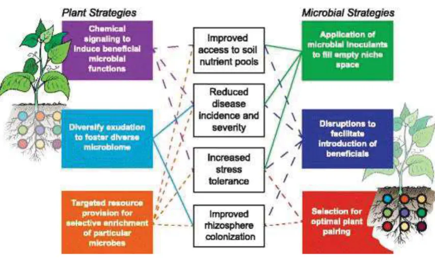
Figure 13: Strategies to reduce chemical inputs and increasing yields from plant and microbial side ... 46
Chapitre 2
Monitoring of rice transcriptional responses to contrasted colonizing patterns of phytobeneficial Burkholderia s.l. reveals a temporal shift in JA systemic response Figure 1: Colonization of the roots of hydroponically grown rice plants by Bv and Pk ... 74Figure 2: Endophytic colonization of the roots of hydroponically grown rice plants by Bv and Pk ... 74
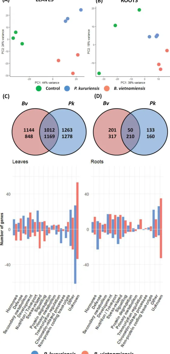
Figure 3: Comparative analysis of leaves and roots transcriptomes in response to Bv and Pk colonization ... 78
Figure 4: Functional categories of rice DEGs upon Bv and Pk colonization ... 78
Figure 5: Confirmation of genes regulated upon Bv and Pk colonization ... 86
Figure 6: JA-related co-expression network transcriptional regulations induced by Pk and Bv root colonization... 94
Figure 7: Local and systemic transcriptional regulations of rice in response to Pk and Bv root colonization ... 100
Supplementary figure 1: Rice roots colonization hotspots of P. kururiensis and B. vietnamiensis ... 108
Supplementary figure 2: Impact of the inoculation on rice biomass ... 109
Supplementary figure 3: Enrichment analysis of Biological GO terms of common leaves and roots transcriptomes in response to B. vietnamiensis and P. kururiensis colonization ... 110
Supplementary figure 4: GO terms enrichment analysis of strain-specific leaves transcriptomes ... 111
Supplementary figure 5 GO terms enrichment analysis of strain-specific roots transcriptomes ... 112
Chapitre 3
Transcriptomic response of rice to root colonization by the model endophyte Paraburkholderia phytofirmans PsJN Figure 1: Colonization of rice roots by PsJN ... 138Figure 2: GO terms enrichment analysis of DEGs in rice roots in response to PsJN ... 140
Figure 3: GO terms enrichment analysis of DEGs in rice leaves in response to PsJN ... 140
Figure 4: Functional categories of rice DEGs upon PsJN colonization ... 142
Figure 5: PsJN colonization increases rice photosynthetic efficiency ... 150
Complementary Figure: Comparative analysis of rice transcriptomic response to P. phytofirmans, P. kururiensis and B. vietnamiensis ... 170
Chapitre 4
Comparative analysis of rice defense response to rhizospheric, endophytic and pathogenic Burkholderia s.l. strains and model PGPR Figure 1: Phylogenetic analysis of the strains used in this study ... 182Figure 2: Comparative analysis of rice root colonization ... 184
Figure 3: Relative abundance of root-associated and endophytic inoculated strains ... 186
Figure 4: Temporal analysis of defense-related genes expression in roots ... 188
Figure 5: Principal component analysis of the expression six defense-related genes expressed in inoculated roots ... 188
Figure 6: Temporal analysis of JA-related genes expression in leaves in response to root-inoculation ... 190
Complementary Figure: Impact of the root inoculation of Burkholderia s.l. strains on rice grain yield and mean grain weight ... 202
LISTE DES TABLEAUX
Chapitre 1
Synthèse bibliographique
Table 1: Non exhaustive list of bacterial and fungal endophytic strains with published genome
... 14
Table 2 : Non-exhaustive list of microbial taxons comprising Plant Growth Promoting Microbes ... 20
Table 3: Studies analyzing transcriptomic response of Arabidopsis thaliana to bacterial associative symbiosis ... 38
Table 4 : Studies analyzing transcriptomic response of monocot model species to bacterial associative symbiosis ... 38
Table 5: Differentially expressed plant genes in response to bacterial associative symbiosis revealed by transcriptomic approaches ... 40
Chapitre 2
Monitoring of rice transcriptional responses to contrasted colonizing patterns of phytobeneficial Burkholderia s.l. reveals a temporal shift in JA systemic response Table 1 : Summary of RNA-Seq data generated for rice transcriptome ... 76Table 2: Defense-related DEGs in leaves in response to P. kururiensis and B. vietnamiensis. Presented genes are part of the top 200 up and down-regulated significantly differentially expressed genes ... 80
Table 3: Defense-related DEGs in roots in response to P. kururiensis and B. vietnamiensis. Presented genes are part of the top 200 up and down-regulated significantly differentially expressed genes ... 82
Table 4: Hormone-related DEGs in leaves in response to P. kururiensis and B. vietnamiensis. Presented genes are part of the top 200 up and down-regulated significantly differentially expressed genes ... 88
Table 5: Hormone-related DEGs in roots in response to P. kururiensis and B. vietnamiensis. Presented genes are part of the top 200 up and down-regulated significantly differentially expressed genes ... 90
Supplementary Table 1: Hydroponic medium ... 113
Supplementary Table 2: Bacterial strains used in this study ... 114
Supplementary Table 3: Primers used in this study ... 115
Supplementary Table 4a: Gene Ontology term enrichment analysis of commonly up-regulated DEGs in leaves ... 116
Supplementary Table 4b: Gene Ontology term enrichment analysis of commonly down-regulated DEGs in leaves ... 118
Supplementary Table 4c: Gene Ontology term enrichment analysis of commonly up-regulated DEGs in roots ... 122
Supplementary Table 4d: Gene Ontology term enrichment analysis of commonly down-regulated DEGs in roots ... 124
Supplementary Table 5: Gene Ontology term enrichment analysis of differentially
expressed genes specifically regulated by each strain in leaves ... 126
Supplementary Table 6: Gene Ontology term enrichment analysis of differentially expressed genes specifically regulated by each strain in roots ... 128
Supplementary Table 9: Genes used to confirm RNA-Seq and their function ... 130
Supplementary Table 10: Expression profiles of the JA network in leaves ... 130
Chapitre 3
Transcriptomic response of rice to root colonization by the model endophyte Paraburkholderia phytofirmans PsJN Table 1: List of defense-related DEGs in rice roots in response to PsJN ... 144Table 2: List of hormone-related DEGs in rice roots in response to PsJN ... 146
Table 3: List of photosynthesis-related DEGs in rice leaves in response to PsJN ... 148
Supplementary Table 1: Summary of RNA-Seq data generated for rice transcriptomes .. 160
Supplementary Table 4: GO term enrichment analysis ... 161
Supplementary Table 5: KEGG pathway enrichment analysis ... 165
Supplementary Table 7: List of signaling-related DEGs in rice roots in response to PsJN ... 166
Supplementary Table 8: List of transport-related DEGs in rice roots in response to PsJN ... 168
Chapitre 4
Comparative analysis of rice defense response to rhizospheric, endophytic and pathogenic Burkholderia s.l. strains and model PGPR Table 1: List of strains used in this study ... 182Table 2: Rice root colonization patterns summary ... 185
Supplementary Table 1: List and growth conditions of fluorescent strains ... 200
Abbréviations
ABA : Acide Absicissique ACC : Acide1- aminocyclopropane-1-carboxylic acid ACO : ACC oxidase ACS : Acide 1-aminocyclopropane Synthase
AM : Arbuscular Mycorrhiza
AOS : Allene Oxide Synthase
IAA : Indole-3-Acetic Acid ARF : Auxin Response Factor
BR : Brassinosteroid CFU : Colony Forming Unit CK : Cytokinine
DEG : Differentially Expressed Gene
DNA : Desoxyribonucleic Acid
ERF : Ethylene Responsive Factors
ETI : Effector-Triggered Immunity
ETS : Effector-Triggered Susceptibility
FDR : False Discovery Rate Flg22 : Flagellin 22 FLS2 : Flg sensing 2 GA : Gibberellin GO : Gene Ontology HR : Hypersensitive Response
ISR : Induced Systemic Resistance
JA : Jasmonic Acid
JAZ : Jasmonate ZIM (zinc-finger inflorescence
meristem) domain
KEGG : Kyoto
Encyclopedia of Genes and Genomes
LRR : Leucin Rich Repeat LRR-RLK : Leucin Rich Repeat-Receptor Like Kinase
LysM : Lysin Motif MAMP : Microbe-Associated Molecular Pattern MAPK : Mitogen-Activated Protein Kinase MPKK : Mitogen-activated Protein Kinase Kinase MTI : MAMP-Triggered Immunity
NB-ARC : Nucleotide Binding-APAF-1, R proteins, and CED-4 NBS-LRR : Nucleotide-Binding Site-Leucine Rich Repeat
NGS: Next-Generation Sequencing
NLR : Nucleotide-binding domain, leucine-rich repeat containing PAL : Phenylalanine Ammonia-Lyase PAMP : Pathogen-Associated Molecular Pattern PBZ : Probenazole Inducible Protein
PIN : pin Formed PGPF : Plant Growth-Promoting Fungi PGPM : Plant Growth-Promoting Microbe PGPR : Plant Growth-Promoting Rhizobacteria PRR : Pattern Recognition Receptor PR : Pathogenesis Related PTI : PAMP-Triggered Immunity qPCR : Quantitative Polymerase Chain Reaction RT-qPCR : Reverse
Transcription Quantitative Polymerase Chain Reaction RLK : Receptor-Like Kinase RLP : Receptor-like protein RNA : Ribonucleic Acid RNA-Seq : RNA
Sequencing
ROS : Reactive Oxygen Species
RR : Response Regulator SA : Salicylic Acid SAR : Systemic Acquired Resistance
SL : Strigolactone WAK : Wall-Associated Kinase
1 Introduction générale
L’une des caractéristiques fondamentales des végétaux est leur régime énergétique autotrophe: en transformant l’énergie provenant du rayonnement solaire en énergie chimique utilisée pour la fixation du CO2, les plantes alimentent l’ensemble des réseaux trophiques
continentaux en ressources organiques exploitables. Ayant par conséquent un rôle central dans le fonctionnement des écosystèmes terrestres, les plantes sont très attractives pour une grande diversité d’organismes vivants. De ce fait les plantes sont continuellement confrontées à une abondance de microorganismes regroupée sous le terme de « microbiote ». On retrouve au sein de ces communautés des microorganismes entretenant des interactions très diverses, pouvant être bénéfique, neutre ou délétère, pour la plante hôte. Quelque soit le type d’interaction mis en place entre une plante et un microorganisme donné, des mécanismes moléculaires complexes sont mis en jeu. En particulier, les plantes sont capables de reconnaitre des motifs moléculaires microbiens conservés qui induisent l’activation d’une réponse immunitaire dite « non-hôte ». Cette réponse immunitaire basale a été largement étudiée dans des systèmes expérimentaux impliquant des microorganismes mutualistes et pathogènes. En effet, cette réponse immunitaire peut-être évitée, inhibée ou modulée par les microorganismes via différents mécanismes moléculaires.
Néanmoins, dans le contexte d’interactions facultatives appelées « symbioses associatives » entre les plantes et des microorganismes bénéfiques (appelées aussi PGPM, pour Plant Growth Promoting Microbe), qui n’impliquent pas la formation d'organes spécialisés, les processus de régulation immunitaire ont très peu été étudiés. Ces associations améliorent la croissance des plantes par différents mécanismes pouvant être hormonaux, ou passer par des apports nutritionnels supplémentaires ou encore en augmentant la tolérance des plantes à des bioagresseurs. Ainsi, dans le contexte actuel de transition vers une agriculture visant à limiter l’utilisation d’intrants chimiques comme engrais et pesticides, ce type d'interactions, plante-microorganismes bénéfiques, émerge comme une solution prometteuse. Cependant afin de traduire ce potentiel en réelle application agronomique, une profonde compréhension du fonctionnement de ces symbioses est nécessaire afin d'en tirer le meilleur parti. En effet, le transfert d’effets bénéfiques mesurés dans des conditions contrôlées de laboratoire vers le champ se solde souvent par des échecs. L'une des questions fondamentales actuelles dans ce champ de recherche est de déterminer comment les plantes sont capables de reconnaitre et de favoriser des microorganismes bénéfiques tout en empêchant la colonisation de ses tissus par des microorganismes délétères. Pour se faire, étudier la réponse des plantes face à des microorganismes entretenant avec leur hôte des interactions variées est nécessaire. Cependant,
Rapport de gestion
2015
2 Phylogénie des Burkholderia s.l. adapté de Estrada-de los Santos et al., 2018
Phylogénie basé sur les séquences d’acides aminés de 106 gènes concaténés de 122 souches de Burkholderia s .l. parmi les génomes disponibles selon la méthode du maximum de vraisemblance.Les couleurs indiquent les environnements d’isolement et les niches écologiques générales des différentes souches. La barre d’échelle indique le nombre de substitution par site.
3 comparer l'interaction entre une plante donnée et des microorganismes très éloignés au niveau phylogénétique pourrait masquer l'effet du type d'interaction mis en place. Néanmoins, il existe quelques taxons regroupant des espèces bénéfiques et pathogènes de plantes (Pseudomonas, Stenotrophomonas, Pantoae, Burkholderia, Herbaspirillum entre autres) permettant ainsi de comparer la réponse des plantes à des espèces relativement proches évolutivement.
Parmi ces taxons, le genre Burkholderia sensu lato (s.l.), une sous-classe des bétaprotéobactéries, présente des caractéristiques intéressantes. Premièrement, c'est un genre ubiquiste, on retrouve des membres de ce genre dans une grande diversité d'environnements, interagissant avec de nombreux hôtes, en tant que pathogènes, symbiotes bénéfiques ou commensaux (Coenye et Vandamme, 2003). Des études phylogénétiques récentes ont amené le genre Burkholderia s.l. à être divisé en plusieurs genres. En particulier, ces études ont amené à la création de deux genres regroupant des espèces bactériennes entretenant avec les plantes des types d'interactions opposées (Beukes et al., 2017; Estrada-de los Santos et al. 2016; Sawana et al. 2014). Le genre Paraburkholderia regroupent des espèces environnementales et symbiotiques de plantes (Kaur et al., 2017) dont certains membres ont montré des effets bénéfiques sur la croissance de leur hôte soit par la formation de nodules fixateurs d'azote (Gyaneshwar et al., 2011) soit en formant des symbioses associatives (Voir phylogénie des Burkholderia s.l.). Tandis que le genre Burkholderia sensu stricto (s.s.) regroupe des espèces bactériennes opportunistes pour l’homme, en particulier le complexe
B. cepacia (Vial et al., 2011), ainsi que pathogènes de mammifères et de plantes (Eberl et
Vandamme, 2016). De plus, on retrouve des membres des deux clades naturellement associés aux racines de plantes, indiquant que la capacité de colonisation de l'environnement "plante" est partagée par ces deux lignées bactériennes (Mannaa et al., 2018). D'autre genres bactériens ont été récemment définis, plus anecdotiques en termes de nombre de taxons les composant, ils regroupent également des espèces avec des écologies particulières (Estrada-de los Santos
et al., 2018). Le genre Mycetohabitans regroupe des endosymbiotes de champignons, le genre Caballeronia des espèces dites environnementales, le genre Trinickia des espèces retrouvées
dans le sol et associées aux plantes, enfin, le genre Robbsia permet d'accommoder phylogénétiquement une espèce pathogène, R. andropogonis (Lopes-Santos et al., 2017). Les genres Burkholderia s.s. et Paraburkholderia regroupent des espèces capables de coloniser la zone proche des racines, la rhizosphère, ainsi que leur surface, le rhizoplan, mais également les tissus internes des plantes, l'endosphère. En particulier, l'espèce P. phytofirmans est considéré comme un modèle d'endophyte bactérien, elle très étudié au vu de ses effets
4 bénéfiques pour les plantes (Esmaeel et al., 2018). En effet, cette espèce bactérienne est capable de coloniser l'endosphère d'une grande diversité de plantes, allant jusqu'à coloniser les fruits de la vigne (Vitis vinifera) et les tissus des plantes des générations suivantes (Compant
et al., 2008). Au vu de ces particularités, le genre Burkholderia s.l. est un modèle de choix
pour procéder à des études comparatives afin de décrire les facteurs biologiques discriminant les interactions bénéfiques et délétères dans les interactions plante-bactéries.
Du côté du modèle végétal, le riz (Oryza sativa) émerge comme une plante hôte privilégiée pour étudier cette frontière entre symbiose bénéfique et pathogénie au sein de ce taxon. En effet, plusieurs espèces de Burkholderia s.s. sont des bactéries phytopathogènes du riz comme
B. glumae, B. plantarii et B. gladioli (Maeda et al., 2006). De plus, deux souches de Burkholderia s.l., capables de fixer l'azote atmosphérique ont été isolées du riz. Paraburkholderia kururiensis M130, une souche endophyte du riz (Baldani et al., 1997) et la
souche Burkholderia vietnamiensis TVV75T (Van et al., 1996), qui appartient au complexe B.
cepacia. Ces deux souches ont montré des effets bénéfiques sur la croissance et le rendement
du riz lorsqu'elle sont inoculées sur les racines de cette plante (Govindarajan et al., 2008; Mattos et al., 2008). Ainsi, l’étude comparative de ces deux modèles permettrait d’étudier comment des interactions bénéfiques peuvent émerger de contextes phylogénétiques différents en terme d’écologie. Finalement, le riz en plus d'être la céréale la plus importante en termes de consommation humaine -elle est la ressource nutritionnelle principale pour plus de la moitié de la population mondiale (Muthayya et al., 2014)-, est la monocotylédone modèle de la communauté scientifique. C'est la première plante d'intérêt agronomique à avoir vu son génome séquencé par un consortium international au début des années 2000 et ce pour les deux sous-espèces d'Oryza sativa: respectivement les sous-espèces japonica (Goff et al., 2002) et indica (Yu et al., 2002). De plus, de nombreuses ressources génétiques et génomiques ont depuis été développées pour l'étude de cette plante (Delseny et al., 2001; Huang et al., 2013; Kawahara et al., 2013; Li et al., 2017; Sakai et al., 2013). Enfin, le riz est également un modèle pour l'étude des interactions plante-microorganismes. En effet, l'étude de certains des pathogènes du riz, comme le champignon Magnaporthe oryzae ou les bactéries Xanthomonas oryzae, ont permis l'établissement de pathosystèmes de première importance au sein de la communauté scientifique (Niño-Liu et al., 2006; Wilson et Talbot, 2009).
En tirant partie de ce modèle expérimental riz-Burkholderia, ces travaux de thèse ont eu pour objectif de décrire les réponses physiologiques et transcriptionnelles du riz lors de l'interaction avec des bactéries aux contextes phylogénétiques différents entretenant
5 avec le riz des types d'interactions variés. Pour ce faire, cela a nécessité la description des différents types d'interactions, en particulier en termes de colonisation des tissus racinaires. Par la suite, des analyses transcriptomiques des feuilles et des racines de riz ont permis de mettre en évidence les processus physiologiques régulés au cours de la mise en place des différentes interactions. Par ces approches, ces travaux de thèse cherchent à répondre aux questions suivantes :
1. Quelle sont les régulations transcriptionnelles du riz au cours de l’interaction avec des bactéries endophytes bénéfiques ayant des contextes phylogénétiques contrastés ? 2. Quelle est la réponse du riz à une bactérie endophyte modèle par rapport à des
endophytes lui étant naturellement associé ?
3. Quelle est la réponse du riz à une diversité de souches ayant des écologies contrastées avec leur hôte ?
Ce manuscrit se décline en 5 chapitres. Le premier chapitre est une synthèse bibliographique qui a pour but de réunir les connaissances actuelles sur l'importance du microbiote des plantes ainsi que le changement de paradigme que ces nouvelles connaissances amènent dans la compréhension du fonctionnement de l’interaction entre les végétaux et leur environnement biotique. Ce chapitre traite (1) des caractéristiques du microbiote des plantes, (2) de la réponse des plantes aux différents types d'interactions plante-microorganismes, (3) de l'état des connaissances dans l'étude des régulations physiologiques induites par les plantes au cours de symbioses associatives et en particulier les régulations transcriptionnelles des monocotylédones au cours de l’interaction avec des bactéries PGPR et enfin (4) du potentiel du microbiote ainsi que les perspectives d'applications agronomiques.
Le deuxième chapitre traite de l'analyse comparative de la réponse transcriptionnelle du riz lors de l'interaction avec deux souches bénéfiques de Burkholderia s.l. aux contextes phylogénétiques opposés en terme d'écologie: Paraburkholderia kururiensis M130 et
Burkholderia vietnamiensis TVV75T. Le troisième chapitre traite des réponses physiologiques et transcriptionnelle du riz lors de l'interaction avec une bactérie endophyte à large spectre d'hôte: Paraburkholderia phytofirmans PsJNT, en comparaison avec les souches isolées du riz. Le quatrième chapitre traite de la réponse transcriptionelle du riz face à une diversité de souches de Burkholderia s.l. isolées du riz, entretenant avec leur hôte des interactions allant de la symbiose bénéfique à la pathogénie. Enfin, la cinquième partie correspond à une discussion générale de l'ensemble des résultats, accompagnée des perspectives découlant de ces travaux de thèse.
6 Les chapitres 2, 3 et 4 sont rédigés sous forme d'articles scientifiques et sont par conséquent en langue anglaise. De même, en vue d’une publication d’article de revue, le premier chapitre également est rédigé en anglais. Néanmoins, les méthodes appliquées ainsi que les principaux résultats de chaque article sont résumés en préambule des chapitres 2, 3 et 4 en langue française.
Bibliographie introduction générale
Baldani, V. L. D., Oliveira, E., Balota, E., Baldani, J. I., Kirchhof, G., and Dobereiner, J. (1997). Burkholderia brasilensis sp. nov., uma nova especie de bacteria diazotrofica endofitica. An. Acad. Bras. Cienc. 69, 116.
Coenye, T., and Vandamme, P. (2003). Diversity and significance of Burkholderia species occupying diverse ecological niches. Environ. Microbiol. 5, 719–729.
doi:10.1046/j.1462-2920.2003.00471.x.
Compant, S., Kaplan, H., Sessitsch, A., Nowak, J., Ait Barka, E., and Clément, C. (2008). Endophytic colonization of Vitis vinifera L. by Burkholderia phytofirmans strain PsJN: from the rhizosphere to inflorescence tissues. FEMS Microbiol. Ecol. 63, 84–93. doi:10.1111/j.1574-6941.2007.00410.x.
Delseny, M., Salses, J., Cooke, R., Sallaud, C., Regad, F., Lagoda, P., et al. (2001). Rice genomics: Present and future. Plant Physiol. Biochem. 39, 323–334. doi:10.1016/S0981-9428(01)01245-1.
Eberl, L., and Vandamme, P. (2016). Members of the genus Burkholderia: good and bad guys. F1000Research 5. doi:10.12688/f1000research.8221.1.
Esmaeel, Q., Miotto, L., Rondeau, M., Leclère, V., Clément, C., Jacquard, C., et al. (2018).
Paraburkholderia phytofirmans PsJN-Plants Interaction: From Perception to the Induced
Mechanisms. Front. Microbiol. 9. doi:10.3389/fmicb.2018.02093.
Estrada-de los Santos, P., Rojas-Rojas, F. U., Tapia-Garcia, E. Y., Vasquez-Murrieta, M. S., and Hirsch, A. M. (2016). To split or not to split: an opinion on dividing the genus
Burkholderia. Ann. Microbiol. 66, 1303–1314. doi:10.1007/s13213-015-1183-1.
Estrada-de los Santos, P., Palmer, M., Chávez-Ramírez, B., Beukes, C., Steenkamp, E., Briscoe, L., et al. (2018) Whole Genome Analyses Suggests that Burkholderia sensu lato Contains Two Additional Novel Genera (Mycetohabitans gen. nov., and Trinickia gen.
nov.): Implications for the Evolution of Diazotrophy and Nodulation in the Burkholderiaceae. Genes. 9:389. doi: 10.3390/genes9080389.
Goff, S. A., Ricke, D., Lan, T.-H., Presting, G., Wang, R., Dunn, M., et al. (2002). A draft sequence of the rice genome (Oryza sativa L. ssp. japonica). Science (80-. ). 296, 92– 100. doi:10.1126/science.1068275.
Govindarajan, M., Balandreau, J., Kwon, S.-W., Weon, H.-Y., and Lakshminarasimhan, C. (2008). Effects of the Inoculation of Burkholderia vietnamensis and Related Endophytic Diazotrophic Bacteria on Grain Yield of Rice. Microb. Ecol. 55, 21–37.
doi:10.1007/s00248-007-9247-9.
Gyaneshwar, P., Hirsch, A. M., Moulin, L., Chen, W.-M., Elliott, G. N., Bontemps, C., et al. (2011). Legume-Nodulating Betaproteobacteria: Diversity, Host Range, and Future Prospects. Mol. Plant-Microbe Interact. 24, 1276–1288. doi:10.1094/MPMI-06-11-0172. Huang, X., Lu, T., and Han, B. (2013). Resequencing rice genomes: an emerging new era of
rice genomics. Trends Genet. 29, 225–232. doi:10.1016/j.tig.2012.12.001. Kaur, C., Selvakumar, G., and Ganeshamurthy, A. N. (2017). “Burkholderia to
Paraburkholderia: The Journey of a Plant-Beneficial-Environmental Bacterium,” in Recent advances in Applied Microbiology (Singapore: Springer Singapore), 213–228.
7 Kawahara, Y., Bastide, M. de la, Hamilton, J. P., Kanamori, H., McCombie, W. R., Ouyang,
S., et al. (2013). Improvement of the Oryza sativa Nipponbare reference genome using next generation sequence and optical map data. Rice 6, 1–10. doi:10.1186/1939-8433-6-4.
Li, G., Jain, R., Chern, M., Pham, N. T., Martin, J. A., Wei, T., et al. (2017). The Sequences of 1,504 Mutants in the Model Rice Variety Kitaake Facilitate Rapid Functional
Genomic Studies. Plant Cell, tpc.00154.2017. doi:10.1105/tpc.17.00154.
Lopes-Santos, L., Castro, D. B. A., Ferreira-Tonin, M., Corrêa, D. B. A., Weir, B. S., Park, D., et al. (2017). Reassessment of the taxonomic position of Burkholderia andropogonis and description of Robbsia andropogonis gen. nov., comb. nov. Antonie Van
Leeuwenhoek 110, 727–736. doi:10.1007/s10482-017-0842-6.
Maeda, Y., Shinohara, H., Kiba, A., Ohnishi, K., Furuya, N., Kawamura, Y., et al. (2006). Phylogenetic study and multiplex PCR-based detection of Burkholderia plantarii,
Burkholderia glumae and Burkholderia gladioli using gyrB and rpoD sequences. Int. J. Syst. Evol. Microbiol. 56, 1031–1038. doi:10.1099/ijs.0.64184-0.
Mannaa, M., Park, I., and Seo, Y.-S. (2018). Genomic Features and Insights into the
Taxonomy, Virulence, and Benevolence of Plant-Associated Burkholderia Species. Int.
J. Mol. Sci. 20, 121. doi:10.3390/ijms20010121.
Mattos, K. A., Pádua, V. L. M., Romeiro, A., Hallack, L. F., Neves, B. C., Ulisses, T. M. U.,
et al. (2008). Endophytic colonization of rice (Oryza sativa L.) by the diazotrophic
bacterium Burkholderia kururiensis and its ability to enhance plant growth. An. Acad.
Bras. Cienc. 80, 477–493.
Muthayya, S., Sugimoto, J.D., Montgomery, S. and Maberly, G.F. (2014). An overview of global rice production, supply, trade, and consumption. Ann. N.Y. Acad. Sci. 1324, 7-14. Niño-Liu, D. O., Ronald, P. C., and Bogdanove, A. J. (2006). Xanthomonas oryzae pathovars:
Model pathogens of a model crop. Mol. Plant Pathol. 7, 303–324. doi:10.1111/j.1364-3703.2006.00344.x.
Sakai, H., Lee, S. S., Tanaka, T., Numa, H., Kim, J., Kawahara, Y., et al. (2013). Rice Annotation Project Database (RAP-DB): An Integrative and Interactive Database for Rice Genomics. Plant Cell Physiol. 54, e6–e6. doi:10.1093/pcp/pcs183.
Sawana, A., Adeolu, M., and Gupta, R. S. (2014). Molecular signatures and phylogenomic analysis of the genus Burkholderia: proposal for division of this genus into the emended genus Burkholderia containing pathogenic organisms and a new genus Paraburkholderia
gen. nov. harboring environmental species. Front. Genet. 5, 429.
doi:10.3389/fgene.2014.00429.
Van, V. T., Berge, O., Balandreau, J., Kê, S. N., and Heulin, T. (1996). Isolation and nitrogenase activity of Burkholderia vietnamiensis, a nitrogen-fixing bacterium
associated with rice (Oryza sativa L) on a sulphate acid soil of Vietnam. Agronomie 8, 479–491. Available at: https://www.infona.pl//resource/bwmeta1.element.elsevier-ae778a6a-2fac-3889-ad50-821abb692f43
Vial, L., Chapalain, A., Groleau, M.-C., and Déziel, E. (2011). The various lifestyles of the
Burkholderia cepacia complex species: a tribute to adaptation. Environ. Microbiol. 13,
1–12. doi:10.1111/j.1462-2920.2010.02343.x.
Wilson, R. A., and Talbot, N. J. (2009). Under pressure: Investigating the biology of plant infection by Magnaporthe oryzae. Nat. Rev. Microbiol. 7, 185–195.
doi:10.1038/nrmicro2032.
Yu, J., Hu, S., Wang, J., Wong, G. K. S., Li, S., Liu, B., et al. (2002). A draft sequence of the rice genome (Oryza sativa L. ssp. indica). Science (80). 296, 79–92.
8
C
HAPITRE
1
9 Multicellular organisms as metaorganisms
The vast majority of living organisms on earth are microorganisms belonging to the three kingdoms of life: Bacteria, Archaea and Eukaryotes. Indeed, microbial life forms represent 5-15% of earth carbon biomass (Bar-On et al., 2018; Kallmeyer et al., 2012; Whitman et al., 1998) and have virtually colonized all habitats. In consequence, since their apparition, multicellular life forms have been confronted to the presence of microorganisms. Indeed, multicellularity creates nutrient niches for microbial colonization. Thus formed niches can be colonized by numerous and highly diverse microorganisms. These microbial communities are regrouped under the term "microbiome", defined as “the ecological communities of commensal, symbiotic and pathogenic microorganisms associated to the tissues of a host”. However, as this concept is not just limited to human-associated microbes, it has been extended to all kinds of environments -other hosts but not only-. Nonetheless, the term evolved to refer to the collection of genomes of all the microorganisms and the host, therefore leading to the concept of “second genome” of hosts. The term “microbiota” is used to refer to the microbial communities associated to a host.
Animal and plant microbiome commonalities
Although the fact that the human body is mostly composed of microbial cells was known for a long time (Savage, 2003), the two past decades revolutionized the way we conceive the evolution and the functioning of multicellular organisms. Indeed, recent technological advances of next-generation sequencing (NGS) enabled to unravel the taxonomic diversity of the microbiota through culture-independent approaches. But most importantly, the role of the microbiome on hosts' functions such as nutrition or health was discovered especially for vertebrates (Chung et al., 2012) and plants (Müller et al., 2016). Interestingly, these two clades have in common the fact that their respective organs dedicated to nutrient acquisition - the gut and roots - are highly colonized by a wide diversity of microbes (Mendes and Raaijmakers, 2015). While the both organs are dominated by the same bacterial phyla (Firmicutes, Bacteroidetes, Proteobacteria and Actinobacteria), lower taxonomic levels shows distinct bacterial microbiota compositions for each organ (Hacquard et al., 2015). However, distinct microbiota composed of different taxa can exhibit functional redundancy; this was
10
Figure 1: Interplay between host and microbes in plant-microbiome interactions (Adapted from Pieterse
et al., 2016)
Plants invest a proportion of their photosynthetically fixed carbon sources in maintaining and shaping the microbial community in and around their roots. In turn, plants profit from microbiome-encoded functions, resulting in improved growth, development, nutrition, and/or immunity. The plant genotype affects root metabolism, immune system functioning, and root exudate composition (orange box and arrows), which in turn influences the activity and structure of the root microbiome. The relative success of soil-borne pathogens and mutualists in their respective association with plant roots is affected by the degree of activity of microbes that antagonize or support them. Such microbe-microbe interactions (blue box) are mediated via strain-specific production and perception of molecules with dedicated antimicrobial or probiotic activities that selectively inhibit or support microbial growth. Collectively, microbiota in and around the root provide important services to the plant, such as improved root architecture, enhanced nutrient uptake, promotion of plant growth, activation of induced systemic resistance (ISR), and suppression of soil-borne pathogens and microbes that stimulate induced systemic susceptibility (ISS) (purple box and arrows).
11 firstly discovered for humans (Huttenhower et al., 2012) and was also observed for plants (Ofek-Lalzar et al., 2014). Although these two organs are not enriched in the same microbial functions, root-associated microbiota was proposed to be a human opportunistic pathogens reservoir (Van Overbeek et al., 2014) as the functions needed for both environments colonization are shared (Chemotaxis, antibiotic resistance, competition for colonization niches and nutrients among others) (Berg et al., 2005).
Plant microbiome specificities
In spite of all of these commonalities between plants' and animals' microbiomes, a major physiological difference is the distinct energy production strategies of each host. While animals are heterotrophs and therefore entirely rely on the energy originally captured by other organisms, plants are autotrophs, producing their energy through photosynthesis. This fact has two main consequences: (i) Nutrient acquisition by roots is almost exclusively limited to mineral ions and water uptake (ii) The long-distance transport of photo-assimilates from leaves to the “sink” organs drives plants’ attractiveness for other organisms including microorganisms. This latest is even more accurate for roots, given that 15-60% of plants total photosynthesized C is transferred to the roots (Kuzyakov and Domanski, 2000) of which 40-90% (Lynch and Whipps, 1990) are transferred to the soil through the active process of rhizodeposition of a wide range of molecules (sugars, amino acids, small-molecular weight metabolites, enzymes for example) which influence the interactions between plants and other surrounding organisms (Bais et al., 2006). This has major consequences on the soil microbiota as it provides a high concentration of nutrients for microbes in the surrounding of the root. Consequently, the composition and/or the abundance of root-associated microbiota are different from the soil microbiota which is considered as the main inoculum for root-associated microbes (Bulgarelli et al., 2012). This is called the “rhizosphere effect” and has been described in a number of plants (Grayston et al., 1997; Obraztzova, 1936; Timonin, 1939), the rhizosphere being defined as “the zone of soil under plants’ roots influence” by Lorentz Hiltner (1904) (Hiltner, 1904). This fact led to the idea that plants are able to “shape” their associated microbiota particularly through rhizodeposition (Hartmann et al., 2009; Sasse
et al., 2018). Undoubtedly, numerous factors dynamically influence plant microbiota such as:
the host species (Fitzpatrick et al., 2018; Schlaeppi et al., 2014), abiotic and biotic stresses (Berendsen et al., 2012; Caddell et al., 2019), the nutritional status (Castrillo et al., 2017; Pii
et al., 2016) and the development stage (Dombrowski et al., 2017; Edwards et al., 2018)
12
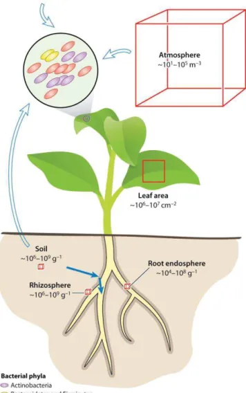
Figure 2: Plant microbiota and origins of plant-associated microbes (from Bulgarelli
et al., 2013)
Bacterial population sizes and phyllosphere community composition. Numbers correspond to estimations of number of bacterial cells in phyllosphere, atmosphere, rhizosphere, and root and soil bacterial communities. Open and solid arrows represent inoculation routes for the phyllosphere and root microbiota, respectively.
Figure 3: Microbial root colonization hotspots (From Liu et al., 2017)
Schematic representation of the bacterial distribution and colonization patterns of the endosphere of a plant root. The emerging sites of lateral roots are among the hotspots of bacterial colonization. Arrows represent the translocation of bacteria inside the xylem and phloem. Endophytic bacteria may engage in different life styles as depicted by different colored ovals.
13 these factors, so that plants recruit microbes from the soil to provide microbial functions beneficial in different contexts (Bakker et al., 2018).
Plant-associated microbial niches
Nonetheless, plants’ roots and their vicinity are not the only plant-associated niches for microbes to colonize. In fact, as humans, virtually all plants’ tissues are colonized by microbes. Consequently, several plant-associated microbial niches have been defined (Turner
et al., 2013) (Figure 2). The most studied niche is the rhizosphere (as defined in previous
paragraph) but the surface of plants’ tissues are also colonized, root surface is called the rhizoplane (Foster, 2003) while leaves’ surface is referred as the phyllosphere (Lindow and Brandl, 2003; Vorholt, 2012). Microorganisms colonizing the surface of plants are defined as epiphytes in opposition to endophytes which are able to colonize the interior of plants tissues, referred to as the endosphere. The concept of endophytes is a rather old one as it was firstly proposed in 1809 (Link, 1809), endophytic microbes were termed “entophytae” at this time. The first mention of endophytes is attributed to M.L.V. Galippe in 1887 (Compant et al., 2012), which proposed the pioneering idea that endophytes are soil-derived microorganisms. His work was extensively criticized in the 19th century following Pasteur’s view that a healthy organism is one free of any microbial colonization. This idea was since largely rejected (Partida-Martínez and Heil, 2011) as microbe-free or “axenic” organisms exhibit abnormal physiological functioning especially for nutrient uptake and immunity. Also, misleading ways to define endophytes have been used, in particular the fact that endophytes “are able to colonize inner tissues without causing any harm” (Wilson, 2006). In fact, endophytes can be pathogenic as well as non-pathogenic and each category harbor major similarities in terms of colonization processes and genomic repertoires (Brader et al., 2017). Among the diversity of endophytes, some are restricted to the interior of roots (“endorhizosphere”) while others are able to systemically colonize plants in the stems and leaves particularly by colonizing the xylem and being transported following the transpiration flow (Afzal et al., 2019; Compant et al., 2010) (Table 1, Figure 3).
Finally, the most recently defined niche is the spermosphere: indeed, some microbes are able to colonize seeds -both on the surface and the interior- and can also be vertically transmitted (Shade et al., 2017). The microbial diversity of these different niches has been studied through the defining of the core root microbiome of several accessions of Arabidopsis
14 Table 1: Non exhaustive list of bacterial and fungal endophytic strains with published genome
Strain Host plants PGP activities Reference
Bacterial endophytes
Azoarcus sp. BH72 Rice Nitrogen fixation Krause et al. (2006) Azospirillum
lipoferum 4B Rice, maize, wheat
Nitrogen fixation,
phytohormone secretion Wisniewski-Dyé et al. (2011) Azospirillum sp. B510 Rice Nitrogen fixation,
phytohormone secretion Kaneko et al. (2010) Burkholderia phytofirmans PsJN Potato, tomato, maize, barley, onion, canola, grapevine
IAA synthesis, ACC
deaminase Weilharter et al. (2011)
Burkholderia spp.
KJ006 Rice
ACC
deaminase, nif gene
cluster, antifungal action (indirect PGP)
Kwak et al. (2012) Enterobacter cloacae
ENHKU01 Pepper Unkwon role in PGP Liu et al. (2012) Enterobacter sp. 638 Poplar Siderophore, IAA,
Biocontrol Taghavi et al. (2009) Gluconacetobacter
diazotrophicus PaI5
Sugarcane, rice, Coffea, Camellia
Nitrogen fixation, auxin
synthesis Bertalan et al. (2009) Herbaspirillum
seropedicae SmR1 Rice
Nitrogen fixation, IAA synthesis, ACC deaminase
Pedrosa et al. (2011) Klebsiella
pneumoniae 342 Maize, wheat Nitrogen fixation Fouts et al. (2008)
Paraburkholderia
kururiensis M130 Rice
Nitrogen fixation, IAA synthesis, ACC deaminase
Coutinho et al (2013) Paraburkholderia
tropica Ppe8 Sugarcane Nitrogen fixation da Silva et al. (2018) Pseudomonas
putida W619 Poplar
IAA synthesis, ACC
deaminase Taghavi et al. (2009) Pseudomonas
stuzeri A1501 Rice Nitrogen fixation Yan et al. (2008)
Serratia
proteamaculans 568 Soybean
IAA synthesis, ACC
deaminase, Biocontrol Taghavi et al. (2009) Stenotrophomonas
maltophilia R551-3 Poplar
IAA synthesis, ACC
deaminase Taghavi et al. (2009) Paenibacillus
polymyxa M1 Wheat
NRPS and lipopeptide
synthesis Eastman et al. (2014)
Fungal endophytes
Epichloë festucae Pooideae grasses Alkaloïds synthesis Winter et al. (2018) Piriformospora indica Brassicaceae,
Barley Stress tolerance Zuccaro et al. (2011) Gaeumannomyces sp.
JS-464
Phragmites communis
Secondary metabolites
synthesis Kim et al. (2017) Trichoderma sp. Various plants Biocontrol Mukherjee et al. (2013)
15
al., 2015). The analysis of the relative abundance of core taxa revealed a bottleneck in the
global species richness from the rhizosphere to the endosphere. This observation was attributed to an increasing selective gradient from the rhizosphere to the endosphere (Bulgarelli et al., 2013) for microbes holding at the same time rhizoplane, rhizosphere and endosphere-competences. This can be extrapolated to the microbes able to colonize the interior of the above-ground part of the plants.
Different types of plant-microbe interactions
Within the different microbial niches previously introduced, microorganisms exhibiting various ecological interactions with plants can be found. Classically, three extreme categories of plant-associated microorganisms are defined: “pathogens”, “parasites” and “mutualists”. Also, we will here use the term “symbiosis” as defined by “the living together of two dissimilar organisms in intimate association or close union”. Therefore, in the broad sense, interactions ranging from mutualism, where both plant and microbe benefits, commensalism, where the microbe benefits while not negatively impacting the plant, to parasitism, where the microbes benefits at host’ expense, can all be considered as symbiotic (Figure 4) (Newton et
al., 2010). Another distinction can be established between parasites and pathogens, while the
latter can actively damage tissues and hijack its host physiology for their own trophic benefit, one could consider that parasites indirectly negatively impact their host by exploiting the niche of hosts’ tissues (Newton et al., 2010). While these definitions are useful, they are limited to have broad sense in the diversity of host-microbes interactions, particularly in the case of interactions changing categories throughout the microbe life cycle (Newton et al., 2010). Therefore, a “continuum” vision of plant-microbe interactions from detrimental to beneficial was proposed to accommodate recent findings and particular study cases (Hirsch, 2004). This statement has a special relevance in the light of the recent unraveling of the plant microbiome composition. Indeed, the concept of commensalism is central to the “metaorganism” vision of plant microbiome (Berg et al., 2016; Bosch and McFall-Ngai, 2011). Effectively, commensals can have direct or indirect effects in particular environmental conditions impacting host fitness consequently not being commensals anymore. The previously introduced idea that plants are in fact metaorganisms led to the concept of holobiont, referring as the unit of selection in evolution (Zilber-Rosenberg and Rosenberg, 2008). Historically, the understanding of plant-microbe interactions came from the analysis of plant-pathogen (Gabriel and Rolfe, 1990; Mansfield et al., 2012) and plant-mutualist models (Bonfante and Genre, 2010; Douglas Cook, 1999). This can be due to the economic
16
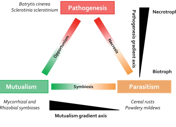
Figure 4: Trophic space occupied by microorganisms in association with plants (adapted from Newton et
al., 2010)
The range of trophic relationships of example microbe–plant associations is represented as space occupied between the three key trophic states of pathogenicity, mutualism and parasitism at the corners of the triangle. The vertical axis represents a gradient of pathogenesis from necrotrophy to biotrophy. The horizontal axis represents a gradient from mutualism to parasitism for symbiotic relationships.
17 importance of such interactions -in particular for plant diseases-. Another major reason probably is the strong phenotypes that can be relatively easily analyzed in such models. Indeed, this statement can be argued in opposition to the discrete and highly context-dependent phenotypes driven by other members of the plant microbiome.
Plant-beneficial microbial interactions Main plant-mutualist models overview
As previously stated, the first extensively models used to study the interactions between plant and beneficial microbes are mutualistic ones. The most important, in terms of evolutionary, ecological and economical relevance is the plant-mycorrhiza model. The name of this symbiosis (from the Greek “mycos”, meaning fungus and “rhiza” meaning root) clearly states that this association give rise to a new organism composed of both the fungus and the root. It is, indeed, the most widespread plant-microbe symbiosis as 70-90% of land plants are associated to mycorrhiza (Parniske, 2008) and also the most ancient, as land plants ancestors were apparently associated with similar fungal structures (Remy et al., 1994). This is most likely due to the fact that such association was needed for land colonization by plants (Selosse and Le Tacon, 1998), indeed these fungi provide additional potential of nutrients and water uptake for land plants (Harley, 1989). Probably the most crucial role of mycorrhiza for plants is their help for the uptake of phosphorus (P) from soil. This element is an essential plant nutrient and one of the most limiting, especially in soil. Indeed, roots can only uptake inorganic P directly while this form is usually in minority in soils and therefore not available for plants (Hinsinger, 2001). As they can transform P in chemical forms available for plants, mycorrhiza are highly crucial for plant nutrition (López-Arredondo et al., 2014). Two types of mycorrhiza can be distinguished: (i) Ectomycorrhiza, mainly found in association with trees in temperate forests, remains outside of plant cells. (ii) Endomycorrhiza, for which, part of the fungal hyphae is endocellular although a double membrane is formed by the invagination of plant cell membrane (Parniske, 2008). In the case of the most largely distributed endomycorrhiza, arbuscular mycorrhiza (AM), the formation of so-called “arbuscules” enables efficient nutrient exchanges between host and symbiont.
The second most extensively studied plant-mutualist model, the legume-rhizobium symbiosis, also implies endocellular colonization and improves nutrient availability for plants (Oldroyd
et al., 2011). In this case, the symbionts are Proteobacteria (from alpha and beta-subclasses)
18
Figure 5: Schema illustrating the common symbiosis signalling pathway (CSSP) (From Barker et al., 2017) A number of plant genes and secondary messengers are required for the successful functioning of the conserved CSSP core module, first discovered in the model legume species Lotus japonicus (Lj) and Medicago truncatula (Mt), and now extended to plant hosts of all known root endosymbiotic associations including the rhizobial/legume, arbuscular mycorrhizal (AM) and actinorhizal symbioses. In the case of the rhizobial and AM symbioses the CSSP is activated following symbiotic signal perception (NF, Nod factors; MF, Myc factors) by plasma membrane (PM) localized LysM-RLK receptors, most probably part of a larger complex including LjSYMRK/MtDMI2. Following signal transduction from the PM to the nucleus, nuclear membrane cation channels known as LjCASTOR/LjPOLLUX/MtDMI1, likely in association with cyclic nucleotide gated-calcium channel complex (CNGC) required for rapid Ca2+ release and the initiation of nucleoplasmic Ca2+ spiking. The subsequent decoding of the intranuclear Ca2+ oscillatory response involves two key associated components (LjCCaMK/MtDMI3 and LjCYCLOPS/MtIPD3). Binding of Ca2+ to the calcium and calmodulin-dependent kinase CCaMK leads to phosphorylation of the coiled-coil protein CYCLOPS. Finally, the activation of a downstream signalling cascade via a repertoire of GRAS/ERF transcription factors results in the synthesis of the suite of proteins required for the transcriptional re-modelling of the epidermal cell in preparation for apoplastic infection. In the case of the N-fixing actinorhizal association, the nature of both the Frankia signal (AF, actinorhizal factor) and the corresponding host receptor remain to be determined, and that direct evidence for an essential role in microbial/host signalling has only been demonstrated so far for CgSYMRK and CgCCaMK, although orthologues for both CASTOR/POLLUX/DMI1 and CYCLOPS/IPD3 have been found in Casuarina glauca.
19 nutrition. This implies the formation of specialized organs, called “nodules”, which are inhabited by the rhizobial cells (Brewin, 1991). This organ provides the required environment for the so-called “fixation of atmospheric N2” by the bacterial population.
Extensive years of research enabled to deeply appreciate the underlying molecular and developmental processes implicated in the establishment of these two mutualistic symbioses (Oldroyd, 2013) (Figure 5). The first step is the perception by microbes of plant-secreted flavonoids (Abdel-Lateif et al., 2012) which will trigger the production of symbiotic signals by microbes. So called Nod and Myc factors which are (Lipo-)chitooligosaccharides derivatives (Gough and Cullimore, 2011) will be in turn recognize by the plant through receptor-like kinases receptor molecules of the LysM and SYMRK family (Gust et al., 2012; Oldroyd, 2013). This molecular interaction will lead, through yet unclear mechanisms, to an essential symbiotic nuclear calcium oscillation dependent on cation channels located on the nuclear membranes. This will trigger transcriptional reprogramming leading to developmental processes which enable the symbionts endocellular accommodation through the formation of specialized organs: nodules, arbuscules and actinorhizal roots. Of particular interest is the fact that a large part of the genes implicated in the previously described molecular events are highly conserved among plant-microbe symbiosis (Figure 5). This symbiotic "toolkit" is referred as the common symbiosis signaling pathway. Some of these genes were even already present, for the early signaling steps, in the algal ancestor of land plants (Delaux et al., 2015).
Plant-microbe associative symbioses
These mutualistic plant-microbe symbioses can be considered as highly “advanced” ones, as undoubtedly, it is the result of a long-term co-evolution process (Martínez-Romero, 2009). This could not have happened if very important benefits were not shared by both partners. Although it is less clear for the mycorrhizal symbiosis, as P is commonly not accessible to land plants, it is very clear for rhizobial symbiosis, which appears to be an “extreme example”. Indeed, the biological nitrogen fixation process is very costly in terms of required energy (Atkins, 1984), therefore, if plants can fulfill their N nutrition “on their own”, they would have “no interest” in the investment needed for the establishment of an “efficient symbiosis”. Another fact supporting this view is that the availability of N negatively impacts the beneficial effects of this mutualistic association for the plant (Heath and Tiffin, 2007). However, in contrast to these two classical cases, numerous other examples of beneficial plant-microbe examples exist and have also been extensively studied.
20 Table 2 : Non-exhaustive list of microbial taxons comprising Plant Growth Promoting Microbes (From Ahemad et al., 2014, Hayat et al., 2010, Hossain et al., 2017 and Jousset, 2017)
PGPR taxons Genus PGPF taxon Genus Protists taxon Genus
Proteobacteria Acetobacter Ascomycota Aspergillus Alveolata Tetrahymena Achromobacter Aureobasidium Rhizaria Cercomonas
Acinetobacter Chaetomium Heteromita
Azoarcus Cladosporium Euglenozoa Bodo Azomonas Colletotrichum Excavata Jakoba
Azospirillum Exophiala Amoebozoa Acanthamoeba Bradyrhizobium Penicillium Oomycota Phytophthora
Brevundimonas Trichoderma Pythium
Burkholderia Fusarium Citrobacter Gliocladium Delftia Phoma Enterobacter Phomopsis Erwinia Purpureocillium Gluconacetobacter Talaromyces Herbaspirillum Basidiomycota Limonomyces
Klebsiella Rhodotorula
Kluyvera Rhizoctonia
Mesorhizobium Piriformospora Ochrobactrum Zygomycota Mucor
Pantoae Rhizopus Paraburkholderia Phyllobacterium Pseudomonas Psychrobacter Rahnella Rhizobium Serratia Sinorhizobium Sphingomonas Stenotrophomonas Variovorax Xanthomonas Bacteroidetes Flavobacterium Actinobacteria Arthrobacter Cellulomonas Mycobacterium Rhodococcus Firmicutes Bacillus Brevibacillus Paenibacillus
21 All of them have been regrouped under the name of associative symbioses or cooperations (Barea et al., 2005). These interactions are defined as facultative, exhibit large host and symbionts spectrum and do not require the formation of specialized organs (Hayat et al., 2010) in clear opposition to the previously introduced plant-microbe mutualistic interactions. In terms of phylums, multiple clades of microbes can be implicated in such interactions : The most studied ones are bacteria (Hayat et al., 2010), fungi (Hossain et al., 2017) and protists (Gao et al., 2019) (Table 2). However, the possibility to identify plant-beneficial oomycetes, archeabacteria and even viruses can't be rule out as such organisms are also important parts of the plant microbiome. Although facultative beneficial microbes can inhabit the phyllosphere, most studies focused on root and rhizosphere microbiome. In order to exhibit beneficial effects while interacting with plants, microbes must firstly harbor specific traits to interact with plants. A number of functions have been linked to rhizosphere competence, for example: motility, attachment, stress resistance, production of secondary metabolites (Barret et al., 2011; Compant et al., 2010). However harboring these functions is not sufficient to exhibit strong beneficial effects in plants. A wide diversity of functions and activities has been described as "plant-growth promoting traits". Their identification mainly emanates from the study of so-called Plant-Growth Promoting Rhizobacteria (PGPR), an expression coined by J.W. Kloepper in the late 1970s (Kloepper and Schroth, 1978; Suslow et al., 1979). Other terms followed this first one: Plant-Growth Promoting Bacteria (PGPB), Plant-Growth Promoting Fungi (PGPF). It is, however, interesting to recognize that the concept of "bacterial fertilization" have already been extensively studied and applied in the soviet union in the 1950s (Brown, 1974).
Plant Growth-Promoting traits
PGP traits can be defined as any biochemical activity known to improve plant growth also encompassing the increased tolerance to diseases (Glick, 2012; Lugtenberg and Kamilova, 2009). Two main categories of PGP traits can be distinguished: Those having a direct impact on plant growth and those having indirect ones. This distinction is based on the fact that the indirect mechanisms exhibit growth promotion exclusively in the context of detrimental biotic interactions (pathogens, parasites, herbivores for example) (Figure 6).
The most studied process is probably the direct improvement of plant nutrition especially for the uptake of N, P and Fe although the nutrition in other (micro-)nutrients can also be
22 Figure 6: Mechanisms of Plant Growth Promoting Microbes in enhancing plant growth (Adapted from Nazir et al., 2018)
Schematic representation of plant growth promoting traits separated between direct (green box) and indirect effects (blue and red boxes), the latter being dependent on the presence of pathogens. In exchange to these growth promotions microbial functions, plants provide photosynthates typically through root exudation. ACC: 1-aminocyclopropane-1-carboxylate, P: Phosphate, ISR : Induced Systemic Resistance