HAL Id: hal-01478354
https://hal.archives-ouvertes.fr/hal-01478354
Submitted on 8 Oct 2018
HAL is a multi-disciplinary open access
archive for the deposit and dissemination of
sci-entific research documents, whether they are
pub-lished or not. The documents may come from
teaching and research institutions in France or
abroad, or from public or private research centers.
L’archive ouverte pluridisciplinaire HAL, est
destinée au dépôt et à la diffusion de documents
scientifiques de niveau recherche, publiés ou non,
émanant des établissements d’enseignement et de
recherche français ou étrangers, des laboratoires
publics ou privés.
Distributed under a Creative Commons Attribution| 4.0 International License
translocation of GLUT4 to the cell surface which is
maintained in insulin resistance
Marion Berenguer, Laurène Martinez, Sophie Giorgetti-Peraldi, Yannick Le
Marchand-Brustel, Roland Govers
To cite this version:
Marion Berenguer, Laurène Martinez, Sophie Giorgetti-Peraldi, Yannick Le Marchand-Brustel, Roland
Govers. A serum factor induces insulin-independent translocation of GLUT4 to the cell surface which
is maintained in insulin resistance. PLoS ONE, Public Library of Science, 2010, 5 (12), pp.e15560.
�10.1371/journal.pone.0015560�. �hal-01478354�
A Serum Factor Induces Insulin-Independent
Translocation of GLUT4 to the Cell Surface which Is
Maintained in Insulin Resistance
Marion Berenguer1,3, Laure`ne Martinez1,3, Sophie Giorgetti-Peraldi2,3, Yannick Le Marchand-Brustel1,2,3, Roland Govers1,3*
1 INSERM, U895, Mediterranean Research Center for Molecular Medicine (C3M), Avenir Team 9, Nice, France, 2 INSERM, U895, Mediterranean Research Center for Molecular Medicine (C3M), Team 7, Nice, France,3 University of Nice-Sophia-Antipolis, Faculty of Medicine, Signaling and pathologies (IFR50), Nice, France
Abstract
In response to insulin, glucose transporter GLUT4 translocates from intracellular compartments towards the plasma membrane where it enhances cellular glucose uptake. Here, we show that sera from various species contain a factor that dose-dependently induces GLUT4 translocation and glucose uptake in 3T3-L1 adipocytes, human adipocytes, myoblasts and myotubes. Notably, the effect of this factor on GLUT4 is fully maintained in insulin-resistant cells. Our studies demonstrate that the serum-induced increase in cell surface GLUT4 levels is not due to inhibition of its internalization and is not mediated by insulin, PDGF, IGF-1, or HGF. Similarly to insulin, serum also augments cell surface levels of GLUT1 and TfR. Remarkably, the acute effect of serum on GLUT4 is largely additive to that of insulin, while it also sensitizes the cells to insulin. In accordance with these findings, serum does not appear to activate the same repertoire of downstream signaling molecules that are implicated in insulin-induced GLUT4 translocation. We conclude that in addition to insulin, at least one other biological proteinaceous factor exists that contributes to GLUT4 regulation and still functions in insulin resistance. The challenge now is to identify this factor.
Citation: Berenguer M, Martinez L, Giorgetti-Peraldi S, Le Marchand-Brustel Y, Govers R (2010) A Serum Factor Induces Insulin-Independent Translocation of GLUT4 to the Cell Surface which Is Maintained in Insulin Resistance. PLoS ONE 5(12): e15560. doi:10.1371/journal.pone.0015560
Editor: Jaswinder K. Sethi, University of Cambridge, United Kingdom
Received September 10, 2010; Accepted November 12, 2010; Published December 20, 2010
Copyright: ß 2010 Berenguer et al. This is an open-access article distributed under the terms of the Creative Commons Attribution License, which permits unrestricted use, distribution, and reproduction in any medium, provided the original author and source are credited.
Funding: This work was supported by the Institut National de la Sante´ et de la Recherche Me´dicale (INSERM), the University of Nice-Sophia-Antipolis, the Conseil Re´gional Provence-Alpes-Coˆte d’Azur, the Conseil Ge´ne´ral des Alpes Maritimes, and an Alfediam-Roche Diagnostics award. RG is a recipient of an INSERM Avenir grant and LM is supported by an INSERM/Re´gion Provence-Alpes-Coˆte d’Azur/Servier grant. The funders had no role in study design, data collection and analysis, decision to publish, or preparation of the manuscript.
Competing Interests: The authors have declared that no competing interests exist. * E-mail: govers@unice.fr
Introduction
GLUT4 is the principle glucose transporter that is responsible for the insulin-induced uptake of glucose by muscle and adipose tissue after a meal. The main characteristic feature of GLUT4 is the absence of cell surface recycling in non-stimulated cells [1]. This implicates the presence of a highly efficient cellular mechanism that retains GLUT4 intracellularly. How this retention is organized and which molecules are implicated is currently unknown. It has been postulated that the non-endosomal GLUT4 pool plays a major role in intracellular retention [2]. Nevertheless, albeit to a lesser extent, endosomal GLUT4 has been demon-strated to be retained intracellularly and responsive to insulin [3,4]. In accordance, GLUT1 and the transferrin receptor (TfR), both localized exclusively in endosomes, also translocate to the plasma membrane upon insulin stimulation [2,5]. GLUT4 is regulated by insulin at various levels. Insulin signaling reduces GLUT4 retention allowing GLUT4 to move towards the cell periphery [6], increases its endosomal recycling [7], and enhances its docking and fusion with the plasma membrane [8]. On the other hand, GLUT4 internalization is hardly regulated by insulin, at least in adipocytes [4,9]. Despite the fact that many players in insulin signaling and GLUT4 traffic are known, it remains elusive how these two pathways intercommunicate.
In insulin resistance, a condition related to type 2 diabetes, insulin no longer leads to an efficient translocation of GLUT4 towards the cell surface. Insulin resistance has been associated with a number of cellular phenomena which are likely to be linked. First, the level of reactive oxygen species (ROS) is increased under conditions of insulin resistance, while antioxi-dants are able to ameliorate insulin sensitivity and glucose uptake [10,11]. Second, insulin resistance has been associated with a reduction in the phosphorylation (activity) of insulin signaling molecules [12,13]. Third, increases in O-GlcNAc modification of proteins is linked to insulin resistance [14]. Notably, PKB, IRS-1, munc18c, as well as GLUT4 itself were found to be modified by O-GlcNAcylation [15,16]. Fourth, in insulin resistance, expres-sion levels of molecules implicated in insulin-induced GLUT4 translocation are decreased [17,18]. Finally, insulin resistance has been associated with a change in the intracellular localization of GLUT4 [19,20].
Taken together, multiple cellular mechanisms contribute to the development of insulin resistance and the associated reduction in GLUT4-mediated glucose uptake. It would be therapeutically relevant to discover novel ways to increase cell surface GLUT4 levels. Here, we present evidence for the existence of a putative factor in serum that displays an insulin-like effect regarding GLUT4 translocation and cellular glucose uptake. Remarkably,
the effect of this factor on GLUT4 is additive to the action of insulin and is fully maintained in insulin resistance.
Materials and Methods Materials
3T3-L1 preadipocytes were obtained from ATCC/LGC Standards (Teddington, UK). Plat-E cells were generously provided by Dr Toshio Kitamura (University of Tokyo, Japan). Bovine and human sera were from PAA (Pasching, Austria). Rabbit serum was withdrawn from New Zealand rabbits. Media and HEPES were from Invitrogen (Carlsbad, CA), insulin from Lilly (Suresnes, France), PDGF-BB, IGF-1, and HGF from PeproTech (Rocky Hill, NJ), and 2-[3H]deoxyglucose from PerkinElmer Life (Waltham, MA). Monoclonal anti-HA antibody was from Covance (Emeryville, CA), mouse IgG antibody from Sigma-Aldrich (St. Louis, MO), polyclonal GLUT4, GLUT1, and insulin receptor (IRb) antibodies and HRP-conjugated secondary antibodies from Santa Cruz (Santa Cruz, CA), antibodies against PAS, ERK, phospho-ERK, Akt, phospho-Akt (T308 and S473), AMPK, phospho-AMPK, and phosphotyrosine from Cell Signal-ing Technology (Danvers, MA), anti-AS160 from AbCam (Cam-bridge, MA), and anti-phospho-AS160, anti-TfR and fluorescent antibodies from Invitrogen. IRS-1 antibody has been described before [21]. Polyclonal antibody against syntaxin 13 was generously provided by Dr Rytis Prekeris (University of Colorado, Denver, CO). Insulin ELISA kit was purchased from Spi-Bio (Montigny le Bretonneux, France) and inhibitors from Sigma (St. Louis, MO; wortmannin, LY294002, genistein), Calbiochem/ Merck (Nottingham, UK; AG1024, Compound C, Akti 1/2, Ro 31-8220, U-73122, okadaic acid, U0126, rapamycin) and Alexis Biochemicals/Enzo Life Sciences (Plymouth Meeting, PA; herbi-mycin A, staurosporine). All chromatography materials were from Sigma. pBABE vector was kindly provided by Dr Hartmut Land (University of Rochester, Rochester, NY) and GLUT1 cDNA by Dr Mike Mueckler (Washington University, St. Louis, MO).
Molecular Biology
The cDNAs encoding HA-GLUT4 (GLUT4 with an HA epitope tag in its first luminal domain) and HA-TfR (TfR with an HA tag at its C-terminus) inserted in pBABE-puro vector have been described elsewhere [22,23]. A pBABE vector containing the cDNA encoding human GLUT1 with an HA epitope tag in its first luminal domain between residues 58 and 59 was constructed by PCR using the oligos 59-GATCGACTAGGGTCCATAGATA- CGGAGAATCAATATTACCAGAGATCGATTATCCGTAC- GATGTTCCTGATTATGCTGAGACCACGCTCACCACGC-TCTGG and 39-GATCGTCGACCTCGAGTCACACTTGG-GAATCAGCCCC. The sequence of the PCR parts was verified by sequence analysis (Cogenics/Beckman Coulter).
Cell Culture
Preadipocytes were cultured and differentiated as described before [23]. Differentiated cultures contained at least 95% adipocytes. To express HA-tagged molecules in adipocytes, preadipocytes were infected with retrovirus as described before [22], except that Plat-E cells were used for the production of virus [24]. L6 myoblasts were infected with retrovirus for the expression of HA-GLUT4 and cultured and differentiated as described previously [22,25].
Pervanadate was freshly prepared by combining 639ml PBS, 300ml 100 mM sodium vanadate and 61ml 3% H2O2. After
15 minutes, the pervanadate was used at a concentration of 100mM [26].
For the induction of insulin resistance, a previously described procedure was used [27], except that adipocytes were incubated for 24 hr with 100 nM insulin.
Human adipocytes were cultured and differentiated as de-scribed elsewhere [21].
Fluorescence-based techniques
The fluorescence-based assay for the detection of cell surface GLUT4 levels and the method to measure GLUT4 internalization have been described previously [22,28]. In morphological studies, cells were analyzed using a Zeiss LSM 510 confocal laser scanning microscope (Carl Zeiss, Go¨ttingen, Germany) in the C3M Cell Imaging Facility MICA.
Glucose uptake
Adipocytes, grown in gelatin-coated 12 well plates, were incubated for 2 hours in DMEM with 0.2% BSA and for 5 minutes in KRP (12.5 mM HEPES pH 7.4, 120 mM NaCl, 6 mM KCl, 1.2 mM MgSO4, 1 mM CaCl2, 0.4 mM NaH2PO4,
0.6 mM Na2HPO4). Cells were treated or not with insulin or
serum for 20 minutes. To control cells, 50mM cytochalasin B was added (assay background). Cells were incubated for 3 minutes with 0.1 mM 2-[3H]deoxyglucose (0.28mCi/well), extensively washed with ice-cold phosphate-buffered saline (PBS), and lysed in 1% Triton X-100. Radioactivity was measured by scintillation counting. To be able to compare serum-induced with insulin-induced 2-DOG uptake, either unlabeled glucose was added to the insulin incubations (Figure 2) or sera were dialyzed (Figure 3) in order to have similar glucose concentrations during the uptake.
Immunoblotting and immunopurification
3T3-L1 adipocytes were serum-starved for 2 h, incubated for 5 minutes with or without 100 nM insulin or 25% FBS and lysed in ice-cold lysis buffer [29]. For immunopurification (IP), protein A agarose beads (Roche Diagnostics, Meylan, France) were incu-bated for 1 hr with 5mg of IRS-1 or IR antibodies at room temperature and for 16 hr with 1 mg of adipocyte lysate at 4uC, subsequently. For control IPs, antibody-bound beads were incubated in the absence of lysate. HRP-conjugated secondary antibodies were visualized using chemiluminescence reagent (Roche Diagnostics) and a CCD camera-based imager (LAS-3000, Fujifilm; St. Quentin en Yvelines, France). Relative intensities were quantitated using MultiGauge software (Fujifilm).
Column chromatography
Human serum (0.5 ml) was fractionated on a 24 ml Sephacryl 200-HR column using an elution buffer consisting of 150 mM NaCl and 10 mM tris pH 7.4 and a flow rate of 5.0 ml/hr. Calibration markers blue dextran (void volume; 2000 kDa), amylase (200 kDa), alcohol dehydrogenase (150 kDa), bovine serum albumin (67 kDa), ovalbumine (43 kDa), chymotrypsinogen (26 kDa), RNase A (14 kDa) and 2-[3H]deoxyglucose (0.2 kDa) were fractionated using identical volumes and flow rates. Serum fractions (0.75 ml) were concentrated 5 times using 3K Amicon Ultra concentrators before subjecting to the fluorescence-based GLUT4 assay described above.
Statistics
All data are presented as average 6 SD. Experiments were performed at least three times. Representative experiments are shown. Comparisons between data sets were evaluated using two-tailed Student’s t-tests and comparisons between dose-response curves were evaluated using nonlinear four-parameter sigmoidal
dose - response curve fittings and F-tests (Graphpad Prism software). Differences between data sets were considered statisti-cally different when P,0.05.
Results
Serum induces the translocation of GLUT4 towards the cell surface
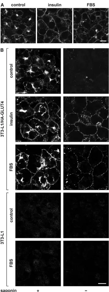
Insulin is the master regulator of glucose homeostasis via its action on intracellular GLUT4 traffic. To test whether there exist other factors in serum that regulate cell surface GLUT4 levels, 3T3-L1 adipocytes were incubated for 20 minutes with 100 nM insulin, 50% fetal bovine serum (FBS), or left untreated. Cells were subsequently immunolabeled with anti-GLUT4 antibody, and analyzed by microscopy (Figure 1A). We observed that both insulin and serum induced the translocation of endogenous GLUT4 towards the cell surface. As this labeling does not distinguish between GLUT4 molecules localized at the cell surface or just beneath, we expressed ectopic GLUT4 in 3T3-L1 adipocytes bearing an HA-epitope tag within its first extracellular domain. Permeabilized and intact cells were immunolabeled with anti-HA antibody (Figure 1B). The permeabilized adipocytes (left panels) showed that both insulin and FBS increased the appearance of HA-GLUT4 near or at the plasma membrane, while the non-permeabilized cells (right panels) clearly demon-strated an increase in the amounts of GLUT4 at the cell surface upon stimulation with insulin or FBS. The absence of signal in control adipocytes (bottom panels), demonstrated that the immunolabel in FBS-treated HA-GLUT4-expressing adipocytes was specific.
Quantitative analysis of serum-induced GLUT4 translocation and glucose uptake
Next, we measured GLUT4 translocation quantitatively, using an assay that is based on the culture and labeling of HA-GLUT4-expressing adipocytes in 96 well plates [4]. Cells were processed as described above but fluorescence was analyzed using a fluorescence microplate reader. This revealed that the effect of FBS on cell surface GLUT4 levels was concentration-dependent (Figure 2A). To determine whether the FBS-induced translocation of GLUT4 was accompanied by an increase in glucose transport across the plasma membrane, cellular glucose uptake was measured (Figure 2B). Similarly to its effect on GLUT4 translocation, FBS induced glucose uptake in a concentration-dependent manner. The arrows in the left panels indicate the concentration of insulin in undiluted serum, demonstrating that the observed effects of FBS were not mediated by insulin (see also Table S1). Remarkably, compared with insulin, the increase in cell surface GLUT4 levels in response to FBS was accompanied by a relatively small increase in glucose uptake. This could be due to a smaller effect of FBS on the activity of GLUT4 [30,31]. Stimulating the cells for various times periods demonstrated that the smaller effect of FBS on glucose transport was independent of the length of stimulation (Figure S1).
Figure 1. Effect of serum on the intracellular localization of GLUT4. 3T3-L1 adipocytes (A) or HA-GLUT4-expressing adipocytes (B) were incubated for 20 minutes with 100 nM insulin, 50% FBS, or left
untreated (control). Upon fixation, cells were immunolabeled with anti-GLUT4 antibody to label endogenous anti-GLUT4 (A) or with anti-HA in the absence (right panels) or presence of saponin (left panels) to label HA-GLUT4 at the cell surface or total cellular HA-HA-GLUT4, respectively (B). Control adipocytes that did not express HA-GLUT4 were used to analyse the specificity of the anti-HA labeling (4 lower panels in B). Bar, 10 mm. doi:10.1371/journal.pone.0015560.g001
The effect of serum on GLUT4 is not due to inhibition of its internalization and is not mediated by PDGF, IGF-1, or HGF
To establish whether the effect of FBS on GLUT4 was due to an increase in exocytosis or to a reduction in internalization, GLUT4 internalization was measured (Figure S2). In brief, HA-GLUT4-expressing adipocytes were incubated with insulin to increase cell surface GLUT4 levels, cooled down on ice, acid stripped to remove insulin, incubated with anti-HA antibody, and washed to remove non-bound antibody. Upon transfer to 37uC, the cells were incubated with or without 25% FBS to allow the antibody to internalize. Following anti-HA immuno-labeling, the cells were scored for the presence of internalized label. Unexpectedly, we observed that FBS increased the GLUT4 internalization rate. Therefore, the positive effect of FBS on cell surface GLUT4 levels must be due to an increased exocytosis rate. Control adipocytes that were incubated with 0.45 M sucrose displayed a large reduction in internalization rate [32].
To determine whether the GLUT4-translocating activity of serum was due to the presence of factors that have previously been
shown to alter cell surface GLUT4 levels or glucose transport under certain conditions, we tested platelet-derived growth factor (PDGF; [33]), insulin-like growth factor-1 (IGF-1; [34]), and hepatocyte growth factor (HGF; [35]) in our cell system (Figure S3). None of these factors increased cell surface GLUT4 levels (Figure S3A), though all factors activated signal transduction pathways (Figure S3B).
The effect of serum on GLUT4 is independent of serum origin and cell type
To determine whether the translocation-inducing serum factor was exclusively present in FBS, we also studied newborn bovine, adult bovine, rabbit and human serum (Figure 3A–D). We observed that in 3T3-L1 adipocytes, all tested sera induced GLUT4 translocation and glucose uptake that could not be accounted for by serum insulin levels (Table S1).
To test whether the action of serum on GLUT4 was dependent on cell type, human adipocytes, rat myoblasts, and rat myotubes were investigated. In in vitro differentiated human adipocytes, FBS and human serum induced an insulin-independent increase in glucose uptake (Figure 3E). In L6 myoblasts and myotubes, both insulin and FBS acutely increased the amount of HA-GLUT4 at the plasma membrane (Figure 3F and G). For both muscle cell models, the ED50 values for insulin and FBS were around 5 nM and 1%, respectively (data not shown). This indicated that, compared with 3T3-L1 adipocytes, these cells were even more sensitive to FBS and that also in these cells FBS induced GLUT4 translocation independent of insulin.
Serum augments also the cell surface levels of GLUT1 and the transferrin receptor (TfR), but only for GLUT4 the increase is additive to the effect of insulin
As insulin also increases the amount of GLUT1 and TfR at the plasma membrane, we investigated whether serum would have a similar effect. Therefore, we analyzed the kinetics of the appearance of GLUT4, GLUT1, and TfR at the cell surface in response to insulin, FBS, or both (Figure 4). As for GLUT4, GLUT1 and TfR proteins were studied that contained an HA epitope tag within their extracellular domain. In particular for GLUT4 and GLUT1, overexpression levels of these HA-tagged molecules were moderate (Figure 4A). Colocalization studies with syntaxin 13 showed that the intracellular localization of HA-tagged GLUT1 did not differ from that of endogenous GLUT1 (Figure S4). While insulin-increased cell surface GLUT4 levels were relatively stable, FBS led to a somewhat transient increase in GLUT4 at the plasma membrane, reaching maximal levels after 7–10 minutes (Figure 4B). Remarkably, the effects of insulin and FBS were largely additive and moreover, the additive effect of FBS did not decline throughout the duration of the experiment. The effect of FBS was not specific to GLUT4 as GLUT1 and TfR levels were also increased at the plasma membrane upon FBS stimulation (Figure 4C and D). An effect of serum on GLUT1 has been demonstrated before [36]. While GLUT1 and TfR were also sensitive to insulin stimulation, in accordance with previous studies [5], the effects of FBS and insulin were not additive. Treatment of HA-GLUT4-expressing adipocytes with various concentrations of insulin in the absence and presence of FBS demonstrated that along the entire insulin concentration range, FBS increased cell surface GLUT4 levels (Figure 4E). These data suggest that the insulin and FBS signaling pathways leading to increases in cell surface GLUT4 levels are largely distinct. Moreover, FBS sensitized the adipocytes to insulin, in
Figure 2. Concentration-dependent GLUT4 translocation and glucose uptake in 3T3-L1 adipocytes in response to insulin and FBS. (A) HA-GLUT4-expressing 3T3-L1 adipocytes cultured in 96 well plates were incubated with the indicated concentrations of insulin (left panel) or FBS (right panel), after which the cells were fixed, incubated with or without saponin, and quantitatively immunolabeled for HA signal as described in Experimental. The amounts of GLUT4 at the plasma membrane (non-permeabilized cells) was expressed as percent-age of total cellular GLUT4 levels (permeabilized cells). (B) 3T3-L1 adipocytes were incubated for 20 minutes with the indicated concen-trations of insulin (left panel) or FBS (right panel), followed by a three minute incubation with radiolabeled 2-DOG. Cellular 2-DOG uptake was determined for each condition and expressed as percentage of glucose uptake in the presence of saturating concentrations of insulin (100 nM). Arrows in left panels indicate the concentration of insulin in undiluted FBS, demonstrating that the effects of FBS are not mediated by insulin. doi:10.1371/journal.pone.0015560.g002
that the insulin dose-response curve shifted leftwards concomitant with a significant reduction in EC50 of 0.57 nM to 0.19 nM (Figure 4F).
Figure 3. The effect of serum on GLUT4 is independent of serum origin and cell type. (A) 3T3-L1 adipocytes were incubated for 20 minutes with the indicated concentrations of fetal, newborn, and adult bovine serum and cell surface GLUT4 levels were determined. (B) 3T3-L1 adipocytes were incubated for 20 minutes with 100 nM insulin, 50% serum or left untreated and cellular glucose uptake was measured and expressed as percentage of insulin-stimulated glucose uptake. (C,
D) Rabbit and human sera were analyzed as under (A) and (B). (E) Human adipocytes were incubated for 20 minutes with various concentrations of insulin, FBS, or human serum (HS) and glucose
Figure 4. The effect of FBS is additive to that of insulin and is not limited to GLUT4. (A) Lysates of 3T3-L1 adipocytes expressing HA-GLUT4, HA-GLUT1, or HA-TfR, and control adipocytes were subjected to SDS-PAGE and immunoblotted using the indicated antibodies. 3T3-L1 adipocytes expressing HA-GLUT4 (B), HA-GLUT1 (C), or HA-TfR (D) were stimulated for various times with 100 nM insulin, 25% FBS or both and relative cell surface GLUT4 levels were determined. (E) Adipocytes were incubated for 20 minutes with the indicated concentrations of insulin in the absence or presence of 25% FBS, and relative cell surface GLUT4 levels were determined. (F) Data from (E) were transformed in dose-response curves in which the effects of the various insulin concentrations were related to the maximal insulin effects (difference between 0 and 100 nM insulin) in the absence and presence of FBS. EC50 of insulin in absence and presence of FBS is 0.57 and 0.19 nM insulin, respectively. P,0.0001.
doi:10.1371/journal.pone.0015560.g004
uptake was measured and expressed as percentage of maximal glucose uptake in response to insulin. (F,G) HA-GLUT4-expressing myoblasts (F) and myotubes (G) were stimulated with either 100 nM insulin or 25% FBS and cell surface GLUT4 levels were determined. Serum insulin concentrations are depicted in Table S1.
doi:10.1371/journal.pone.0015560.g003
Investigation of the additive effects of serum and insulin demonstrates important differences in signaling
In order to obtain evidence for differences in insulin- and serum-induced signaling leading to the translocation of GLUT4, we analyzed their effect on cell surface GLUT4 levels in cells exposed to hyperosmolarity, protein tyrosine phosphatase inhibitor pervanadate, and AMPK activator AICAR (Figure 5). These three conditions have been shown to increase cell surface GLUT4 levels in 3T3-L1 adipocytes, L6 myoblasts, and/or L6 myotubes [26,37,38]. Hyperosmolarity (0.45 M sucrose; [32]) modestly but significantly increased cell surface GLUT4 levels in the absence and presence of insulin and FBS (Figure 5A). While the pervanadate-induced increase in cell surface GLUT4 levels could not be further enhanced by insulin, the effects of pervanadate and FBS were partially additive (Figure 5B). AICAR did not increase the amount of GLUT4 at the cell surface in unstimulated and insulin-stimulated cells (Figure 5C). However, the effect of FBS on GLUT4, in the absence as well in the presence of insulin, was significantly enhanced by AICAR. This confirmed that the signaling pathways that are activated by insulin and FBS and lead to GLUT4 translocation are likely to be distinct.
The serum factor that induces GLUT4 translocation activates signaling pathways that are distinct from those activated by insulin
We next sought further proof that the two signaling pathways are indeed distinct. To this aim, we applied two approaches. In the first approach, we investigated insulin- and serum-induced GLUT4 translocation in the presence of inhibitors of various signaling molecules that are part of the insulin signal transduction cascade (Figure 6A). These compounds inhibit insulin receptor tyrosine kinase activity (AG1024; [39], PI 3-kinase (wortmannin and LY294002; [40]), serine/threonine kinases (staurosporine; [41]), tyrosine kinases (genistein and herbimycin A; [42]), Akt (Akti-1/2; [43]), PKC (Ro 31-8220; [44]), AMPK (compound C; [45]), MEK (U0126; [21]), mTOR (rapamycin; [46]), protein phosphatases 1 and 2A (okadaic acid; [2]), and phospholipase C (U-73122; [47]). This analysis demonstrated that while serum-and insulin-induced GLUT4 translocation were similarly affected by most of these inhibitors, there were three important differences. Tyrphostin AG1024 reduced insulin-induced GLUT4 translocation for more than 50% while it left serum action unaffected. A prolonged exposure of the adipocytes to herbimycin A, a condition that is associated with a reduction in expression and signaling by the insulin receptor in MCF-7 cells [48], decreased serum-induced GLUT4 translocation to a greater extent than translocation induced by insulin. A larger effect on serum action was also found for phospholipase C inhibitor U-73122 [47].
In a second approach, we used Western blot analyses to study the effect of FBS on several signaling molecules that are known to be implicated in GLUT4 translocation (Figure 6B–E). In contrast to insulin, FBS did not induce tyrosine phosphorylation of the insulin receptor and IRS-1 (Figure 6B and C) nor threonine and serine phosphorylation of Akt and its downstream effector AS160 (Figure 6D). We did observe that FBS largely increased phosphorylation of MAP kinases ERK1 and ERK2. However, these MAP kinases do not play a role in GLUT4 translocation [49]. Finally, we analyzed the presence of phosphorylated Akt substrates using an antibody directed against a Phosphorylated Akt Substrate consensus sequence (PAS antibody; Figure 6E). Several proteins displayed immunoreactivity in insulin-stimulated cells.
These proteins were not phosphorylated in FBS-treated cells. This is in accordance with the absence of phosphorylation of Akt substrate AS160. However, in these cells there was one particular protein of approximately 60 kDa that was largely phosphorylated in response to FBS. This protein did not appear to be phosphorylated upon insulin stimulation, making it tempting to speculate that this protein and its upstream kinase may be involved in FBS-induced GLUT4 translocation. These data indicate that while the serum-activated signaling pathways that lead to GLUT4 translocation remain to be revealed, it is clear that they are, at least in part, distinct from those involved in insulin signaling.
Figure 5. The additive effects of FBS and insulin on various stimuli are distinct. (A) Adipocytes were incubated for 1 hour in the absence or presence of 0.45 M sucrose, following a 10 minute incubation with 100 nM insulin, 25% FBS or control medium. Cell surface GLUT4 levels were determined. (B) Cells were incubated with or without 100 mM pervanadate for a total of 20 minutes. Insulin (100 nM) and FBS (25%) were added for the final 10 minutes. Amounts of GLUT4 at the plasma membrane were determined (C) Cells were incubated for 40 minutes with or without 5 mM AICAR, 100 nM insulin, and 25% FBS, upon which relative cell surface GLUT4 levels were established. *P,0.001; **P,0.0001; ns, non-significant.
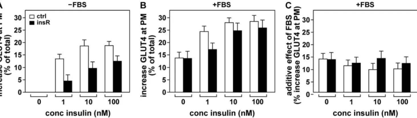
Serum-induced GLUT4 translocation is maintained in insulin resistance
So far, our results suggested that the effect of the serum factor on GLUT4 might be maintained in insulin resistance, as the action of FBS on GLUT4 was largely additive to that of insulin (Figure 4) while distinct signaling pathways appeared to be involved (Figure 5 and 6).
To investigate this, insulin resistant 3T3-L1 adipocytes were incubated with different concentrations of insulin in the absence and presence of 25% FBS and cell surface GLUT4 levels were measured (Figure 7). As expected, insulin resistant cells displayed
reduced GLUT4 translocation in response to insulin (panel A). Notably, the presence of FBS during the insulin incubation largely increased cell surface GLUT4 levels in both insulin-sensitive and insulin-resistant cells (panel B). Calculation of the additive effect of FBS demonstrated that its action on GLUT4 persisted in insulin-resistant cells (panel C). These data demonstrate that the effect of the serum factor is not only independent of insulin but also that its effect is fully preserved in insulin resistance.
The GLUT4-translocating activity of serum fractionates in two peaks in size exclusion chromatography
To investigate the nature of the putative factor in serum, we fractionated human serum on a Sephacryl 200HR column and analyzed the effect of the fractions on cell surface GLUT4 levels (Figure 8). This revealed two major peaks estimated at ,170 kDa and ,33 kDa and a minor peak at around 7 kDa, suggesting that either at least two independent factors contribute to the GLUT4-translocating activity in serum or that part of the implicated factor exists in a protein complex.
Discussion
Here, we present evidence for the existence of GLUT4-translocating activity in serum and show that the responsible factor is not insulin, IGF-1, PDGF, or HGF. Moreover, neither the insulin receptor nor IRS-1 are tyrosine phosphorylated upon serum stimulation. Also, insulin- but not serum-induced GLUT4 translocation is inhibited by tyrphostin AG1024, known to target the IGF-1 receptor and, to a somewhat lesser extent, the insulin receptor [39]. This further confirms that the serum effect is not mediated by insulin or IGF-1. This is important as an insulin-like activity of serum on adipocytes has previously been attributed to NSILA [50], later identified as IGF [51]. We have found insulin-independent GLUT4-translocating activity in all sera that we have tested (i.e. fetal, newborn and adult bovine serum, human serum and rabbit serum). Our studies have demonstrated that multiple adipocyte and muscle cell models display serum-induced GLUT4 translocation. Taken together, this demonstrates that the factor is generally present in serum and acts on all cell types that are implicated in postprandial glucose uptake. Importantly, our data imply a novel signal transduction pathway leading to GLUT4 translocation and glucose uptake, which is preserved in insulin resistance.
Several of our observations demonstrate that the mode of action of FBS is largely different from that of insulin. First, while insulin action regarding GLUT4 was largely restricted in insulin-resistant adipocytes, serum-induced GLUT4 translocation was fully preserved. Second, the effect of FBS is largely additive to that of insulin. Third, while the effect of pervanadate on GLUT4 is partially additive to that of FBS, it is not additive to the effect of insulin. Fourth, incubation of the adipocytes with the AMP kinase activator AICAR increases cell surface GLUT4 levels when added together with FBS but not with insulin. Fifth, our inhibitor studies demonstrated that the FBS effect was much more sensitive to long-term herbimycin A treatment and phospholipase C inhibitor U-73122 than that of insulin. Finally, analyses of the insulin signaling pathway demonstrated that the downstream signaling molecules involved in insulin-induced GLUT4 translocation are not activated by FBS.
Our data demonstrate that there exists an as yet unidentified signaling pathway that leads to GLUT4 recruitment and glucose uptake and that this pathway is activated by a serum factor. Here, we have shown that this pathway does not imply AMP kinase, as serum-induced GLUT4 translocation was not inhibited by AMPK
Figure 6. The intracellular signal transduction pathways involved in FBS-induced GLUT4 translocation are partially different from those of insulin. (A) Adipocytes were preincubated for 30 minutes with 25 mM AG1024, 100 nM wortmannin, 50 mM LY294002, 30 mM staurosporine, 300 mM genistein, 2 mM herbimycin A (‘herbimycin/0.5’), 10 mM Akti-1/2, 10 mM Ro 31-8220, 40 mM compound C, 10 mM U0126, 40 nM rapamycin, 1 mM okadaic acid, or 10 mM U-73122, or for 24 hours with 1.75 mM herbimycin A (‘herbimycin/24’). Insulin (100 nM) or FBS (50%) was added, cells were incubated for a further 10 minutes, and cell surface GLUT4 levels were determined. To demonstrate the effect of the inhibitors, the relative increase in cell surface GLUT4 levels in the presence of inhibitors was expressed as percentage of the increase in the absence of inhibitors. (B–E) Adipocytes were stimulated for 5 minutes with 100 nM insulin, 25% FBS or left untreated. Lysates were either directly subjected to SDS-PAGE and immunoblotting using the indicated antibodies (B,D,E), or first subjected to immunopurification (IP), in the case of the insulin receptor (IR) and IRS-1 (C). *P,0.05; **P,0.01; ***P,0.005.
doi:10.1371/journal.pone.0015560.g006
inhibitor compound C. Moreover, the translocation of GLUT4 in response to FBS was enhanced by AMPK activator AICAR, while FBS itself did not alter phosphorylation of AMPK or its downstream effector acetyl CoA carboxylase (ACC; not shown). The effect of FBS on GLUT4 was inhibited more than 50% by inhibitors of PI 3-kinase as well as by staurosporine, 24 hour herbimycin A treatment, Akti-1/2, and U73122, indicating the involvement of PI 3-kinase, a serine/threonine protein kinase, and phospholipase C. Although serum-induced GLUT4 translocation was inhibited by Akti-1/2, serum did not induce phosphorylation of Akt2 nor of its downstream effector AS160 [52], suggesting that the inhibition by Akti-1/2 may be due to the involvement of a structurally similar serine/threonine kinase whose activity is reduced by Akti-1/2. In accordance with this possibility, serum but not insulin induced the phosphorylation of a ,60 kDa protein that was reactive with the PAS antibody, raised against a phosphorylated Akt substrate consensus sequence. These results
suggest that another member of the AGC kinase family may be involved in the serum action as many members of this family share the same substrate phosphorylation sequence.
At present it is unclear what is the nature of the serum factor(s). The factor is likely to be a protein as the GLUT4-translocating activity is collected in the protein fraction upon ammonium sulfate precipitation (data not shown). Moreover, on a size exclusion column, it fractionates into two major peaks (estimated Mr33 and
170 kDa), while it is not retained on a hydrophobic LH20 column (not shown). This may imply that two proteins are involved. Alternatively, the implicated factor may in part be associated with other proteins. Interestingly, heating FBS for up to 10 minutes at 100uC precipitates 75% of the protein content, while leaving the serum GLUT4-translocating activity intact (not shown), implying thermostability and suggesting that it is not the mere presence of a bulk of protein that induces GLUT4 translocation but that the effect is more specific. This is also evident from our size exclusion chromatography studies, in which GLUT4-translocating activity is present in fractions that contain minimal amounts of protein.
This factor may be regulated in a way similar to insulin in that its serum levels may oscillate depending on food intake. Moreover, its serum levels may be altered in insulin resistance in vivo. To date, we have not yet investigated these issues. The 96 well plate technique that has been used throughout this study will largely aid in addressing these questions and will likely lead to its purification and identification, and to the definition of its (patho)physiological relevance. Unfortunately, the activity-based purification of un-identified proteins from serum has proven often to be a difficult and laborious task.
Taken together, we have demonstrated the presence of a factor (or several factors) in serum that induce GLUT4 translocation in a manner that is largely independent of insulin and insulin signaling and whose effects are fully maintained in insulin resistance. Hence, this factor may prove to have beneficial effects in type 2 diabetes. Now, the challenging task is to identify this factor and to examine its effects on GLUT4 and glucose uptake under normal conditions and conditions of insulin resistance in vitro and in vivo.
Supporting Information
Table S1 Insulin concentrations in the sera used in the described studies.
(TIF)
Figure 7. The effect of FBS on GLUT4 persists in insulin-resistant adipocytes. 3T3-L1 adipocytes rendered insulin-resistant by a 24 hour 100 nM insulin treatment (black bars, ‘insR’) or control adipocytes (white bars, ‘ctrl’) were incubated for 20 minutes with the indicated concentrations of insulin in the absence (A) or presence of 25% FBS (B) and increases in cell surface GLUT4 levels were determined. (C) Values of (A) were subtracted from those of (B) to calculate the additive effect of FBS. In (A), control and insulin-resistant cells were significantly different for all three insulin concentrations (P,0.005), while in (C) there were no significant differences between the cells.
doi:10.1371/journal.pone.0015560.g007
Figure 8. Size exclusion chromatography of serum reveals that multiple proteins or protein complexes are involved in serum-induced GLUT4 translocation. Human serum to which a trace amount of 2-[3H]deoxyglucose was added was fractionated as described in Experimental. Fractions were evaluated for their effect on cell surface GLUT4 levels. The maximum increase was set to 100%. Protein concentrations and 2-DOG were also expressed as percentage of maximum. The void volume (Vo) and the elution profile of the calibration standards (200-14 kDa) are indicated. The (0.2 kDa) 2-DOG peak marks the end of the elution process.
Figure S1 The relative effect of serum on cellular glucose uptake is lower than its effect on cell surface GLUT4 levels. FBS was extensively dialyzed against KRP buffer (cut-off 3 kDa). 3T3-L1 adipocytes expressing HA-GLUT4 (A) or not expressing HA-GLUT4 (B) were serum starved for 2 h in DMEM containing 0.2% BSA, washed with KRP, and incubated for the indicated time periods in the absence (open triangles) or presence of 100 nM insulin (open squares), 25% FBS (filled triangles), 50% FBS (filled squares), or 100% FBS (filled diamonds) in KRP. Subsequently, (A) non-permeabilized fixed cells were immunolabeled with anti-HA and fluorescent secondary antibod-ies followed by measurement of the fluorescence (arbitrary units) or (B) the uptake of radiolabeled 2DOG during a 2 min incubation was measured. The dashed line in the panels represents basal cell surface GLUT4 levels/2DOG uptake.
(TIF)
Figure S2 Serum does not reduce GLUT4 internaliza-tion. 3T3-L1 adipocytes expressing HA-GLUT4 were incubated for 20 min with 100 nM insulin, followed by cooling down on ice, removal of insulin, and cell surface labeling of the cells with anti-HA antibody. Excess antibody was removed, cells were transferred to 37uC, and fixed after various time periods. Permeabilized cells were immunolabeled with fluorescent goat-anti-mouse antibody and analyzed by fluorescence microscopy. Cells were scored for the presence of internalized anti-HA label. Hyperosmolarity (0.45 M sucrose) was included as positive control.
(TIF)
Figure S3 The effect of FBS on GLUT4 is not mediated by PDGF, IGF-1, or HGF. (A) Adipocytes were incubated for
20 minutes with various concentrations of the indicated ligands and relative cell surface GLUT4 levels were determined. (B) 3T3-L1 adipocytes and preadipocytes were incubated for 5 minutes with 100 nM insulin, 2 nM PDGF-BB, 25 nM IGF-1, or 2.6 nM HGF, and lysate samples were subjected to SDS-PAGE and immunoblotting using phospho-ERK and ERK antibodies. (TIF)
Figure S4 HA-tagged GLUT1 is correctly localized. 3T3-L1 adipocytes expressing GLUT1 with an HA-epitope tag in its first extracellular domain were immunolabeled using anti-HA and anti-syntaxin 13 antibodies. Control adipocytes were immunola-beled using anti-GLUT1 and anti-syntaxin 13 antibodies. Note that the localization of HA-GLUT1 is similar compared with endogenous GLUT1. Bar, 5mm.
(TIF)
Acknowledgments
We are greatly indebted to Teresa Gonzalez for human adipocyte cultures and thank Rytis Prekeris for anti-syntaxin13 antibody, Toshio Kitamura for Plat-E cells, Hartmut Land for pBABE vector, Mike Mueckler for GLUT1 cDNA, Gilles Flatau and Heidrun Rhode (Friedrich Schiller University, Jena, Germany) for helpful discussions, Christine Bruce for critical reading of this manuscript, and acknowledge the C3M Cell Imaging Facility MICA.
Author Contributions
Conceived and designed the experiments: RG MB YLMB. Performed the experiments: MB LM RG. Analyzed the data: MB SGP YLMB RG. Wrote the paper: RG.
References
1. Bryant NJ, Govers R, James DE (2002) Regulated transport of the glucose transporter GLUT4. Nat Rev Mol Cell Biol 3: 267–277.
2. Livingstone C, James DE, Rice JE, Hanpeter D, Gould GW (1996) Compartment ablation analysis of the insulin-responsive glucose transporter (GLUT4) in 3T3-L1 adipocytes. Biochem J 315: 487–495.
3. Johnson AO, Subtil A, Petrush R, Kobylarz K, Keller SR, et al. (1998) Identification of an insulin-responsive, slow endocytic recycling mechanism in Chinese hamster ovary cells. J Biol Chem 273: 17968–17977.
4. Govers R, Coster AC, James DE (2004) Insulin increases cell surface GLUT4 levels by dose dependently discharging GLUT4 into a cell surface recycling pathway. Mol Cell Biol 24: 6456–6466.
5. Foran PG, Fletcher LM, Oatey PB, Mohammed N, Dolly JO, et al. (1999) Protein kinase B stimulates the translocation of GLUT4 but not GLUT1 or transferrin receptors in 3T3-L1 adipocytes by a pathway involving SNAP-23, synaptobrevin-2, and/or cellubrevin. J Biol Chem 274: 28087–28095. 6. Inoue M, Chang L, Hwang J, Chiang SH, Saltiel AR (2003) The exocyst
complex is required for targeting of Glut4 to the plasma membrane by insulin. Nature 422: 629–633.
7. Foster LJ, Li D, Randhawa VK, Klip A (2001) Insulin Accelerates Inter-endosomal GLUT4 Traffic via Phosphatidylinositol 3-Kinase and Protein Kinase B. J Biol Chem 276: 44212–44221.
8. Bai L, Wang Y, Fan J, Chen Y, Ji W, et al. (2007) Dissecting multiple steps of GLUT4 trafficking and identifying the sites of insulin action. Cell Metab 5: 47–57.
9. Satoh S, Nishimura H, Clark AE, Kozka IJ, Vannucci SJ, et al. (1993) Use of bismannose photolabel to elucidate insulin-regulated GLUT4 subcellular trafficking kinetics in rat adipose cells. Evidence that exocytosis is a critical site of hormone action. J Biol Chem 268: 17820–17829.
10. Rudich A, Tirosh A, Potashnik R, Hemi R, Kanety H, et al. (1998) Prolonged oxidative stress impairs insulin-induced GLUT4 translocation in 3T3-L1 adipocytes. Diabetes 47: 1562–1569.
11. Houstis N, Rosen ED, Lander ES (2006) Reactive oxygen species have a causal role in multiple forms of insulin resistance. Nature 440: 944–948.
12. Kanety H, Feinstein R, Papa MZ, Hemi R, Karasik A (1995) Tumor necrosis factor alpha-induced phosphorylation of insulin receptor substrate-1 (IRS-1). Possible mechanism for suppression of insulin-stimulated tyrosine phosphoryla-tion of IRS-1. J Biol Chem 270: 23780–23784.
13. Hotamisligil GS, Peraldi P, Budavari A, Ellis R, White MF, et al. (1996) IRS-1-mediated inhibition of insulin receptor tyrosine kinase activity in TNF-alpha-and obesity-induced insulin resistance. Science 271: 665–668.
14. Yang X, Ongusaha PP, Miles PD, Havstad JC, Zhang F, et al. (2008) Phosphoinositide signalling links O-GlcNAc transferase to insulin resistance. Nature 451: 964–969.
15. Chen G, Liu P, Thurmond DC, Elmendorf JS (2003) Glucosamine-induced insulin resistance is coupled to O-linked glycosylation of Munc18c. FEBS Lett 534: 54–60.
16. Park SY, Ryu J, Lee W (2005) O-GlcNAc modification on IRS-1 and Akt2 by PUGNAc inhibits their phosphorylation and induces insulin resistance in rat primary adipocytes. Exp Mol Med 37: 220–229.
17. Thomson MJ, Williams MG, Frost SC (1997) Development of insulin resistance in 3T3-L1 adipocytes. J Biol Chem 272: 7759–7764.
18. Ruan H, Hacohen N, Golub TR, Van Parijs L, Lodish HF (2002) Tumor necrosis factor-alpha suppresses adipocyte-specific genes and activates expression of preadipocyte genes in 3T3-L1 adipocytes: nuclear factor-kappaB activation by TNF-alpha is obligatory. Diabetes 51: 1319–1336.
19. Maier VH, Gould GW (2000) Long-term insulin treatment of 3T3-L1 adipocytes results in mis-targeting of GLUT4: implications for insulin-stimulated glucose transport. Diabetologia 43: 1273–1281.
20. Maianu L, Keller SR, Garvey WT (2001) Adipocytes exhibit abnormal subcellular distribution and translocation of vesicles containing glucose transporter 4 and insulin-regulated aminopeptidase in type 2 diabetes mellitus: implications regarding defects in vesicle trafficking. J Clin Endocrinol Metab 86: 5450–5456.
21. Jager J, Gremeaux T, Cormont M, Le Marchand-Brustel Y, Tanti JF (2007) Interleukin-1beta-induced insulin resistance in adipocytes through down-regulation of insulin receptor substrate-1 expression. Endocrinology 148: 241–251.
22. Govers R, James DE, Coster AC (2008) High-throughput analysis of the dynamics of recycling cell surface proteins. Methods Mol Biol 440: 129–146. 23. Martinez L, Berenguer M, Bruce MC, Le Marchand-Brustel Y, Govers R (2010)
Rosiglitazone increases cell surface GLUT4 levels in 3T3-L1 adipocytes through an enhancement of endosomal recycling. Biochem Pharmacol 79: 1300–1309. 24. Morita S, Kojima T, Kitamura T (2000) Plat-E: an efficient and stable system for
transient packaging of retroviruses. Gene Ther 7: 1063–1066.
25. Somwar R, Kim DY, Sweeney G, Huang C, Niu W, et al. (2001) GLUT4 translocation precedes the stimulation of glucose uptake by insulin in muscle cells: potential activation of GLUT4 via p38 mitogen-activated protein kinase. Biochem J 359: 639–649.
26. Tsiani E, Bogdanovic E, Sorisky A, Nagy L, Fantus IG (1998) Tyrosine phosphatase inhibitors, vanadate and pervanadate, stimulate glucose transport
and GLUT translocation in muscle cells by a mechanism independent of phosphatidylinositol 3-kinase and protein kinase C. Diabetes 47: 1676–1686. 27. Kozka IJ, Clark AE, Holman GD (1991) Chronic treatment with insulin
selectively down-regulates cell-surface GLUT4 glucose transporters in 3T3-L1 adipocytes. J Biol Chem 266: 11726–11731.
28. Williams D, Hicks SW, Machamer CE, Pessin JE (2006) Golgin-160 is required for the Golgi membrane sorting of the insulin-responsive glucose transporter GLUT4 in adipocytes. Mol Biol Cell 17: 5346–5355.
29. Regazzetti C, Peraldi P, Gremeaux T, Najem-Lendom R, Ben-Sahra I, et al. (2009) Hypoxia decreases insulin signaling pathways in adipocytes. Diabetes 58: 95–103.
30. Hausdorff SF, Fingar DC, Morioka K, Garza LA, Whiteman EL, et al. (1999) Identification of wortmannin-sensitive targets in 3T3-L1 adipocytes. Dissocia-tionoOf insulin-stimulated glucose uptake and glut4 translocation. J Biol Chem 274: 24677–24684.
31. Funaki M, Randhawa P, Janmey PA (2004) Separation of insulin signaling into distinct GLUT4 translocation and activation steps. Mol Cell Biol 24: 7567–7577.
32. Li D, Randhawa VK, Patel N, Hayashi M, Klip A (2001) Hyperosmolarity reduces GLUT4 endocytosis and increases its exocytosis from a VAMP2-independent pool in l6 muscle cells. J Biol Chem 276: 22883–22891. 33. Ricort JM, Tanti JF, Van Obberghen E, Le Marchand-Brustel Y (1996)
Different effects of insulin and platelet-derived growth factor on phosphatidy-linositol 3-kinase at the subcellular level in 3T3-L1 adipocytes. A possible explanation for their specific effects on glucose transport. Eur J Biochem 239: 17–22.
34. Manchem VP, Goldfine ID, Kohanski RA, Cristobal CP, Lum RT, et al. (2001) A novel small molecule that directly sensitizes the insulin receptor in vitro and in vivo. Diabetes 50: 824–830.
35. Bertola A, Bonnafous S, Cormont M, Anty R, Tanti JF, et al. (2007) Hepatocyte growth factor induces glucose uptake in 3T3-L1 adipocytes through A Gab1/ phosphatidylinositol 3-kinase/Glut4 pathway. J Biol Chem 282: 10325–10332. 36. Al-Khalili L, Cartee GD, Krook A (2003) RNA interference-mediated reduction in GLUT1 inhibits serum-induced glucose transport in primary human skeletal muscle cells. Biochem Biophys Res Commun 307: 127–132.
37. Kotani K, Ogawa W, Matsumoto M, Kitamura T, Sakaue H, et al. (1998) Requirement of atypical protein kinase clambda for insulin stimulation of glucose uptake but not for Akt activation in 3T3-L1 adipocytes. Mol Cell Biol 18: 6971–6982.
38. Thong FS, Bilan PJ, Klip A (2007) The Rab GTPase-activating protein AS160 integrates Akt, protein kinase C, and AMP-activated protein kinase signals regulating GLUT4 traffic. Diabetes 56: 414–423.
39. Parrizas M, Gazit A, Levitzki A, Wertheimer E, LeRoith D (1997) Specific inhibition of insulin-like growth factor-1 and insulin receptor tyrosine kinase activity and biological function by tyrphostins. Endocrinology 138: 1427–1433.
40. Kotani K, Carozzi AJ, Sakaue H, Hara K, Robinson LJ, et al. (1995) Requirement for phosphoinositide 3-kinase in insulin-stimulated GLUT4 translocation in 3T3-L1 adipocytes. Biochem Biophys Res Commun 209: 343–348.
41. Nishimura H, Simpson IA (1994) Staurosporine inhibits phorbol 12-myristate 13-acetate- and insulin-stimulated translocation of GLUT1 and GLUT4 glucose transporters in rat adipose cells. Biochem J 302: 271–277.
42. Elmendorf JS, Chen D, Pessin JE (1998) Guanosine 59-O-(3-thiotriphosphate) (GTPgammaS) stimulation of GLUT4 translocation is tyrosine kinase-depen-dent. J Biol Chem 273: 13289–13296.
43. Hoehn KL, Hohnen-Behrens C, Cederberg A, Wu LE, Turner N, et al. (2008) IRS1-independent defects define major nodes of insulin resistance. Cell Metab 7: 421–433.
44. Standaert ML, Bandyopadhyay G, Perez L, Price D, Galloway L, et al. (1999) Insulin activates protein kinases C-zeta and C-lambda by an autophosphoryla-tion-dependent mechanism and stimulates their translocation to GLUT4 vesicles and other membrane fractions in rat adipocytes. J Biol Chem 274: 25308–25316.
45. Yamaguchi S, Katahira H, Ozawa S, Nakamichi Y, Tanaka T, et al. (2005) Activators of AMP-activated protein kinase enhance GLUT4 translocation and its glucose transport activity in 3T3-L1 adipocytes. Am J Physiol Endocrinol Metab 289: E643–649.
46. Bogan JS, McKee AE, Lodish HF (2001) Insulin-responsive compartments containing GLUT4 in 3T3-L1 and CHO cells: regulation by amino acid concentrations. Mol Cell Biol 21: 4785–4806.
47. Kayali AG, Eichhorn J, Haruta T, Morris AJ, Nelson JG, et al. (1998) Association of the insulin receptor with phospholipase C-gamma (PLCgamma) in 3T3-L1 adipocytes suggests a role for PLCgamma in metabolic signaling by insulin. J Biol Chem 273: 13808–13818.
48. Sepp-Lorenzino L, Ma Z, Lebwohl DE, Vinitsky A, Rosen N (1995) Herbimycin A induces the 20 S proteasome- and ubiquitin-dependent degradation of receptor tyrosine kinases. J Biol Chem 270: 16580–16587.
49. Taniguchi CM, Emanuelli B, Kahn CR (2006) Critical nodes in signalling pathways: insights into insulin action. Nat Rev Mol Cell Biol 7: 85–96. 50. Froesch ER, Buergi H, Ramseier EB, Bally P, Labhart A (1963)
Antibody-Suppressible and Nonsuppressible Insulin-Like Activities in Human Serum and Their Physiologic Significance. an Insulin Assay With Adipose Tissue of Increased Precision and Specificity. J Clin Invest 42: 1816–1834.
51. Rinderknecht E, Humbel RE (1976) Amino-terminal sequences of two polypeptides from human serum with nonsuppressible insulin-like and cell-growth-promoting activities: evidence for structural homology with insulin B chain. Proc Natl Acad Sci U S A 73: 4379–4381.
52. Kane S, Sano H, Liu SC, Asara JM, Lane WS, et al. (2002) A method to identify serine kinase substrates. Akt phosphorylates a novel adipocyte protein with a Rab GTPase-activating protein (GAP) domain. J Biol Chem 277: 22115–22118.