HAL Id: hal-01404918
https://hal.sorbonne-universite.fr/hal-01404918
Submitted on 29 Nov 2016
HAL is a multi-disciplinary open access
archive for the deposit and dissemination of
sci-entific research documents, whether they are
pub-lished or not. The documents may come from
teaching and research institutions in France or
abroad, or from public or private research centers.
L’archive ouverte pluridisciplinaire HAL, est
destinée au dépôt et à la diffusion de documents
scientifiques de niveau recherche, publiés ou non,
émanant des établissements d’enseignement et de
recherche français ou étrangers, des laboratoires
publics ou privés.
Distributed under a Creative Commons Attribution| 4.0 International License
Long-term exposure to Myozyme results in a decrease of
anti-drug antibodies in late-onset Pompe disease patients
Elisa Masat, Pascal Laforêt, Marie de Antonio, Guillaume Corre, Barbara
Perniconi, Nadjib Taouagh, Kuberaka Mariampillai, Damien Amelin,
Wladimir Mauhin, Jean-Yves Hogrel, et al.
To cite this version:
Elisa Masat, Pascal Laforêt, Marie de Antonio, Guillaume Corre, Barbara Perniconi, et al.. Long-term
exposure to Myozyme results in a decrease of anti-drug antibodies in late-onset Pompe disease patients.
Scientific Reports, Nature Publishing Group, 2016, 6, pp.36182. �10.1038/srep36182�. �hal-01404918�
Long-term exposure to Myozyme
results in a decrease of anti-drug
antibodies in late-onset Pompe
disease patients
Elisa Masat
1, Pascal Laforêt
1,2, Marie De Antonio
3, Guillaume Corre
4, Barbara Perniconi
2,
Nadjib Taouagh
2, Kuberaka Mariampillai
5, Damien Amelin
1, Wladimir Mauhin
1,
Jean-Yves Hogrel
6, Catherine Caillaud
7, Giuseppe Ronzitti
4, Francesco Puzzo
4,
Klaudia Kuranda
1, Pasqualina Colella
4, Roberto Mallone
8,9,10, Olivier Benveniste
1,5,
Federico Mingozzi
1,4& French Pompe Registry Study Group
#Immunogenicity of recombinant human acid-alpha glucosidase (rhGAA) in enzyme replacement therapy (ERT) is a safety and efficacy concern in the management of late-onset Pompe disease (LOPD). However, long-term effects of ERT on humoral and cellular responses to rhGAA are still poorly understood. To better understand the impact of immunogenicity of rhGAA on the efficacy of ERT, clinical data and blood samples from LOPD patients undergoing ERT for >4 years (n = 28) or untreated (n = 10) were collected and analyzed. In treated LOPD patients, anti-rhGAA antibodies peaked within the first 1000 days of ERT, while long-term exposure to rhGAA resulted in clearance of antibodies with residual production of non-neutralizing IgG. Analysis of T cell responses to rhGAA showed detectable T cell reactivity only after in vitro restimulation. Upregulation of several cytokines and chemokines was detectable in both treated and untreated LOPD subjects, while IL2 secretion was detectable only in subjects who received ERT. These results indicate that long-term ERT in LOPD patients results in a decrease in antibody titers and residual production of non-inhibitory IgGs. Immune responses to GAA following long-term ERT do not seem to affect efficacy of ERT and are consistent with an immunomodulatory effect possibly mediated by regulatory T cells.
Pompe Disease (PD) is an autosomal recessive myopathy caused by a deficiency in the lysosomal enzyme acid α -glucosidase (GAA), which results in an abnormal storage of glycogen in several tissues1,2. The infantile form of
PD is the most severe and, if not treated, is associated with early lethality3,4. The adult form of the disease, known
as late onset Pompe disease (LOPD), is compatible with life, although it is associated with progressive deteriora-tion of skeletal muscle funcdeteriora-tion, leading in some cases to significant disability and need for assisted ventiladeteriora-tion5,6.
The approval of recombinant human GAA (rhGAA) (Myozyme) for the treatment of Pompe disease resulted in a significant improvement of both life expectancy and quality of life of infantile PD patients7, although long-term
follow up of children treated with enzyme replacement therapy (ERT) revealed occurrence of symptoms resulting from the incomplete correction of the enzyme deficiency in certain tissues8. Approval of ERT for LOPD patients
followed that of pediatric subjects few years later, and long-term benefit of ERT in this population is still being assessed9–11.
1University Pierre and Marie Curie, INSERM, UMR974, Paris, France. 2Paris-Est neuromuscular center, Institute of
Myology, Pitié-Salpêtrière Hospital, AP-HP, Paris, France. 3University Paris Descartes, INSERM, UMR1138, Paris,
France. 4Genethon, INSERM, UMR951, Evry, France. 5Department of Internal Medicine and Clinical Immunology,
DHUI2B, Pitié-Salpêtrière Hospital, AP-HP, Paris, France. 6Neuromuscular Physiology and Evaluation Lab, Institute
of Myology, Paris, France. 7Department of Metabolic Biochemistry, Necker Hospital, Paris, France. 8Institute Cochin,
INSERM U1016, CNRS UMR8104, Paris, France. 9University Paris Descartes, Faculty of Medicine, Paris, France. 10Department of diabetology, Cochin Hospital, AP-HP, Paris, France.#A comprehensive list of consortium members
appears at the end of the paper. Correspondence and requests for materials should be addressed to F.M. (email: fmingozzi@genethon.fr)
received: 27 April 2016 Accepted: 27 September 2016 Published: 04 November 2016
www.nature.com/scientificreports/
One important common side effect of ERT for Pompe disease is the induction of antibody responses against the infused protein, a phenomenon particularly frequent in infantile patients who are cross-reactive immunolog-ical material (CRIM)-negative4,12, that is associated with lack of efficacy and poor prognosis13,14. Similarly, despite
being CRIM positive, anti-rhGAA antibodies in response to ERT are found also in LOPD patients, although their role is unclear when it comes to the clinical response to enzyme supplementation15. In addition to neutralizing
antibody responses, life-threatening allergic reactions associated with the production of immunoglobulin (Ig) E specific to the enzyme have been reported to occur following the infusion of rhGAA16.
Unlike for infantile PD and for other lysosomal storage disorders17,18, little is known on the impact of immune
responses to rhGAA in LOPD subjects undergoing ERT. Moreover, mechanistic insights into the immuno-genicity of rhGAA are lacking. Limited studies in human peripheral blood mononuclear cells (PBMC) showed dose-dependent increase in interferon gamma (IFNγ ) and tumor necrosis factor alpha (TNFα ) production in CD4+ and CD8+ T cells in LOPD patients receiving ERT19. Additional studies in GAA-null mice treated with
rhGAA showed high frequency of T cells producing interleukin (IL) 4 in response to rhGAA, highlighting a pre-dominantly T helper (Th) 2-driven immune response20–22.
Here we characterized the immune responses to rhGAA in a large cohort of LOPD subjects either receiving long-term ERT or untreated. In this study we demonstrate that rhGAA infusion results in the early production of high-titer antibodies in a subset of subjects, however antibodies appear to drop over time with continuation of ERT. IgG subclass characterization shows production of non-inhibitory antibodies with no evident effect on enzyme activity or uptake, while rhGAA-specific T cell activation profile is consistent with immune modulation, possibly mediated by regulatory T cells.
Results
Long-term ERT results in clearance of anti-rhGAA antibodies in LOPD patients.
To understand the effect of long-term ERT on humoral responses to rhGAA, anti-rhGAA antibody data from LOPD subjects (n = 24) who received ERT with Myozyme at least 3 years (three antibody measurements per year) and developed a response to the enzyme were collected and analyzed. Of these subjects, five had peak antibody titers ≥ 1:25,000, twelve had peak antibody titers ≥ 1:6,400 and < 1:25,000, and seven developed peak antibody titers < 1:6,400. No clear correlation between GAA gene mutations (Supplementary Table S1) and peak antibody titers was found (data not shown). 17 out of 24 patients showed the highest antibody titer in the first 1,000 days on ERT (Fig. 1A),Figure 1. Long-term ERT in LOPD patients results in a decrease of anti-rhGAA antibodies. (A) Measurement
of anti-rhGAA antibody titers over time (n = 24 subjects). (B) Comparison of antibody titers measured in the first 500 days of ERT (n = 108 measurements) with titers measured between 500 and 1,000 days of ERT (n = 82 measurements), 1,000 and 1,500 days of ERT (n = 64 measurements), and 1,500 and 2,000 days of ERT (n = 46 measurements). For each time period, the same number of measurements for each subject was included in the analysis. ANOVA analysis was used to compare the measurements over time (*P < 0.05). Antibody titers are expressed as reciprocal dilution as previously described10.
with one subject reaching a titer of > 1:200,000, followed by a decrease to levels close to baseline for most sub-jects. Statistical analysis of data, performed by comparison of titers measured during the first 500 days of ERT (108 measurements) with those collected between 501 and 1,000 days (82 measurements), between 1,001 and 1,500 days (64 measurements), and between 1,500 and 2,000 days of ERT (46 measurements), confirmed that the changes in antibody titers were significant (Fig. 1B). These results indicate that long-term ERT in LOPD subjects is associated with a decrease of anti-rhGAA antibodies.
Long-term ERT in LOPD subjects results in development of non-neutralizing anti-rhGAA IgG1
and IgG4.
To better understand the nature of anti-rhGAA antibody responses observed in the LOPD patients enrolled in the study, serum samples were tested using an antibody binding assay specific for anti-rhGAA IgG sub-classes, IgM, and IgE (Fig. 2). Healthy donors (HD) and untreated LOPD subjects did not display significant levels of antibodies specific to rhGAA, whereas a subset of LOPD subjects who received long-term ERT had elevated lev-els of anti-rhGAA IgG1 and IgG4 antibodies (Fig. 2D). No significant levlev-els of IgG2, IgG3, IgM, and IgE antibod-ies were detected. As IgG4 have been reported in association with inhibitor activity23, seven samples, displayingthe highest anti-rhGAA antibody titers, were then tested for neutralizing activity on both rhGAA enzyme activity and cell uptake assays and no inhibitory activity was measured (Supplementary Fig. S1A). In agreement with these results, follow up of six-minute walk test (6MWT) and forced vital capacity (FVC) in IgG4 positive subjects showed a similar trend to the subjects from the same cohort (Supplementary Fig. S1B,C, and vide infra). These results confirm the presence of anti-rhGAA antibodies in CRIM-positive LOPD subjects even after long-term ERT. They also indicate that repeated exposures to the rhGAA antigen results in production of non-neutralizing antibodies including IgG4, an antibody subclass previously associated with activation of regulatory B cells24,25. Figure 2. Anti-rhGAA IgG1 and IgG4 are the most prevalent subclasses of antibodies developed in response to long-term ERT. (A–F) Anti-rhGAA antibodies measured with a capture assay specific for IgG1,
IgG2, IgG3, IgG4, IgM, and IgE antibodies. Estimated antibody concentrations are reported for each subject. Average and standard deviation are indicated for each of the study cohorts: LOPD subjects receiving ERT (Treated, n = 28), untreated LOPD subjects (Untreated, n = 10), and healthy donors (HD, n = 43). Unpaired two-tailed t-test was used to compare results across the study cohorts (**P < 0.01).
www.nature.com/scientificreports/
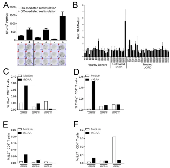
DC-mediated restimulation of PBMCs results in detection of T cell reactivity to rhGAA.
IgG4 production has been associated with induction of immunological tolerance24. To test whether a similarmecha-nism mediated the observed decrease in anti-GAA antibody titers in LOPD subjects undergoing ERT, and to study T cell reactivity to rhGAA in treated and untreated LOPD subjects, an IFNγ ELISpot assay specific for the enzyme was established. Initial screening of PBMCs isolated from treated LOPD subjects revealed lack of reactivity to rhGAA, even in subjects with detectable anti-rhGAA antibodies (data not shown). To enhance T cell reactivity to rhGAA, we used a dendritic cell (DC) activation protocol optimized to study low-frequency T cells26. Following
restimulation with rhGAA and specific cytokines and chemokines prior to the ELISpot assay, we were able to measure production of IFN-γ in response to rhGAA in some subjects (Fig. 3A). Five out of twenty-eight treated LOPD subjects showed detectable T cell reactivity to rhGAA, however no correlation was found between produc-tion of IFNγ and anti-rhGAA antibody titers (Fig. 3B). A positive T cell response to rhGAA was detected also in one untreated LOPD subject and one HD, an observation previously made for other antigens such as coagulation factor VIII in HD and in hemophilia A subjects27. Additional analyses of treated LOPD PBMCs restimulated in
vitro with rhGAA for 48 hours followed by intracellular cytokine staining (ICS), showed a complex pattern of
acti-vation of CD4+ T cells, with production of IFNγ , TNFα , IL2, and IL17 in some subjects (Fig. 3C–F). No response
to rhGAA was detectable in CD8+ T cells (data not shown). These results indicate that circulating GAA-reactive T
cells can be found in peripheral blood, although detectable only after restimulation with the antigen28. Figure 3. T cell reactivity to rhGAA is detectable after restimulation of PBMCs with the antigen.
(A) Comparison of IFNγ ELISpot count in PBMCs isolated from LOPD subjects receiving ERT before and after DC-mediated restimulation. Cells were either directly plated in the ELISpot assay (− ) or restimulated
in vitro with 10 μ g/ml of rhGAA prior to the assay (+ ). Results are expressed in spot forming units (SFU) per
106 cells. Results are reported as average of triplicate testing + /− standard deviation. Images of the test wells
in the IFNγ ELISpot assay are shown below the histogram plot. (B) Combined results for all subjects screened with the IFNγ ELISpot assay after PBMC restimulation. PBMCs from LOPD subjects receiving ERT (Treated, n = 28), untreated LOPD subjects (Untreated, n = 10), and HD (n = 15) were tested in the assay. Results are expressed as ratio between SFU/106 cells in rhGAA-restimulated cells vs. medium control. (C–F) PBMCs from
LOPD subjects receiving ERT (n = 3) were restimulated with 10 μ g/ml of rhGAA for 48h and then stained intracellularly for IFNγ , TNFα , IL2, and IL17. Results are reported as percentage of CD4+ T cells secreting a
IL2 is the cytokine signature of T cell reactivity to rhGAA in treated LOPD subjects.
To better define the profile of T cells reactive to rhGAA in all subjects studied, we collected supernatant from PBMCs after DC-mediated restimulation and tested them against an array of cytokines and chemokines using the Luminex platform (Fig. 4 and Supplementary Fig. S2). In this assay, significantly elevated levels of IL2 were detected only in treated LOPD subjects (Fig. 4A). IL10 was also slightly upregulated in subjects receiving ERT, although not at significant levels (Fig. 4B). Conversely, significant amounts of cytokines were found in both treated and untreated LOPD subjects compared with HD (Fig. 4C–L). A Pearson correlation matrix was used to analyze the data deriv-ing from the cytokine and chemokine production in treated and untreated LOPD subject and the data derivderiv-ing from the HD, confirming the presence of a correlation among cytokines in responses to rhGAA in both treated and untreated LOPD subjects (Fig. 4M). These results may reflect the proinflammatory state associated with the disease19,29, which was previously described in infantile Pompe patients30. Alternatively, the reactivity detectedagainst rhGAA in both treated and untreated LOPD patients may reflect an underlying immune response result-ing from the expression of the endogenous mutant GAA protein.
The secretion of IL2 after DC-mediated restimulation only in treated LOPD subjects possibly reflects a state of anergy of reactive T cells deriving from long-term exposure to rhGAA31. Alternatively, the detection of slightly
elevated levels of IL10 in treated LOPD subjects may also be evidence of equilibrium between proinflammatory and tolerogenic signals. To address this point, the untouched CD25neg fraction of PBMCs was isolated from three
LOPD subjects displaying high anti-rhGAA IgG4 levels. Following a 48-hour restimulation with rhGAA, both the untouched PBMC and the CD25neg fraction of PBMC was co-cultured with autologous DCs pulsed with rhGAA.
Cells were tested for reactivity to rhGAA in an IFNγ ELISpot assay, showing enhanced T cell reactivity to rhGAA in the CD25neg fraction of PBMC (Fig. 4N), suggesting that the induction of peripheral tolerance contributed
to the unresponsiveness to the rhGAA antigen. Similarly, to investigate the role of suppressive IL10-expressing B cells24,32, we depleted CD19+CD24hiCD38hi B cells in PBMC collected from six treated LOPD subjects. The
remaining untouched PBMC fraction was co-cultured with autologous DCs pulsed with rhGAA antigen and then tested for reactivity to rhGAA in an IFNγ ELISpot assay. Depletion of regulatory B cells resulted in a slight enhancement of T cell reactivity to rhGAA in only 1/6 subjects (Fig. 4O), not supporting the involvement of CD19+CD24hiCD38hi B cells in the observed hyporesponsiveness to rhGAA.
Infusion of rhGAA in LOPD subjects is associated with systemic upregulation of cytokines and
chemokines.
To test whether rhGAA infusion is associated with systemic activation of the immune system, we performed a study in ten LOPD subjects undergoing ERT in which we monitored serum cytokines and chemokine levels. For each subject, a serum sample was collected immediately before the infusion of rhGAA and one additional sample was collected at the end of the infusion, after flushing the intravenous line with saline solution. rhGAA administration resulted in a significant upregulation of several cytokines and chemokines, including IL8, monocyte chemotactic protein 1 (MCP1), macrophage inflammatory protein 1 beta (MIP1β ), IL7, and IL13 (Fig. 5). Additional cytokines were upregulated, although the changes measured were not signif-icant; these included IL6, TNFα , IL1β , IFNγ , IL4, IL5, and granulocyte macrophage colony-stimulating factor (GM-CSF) (Supplementary Fig. S3). No clear correlation between reactivity to rhGAA and baseline measure-ments of humoral or cellular responses to rhGAA was evident in the subjects studied (not shown). These results indicate that administration of rhGAA results in early activation of immunity.Long-term clinical outcome of ERT does not correlate with immune responses to rhGAA.
Follow-up measurements of six-minute walk test (6MWT) and forced vital capacity (FVC) in the cohort of treated LOPD subjects (Fig. 6A, B respectively) showed an initial improvement or stabilization of both measure-ments followed by a downward trend. This finding, which possibly reflects a worsening of the disease despite ERT in a subset of patients, will need to be confirmed in larger studies. Notably, a recently published meta-analysis of clinical trials involving LOPD patients undergoing ERT also showed a marked improvement in 6MWT and FVC in the first year of treatment, followed by a subsequent stabilization or decline in performance11.
In our analysis, no correlation was found between the evolution of FVC and 6MWT and the measurements of immune responses to rhGAA after long-term ERT (Fig. 6C), consistent with the fact that no neutralizing anti-body response was detected after long-term ERT. These results confirm that, after long-term ERT, no relationship between clinical endpoints of disease progression and immune responses to rhGAA is found.
Discussion
Immunogenicity of protein-replacement therapeutics represents a major hurdle in the treatment of genetic33 and
acquired34 disorders. Several factors contribute to shape the immune responses to an antigen, including the genetic
background of the host4,35, the immunogenicity of the therapeutic protein36, polymorphisms in genes regulating
immune responses37, and presence of contaminants in the drug substance that can exert an adjuvant effect on
immune responses36. Additionally, underlying inflammation associated with some diseases can enhance immune
responses against protein and gene therapeutics38,39. To date, the mechanisms driving the immunogenicity
of rhGAA in ERT for Pompe disease are largely unknown.
Here we presented a comprehensive characterization of immune responses to rhGAA in a cohort of subjects affected by LOPD, either untreated or undergoing long-term ERT. The data presented demonstrate that in this patient population, despite the presence of residual GAA antigen endogenously expressed40, antibodies responses
are elicited by ERT, in some cases reaching high titers (Fig. 1A and refs 15 and 41). Unlike for other genetic dis-eases35, no clear correlation with the underlying mutation in the disease-causative gene was found, reflecting the
fact that LOPD subjects are typically CRIM-positive, and suggesting that other genetic factors42 may contribute to
shape the response to rhGAA. To this end, secretion of cytokines in response to rhGAA by PBMC collected both from treated and untreated LOPD subjects indicate the existence of an underlying proinflammatory condition
www.nature.com/scientificreports/
Figure 4. Cytokine profiling of supernatant from PBMCs restimulated with rhGAA. Supernatants from
cells restimulated with rhGAA were collected after 48 hours of restimulation in vitro and assayed for cytokine and chemokine production. (A) Levels of IL2 measured in conditioned media in LOPD subjects receiving ERT (Treated, n = 28), untreated LOPD subjects (Untreated, n = 10), and healthy donors (HD, n = 17). Error bars represent the standard deviation of the mean. Mann-Whitney test was used to compare data across the study groups. (B–L) Cytokine and chemokine concentration in media measured with the Luminex array technology; shown are individual values measured in LOPD subjects receiving ERT (Treated, n = 28), untreated LOPD subjects (Untreated, n = 10), and healthy donors (HD, n = 17). Error bars represent the average of a cohort + /− standard deviation. Mann-Whitney test was used to compare data across the study groups. (M) Pearson correlation matrix comparing measurements of cytokine and chemokine production in responses to rhGAA in treated (n = 28) and untreated (n = 10) LOPD subjects, and HD (n = 15). Numbers in the table represent the correlation coefficient between two variables with the relative p value (t-test). (N) Depletion of CD25+ in
PBMCs from treated LOPD subjects. The untouched CD25neg PBMC fraction was co-cultured with autologous
DCs pulsed with rhGAA antigen or unpulsed DCs as negative control, followed by IFNγ ELISpot. Results of the IFNγ ELISpot assay are shown as average of spot forming units (SFU) per 106 cells plated in the assay + /−
standard deviation of triplicate testing. (O) Sorted untouched CD19+CD24negCD38neg PBMC fraction from
treated LOPD subjects co-cultured with autologous DCs pulsed with rhGAA antigen (or negative control) followed by IFNγ ELISpot. Results of are shown as average SFU/106 cells + /− standard deviation of triplicate
associated with the disease, regardless of ERT. This is possibly the consequence of impaired autophagy43 that
characterizes Pompe disease and other lysosomal storage diseases44,45. Alternatively, as previously observed in
Duchenne muscular dystrophy patients38, residual expression of the endogenous GAA in these subjects may
also result in an increased immunogenicity of rhGAA. These results were also confirmed in vivo, as we showed that infusion of rhGAA in LOPD patients triggered immediate systemic secretion of several cytokines and chemokines.
In our cohort of subjects, despite the fact that some subjects reached anti-rhGAA antibody titers > 1:200,000 during the first ~1,000 days of ERT, long-term exposure to rhGAA in the absence of immunomodulatory interventions resulted in a marked decrease in antibody levels, with no late occurrence of antibodies, except for non-neutralizing IgG1 and IgG4. This outcome of ERT is markedly different that observed in infantile Pompe patients, who need adjuvant immunomodulatory therapy to prevent or eradicate immune responses to rhGAA13,46,47.
Differently from ERT in hemophilia A, where detection of IgG4 is typically associated with presence of inhib-itor23,48,49, the presence of increased levels of non-neutralizing IgG4 antibodies, in the absence of IgE antibodies,
in treated LOPD subjects compared with HD and untreated LOPD subjects possibly reflects the desensitization to the immunogen24,50–52. Alternatively, the production of GAA-specific IgG4 may simply reflect the chronic
exposure to antigen53. Whether the residual anti-rhGAA IgG titers found in our patient cohort affect clearance or
bioavailability of the protein, remain to be established54.
Immune tolerance associated with ERT has been previously described in humans affected by diseases like mucopolysaccharidosis type I55,56 and hemophilia B49. Studies in mice20,21 support the idea that CD4+ T helper
cells are key to the development of humoral responses to rhGAA, confirmed by the fact that targeting these cells with non-depleting monoclonal antibodies results in avoidance of antibody formation to the protein57. In
our study, detection of CD4+ T cell reactivity only after mature DC-mediated restimulation26 of PBMCs
high-lights a state of hyporesponsiveness of T cells, which can produce large amounts of proinflammatory cytokines when encountering the rhGAA antigen in the context of the appropriate stimulus. IL2, in particular appears to be the cytokine signature in LOPD subjects on ERT. Production of IgG4 antibodies has been associated with the presence of IL10-secreting B regulatory 1 (Br1) cells24. Detection of slightly elevated levels of IL10
fol-lowing PBMC restimulation may indicate the existence of some mechanism of active suppression of immune responses to rhGAA that contributes to the development of immune tolerance to the antigen. Indeed, depletion of CD4+CD25+ T cells from PBMCs did resulted in enhanced T cell reactivity to rhGAA, while little to no effect was
observed when CD24hiCD38hi B cells32 were depleted. While these results are preliminary, they support the role of
regulatory T cells in the development of peripheral tolerance to rhGAA following ERT. More detailed studies will also help to fully address the role of CD73−CD25+CD71+ Br1 cells24 in Pompe disease patients undergoing ERT.
Finally, it is not surprising that the measurements of immune responses after long-term exposure to ERT do not correlate with the longitudinal analysis of 6MWT and FVC. This is likely to be the results of multiple factors, including (1) all subjects after long-term ERT had low residual levels of anti-rhGAA antibodies which were not neutralizing, (2) the fact that the immune responses measured were more consistent with a state of unresponsive-ness, rather than immunity, to rhGAA and (3) the fact that the immune assays were performed far in time from the peak of antibody responses to rhGAA.
Figure 5. Serum cytokine and chemokine profiling of LOPD subjects before and after ERT. (A–E) Luminex
cytokine and chemokine measurements before (PRE) and immediately after (POST) rhGAA administration (over the course of four hours) in LOPD subjects (n = 10). Shown are cytokines for which a significant change in serum levels was measured at the end of ERT. Paired t-test was used to compare data analysis PRE vs. POST infusion (*P < 0.05; **P < 0.01).
www.nature.com/scientificreports/
In summary, exposure to rhGAA via ERT in LOPD subjects results in early development of antibodies to the protein, which subsequently mostly disappear over time. The presence of residual levels of non-neutralizing rhGAA-specific IgG4 antibodies, and the T cell activation profile consistent with hyporesponsiveness to the anti-gen, reflect an immunomodulatory effect mediated by the long-term exposure to the antianti-gen, possibly via the induction of regulatory T cells. These data indicate that antibody formation induced by long-term ERT is not a major concern in LOPD patients. Immune tolerance induction protocols based on high-dose antigen expo-sure58,59 and/or pharmacological interventions60 are valuable tools to manage detrimental humoral immune
responses encountered in ERT. Conversely, in LOPD patients, acute reactions to the enzyme are among the most relevant immune complications of ERT61,62, thus not warranting the use of immunomodulatory strategies to
eradicate anti-GAA antibodies.
Materials and Methods
Human samples.
LOPD subjects included in this study were part of the French registry of adult Pompe disease patients. Approvals from the competent health authorities and the local Ethical Committee of the Pitie-Salpetriere Hospital, Paris, were obtained prior to the initiation of the study (CNIL N/Ref MMS/CWR/AR155497; CCTIRS N: 14.520; CCP approval 25/06/2014). Informed consent was given by each participant prior to inclusion in the study. All the experiments were carried out in accordance with the relevant guidelines for clinical investi-gation in France and Europe. Study inclusion criteria were diagnosis of PD defined as deficiency of GAA enzyme activity measured in blood or skin fibroblasts. PD diagnosis was confirmed by GAA gene sequencing. Exclusion criteria were age older than 80 years, ongoing or recent (less than 3 months) immunosuppressive treatment,Figure 6. Clinical outcome measures and statistical analysis of results. (A) % forced vital capacity (FVC)
measurements, expressed as % predicted in healthy individuals, over a period of 4 years in 21 treated LOPD subjects; the red line represents the average evolution of the measurement. The estimated slope is − 1.16% per year; the estimated intercept is 58.9%; p value is given by linear mixed models (see Methods). The following equations were used to calculate the % FVC: [FVC predicted for males = 0.060 * height − 0,0214 * age – 4.65]; [FVC predicted for females = 0.0491 * height − 0.0216 * age – 3.59]; [FVC (%) = FVC observed/FVC
predicted * 100] (B) Six-minute walk test (6MWT), expressed as % predicted in healthy individuals, measured over a period of 4 years in 19 subjects, the red line represents the average evolution of the measurement. The estimated slope is − 2.06% per year; the estimated intercept is 59.6%; p value is given by linear mixed models (see Methods) (***P < 0.001). The following equations were used to calculate the % 6MWT: [6MWT predicted for males = − 309–5.02 * age − 1.76 * weight + 7.57 * height]; [6MWT predicted for females = 667–5.78 * age − 2.29 * weight + 2.11 * height]; [6MWT (%) = 6MWT observed/6MWT predicted * 100]. (C) Spearman correlation of measurements of immune responses to rhGAA with clinical outcome measures recorded at the time of blood collection. The numbers in the table represent the correlation coefficient between two variables with the relative p value (t-test) (NS, not significant).
malignancy not in remission, immunodeficiency, or autoimmunity. Blood samples were collected via venipunc-ture in heparin tubes and used to isolate serum and PBMCs.
Medical history was collected at the time of study inclusion, which included measurements of disease progres-sion such as 6MWT and FVC9,10, for which data were collected through validated testing across all clinical centers
participating to the study. Historical measurements of anti-rhGAA antibodies were also collected. Samples from 28 LOPD subjects receiving biweekly infusion with rhGAA at 20 mg/kg (Treated LOPD) were collected. Treated LOPD (average age 58.2 years, standard deviation 2.4 years; median 57.5 years) were on ERT for 6.5 years on average (standard deviation 2.4; median 7), 13 males and 15 females participated to the study. Additional sam-ples from 10 LOPD subjects (4 males and 6 females) who were not on ERT (Untreated LOPD, average age 53.4 years, standard deviation 17.2 years; median 48 years) were also collected, together with 43 healthy donors (HD). Almost all the LOPD subjects carried the mutation c.32-13T > G in at least one allele. Details of the LOPD sub-jects enrolled in this study are given in Supplementary Table S1. De-identified HD blood samples (n = 43) were collected through the French blood bank (Etablissement Française du Sang, EFS). De-identified PD patient fibro-blasts lacking GAA activity were obtained from the Coriell Institute repository (Camden, NJ, USA) and used in the rhGAA uptake assay.
Antibody assays.
Antibody titers over time in treated subjects were determined as previously described10.To study the IgG subclasses specific to GAA, 96-well Nunc Polysorp Immunoplate microplates (Dutscher, Paris, France) were coated with rhGAA to a final concentration of 1 μ g/ml. A standard curve made of purified human IgG (Gamunex, Grifolds, Meyreuil, France), purified human IgG1 (Life Technologies, Saint Aubin, France), IgG2, IgG3, IgG4, IgE (Abcam, Paris, France), or IgM (Sigma-Aldrich, Lyon, France) was added directly to the plates. Plates were coated overnight at 4 °C. The next day, after blocking the plates, serum samples were added at dilu-tions of 1:10 and 1:20 in duplicate and incubated overnight at 4 °C. Monoclonal anti-human Ig biotin-conjugated antibodies specific for each subclass (all from Sigma-Aldrich, Lyon, France) were added to the plates. Binding of the secondary antibodies was detected with alkaline phosphatase-conjugated streptavidin (Sigma-Aldrich, Lyon, France). The enzymatic reaction was developed with p-nitrophenyl phosphate (Sigma-Aldrich, Lyon, France) substrate and color development was read at 405 nm using a microplate reader (MRX Relevation, DYNEX Technologies, Denkendorf, Germany). Anti-rhGAA antibody concentration was determined against the Ig subclass-specific standard curve using 4-parameters regression.
To evaluate the neutralizing activity of serum anti-rhGAA antibodies, a GAA activity assay was established as previously described63.
Short-term T cell restimulation with dendritic cell (DC) induction and cell selection.
PBMCs were restimulated in vitro according to a protocol designed to enhance detection of antigen-specific T cell responses26.PBMCs from the three cohorts studied were stimulated with rhGAA to a final concentration of 10 μ g/ml, or medium without antigen. Cells recovered after the short-term restimulation protocols were used to perform antigen-specific T cell assays.
CD25+ T cell depletion was performed prior to restimulation of PBMC with antigen using magnetic beads
conjugated with anti-CD25 antibodies (Miltenyi Biotec, Bergisch Gladbach, Germany). Flow cytometry was used to assess the efficacy of depletion. PBMC depleted of CD19+CD24hiCD38hi B cells were obtained by staining cells
with anti-CD19, CD24, and CD38 antibodies (all BD Biosciences, Le Pont de Claix, France) following by sorting on a FACSAria (BD Biosciences).
Antigen-specific T cell assays.
The IFNγ ELISpot assay was performed as previously described64. rhGAAwas used at a concentration of 10 μ g/ml, medium only served as negative control, and a mix of 0.05 μ g/ml of phorbol 12-myristate 13-acetate (PMA, Sigma-Aldrich, Lyon, France) and 1 μ g/ml of ionomycin (Sigma-Aldrich, Lyon, France) was used as positive control. All antigens and controls were tested in triplicate.
Intracellular cytokine staining (ICS) was performed by incubating PBMCs from treated LOPD with rhGAA protein (10 μ g/ml). Brefeldin A (Sigma-Aldrich, Lyon, France) and monensin (GolgiStop, BD Biosciences, Le Pont de Claix, France) were added to the cultures at a concentration of 1 μ g/ml and 2 μ M, respectively. A mix of PMA (0.05 μ g/ml) and ionomycin (1 μ g/ml) was used as positive control in the ICS assay. At a 4-hour incuba-tion with the antigen, cells were harvested and a mix of fluorescently-labeled antibodies specific for the surface markers CD4 (PerCP-Cy5.5, 1:100, eBioscience, Paris, France), and CD8 (Alexa Fluor-700, 1:100, eBioscience, Paris, France), was added to the cells. An amine-reactive aqua dye (Life Technologies, Saint Aubin, France) was used as viability stain. Cells were then fixed and permeabilized with Cytofix/Cytoperm (BD Biosciences Le Pont de Claix, France) and intracellular staining was performed using antibodies against IL2 (PE-Cy7, 1:100 dilution, eBiosciences, Paris, France), IL17 (BV785, 1:20 dilution, BD Biosciences, Paris, France), IFNγ (PE, 1:100 dilution, eBiosciences, Paris, France), TNFα (APC, 1:100 dilution, BD Biosciences, Le Pont de Claix, France). Cells were acquired on a BD LSRFortessa flow cytometer (BD Biosciences, Le Pont de Claix, France). Data analysis was per-formed using Flowjo software version 10 (Tree star, Ashland, OR, United States).
Cytokine and chemokine quantitation.
Supernatants from PBMC restimulation in vitro and serum samples from LOPD subjects were used for the detection of cytokines and chemokines using a multiplexed bead immunoassay based on the Luminex technology. The levels of 17 cytokines (IL1β , IL2, IL4, IL5, IL6, IL7, IL8, IL10, IL12, IL13, IL17, G-CSF, GM-CSF, IFNγ , MCP1, MIP1β , and TNFα ) were measured with the Bio-Plex Pro human cytokine 17-plex immunoassay (BioRad, Marnes-la-Coquette, France) following the manufacturer’s instructions. A Bioplex 200 system (BioRad) was used to analyze the samples.www.nature.com/scientificreports/
Statistical analysis.
All data are shown as mean value + /− standard deviation. Data analysis was performed as indicated in figure legends using GraphPad Prism version 6 (GraphPad Software Inc., La Jolla, CA, USA). P values lower than 0.05 were considered significant.Hierarchical clustering of the levels of cytokines and chemokines in HD and PD patients was performed with the StatistiXL software (Digital River, UK) using the Pearson correlation coefficient.
Data deriving from the immune assays (anti-rhGAA IgG levels, cytokine profiles, IFNγ ELISpot assay) and the latest measurements of clinical endpoints were used to calculate the correlation matrix using the Spearman coefficient with the free R software (Version 2.15.2, 32) and the Psych package (Version 1.5.8, Procedures for Personality and Psychological Research, Northwestern University, Evanston, Illinois, USA, http://CRAN.R-roject. org/package= psych). In both correlation matrices the correlation coefficient r was transformed to t using the for-mula t = r × [√ (n − 2)/√ (1 − r2)] for statistical testing and the p value adjusted for multiple comparisons using the false discovery rate method.
Only LOPD with a minimum of four years of clinical follow-up for 6MWT and FVC were included in the longitudinal analysis of clinical data. Subjects with a follow-up of < 4 years or who started and then discontinued ERT were excluded from the analysis (FVC, n = 21; 6MWT, n = 19). A linear mixed model was used to take into account the within-patient correlation and the between-patient variability as previously described65 with the
free R software (Version 2.15.2, 32) and the nlme package (Version 3.1-125, Linear and Nonlinear Mixed Effects Models, http://CRAN.R-project.org/package= nlme).
References
1. van der Ploeg, A. T. & Reuser, A. J. Pompe’s disease. Lancet 372, 1342–1353, doi: 10.1016/S0140-6736(08)61555-X (2008). 2. Hirschhorn, R. a. R. AJJ. In The Metabolic and Molecular Basis of Inherited Disease (ed A. L. Beaudet, C. R. Scriver, W. S. Sly, D. Valle)
3389–3420 (McGraw-Hill, 2001).
3. Kroos, M., Hoogeveen-Westerveld, M., van der Ploeg, A. & Reuser, A. J. The genotype-phenotype correlation in Pompe disease. Am
J Med Genet C Semin Med Genet 160C, 59–68, doi: 10.1002/ajmg.c.31318 (2012).
4. Kishnani, P. S. et al. Cross-reactive immunologic material status affects treatment outcomes in Pompe disease infants. Mol Genet
Metab 99, 26–33, doi: 10.1016/j.ymgme.2009.08.003 (2010).
5. Orlikowski, D. et al. Recombinant human acid alpha-glucosidase (rhGAA) in adult patients with severe respiratory failure due to Pompe disease. Neuromuscul Disord 21, 477–482, doi: 10.1016/j.nmd.2011.04.001 (2011).
6. Teener, J. W. Late-onset Pompe’s disease. Semin Neurol 32, 506–511, doi: 10.1055/s-0033-1334469 (2012).
7. Kishnani, P. S. et al. Recombinant human acid [alpha]-glucosidase: major clinical benefits in infantile-onset Pompe disease.
Neurology 68, 99–109, doi: 10.1212/01.wnl.0000251268.41188.04 (2007).
8. Prater, S. N. et al. The emerging phenotype of long-term survivors with infantile Pompe disease. Genet Med 14, 800–810, doi: 10.1038/gim.2012.44 (2012).
9. van der Ploeg, A. T. et al. Open-label extension study following the Late-Onset Treatment Study (LOTS) of alglucosidase alfa. Mol
Genet Metab 107, 456–461, doi: 10.1016/j.ymgme.2012.09.015 (2012).
10. van der Ploeg, A. T. et al. A randomized study of alglucosidase alfa in late-onset Pompe’s disease. N Engl J Med 362, 1396–1406, doi: 10.1056/NEJMoa0909859 (2010).
11. Schoser, B. et al. Survival and long-term outcomes in late-onset Pompe disease following alglucosidase alfa treatment: a systematic review and meta-analysis. J Neurol, doi: 10.1007/s00415-016-8219-8 (2016).
12. Mendelsohn, N. J., Messinger, Y. H., Rosenberg, A. S. & Kishnani, P. S. Elimination of antibodies to recombinant enzyme in Pompe’s disease. N Engl J Med 360, 194–195, doi: 10.1056/NEJMc0806809 (2009).
13. Banugaria, S. G. et al. Persistence of high sustained antibodies to enzyme replacement therapy despite extensive immunomodulatory therapy in an infant with Pompe disease: need for agents to target antibody-secreting plasma cells. Mol Genet Metab 105, 677–680, doi: 10.1016/j.ymgme.2012.01.019 (2012).
14. Nicolino, M. et al. Clinical outcomes after long-term treatment with alglucosidase alfa in infants and children with advanced Pompe disease. Genet Med 11, 210–219, doi: 10.1097/GIM.0b013e31819d0996 (2009).
15. de Vries, J. M. et al. High antibody titer in an adult with Pompe disease affects treatment with alglucosidase alfa. Mol Genet Metab
101, 338–345, doi: 10.1016/j.ymgme.2010.08.009 (2010).
16. El-Gharbawy, A. H. et al. An individually, modified approach to desensitize infants and young children with Pompe disease, and significant reactions to alglucosidase alfa infusions. Mol Genet Metab 104, 118–122, doi: 10.1016/j.ymgme.2011.07.004 (2011). 17. Linthorst, G. E., Hollak, C. E., Donker-Koopman, W. E., Strijland, A. & Aerts, J. M. Enzyme therapy for Fabry disease: neutralizing
antibodies toward agalsidase alpha and beta. Kidney Int 66, 1589–1595, doi: 10.1111/j.1523-1755.2004.00924.x (2004).
18. Germain, D. P., Kaneski, C. R. & Brady, R. O. Mutation analysis of the acid beta-glucosidase gene in a patient with type 3 Gaucher disease and neutralizing antibody to alglucerase. Mutat Res 483, 89–94 (2001).
19. Banati, M., Hosszu, Z., Trauninger, A., Szereday, L. & Illes, Z. Enzyme replacement therapy induces T-cell responses in late-onset Pompe disease. Muscle Nerve 44, 720–726, doi: 10.1002/mus.22136 (2011).
20. Nayak, S. et al. Immune responses and hypercoagulation in ERT for Pompe disease are mutation and rhGAA dose dependent. PLoS
One 9, e98336, doi: 10.1371/journal.pone.0098336 (2014).
21. Nayak, S. et al. Mapping the T helper cell response to acid alpha-glucosidase in Pompe mice. Mol Genet Metab 106, 189–195, doi: 10.1016/j.ymgme.2012.03.009 (2012).
22. Sun, B. et al. Immunomodulatory gene therapy prevents antibody formation and lethal hypersensitivity reactions in murine pompe disease. Mol Ther 18, 353–360, doi: 10.1038/mt.2009.195 (2010).
23. Whelan, S. F. et al. Distinct characteristics of antibody responses against factor VIII in healthy individuals and in different cohorts of hemophilia A patients. Blood 121, 1039–1048, doi: 10.1182/blood-2012-07-444877 (2013).
24. van de Veen, W. et al. IgG4 production is confined to human IL-10-producing regulatory B cells that suppress antigen-specific immune responses. J Allergy Clin Immunol 131, 1204–1212, doi: 10.1016/j.jaci.2013.01.014 (2013).
25. Meiler, F., Klunker, S., Zimmermann, M., Akdis, C. A. & Akdis, M. Distinct regulation of IgE, IgG4 and IgA by T regulatory cells and toll-like receptors. Allergy 63, 1455–1463, doi: 10.1111/j.1398-9995.2008.01774.x (2008).
26. Martinuzzi, E. et al. acDCs enhance human antigen-specific T-cell responses. Blood 118, 2128–2137, doi: 10.1182/ blood-2010-12-326231 (2011).
27. Hu, G., Guo, D., Key, N. S. & Conti-Fine, B. M. Cytokine production by CD4+ T cells specific for coagulation factor VIII in healthy subjects and haemophilia A patients. Thromb Haemost 97, 788–794 (2007).
28. Knoechel, B. et al. Functional and molecular comparison of anergic and regulatory T lymphocytes. J Immunol 176, 6473–6483 (2006).
29. Moran, E. M. & Mastaglia, F. L. Cytokines in immune-mediated inflammatory myopathies: cellular sources, multiple actions and therapeutic implications. Clin Exp Immunol 178, 405–415, doi: 10.1111/cei.12445 (2014).
30. Palermo, A. T. et al. Transcriptional response to GAA deficiency (Pompe disease) in infantile-onset patients. Mol Genet Metab 106, 287–300, doi: 10.1016/j.ymgme.2012.05.004 (2012).
31. Prendergast, C. T., Sanin, D. E. & Mountford, A. P. CD4 T cell hypo-responsiveness induced by schistosome larvae is not dependent upon eosinophils but may involve connective tissue mast cells. Parasite Immunol, doi: 10.1111/pim.12300 (2015).
32. Blair, P. A. et al. CD19(+ )CD24(hi)CD38(hi) B cells exhibit regulatory capacity in healthy individuals but are functionally impaired in systemic Lupus Erythematosus patients. Immunity 32, 129–140, doi: 10.1016/j.immuni.2009.11.009 (2010).
33. Kishnani, P. S. et al. Immune response to enzyme replacement therapies in lysosomal storage diseases and the role of immune tolerance induction. Mol Genet Metab, doi: 10.1016/j.ymgme.2015.11.001 (2015).
34. Keiserman, M. et al. The effect of antidrug antibodies on the sustainable efficacy of biologic therapies in rheumatoid arthritis: practical consequences. Expert Rev Clin Immunol 10, 1049–1057, doi: 10.1586/1744666X.2014.926219 (2014).
35. Fakharzadeh, S. S. & Kazazian, H. H., Jr. Correlation between factor VIII genotype and inhibitor development in hemophilia A.
Semin Thromb Hemost 26, 167–171, doi: 10.1055/s-2000-9819 (2000).
36. Shankar, G., Pendley, C. & Stein, K. E. A risk-based bioanalytical strategy for the assessment of antibody immune responses against biological drugs. Nat Biotechnol 25, 555–561, doi: 10.1038/nbt1303 (2007).
37. Bartelds, G. M. et al. Anti-adalimumab antibodies in rheumatoid arthritis patients are associated with interleukin-10 gene polymorphisms. Arthritis Rheum 60, 2541–2542, doi: 10.1002/art.24709 (2009).
38. Flanigan, K. M. et al. Anti-dystrophin T cell responses in Duchenne muscular dystrophy: prevalence and a glucocorticoid treatment effect. Hum Gene Ther 24, 797–806, doi: 10.1089/hum.2013.092 (2013).
39. Mendell, J. R. et al. Dystrophin immunity in Duchenne’s muscular dystrophy. N Engl J Med 363, 1429–1437, doi: 10.1056/ NEJMoa1000228 (2010).
40. Disease, A. W. G. o. M. o. P. et al. Pompe disease diagnosis and management guideline. Genet Med 8, 267–288, doi: 10.109701. gim.0000218152.87434.f3 (2006).
41. Patel, T. T. et al. The impact of antibodies in late-onset Pompe disease: a case series and literature review. Mol Genet Metab 106, 301–309, doi: 10.1016/j.ymgme.2012.04.027 (2012).
42. Wroblewska, A., Reipert, B. M., Pratt, K. P. & Voorberg, J. Dangerous liaisons: how the immune system deals with factor VIII. J
Thromb Haemost 11, 47–55, doi: 10.1111/jth.12065 (2013).
43. Nascimbeni, A. C., Fanin, M., Tasca, E., Angelini, C. & Sandri, M. Impaired autophagy affects acid alpha-glucosidase processing and enzyme replacement therapy efficacy in late-onset glycogen storage disease type II. Neuropathol Appl Neurobiol 41, 672–675, doi: 10.1111/nan.12214 (2015).
44. Lucia, A. et al. McArdle disease: another systemic low-inflammation disorder? Neurosci Lett 431, 106–111, doi: 10.1016/j. neulet.2007.11.028 (2008).
45. Allen, M. J., Myer, B. J., Khokher, A. M., Rushton, N. & Cox, T. M. Pro-inflammatory cytokines and the pathogenesis of Gaucher’s disease: increased release of interleukin-6 and interleukin-10. QJM 90, 19–25 (1997).
46. Messinger, Y. H. et al. Successful immune tolerance induction to enzyme replacement therapy in CRIM-negative infantile Pompe disease. Genet Med 14, 135–142, doi: 10.1038/gim.2011.4 (2012).
47. Banugaria, S. G. et al. Bortezomib in the rapid reduction of high sustained antibody titers in disorders treated with therapeutic protein: lessons learned from Pompe disease. Genet Med 15, 123–131, doi: 10.1038/gim.2012.110 (2013).
48. Montalvao, S. A. et al. A longitudinal evaluation of anti-FVIII antibodies demonstrated IgG4 subclass is mainly correlated with high-titre inhibitor in haemophilia A patients. Haemophilia 21, 686–692, doi: 10.1111/hae.12646 (2015).
49. Moorehead, P. C. et al. Rapid acquisition of immunologic tolerance to factor VIII and disappearance of anti-factor VIII IgG4 after prophylactic therapy in a hemophilia A patient with high-titer factor VIII inhibitor. J Pediatr Hematol Oncol 37, e220–e222, doi: 10.1097/MPH.0000000000000287 (2015).
50. Akdis, C. A. & Akdis, M. Mechanisms of immune tolerance to allergens: role of IL-10 and Tregs. J Clin Invest 124, 4678–4680, doi: 10.1172/JCI78891 (2014).
51. Renand, A. et al. Chronic cat allergen exposure induces a TH2 cell-dependent IgG4 response related to low sensitization. J Allergy
Clin Immunol 136, 1627–1635, e1613, doi: 10.1016/j.jaci.2015.07.031 (2015).
52. Santos, A. F. et al. IgG4 inhibits peanut-induced basophil and mast cell activation in peanut-tolerant children sensitized to peanut major allergens. J Allergy Clin Immunol 135, 1249–1256, doi: 10.1016/j.jaci.2015.01.012 (2015).
53. Aalberse, R. C., van der Gaag, R. & van Leeuwen, J. Serologic aspects of IgG4 antibodies. I. Prolonged immunization results in an IgG4-restricted response. J Immunol 130, 722–726 (1983).
54. Dazzi, F. et al. High incidence of anti-FVIII antibodies against non-coagulant epitopes in haemophilia A patients: a possible role for the half-life of transfused FVIII. Br J Haematol 93, 688–693 (1996).
55. Dickson, P. et al. Immune tolerance improves the efficacy of enzyme replacement therapy in canine mucopolysaccharidosis I. J Clin
Invest 118, 2868–2876, doi: 10.1172/JCI34676 (2008).
56. Kakavanos, R., Turner, C. T., Hopwood, J. J., Kakkis, E. D. & Brooks, D. A. Immune tolerance after long-term enzyme-replacement therapy among patients who have mucopolysaccharidosis I. Lancet 361, 1608–1613, doi: 10.1016/S0140-6736(03)13311-9 (2003). 57. Han, S. O. et al. Enhanced efficacy from gene therapy in Pompe disease using coreceptor blockade. Hum Gene Ther 26, 26–35, doi:
10.1089/hum.2014.115 (2015).
58. Oldenburg, J., Schwaab, R. & Brackmann, H. H. Induction of immune tolerance in haemophilia A inhibitor patients by the ‘Bonn Protocol’: predictive parameter for therapy duration and outcome. Vox Sang 77 Suppl 1, 49–54, doi: 56717 (1999).
59. Singh, N. J. & Schwartz, R. H. The strength of persistent antigenic stimulation modulates adaptive tolerance in peripheral CD4+ T cells. J Exp Med 198, 1107–1117, doi: 10.1084/jem.20030913 (2003).
60. Kazi, Z. B. et al. Durable and sustained immune tolerance to ERT in Pompe disease with entrenched immune responses. JCI Insight
1, doi: 10.1172/jci.insight.86821 (2016).
61. Lipinski, S. E., Lipinski, M. J., Burnette, A., Platts-Mills, T. A. & Wilson, W. G. Desensitization of an adult patient with Pompe disease and a history of anaphylaxis to alglucosidase alfa. Mol Genet Metab 98, 319–321, doi: 10.1016/j.ymgme.2009.07.001 (2009). 62. Park, J. S., Kim, H. G., Shin, J. H., Choi, Y. C. & Kim, D. S. Effect of enzyme replacement therapy in late onset Pompe disease: open
pilot study of 48 weeks follow-up. Neurol Sci 36, 599–605, doi: 10.1007/s10072-014-2000-5 (2015).
63. Galjaard, H., Mekes, M., Josselin de Jong, J. D. & Niermeijer, M. F. A method for rapid prenatal diagnosis of glycogenosis II (Pompe’s disease). Clin Chim Acta 49, 361–375 (1973).
64. Mingozzi, F. et al. AAV-1-mediated gene transfer to skeletal muscle in humans results in dose-dependent activation of capsid-specific T cells. Blood 114, 2077–2086, doi: 10.1182/blood-2008-07-167510 (2009).
65. Verbeke, G. & Molenberghs, G. Linear Mixed Models for Longitudinal Data. Springer, New York, NY, USA (2000).
Acknowledgements
The authors thank Dr. David Scott for critical review of the manuscript. Supported by a grant from the Vaincre les Maladies Lysosomales (VML) association, the French Muscular Dystrophy Association (AFM). This project has received funding from the European Union’s Horizon 2020 research and innovation programme under grant agreement No. 667751.
www.nature.com/scientificreports/
Author Contributions
E.M., P.L., O.B. and F.M. contributed to experimental design, results interpretation, and manuscript writing. E.M., M.D.A., G.C., B.P., N.T., K.M., D.A., W.M., J.-Y.H., C.C., G.R., F.P., K.K. P.C. and R.M. contributed to experimental design, data acquisition and analysis, and interpretation of results. O.B. and P.L. and the French Pompe registry contributed to the collection of human samples and relative clinical history: G.B., A.L.B.M., A.B., B.E., S.L.L., T.S., A.C., V.D., F.B., F.B., Y.C., F.C., P.C., I.D., A.E.L., L.F., A.F., D.H., X.F., G.S., R.F., M.P., D.G., K.B., N.G.F., H.J., P.L., A.L., A.L., A.M., Y.P., M.C.M.M., A.N.P., C.N., D.O., N.P., P.P., J.P., F.L., H.P., A.D., D.R., S.S., C.D., E.S.C., J.P., V.T., D.V. and F.Z.
Additional Information
Supplementary information accompanies this paper at http://www.nature.com/srep
Competing financial interests: Pascal Laforet has received grants and honoraria from Genzyme Company, and
honoraria from BioMarin and Amicus Therapeutics and all the other authors declare no competing financial interests.
How to cite this article: Masat, E. et al. Long-term exposure to Myozyme results in a decrease of anti-drug
antibodies in late-onset Pompe disease patients. Sci. Rep. 6, 36182; doi: 10.1038/srep36182 (2016).
Publisher's note: Springer Nature remains neutral with regard to jurisdictional claims in published maps and
institutional affiliations.
This work is licensed under a Creative Commons Attribution 4.0 International License. The images or other third party material in this article are included in the article’s Creative Commons license, unless indicated otherwise in the credit line; if the material is not included under the Creative Commons license, users will need to obtain permission from the license holder to reproduce the material. To view a copy of this license, visit http://creativecommons.org/licenses/by/4.0/
Consortia
French Pompe Registry Study Group
G. Bassez
11, A. L. Bedat-Millet
12, A. Behin
13, B. Eymard
13, S. Leonard-Louis
13, T. Stojkovic
13,
A. Canal
13, V. Decostre
13, F. Bouhour
14, F. Boyer
15, Y. Castaing
16, F. Chapon
17, P. Cintas
18, I. Durieu
19,
A. Echaniz-Laguna
20, L. Feasson
21, A. Furby
21, D. Hamroun
22, X. Ferrer
23, G. Solé
23, R. Froissart
24,
M. Piraud
24, D. Germain
25, K. Benistan
25, N. Guffon-Fouilhoux
26, H. Journel
27, P. Labauge
28,
A. Lacour
29, A. Levy
30, A. Magot
31, Y. Péréon
31, M.-C. Minot-Myhié
32, A. Nadaj-Pakleza
33,
C. Nathier
33, D. Orlikowski
34, N. Pellegrini
32, P. Petiot
36, J. Praline
37, F. Lofaso
38, H. Prigent
38,
A. Dutry
39, D. Renard
39, S. Sacconi
40, C. Desnuelle
40, E. Salort-Campana
41, J. Pouget
41,
V. Tiffreau
42, D. Vincent
43, F. Zagnoli
4411Centre de référence de pathologie musculaire Paris-Ouest, Hôpital Henri-Mondor, Créteil. 12Centre de
compétence de pathologie neuromusculaire, CHU Charles Nicolle, Rouen. 13Centre de référence de pathologie
neuromusculaire Paris-Est, institut de Myologie, groupe hospitalier Pitie -Salpetrière, Paris. 14Service ENMG
et Pathologies Neuromusculaires, Hopital Neurologique Pierre Wertheimer, GHE Lyon, Bron. 15Service de
médecine physique et de réadaptation, CHU de Reims. 16Service de réanimation, CHU de Bordeaux, Bordeaux. 17Centre de compétence des maladies neuromusculaires, CHU de Caen. 18Centre SLA et maladies
neuromus-culaires, CHU de Toulouse-Rangueil, Toulouse, France. 19Service de médecine interne, centre hospitalier Lyon
Sud, Pierre-Bénit. 20Département de neurologie, hôpitaux universitaires de Strasbourg, hôpital de Hautepierre,
Strasbourg. 21Centre de référence des maladies neuromusculaires rares Rhône-Alpes, Hôpital Nord, CHU de
Saint-Etienne. 22Centre Hospitalo-Universitaire de Montpellier. Hôpital Arnaud de Villeneuve, 34000. Montpellier. 23Centre de référence des maladies neuromusculaires, CHU de Bordeaux-GH Sud - hôpital Haut-Levesque, Pessac. 24Centre de biologie et pathologie Est, hospices civils de Lyon, Bron. 25Service de génétique médicale, Hôpital
Raymond-Poincaré, Garches. 26Unité des maladies métaboliques, département de pédiatrie, CHU de Lyon-GH
Est hôpital Femme-Mère-Enfant, Bron. 27Génétique médicale, centre hospitalier Bretagne-Atlantique, Vannes. 28Département de Neurologie, CHU de Montpellier, Montpellier. 29Service de Neurologie D et centre de réference
des maladies rares neuromusculaires, C.H.R.U Lille, Hopital Roger Salengro, Lille. 30Service de pneumologie, centre
hospitalier Jacques-Cœur, Bourges. 31Centre de référence des maladies neuromusculaires Nantes-Angers, Hôtel
Dieu, Nantes. 32Service neurologie, CHU de Rennes, Rennes. 33Centre de référence des maladies neuromusculaires
Nantes/Angers, Service de neurologie, CHU d’Angers, Angers. 34Service de réanimation et unité de ventilation à
domicile, CICIT 805, Hôpital Raymond Poincaré, Garches. 35Service de soins de suite et de réadaptation neurologie,
GHI du Vexin, Aincourt. 36Centre de référence maladies neuromusculaires de la région Rhône-Alpes, hôpital de la
Croix-Rousse, Lyon. 37Centre de compétences maladies neuromusculaires, CHRU de Tours, Tours. 38Département
d’explorations fonctionnelles, Hôpital Raymond-Poincaré, Garches. 39CHU de Nîmes, hôpital Caremeau, Nîmes. 40Centre de réference des maladies Neuromusculaires et SLA, Hôpital Archet1 BP 3079, Nice. 41Centre de référence
des maladies neuromusculaires et de la SLA, Hôpital de la Timone, Aix-Marseille Université, Marseille. 42Centre
de référence des maladies neuromusculaires, CHRU de Lille, Lille. 43Service de neurologie, groupe hospitalier La