Dysfunction of Rapid Neural Adaptation in Dyslexia
The MIT Faculty has made this article openly available.
Please share
how this access benefits you. Your story matters.
Citation
Perrachione, Tyler K. et al. "Dysfunction of Rapid Neural Adaptation
in Dyslexia." Neuron 92, 6 (December 2016): P1383-1397 © 2016
Elsevier Inc
As Published
http://dx.doi.org/10.1016/j.neuron.2016.11.020
Publisher
Elsevier BV
Version
Author's final manuscript
Citable link
https://hdl.handle.net/1721.1/126439
Terms of Use
Creative Commons Attribution-NonCommercial-NoDerivs License
Dysfunction of rapid neural adaptation in dyslexia
Tyler K. Perrachione1,3,4,*, Stephanie N. Del Tufo1,3, Rebecca Winter3, Jack Murtagh3,
Abigail Cyr3, Patricia Chang3, Kelly Halverson3, Satrajit S. Ghosh2,3, Joanna A. Christodoulou1,5, and John D.E. Gabrieli1,3,*
1Department of Brain and Cognitive Sciences, Massachusetts Institute of Technology, Cambridge,
MA 02139, USA
2Research Laboratory of Electronics, Massachusetts Institute of Technology, Cambridge, MA
02139, USA
3McGovern Institute for Brain Research, Massachusetts Institute of Technology, Cambridge, MA
02139, USA
Summary
Identification of specific neurophysiological dysfunctions resulting in selective reading difficulty (dyslexia) has remained elusive. In addition to impaired reading development, individuals with dyslexia frequently exhibit behavioral deficits in perceptual adaptation. Here, we assessed neurophysiological adaptation to stimulus repetition in adults and children with dyslexia for a wide variety of stimuli – spoken words, written words, visual objects, and faces. For every stimulus type, individuals with dyslexia exhibited significantly diminished neural adaptation compared to controls in stimulus-specific cortical areas. Better reading skills in adults and children with dyslexia were associated with greater repetition-induced neural adaptation. These results highlight a dysfunction of rapid neural adaptation as a core neurophysiological difference in dyslexia that may underlie impaired reading development. Reduced neurophysiological adaptation may relate to prior reports of reduced behavioral adaptation in dyslexia, and may reveal a
difference in brain functions that ultimately results in a specific reading impairment.
eTOC Blurb
Perrachione et al. studied neurophysiological adaptation to stimulus repetition in adults and children with dyslexia, finding reduced adaptation across a variety of diverse stimuli.
*Contact: Tyler Perrachione, Ph.D., [email protected], (Lead Contact). John Gabrieli, Ph.D., [email protected].
4Present Address: Department of Speech, Language, and Hearing Sciences, Boston University, Boston, MA 02215, USA
5Present Address: Department of Communication Sciences and Disorders, MGH Institute of Health Professions, Boston, MA 02129, USA
AUTHOR CONTRIBUTIONS
TP and JG designed the study. SD and RW conducted behavioral assessments and characterization of participants in Experiments 1 and 2; PC, AC, KH, and JC did so for participants in Exp. 3. TP, SD, and RW collected fMRI data in Experiments 1 and 2; TP, JM, AC, and JC did so for Exp. 3. TP and SG analyzed the data. TP and JG wrote the manuscript. All authors read and approved the manuscript.
Publisher's Disclaimer: This is a PDF file of an unedited manuscript that has been accepted for publication. As a service to our
HHS Public Access
Author manuscript
Neuron
. Author manuscript; available in PMC 2017 December 21. Published in final edited form as:Neuron. 2016 December 21; 92(6): 1383–1397. doi:10.1016/j.neuron.2016.11.020.
A
uthor Man
uscr
ipt
A
uthor Man
uscr
ipt
A
uthor Man
uscr
ipt
A
uthor Man
uscr
ipt
Dysfunctional adaptation in representing consistent features of stimuli may be a core neural signature of dyslexia.
INTRODUCTION
Dyslexia is a neurological disorder that specifically impairs the development of expert reading skills (Gabrieli, 2009; Lyon et al., 2003). However, because reading is a relatively recent cultural invention rather than an adaptation honed by natural selection, any impairment in reading development must arise from some other, more fundamental difference in the structure or function of the dyslexic brain. Research in functional brain imaging has elaborated a core system of visual and language areas that underlie reading (Price, 2012; Rueckl et al., 2015; Schlaggar and McCandliss, 2007; Wandell et al., 2012), and shown that this reading network is altered in individuals with dyslexia (Norton et al., 2015; Paulesu et al. 2014; Pollack et al., 2015; Shaywitz et al. 1998), but so far has produced scant evidence for how basic neurobiological processes may be disrupted in individuals with dyslexia in a way that explains how the cognitive or perceptual precursors to reading are impaired. Behavioral research has not gone much further: although impaired reading development is most commonly associated with disordered phonological processing
(Bradley and Bryant, 1983), this leaves open the question of how such processing itself came to be impaired.
Learning to read is a complex process, involving many aspects of vision, language, motor control (eye movements), and attention. It is unlikely, therefore, that there is a single mechanistic explanation for dyslexia. Nevertheless, there is a large body of evidence that, on average, individuals with dyslexia show deficits in rapid perceptual and motor learning on nonverbal tasks. Unlike typical readers, who demonstrate enhanced perceptual thresholds in discrimination tasks when a target stimulus is held constant throughout an experiment (Braida et al., 1984), such perceptual enhancements are frequently reduced or absent in dyslexia (Ahissar et al., 2006). This failure to “anchor” to perceptual consistency in dyslexia has also been observed for a wide variety of stimuli and tasks (Ben-Yehudah and Ahissar, 2004; Oganian and Ahissar, 2012) and has been advanced as a potential core deficit in this disorder (Ahissar, 2007). Similarly, individuals with dyslexia tend to exhibit reduced implicit learning in both perceptual (Gabay and Holt, 2015) and perceptual-motor tasks (Lum et al., 2013; Menghini et al., 2006; Stoodley et al., 2008). In general, individuals with dyslexia tend to exhibit a reduced ability to exploit regularities in stimuli to enhance performance. These nonverbal deficits in individuals with dyslexia may be related to known cortical mechanisms of perceptual learning in animals. Rapid neural adaptation to perceptual context has been associated with improved detection behaviors in animal models (Edeline et al., 1993; Fritz et al., 2003; Jääskeläinen et al., 2007). Moreover, neural adaptation in sensory cortices to the consistent features of perceptual noise has been shown to be an important mechanism for improving perception in adverse conditions (Atiani et al., 2009). A large behavioral literature now shows that perceptual noise is significantly more detrimental to individuals with dyslexia than controls across auditory, visual, verbal, and non-verbal tasks (Chait et al., 2007; Sperling et al., 2005, 2006; Ziegler et al., 2009), with neural evidence
A
uthor Man
uscr
ipt
A
uthor Man
uscr
ipt
A
uthor Man
uscr
ipt
A
uthor Man
uscr
ipt
also showing noise-exclusion deficits in dyslexia (White-Schwoch et al., 2015; Zhang et al., 2013). Based on these behavioral effects in dyslexia, and corresponding neurophysiological effects in animal models and humans, we hypothesized that rapid neural adaptation may be dysfunctional in individuals with dyslexia.
Neural adaptation can be assessed in human participants via functional magnetic resonance imaging (fMRI) paradigms that measure the difference in blood oxygenation level dependent (BOLD) signals between blocks of repeated stimuli (“adaptation”) and blocks of numerous, distinct stimuli without repetition (Grill-Spector and Malach, 2001; Krekelberg et al., 2006). Adaptation fMRI is a powerful tool for investigating neurophysiological function in vivo: there is a strong correspondence between regionally localized BOLD adaptation effects and the stimulus selectivity of individual neurons (Bell et al., 2011; Sawamura et al., 2005, 2006), and adaptation paradigms have been used extensively to map stimulus selectivity in visual and auditory cortices (Chandrasekaran et al., 2011; Weiner et al., 2010). Adaptation paradigms in fMRI also have several advantages over alternative methods for interrogating neural adaptation, such as the mismatch negativity (MMN) and other scalp
electrophysiology measures: namely, adaptation fMRI can ascertain not only the magnitude of adaptation, but also its precise spatial localization. Likewise, it can assess diverse perceptual domains while using consistent stimulation paradigms.
A prominent, ecological example of rapid perceptual adaptation in human behavior is adaptation to a speaker’s voice. Listeners rapidly learn the correspondence between a speaker’s idiosyncratic phonetics and their long-term phonological representations, which makes speech perception faster and more accurate (Mullennix and Pisoni, 1990; Nygaard et al., 1994). Neuroimaging experiments of speech perception have shown that listening to speech from a consistent speaker results in adaptation (reduced activation) in auditory cortices (Belin and Zatorre, 2003; Wong et al., 2004). In Experiment 1, we measured neurophysiological adaptation to speech from a consistent speaker versus multiple different speakers while participants performed a speech perception task (auditory word-to-picture matching). We hypothesized that individuals with dyslexia would exhibit diminished neurophysiological adaptation to phonetic consistency during speech perception compared to controls, following their behavioral impairments in this domain (Perrachione et al., 2011). We further sought to determine whether neural adaptation deficits in dyslexia are specific to phonetic/phonological processing of speech, or whether they might be observed for repeated stimuli more generally. In four additional experiments (Experiments 2a–d), we measured neurophysiological adaptation to the repeated presentation of a single stimulus token versus multiple, different tokens of that stimulus category for (a) spoken words, (b) written words, (c) photographs of objects, and (d) photographs of faces. Different conclusions about the role that adaptation deficits may play in reading impairment can be drawn based on the stimulus types for which diminished adaptation is observed. If adaptation deficits are not observed for any conditions in Experiment 2, we can conclude they are related specifically to phonetic/phonological learning. If they are observed for spoken, but not written words, we can conclude adaptation deficits are specific to auditory processing of speech, whereas, if adaptation is diminished for both spoken and written words, but not objects or faces, we can infer a core dysfunction of linguistic processing in dyslexia. However, if adaptation is also
A
uthor Man
uscr
ipt
A
uthor Man
uscr
ipt
A
uthor Man
uscr
ipt
A
uthor Man
uscr
ipt
diminished for the non-linguistic stimulus categories of visual objects and faces, we must consider that dysfunction of rapid neural adaptation during perceptual processing may be a generalized property of the brain in dyslexia. Finally, in Experiment 3, we investigated whether diminished neural adaptation was also present in young children with dyslexia. We hypothesized that, if dysfunctional neurophysiological adaptation underlies reading impairment (rather than being a response to the impairment), it should be observed even in early stages of reading development.
RESULTS
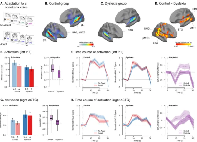
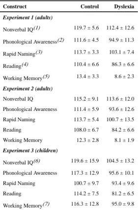
Experiment 1Adaptation to the consistent phonetic-phonological correspondence of speech from a single talker is a hallmark of abstract phonological processing in speech perception (Mullennix and Pisoni, 1990; Nygaard et al., 1994). We measured neurophysiological adaptation to the consistent phonetic features of speech in a block-design, sparse-sampling fMRI paradigm in which listeners heard spoken words and matched them to pictures (Figs. 1A and S1). In each block, we varied whether words were spoken by a single voice (“Adapt” condition) vs. multiple different voices (“No-Adapt” condition), with the expectation that listeners would show neural adaptation to the consistent voice (Wong et al., 2004). Adults with dyslexia (defined as a lifelong history of reading impairment and current performance in the bottom 25th percentile on two or more subtests of reading speed or accuracy) and control adults participated in this experiment (Tables 1 and S1).
Participants successfully maintained attention to the auditory stimuli throughout the word-to-picture matching task, as indicated by near-ceiling accuracy in both groups (control = 99.2%; dyslexia = 98.8%). A repeated-measures ANOVA for effects of group and condition revealed significantly greater accuracy in controls (F1,33 = 5.14, p = 0.03, η2 = 0.07), but no
effect of condition (p = 0.64) or interaction (p = 0.81). The same test for response time revealed a significant effect of condition (F1,33 = 53.62, p < 0.0001, η2 = 0.18) – with faster
response times in the Adapt condition (502ms vs. 563ms) – but no effect of group (p = 0.18) or interaction (p = 0.50).
In the control group, significant neural adaptation (No-Adapt > Adapt contrast) was
observed in two bilateral clusters, each extending throughout superior temporal gyrus (STG; including Heschl’s gyrus (HG) and planum temporale (PT)) and into posterior middle temporal gyrus (pMTG; Fig. 1B). In the dyslexia group, the magnitude and extent of adaptation were markedly reduced, with smaller clusters of significant adaptation encompassing only bilateral HG, PT, and right pSTG (Fig. 1C). There was no repetition-related enhancement (Adapt > No-Adapt) in either group. There were no overall group differences in the basic Task > Rest contrast (Fig. S1).
Compared to the control group, there was significantly less adaptation in the dyslexia group in clusters encompassing STG, PT, suparmarginal gyrus (SMG), and pMTG bilaterally (Fig. 1D). There were no clusters in which the dyslexia group showed more adaptation than controls. The group difference in adaptation was due an increasing difference between the Adapt and No-Adapt conditions over the course of stimulation in the control group, whereas
A
uthor Man
uscr
ipt
A
uthor Man
uscr
ipt
A
uthor Man
uscr
ipt
A
uthor Man
uscr
ipt
the dyslexia group showed similar response magnitude to both conditions throughout (Fig. 1E–H and Table S4).
We further explored how the magnitude of auditory adaptation in individuals with dyslexia was related to their reading abilities. Better core reading abilities in the dyslexia group, as measured by efficiency applying phonological and structural rules in decoding novel word forms (Woodcock, 1998), were associated with greater adaptation in both right (r = 0.56, p < 0.02) and left (r = 0.54, p < 0.03) planum temporale – an area known to be involved in phonetic-phonological abstraction in speech-sound processing (Graves et al., 2008; Griffiths and Warren, 2002).
Experiment 2
Following the discovery in Experiment 1 of significantly diminished auditory adaptation in dyslexia to the phonetic-phonological correspondence of speech, we conducted four follow-up experiments intended to determine the extent of neurophysiological adaptation
differences in dyslexia. We investigated whether adaptation differences would be limited to auditory stimuli or to stimuli with linguistic content, or whether diminished adaptation would be observed for the repetition of stimuli of any kind, indicating dysfunctional adaptation as a generalized feature of information processing in the dyslexic brain. A new sample of adult participants with and without dyslexia was recruited for these experiments, with the same inclusionary criteria as Experiment 1 (Tables 1 and S2).
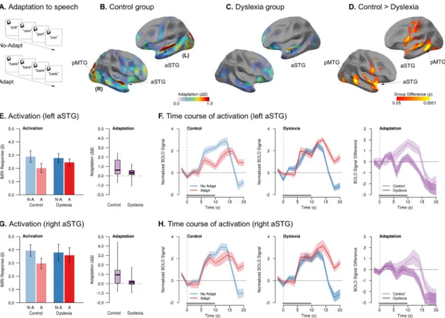
Experiment 2a: Spoken Words—We first investigated whether adaptation in the brains of adults with and without dyslexia would differ to a more obvious repetition of auditory stimuli than the subtle differences between talkers’ voices used in Experiment 1. In this experiment, we measured neurophysiological adaptation to blocks with the repeated presentation of a single spoken word (“Adapt”) vs. blocks with multiple different spoken words (“No-Adapt”) from a single speaker (Fig. 2A).
In the control group, hearing multiple repetitions of the same word resulted in significant adaptation (No-Adapt > Adapt) in left anterior STG and dorsal superior temporal sulcus (STS), as well as right aSTG, pMTG, and frontal operculum (FOC) (Fig. 2B). As before, the magnitude and extent of adaptation were markedly reduced in the dyslexia group, with smaller clusters of significant adaptation encompassing only left aSTG and right FOC. In both groups, there was a single cluster of repetition-related enhancement (greater BOLD response in the Adapt than No-Adapt condition) in left anterior supramarginal gyrus. There were no overall differences in the groups’ task-related activations (Fig. S1C,D).
The dyslexia group again exhibited significantly reduced adaptation compared to controls throughout perisylvian speech areas, including left STG, pMTG, and ventral premotor cortex, as well as right aSTG, planum polare, ventral premotor cortex, and pMTG. There were no clusters in which the dyslexia group showed more adaptation than controls. Whereas stimulus repetition attenuated neurophysiological response in the control group, individuals with dyslexia showed no such distinction in response magnitude (Fig. 2E,G). Likewise, whereas the magnitude of adaptation increased over time in controls, even
A
uthor Man
uscr
ipt
A
uthor Man
uscr
ipt
A
uthor Man
uscr
ipt
A
uthor Man
uscr
ipt
multiple repetitions of a single adapting stimulus did not attenuate the response in dyslexia (Fig. 2F,H, Table S4).
As in Experiment 1, we investigated whether the magnitude of neural adaptation in individuals with dyslexia was related to their reading abilities. We observed a positive correlation between greater adaptation in left PT and better reading skills (Woodcock, 1998) in individuals with dyslexia (r = 0.42, p = 0.05).
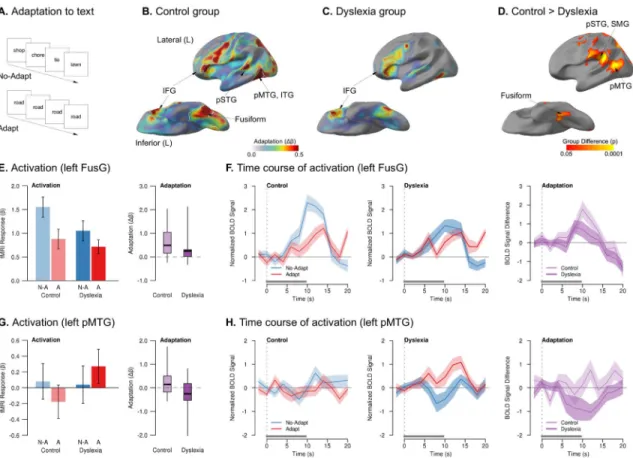
Experiment 2b: Written Words—We next investigated whether the control and dyslexia groups would differ in neural adaptation to the repeated presentation of written words (text) – still linguistic, but now visual stimuli. We measured neurophysiological adaptation to text by contrasting blocks of viewing multiple different written words versus blocks with the repeated presentation of a single written word (Fig. 3A).
In the control group, significant adaptation to the repeated presentation of a written word was observed in temporal (fusiform gyrus (FusG), inferior temporal gyrus (ITG), pSTG, and pMTG), frontal (inferior frontal gyrus (IFG), FOC, premotor cortex, and pre-supplementary motor area (SMA)), and visual (pericalcarine) cortices – all in the left hemisphere only (Fig. 3B). In dyslexia, the only cluster of significant adaptation to repeated visual words was found in left FOC (Fig. 3C). The dyslexia group also showed two clusters of significant enhancement in right pMTG and bilateral precuneus, with both areas also showing task-related deactivations (Fig. S2) (Buckner et al., 2008). Although there was a trend towards overall less activation to text stimuli in dyslexia, this Task > Rest group difference was not significant (Fig. S2C,D).
Compared to the control group, the dyslexia group exhibited significantly attenuated adaptation throughout FusG, pMTG, PT, SMG, and occipital cortex (Fig. 3D) – left hemisphere areas comprising the core of a network for reading (Dehaene and Cohen, 2011; McCandliss et al., 2003; Price, 2012; Price and Devlin, 2011). Additional clusters of significantly reduced adaptation were found in right insula, left motor cortex, and right angular gyrus (AG). There were no clusters in which the dyslexia group showed more adaptation than controls. Adaptation differences in FusG were the result of a smaller difference between the No-Adapt and Adapt conditions in the dyslexia group than in controls (Fig. 3E,F), with increasing group differences over time (Table S4). The group difference in pMTG (an area associated with semantic processing; Hickok and Poeppel, 2007) was qualitatively different: whereas the control group showed modest but non-significant adaptation in this region, the dyslexia group showed a trend for enhancement, with greater activation the more times a written word was repeated (Fig. 3G,H, Table S4). Unlike speech stimuli, and unlike Experiment 1, we did not observe any correlation between adaptation in ventral or lateral temporal areas and reading ability in dyslexia.
Experiment 2c: Objects—In addition to linguistic stimuli in auditory and visual modalities, we also investigated whether reduced adaptation in dyslexia would be observed for nonverbal visual stimuli such as color photographs of objects. We measured
neurophysiological adaptation by contrasting blocks of viewing photographs of multiple
A
uthor Man
uscr
ipt
A
uthor Man
uscr
ipt
A
uthor Man
uscr
ipt
A
uthor Man
uscr
ipt
different objects versus blocks with the repeated presentation of the same photograph of a single object (Fig. 4A).
In both control and dyslexia groups, significant adaptation to the repeated presentation of a photograph of an object was observed throughout visual and ventral temporal cortices known to process visual objects (Malach et al., 1995), including ITG, FusG, and lateral occipital cortex (LOC) extending dorsally into superior parietal lobule (SPL), as well as in bilateral inferior frontal sulcus (IFS), FOC, and preSMA (Fig. 4B,C). Both groups also showed significant enhancement in bilateral PT and precuneus (a task-deactivated area), and controls showed enhancement in two other task-deactivated areas: medial prefrontal cortex (MePFC) and superior frontal gyrus (SFG).
The magnitude of adaptation to object repetition in the dyslexia group was significantly less than in the control group throughout occipital and ventral temporal areas, including ITG, FusG, and LOC extending dorsally into SPL (Fig. 4D). Stimulus repetition resulted in a greater reduction of the BOLD response over time in the control group than in the dyslexia group (Fig. 4E–H and Table S4). There were no clusters in task-activated cortex in which the dyslexia group showed more adaptation than the control group. Better reading skills in the dyslexia group were significantly correlated with greater adaptation to repeated visual objects in both left (r = 0.45, p < 0.03) and right LOC (r = 0.42, p < 0.05).
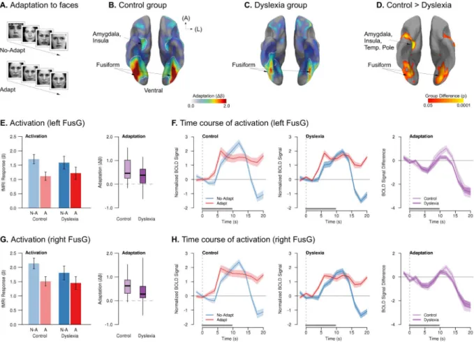
Experiment 2d: Faces—Although putatively nonverbal, objects are nameable, and visual processing of objects may nonetheless involve automatic activation of their linguistic labels (Chabal and Marian, 2015), which may be impaired in dyslexia (Norton and Wolf, 2012; Wolf, 1984); therefore, we lastly investigated whether reduced adaptation in dyslexia would be observed for non-nameable visual stimuli such as photographs of unfamiliar faces. We measured neurophysiological adaptation to faces by contrasting blocks of viewing
photographs of multiple different people’s faces versus blocks with the repeated presentation of the same photograph of a single person’s face (Fig. 5A).
In the control group, repeated presentation of the same picture of a face yielded significant adaptation throughout ventral temporal and visual cortices, including bilateral FusG and LOC, and right amygdala and anterior hippocampus (Fig. 5B). In the dyslexia group, significant adaptation was only observed in smaller bilateral FusG clusters (Fig. 5C). The dyslexia group exhibited significantly less adaptation than the control group in numerous regions associated with face processing (Kanwisher and Yovel, 2006; Tsao and Livingstone, 2008), including bilateral FusG and LOC; right hippocampus, temporal pole, and amygdala; and left premotor cortex and insula (Fig. 5D). As in all other experiments, this group difference was related to a greater reduction of the BOLD response to repeated stimuli in the control group than in the dyslexia group (Fig. 5E–H and Table S4). There were no clusters in which the dyslexia group showed more adaptation than controls, and we did not observe any correlation between adaptation in occipital or ventral temporal areas and reading ability in dyslexia.
To confirm that the group difference in adaptation did not reflect more heterogeneous localization of adaptation effects in the dyslexia group than in the control group, we
A
uthor Man
uscr
ipt
A
uthor Man
uscr
ipt
A
uthor Man
uscr
ipt
A
uthor Man
uscr
ipt
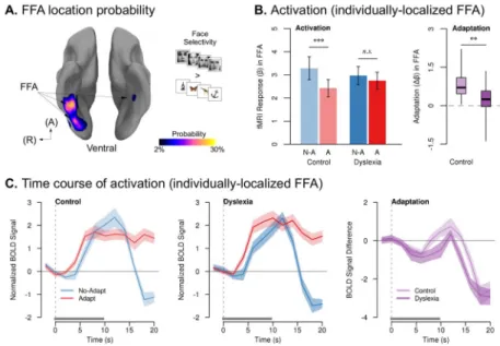
performed an additional analysis that measured adaptation only in the face-selective clusters of each participant. The fusiform face area (FFA; Kanwisher et al., 1997) was localized in individual participants by contrasting activation to faces vs. objects and identifying the anterior-most discrete face-selective cluster in the occipitofusiform region. The FFA was successfully localized in 22 participants in the dyslexia group and 18 participants in the control group (Fig. 6A). The probability of localizing an FFA did not differ by group (χ2 = 0.63, p = 0.43), nor was there a group difference in the threshold at which the FFA cluster could be localized (independent-sample t38 = 0.06, p = 0.95) or the volume of the FFA
(independent-sample t38 = 0.65, p = 0.52), indicating no difference in cortical specialization
for faces between the two groups. The control group showed significant adaptation in their FFAs to repeated faces (paired t17 = 6.13, p < 0.00002), whereas adaptation in the dyslexia
group was not significant (paired t21 = 1.67, p = 0.11). The magnitude of FFA adaptation
was significantly less in the dyslexia group than in controls (independent-sample t38 = 3.37,
p < 0.002) (Fig. 6B). As in the whole-brain analyses, the group difference in adaptation reflected an increasingly smaller response to repeated stimuli in the control group than in dyslexia (Fig. 6C and Table S4). (We attempted a similar analysis for the visual word form area (VWFA; McCandliss et al., 2003) in Experiment 2b, but were unable to reliably isolate this region in our participants using a Words > Objects contrast.)
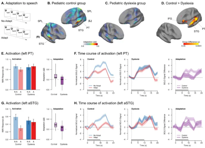
Experiment 3
Having seen robust and apparently domain-general neurophysiological adaptation deficits in adults with dyslexia, we further asked whether reading-ability-related adaptation differences were evident in emerging readers (age 6–9 years). We repeated Experiment 2a with young children with dyslexia and their age-matched peers with typical reading development. We measured neurophysiological adaptation by contrasting blocks of a single, repeated spoken word versus blocks with multiple different spoken words from a single speaker (Fig. 7A). Children with and without dyslexia successfully maintained attention to the auditory stimuli throughout in both conditions, as indicated by near-ceiling accuracy in both groups (control = 98.6%; dyslexia = 97.8%). A repeated-measures ANOVA for effects of group and condition on accuracy revealed no effect of group (F1,43 = 1.64, p = 0.21), no effect of
condition (p = 0.17), and no interaction (p = 0.33). The same test for response time revealed a significant effect of condition (F1,43 = 6.16, p < 0.02, η2 = 0.03) – with faster response
times in the Adapt condition (977ms vs. 1044ms) – but no effect of group (p = 0.59) or interaction (p = 0.56).
In control children, hearing multiple repetitions of the same word resulted in significant adaptation in bilateral STG, PT, and SPL (Fig. 7B). For children with dyslexia, however, there were no areas exhibiting significant adaptation (Fig. 7C). There were no clusters of repetition-related enhancement in either group. Adaptation in children with dyslexia was significantly less than that of controls in left PT, STG, and IFG (Fig. 7D). This group difference was the result of an increasingly large reduction of response to stimulus repetition over time in the control group than in the dyslexia group, who in turn showed almost no response distinction between the Adapt and No-Adapt conditions (Fig. 7E–H and Table S4). There were no clusters in which the dyslexia group showed more adaptation than controls.
A
uthor Man
uscr
ipt
A
uthor Man
uscr
ipt
A
uthor Man
uscr
ipt
A
uthor Man
uscr
ipt
Because the children with dyslexia were in only preliminary stages of reading development, we investigated whether the magnitude of auditory adaptation was related to their
phonological awareness – an important preliterate skill (Bradley and Bryant, 1983) that is a better predictor of long-term reading outcomes than early reading abilities (MacDonald and Cornwall, 1995). We observed positive correlations in the children with dyslexia between the magnitude of adaptation in left PT (r = 0.46, p < 0.04) and left aSTG (r = 0.50, p < 0.025) and their phonological awareness (Wagner et al., 1999).
DISCUSSION
Across six experiments, we found that rapid neural adaptation, as indexed by repetition-induced reduction of the fMRI BOLD signal (Grill-Spector and Malach, 2001), was diminished in children and adults with dyslexia for every stimulus type assessed – auditory language, visual language, visual objects, and faces. This deficit in adaptation was found selectively in the brain regions known to be critically involved in processing each stimulus type (Bell et al., 2011; Chandrasekaran et al., 2011; Kanwisher et al., 1997; Malach et al., 1995; McCandliss et al., 2003; Weiner et al., 2010). Correspondingly, the amount of
preserved adaptation in these stimulus-specific brain regions was related to the reading skills of adults and preliterate skills of children with dyslexia. The breadth of this deficit – across ages, brain regions, and stimulus types – suggests that dysfunction of neural adaptation may be an important neurophysiological difference in many individuals with dyslexia.
Diminished neural adaptation to linguistic stimuli parallels known behavioral deficits in these domains. Adults with dyslexia exhibited less neural adaptation to the speech of a consistent talker, corresponding to their reduced behavioral ability to learn the specific phonetic-phonological features of individual voices (Perrachione et al., 2011) and impaired implicit learning of auditory categories (Gabay and Holt, 2015). Adaptation deficits in dyslexia cannot be ascribed to failure to notice the subtle repetition of stimulus features (voices) in Experiment 1, because even the highly salient, multiple repetitions of single spoken or written words in Experiment 2 resulted in less neural adaptation in dyslexia than controls. Furthermore, these adaptation deficits were found in children with dyslexia early in their literacy development, suggesting that reduced sensitivity to the repetition of language stimuli is present even before reading skills have been extensively trained (Boets, 2014; Goswami, 2015). This parallels the observation that perceptual adaptation is related to preliteracy and language skills even before children begin to learn to read (Banai and Yifat, 2012).
Perhaps more surprisingly, we also observed adaptation deficits to repetition of non-linguistic stimuli such as objects and faces, although strictly perceptual deficits for these stimuli are not generally observed in dyslexia (Rüsseler et al., 2003; cf. Sigurdardottir et al., 2015). This suggests that, in dyslexia, the general capacity for perceptual processes to establish short-term representations of stimulus consistency may be impaired (Ahissar et al., 2006; Chandrasekaran et al., 2009; Hornickel and Kraus, 2013; Jaffe-Dax et al., 2015; Oganian and Ahissar, 2012).
A
uthor Man
uscr
ipt
A
uthor Man
uscr
ipt
A
uthor Man
uscr
ipt
A
uthor Man
uscr
ipt
An impairment in neural adaptation, which reflects the neural processes involved in establishing robust short-term perceptual representations (Alain et al., 2007; Garrido et al., 2009; Jääskeläinen et al., 2007; Khouri and Nelken, 2015) provides a framework for understanding how several other behavioral and neural differences observed in dyslexia may form a constellation of low-level, adaptation-related deficits. First, individuals with dyslexia may have an impairment “anchoring” to consistent stimulus statistics in order to enhance perceptual thresholds (Ahissar et al., 2006; Banai and Ahissar, 2010), a behavioral effect reflected in rapid neural adaptation (Fritz et al., 2003; Garrido et al., 2009). Second,
individuals with dyslexia are also frequently observed to have impairments recognizing both auditory and visual stimuli in the presence of noise (Sperling et al., 2005, 2006; Ziegler et al., 2009). Correspondingly, short-term adaptation of auditory and visual cortices to the statistics of noise facilitate perception in animal models (Atiani et al., 2009), and neural adaptation may also support noise exclusion in humans (Parbery-Clark et al., 2011). Third, neural coding deficits in auditory thalamus and brainstem have been found in dyslexia for tasks that use consistent stimulus or noise features (Chandrasekaran et al., 2009; Díaz et al., 2012). Our observation of dysfunction in cortical adaptation suggests these may be systems-level deficits, given corticofugal signaling is responsible for modulating auditory
representations in thalamus and brainstem (Chandrasekaran et al., 2014; Suga et al., 2002). Finally, individuals with dyslexia may exhibit differences in gamma-band neural
synchronization to auditory stimuli (Lehongre et al., 2011). Synchronous neural activity at these frequencies is induced by neural adaptation (Hansen and Dragoi, 2011), and such neural entrainment to stimulus consistency aids perception (Giraud and Poeppel, 2012; Park et al., 2015). This cluster of adaptation-related impairments may arise from dysfunction in common or related neurobiological mechanisms – namely, ones that constrain the extent to which the dyslexic brain can overcome internal noise and establish the perceptual constancy that underlies short-term perceptual facilitation and supports long-term perceptual learning (Hornickel and Kraus, 2013; Jääskeläinen et al., 2007; Jaffe-Dax et al., 2015).
Could adaptation differences reflect attentional differences?
The suggestion that visual-spatial attention deficits may, in some cases, underlie impaired reading (Franceschini et al., 2012; Vidyasagar and Pammer, 2010) warrants considering whether diminished adaptation in dyslexia might have an attentional origin. There are several reasons why it is unlikely that the present results reflect an impairment in voluntary or intentional deployment of top-down attention. First, between-group differences in adaptation were always observed in the cortical areas specifically implicated in processing stimuli of each type, not areas associated with volitional, top-down attention (Hopfinger et al., 2000; Womelsdorf and Everling, 2015). Second, substantial fMRI adaptation is readily observed in both attentive and passive tasks (Kourtzi and Kanwisher, 2001; Larsson and Smith, 2012; Sawamura et al., 2005). Third, our original observation of adaptation differences (Experiment 1) occurred in a speech perception task where the attentional demands of the Adapt and No-Adapt conditions did not differ.
Attention may nonetheless affect adaptation in subtler ways. Stimuli in Adapt conditions were highly repetitive and thus highly predictable. Perceptual expectations influence the magnitude of neural adaptation (Costa-Faidella et al., 2011; Summerfield et al., 2008;
A
uthor Man
uscr
ipt
A
uthor Man
uscr
ipt
A
uthor Man
uscr
ipt
A
uthor Man
uscr
ipt
Todorovic et al., 2011). Likewise, animal models have shown that rapid changes in neural responses to repeated stimuli require top-down neuromodulatory input (Fritz et al., 2003; Froemke et al., 2007). Diminished adaptation in dyslexia might therefore represent a failure to generate robust, top-down perceptual expectations (Jaffe-Dax et al., 2015): Higher cortical areas may not provide appropriate feedback signals to sensory cortices to facilitate
adaptation (Boets et al., 2013; Saygin et al., 2013; Yeatman et al., 2011). Alternatively, there is some evidence for microanatomical abnormalities that disrupt laminar structure in dyslexia (Galaburda et al., 1994), and disruptions to the local organization of laminar circuits may interfere with the local or long-range connections supporting adaptation, which depends on finely tuned neuromodulatory input (Froemke et al., 2007) and lamina-specific synchronization in sensory cortex (Hansen and Dragoi, 2011).
General neural dysfunction and specific reading impairment
A widely replicated finding in the neuroscience of dyslexia is the observation of reduced activation to print in the canonical reading network, and particularly in left occipitotemporal cortex (Paulesu et al., 2014; Shaywitz et al., 1998). However, the causal connection between developmental dyslexia and reduced sensitivity to print this region remains unclear. On the one hand, dysfunction of left occipitotemporal cortex itself could be a pathway to dyslexia. Alternatively, reduced sensitivity to print in this region could result from developmental differences in other processes that are compromised in dyslexia prior to learning to read, such as phonemic awareness in speech (Bradley and Bryant, 1983) or rapid naming of visual stimuli (Norton and Wolf, 2012). Weaknesses in these preliteracy skills may encumber the functional integration of occipitotemporal cortex into a robust reading network.
Correspondingly, there has been a strong interest in identifying low-level deficits in dyslexia that might serve as neural precursors to explain weaknesses in these preliteracy skills. A challenge for any hypothesis of low-level impairments in dyslexia is to explain how a “general” biological dysfunction gives rise to a “specific” impairment in reading without impacting other complex behaviors. For instance, proponents of various low-level auditory deficits as an explanation for poor reading (Goswami, 2011; Tallal and Piercy, 1973) must offer an account for how these interfere specifically with reading but do not produce apparent disruptions to complex auditory abilities like perceiving speech and music. Likewise, proponents of core visual deficits (Franceschini et al., 2012; Stein, 2001; Vidyasagar and Pammer, 2010) must be able to explain how these impairments affect reading, but do not result in correspondingly serious disruptions to other complex visual behaviors like recognizing objects or driving a car.
A dysfunction of neural adaptation differs from other low-level hypotheses in that it does not posit an impairment in processing specific stimulus features that are critical for other auditory or visual abilities. Instead, it suggests that there may be a general impairment in processes that facilitate perception under adverse or challenging conditions (Jääskeläinen et al., 2007), which, at its core, represents an impairment in mechanisms for rapid perceptual learning – including learning the robust speech-sound categories necessary for sound-to-symbol matching in reading (Bradley and Bryant, 1983). Neural adaptation facilitates behaviors such as detecting stimuli in noise (Atiani et al., 2009; Chandrasekaran et al., 2009;
A
uthor Man
uscr
ipt
A
uthor Man
uscr
ipt
A
uthor Man
uscr
ipt
A
uthor Man
uscr
ipt
Parbery-Clark et al., 2011), discriminating subtle stimulus differences (Edeline et al., 1993; Fritz et al., 2003), and rapid learning of new perceptual categories (Alain et al., 2007; Garrido et al., 2009). Correspondingly, for all of these behaviors facilitated by adaptation, significant deficits have consistently been reported in dyslexia.
Although the brain has evolved to be computationally powerful for solving certain types of problems such as perceiving speech and recognizing objects, learning to read differs in that it is a cultural invention imposing itself upon circuitry that evolved for other purposes (Dehaene et al., 2010; McCandliss et al., 2003). Learning to read depends on the ability to orchestrate, across two perceptual modalities, the complex correspondence between abstract phonological representations of speech sounds and abstract orthographic representations of written symbols – a task that becomes disproportionately more difficult if either type of representation, or access to them, is impaired. Correspondingly, learning to read is one of the most complex examples of human learning, the demands of which are evident from its lengthy and explicit instruction throughout childhood and into adulthood. There is no other human behavior that approaches reading’s demands for coordinating multimodal perceptual representations and cognitive processes. In this way, a general neural dysfunction that is subtly detrimental to other behaviors may be substantially detrimental for learning to read. Extensions and limitations of the fMRI adaptation technique
The specific physiological mechanisms that give rise to adaptation in the BOLD signal are not yet fully understood (Grill-Spector et al., 2006; Krekelberg et al., 2006; Sawamura et al., 2006), and repetition-related fMRI adaptation is likely to reflect a variety of diverse
neurophysiological processes depending on variables like the cortical location (Weiner et al., 2010), stimulation paradigm (Müller et al., 2013), and task demands (Jiang et al., 2013) in which it is observed. Interestingly, fMRI adaptation is attenuated for perceptually noisy stimuli (Turk-Browne et al., 2007), paralleling hypotheses that neural representations themselves may be disproportionately noisy in dyslexia (Hornickel and Kraus, 2013; Jaffe-Dax et al., 2015; Ziegler et al., 2009).
Although the coarseness of fMRI signals precludes this tool from resolving the circuit- or cellular-level source of adaptation impairments in dyslexia by itself, the present observations provide a stronger foundation for the targeted pursuit of neurophysiological differences in dyslexia informed by basic research in animal models. For instance, the necessary role of cholinergic neuromodulation for rapid neural adaptation and consequent behavioral enhancement is well known at the cellular level (Froemke et al., 2007; Suga et al., 2002), with similar effects shown in BOLD imaging (Thiel et al., 2002). Atypical cerebral concentration of neurometabolic precursors to acetylcholine have been found in dyslexia (Bruno et al., 2013; Pugh et al., 2014). Advances in cholinergic radioligands now permit targeted investigation of this neurotransmitter in behavioral adaptation tasks and in dyslexia. Although this example is speculative, our observation of generalized differences in
neurophysiological adaptation contributes to a growing literature from which we can begin to develop a systems- (as opposed to cognitive-) neuroscientific framework for investigating dyslexia.
A
uthor Man
uscr
ipt
A
uthor Man
uscr
ipt
A
uthor Man
uscr
ipt
A
uthor Man
uscr
ipt
The complexity of reading makes successfully learning this skill vulnerable to a broad range of linguistic, visual, and attentional dysfunctions that may occur in variable mixtures in individual children with dyslexia. Correspondingly, although perceptual adaptation deficits in dyslexia have been observed across a large number of studies and diverse range of tasks, some studies have found deficits on only a subset of tasks employed (Beattie et al., 2011), for only a subset of participants (Willburger and Landerl, 2010), or for none at all (Wijnen et al., 2012). The results from the present study, however, provide multiple converging lines of evidence for a substantive relationship between neural adaptation deficits and reading impairment. We not only observed that the degree of neural adaptation in task-relevant brain regions was significantly correlated with reading abilities in adults with dyslexia and with phonological skills in children with dyslexia, we also observed consistent and significant group differences in neural adaptation for all tasks. Such correlations, combined with reliable group differences, correspond to the two ways that neuroimaging and behavioral studies are able to empirically characterize differences between typical and dyslexic brain function.
Finally, although we observed consistently diminished adaptation in dyslexia, and although the magnitude of that adaptation was related to reading ability in dyslexia, our choice to use high-level, perceptually complex stimuli in our adaptation paradigms meant we could not observe whether the degree of neural adaptation was directly related to perceptual adaptation, as would have been possible with a task designed to evaluate perceptual thresholds (Braida et al., 1984). Nevertheless, two-way relationships between neural and perceptual adaptation (Alain et al., 2007), perceptual adaptation and reading abilities (Ahissar et al., 2006; Banai and Yifat, 2012), and neural adaptation and reading impairment (present work) have now been repeatedly demonstrated, and confirmation of the three-way relationship among these three factors can be made in future studies.
An emerging picture of the dyslexic brain
Dyslexia is a specific impairment in developing typical reading abilities. Correspondingly, structural and functional disruptions to the network of brain areas known to support reading are consistently observed in dyslexia (Norton et al., 2015). However, these observations confound cause and consequence, especially since reading is a cultural invention that must make use of existing circuitry evolved for other purposes (Dehaene and Cohen, 2007; Dehaene et al., 2010). In this way, differences between brains that exert more subtle influences on non-reading behaviors are likely to be the culprit in a cascade of perceptual and mnemonic challenges that interfere with the development of typical reading abilities. Recent research has begun to elucidate a cluster of behaviorally-distinct but potentially physiologically-related impairments that are evinced by individuals with reading difficulties and observable in their brains. Through this collection of neural signatures – including unstable neural representations (Hornickel and Kraus, 2013), diminished top-down reflexive control (Boets, 2014; Boets et al., 2013; Chandrasekaran et al., 2009), susceptibility to noise (Sperling et al., 2005; Ziegler et al., 2009), and inability to construct robust short-term perceptual representations (Díaz et al., 2012; Jaffe-Dax et al., 2015; Lehongre et al., 2011) – we are beginning to see that reading impairments can arise from general dysfunction in the processes supported by rapid neural adaptation.
A
uthor Man
uscr
ipt
A
uthor Man
uscr
ipt
A
uthor Man
uscr
ipt
A
uthor Man
uscr
ipt
EXPERIMENTAL PROCEDURES
ParticipantsSeparate groups of adults completed Experiments 1 (N = 19 dyslexia, 19 control) and 2 (N = 24 dyslexia, 23 control). Children with dyslexia (N = 26) and with typical reading
development (N = 25) completed Experiment 3. See Tables S1–S3 for participant assessment and characterization.
Task and Stimuli
In Experiment 1, participants matched auditory words to pictures during a 42-minute, sparse-sampling, block-design fMRI task. In the No-Adapt condition, words were spoken by four different voices; in the Adapt condition, words were spoken by one consistent voice. In Experiments 2 and 3, participants passively attended speech, text, objects, or faces during separate 10-minute, continuous-sampling, block-design fMRI tasks. Eight unique stimuli were presented in No-Adapt blocks; a single stimulus was repeated eight times in Adapt blocks. Children underwent only the speech task.
MRI Analysis
Functional data were preprocessed and modeled in SPM8, Freesurfer, and Nipype. Model and contrast estimation were conducted in EPI space; contrast images were coregistered to participants’ anatomy, then normalized to MNI space. Group-level mixed-effects models assessed adaptation within and between groups.
For full methods details, see Supplemental Experimental Procedures.
Supplementary Material
Refer to Web version on PubMed Central for supplementary material.
Acknowledgments
We thank Chris Moore, Stefanie Shattuck-Hufnagel, Nancy Kanwisher, Bob Desimone, Susan Whitfield-Gabrieli, Carlos Cardenas-Iniguez, Sara Beach, Dasun Peramunage, John Lymberis, Abigail Cyr, Sok Ee Lee, Elizabeth Norton, Bianca Levy, Karl Wolff, Patricia Saxler, Michael Waskom, Pamela Hook, and Cindy Gibbs for their assistance. We thank the Athinoula A. Martinos Imaging Center at the McGovern Institute for Brain Research (MIT) and Steve Shannon, Sheeba Arnold, and Christina Triantafyllou for data collection technical support. This research was supported by the Ellison Medical Foundation and NIH grant UL1RR025758 to JG, and an NSF Graduate Research Fellowship to TP.
References
Ahissar M. Dyslexia and the anchoring-deficit hypothesis. Trends Cogn Sci. 2007; 11:458–465. [PubMed: 17983834]
Ahissar M, Lubin Y, Putter-Katz H, Banai K. Dyslexia and the failure to form a perceptual anchor. Nat Neurosci. 2006; 9:1558–1564. [PubMed: 17115044]
Alain C, Snyder JS, He Y, Reinke KS. Changes in auditory cortex parallel rapid perceptual learning. Cereb Cortex. 2007; 17:1074–1084. [PubMed: 16754653]
A
uthor Man
uscr
ipt
A
uthor Man
uscr
ipt
A
uthor Man
uscr
ipt
A
uthor Man
uscr
ipt
Atiani S, Elhilali M, David SV, Fritz JB, Shamma SA. Task difficulty and performance induce diverse adaptive patterns in gain and shape of primary auditory cortical receptive fields. Neuron. 2009; 61:467–480. [PubMed: 19217382]
Banai K, Ahissar M. On the importance of anchoring and the consequences of its impairment in dyslexia. Dyslexia. 2010; 16:240–257. [PubMed: 20680994]
Banai K, Yifat R. Anchoring in 4- to 6-year-old children relates to predictors of reading. J Exp Child Psychol. 2012; 112:403–416. [PubMed: 22608190]
Beattie RL, Lu ZL, Manis FR. Dyslexic adults can learn from repeated stimulus presentation but have difficulties in excluding external noise. PLOS ONE. 2011; 6:e27893. [PubMed: 22132164] Belin P, Zatorre RJ. Adaptation to speaker’s voice in right anterior temporal lobe. Neuroreport. 2003;
14:2105–2109. [PubMed: 14600506]
Bell AH, Malecek NJ, Morin EL, Hadj-Bouziane F, Tootell RBH, Ungerleider LG. Relationship between functional magnetic resonance imaging-identified regions and neuronal category selectivity. J Neurosci. 2011; 31:12229–12240. [PubMed: 21865466]
Ben-Yehudah G, Ahissar M. Sequential spatial frequency discrimination is consistently impaired among adult dyslexics. Vision Res. 2004; 44:1047–1063. [PubMed: 15031099]
Boets B. Dyslexia: reconciling controversies within an integrative developmental perspective. Trends Cogn Sci. 2014; 18:501–503. [PubMed: 25034040]
Boets B, de Beeck HPO, Vandermosten M, Scott SK, Gillebert CR, Mantini D, Bulthé J, Sunaert S, Wouters J, Ghesquière P. Intact but less accessible phonetic representations in adults with dyslexia. Science. 2013; 342:1251–1254. [PubMed: 24311693]
Bradley L, Bryant PE. Categorizing sounds and learning to read—a causal connection. Nature. 1983; 301:419–421.
Braida LD, Lim JS, Berliner JE, Durlach NI, Rabinowitz WM, Purks SR. Intensity perception. XIII Perceptual anchor model of context-coding. J Acoust Soc Am. 1984; 76:722–731. [PubMed: 6491044]
Bruno JL, Lu ZL, Manis FR. Phonological processing is uniquely associated with neuro-metabolic concentration. NeuroImage. 2013; 67:175–181. [PubMed: 23147236]
Buckner RL, Andrews-Hanna JR, Schacter DL. The brain’s default network. Ann N Y Acad Sci. 2008; 1124:1–38. [PubMed: 18400922]
Chabal S, Marian V. Speakers of different languages process the visual world differently. J Exp Psychol Gen. 2015; 144:539–550. [PubMed: 26030171]
Chait M, Eden G, Poeppel D, Simon JZ, Hill DF, Flowers DL. Delayed detection of tonal targets in background noise in dyslexia. Brain Lang. 2007; 102:80–90. [PubMed: 16887180]
Chandrasekaran B, Hornickel J, Skoe E, Nicol T, Kraus N. Context-dependent encoding in the human auditory brainstem relates to hearing speech in noise: implications for developmental dyslexia. Neuron. 2009; 64:311–319. [PubMed: 19914180]
Chandrasekaran B, Chan AHD, Wong PCM. Neural processing of what and who information in speech. J Cogn Neurosci. 2011; 23:2690–2700. [PubMed: 21268667]
Chandrasekaran B, Skoe E, Kraus N. An integrative model of subcortical auditory plasticity. Brain Topogr. 2014; 27:539–552. [PubMed: 24150692]
Costa-Faidella J, Baldeweg T, Grimm S, Escera C. Interactions between “what” and “when” in the auditory system: temporal predictability enhances repetition suppression. J Neurosci. 2011; 31:18590–18597. [PubMed: 22171057]
Dehaene S, Cohen L. Cultural recycling of cortical maps. Neuron. 2007; 56:384–398. [PubMed: 17964253]
Dehaene S, Cohen L. The unique role of the visual word form area in reading. Trends Cogn Sci. 2011; 15:254–262. [PubMed: 21592844]
Dehaene S, Pegado F, Braga LW, Ventura P, Filho GN, Jobert A, Dehaene-Lambertz G, Kolinsky R, Morais J, Cohen L. How learning to read changes the cortical networks for vision and language. Science. 2010; 330:1359–1364. [PubMed: 21071632]
Díaz B, Hintz F, Kiebel SJ, von Kriegstein K. Dysfunction of the auditory thalamus in developmental dyslexia. Proc Natl Acad Sci. 2012; 109:13841–13846. [PubMed: 22869724]
A
uthor Man
uscr
ipt
A
uthor Man
uscr
ipt
A
uthor Man
uscr
ipt
A
uthor Man
uscr
ipt
Edeline JM, Pham P, Weinberger NM. Rapid development of learning-induced receptive field plasticity in the auditory cortex. Behav Neurosci. 1993; 107:539–551. [PubMed: 8397859]
Franceschini S, Gori S, Ruffino M, Pedrolli K, Facoetti A. A causal link between visual spatial attention and reading acquisition. Curr Biol. 2012; 22:814–819. [PubMed: 22483940] Fritz J, Shamma S, Elhilali M, Klein D. Rapid task-related plasticity of spectrotemporal receptive
fields in primary auditory cortex. Nat Neurosci. 2003; 6:1216–1223. [PubMed: 14583754] Froemke RC, Merzenich MM, Schreiner CE. A synaptic memory trace for cortical receptive field
plasticity. Nature. 2007; 450:425–429. [PubMed: 18004384]
Gabay Y, Holt LL. Incidental learning of sound categories is impaired in developmental dyslexia. Cortex. 2015; 73:131–143. [PubMed: 26409017]
Gabrieli JDE. Dyslexia: a new synergy between education and cognitive neuroscience. Science. 2009; 325:280–283. [PubMed: 19608907]
Galaburda AM, Menard MT, Rosen GD. Evidence for aberrant auditory anatomy in developmental dyslexia. Proc Natl Acad Sci. 1994; 91:8010–8013. [PubMed: 8058748]
Garrido MI, Kilner JM, Kiebel SJ, Stephan KE, Baldeweg T, Friston KJ. Repetition suppression and plasticity in the human brain. NeuroImage. 2009; 48:269–279. [PubMed: 19540921]
Giraud AL, Poeppel D. Cortical oscillations and speech processing: emerging computational principles and operations. Nat Neurosci. 2012; 15:511–517. [PubMed: 22426255]
Goswami U. A temporal sampling framework for developmental dyslexia. Trends Cogn Sci. 2011; 15:3–10. [PubMed: 21093350]
Goswami U. Sensory theories of developmental dyslexia: three challenges for research. Nat Rev Neurosci. 2015; 16:43–54. [PubMed: 25370786]
Graves WW, Grabowski TJ, Mehta S, Gupta P. The left posterior superior temporal gyrus participates specifically in accessing lexical phonology. J Cogn Neurosci. 2008; 20:1698–1710. [PubMed: 18345989]
Griffiths TD, Warren JD. The planum temporale as a computational hub. Trends Neurosci. 2002; 25:348–353. [PubMed: 12079762]
Grill-Spector K, Malach R. fMR-adaptation: a tool for studying the functional properties of human cortical neurons. Acta Psychol. 2001; 107:293–321.
Grill-Spector K, Henson R, Martin A. Repetition and the brain: neural models of stimulus-specific effects. Trends Cogn Sci. 2006; 10:14–23. [PubMed: 16321563]
Hansen BJ, Dragoi V. Adaptation-induced synchronization in laminar cortical circuits. Proc Natl Acad Sci. 2011; 108:10720–10725. [PubMed: 21659632]
Hickok G, Poeppel D. The cortical organization of speech processing. Nat Rev Neurosci. 2007; 8:393– 402. [PubMed: 17431404]
Hopfinger JB, Buonocore MH, Mangun GR. The neural mechanisms of top-down attentional control. Nat Neurosci. 2000; 3:284–291. [PubMed: 10700262]
Hornickel J, Kraus N. Unstable representation of sound: a biological marker of dyslexia. J Neurosci. 2013; 33:3500–3504. [PubMed: 23426677]
Jääskeläinen IP, Ahveninen J, Belliveau JW, Raij T, Sams M. Short-term plasticity in auditory cognition. Trends Neurosci. 2007; 30:653–661. [PubMed: 17981345]
Jaffe-Dax S, Raviv O, Jacoby N, Loewenstein Y, Ahissar M. A computational model of implicit memory captures dyslexics’ perceptual deficits. J Neurosci. 2015; 35:12116–12126. [PubMed: 26338323]
Jiang J, Summerfield C, Egner T. Attention sharpens the distinction between expected and unexpected percepts in the visual brain. J Neurosci. 2013; 33:18438–18447. [PubMed: 24259568]
Kanwisher N, Yovel G. The fusiform face area: a cortical region specialized for the perception of faces. Philos Trans R Soc B Biol Sci. 2006; 361:2109–2128.
Kanwisher N, McDermott J, Chun MM. The fusiform face area: a module in human extrastriate cortex specialized for face perception. J Neurosci. 1997; 17:4302–4311. [PubMed: 9151747]
Khouri L, Nelken I. Detecting the unexpected. Curr Opin Neurobiol. 2015; 35:142–147. [PubMed: 26318534]
A
uthor Man
uscr
ipt
A
uthor Man
uscr
ipt
A
uthor Man
uscr
ipt
A
uthor Man
uscr
ipt
Kourtzi Z, Kanwisher N. Representation of perceived object shape by the human lateral occipital complex. Science. 2001; 293:1506–1509. [PubMed: 11520991]
Krekelberg B, Boynton GM, van Wezel RJA. Adaptation: from single cells to BOLD signals. Trends Neurosci. 2006; 29:250–256. [PubMed: 16529826]
Larsson J, Smith AT. fMRI repetition suppression: neuronal adaptation or stimulus expectation? Cereb Cortex. 2012; 22:567–576. [PubMed: 21690262]
Lehongre K, Ramus F, Villiermet N, Schwartz D, Giraud AL. Altered low-γ sampling in auditory cortex accounts for the three main facets of dyslexia. Neuron. 2011; 72:1080–1090. [PubMed: 22196341]
Lum JAG, Ullman MT, Conti-Ramsden G. Procedural learning is impaired in dyslexia: Evidence from a meta-analysis of serial reaction time studies. Res Dev Disabil. 2013; 34:3460–3476. [PubMed: 23920029]
Lyon GR, Shaywitz SE, Shaywitz BA. A definition of dyslexia. Ann Dyslexia. 2003; 53:1–14. MacDonald GW, Cornwall A. The relationship between phonological awareness and reading and
spelling achievement eleven years later. J Learn Disabil. 1995; 28:523–527. [PubMed: 7595043] Malach R, Reppas JB, Benson RR, Kwong KK, Jiang H, Kennedy WA, Ledden PJ, Brady TJ, Rosen
BR, Tootell RB. Object-related activity revealed by functional magnetic resonance imaging in human occipital cortex. Proc Natl Acad Sci. 1995; 92:8135–8139. [PubMed: 7667258]
McCandliss BD, Cohen L, Dehaene S. The visual word form area: expertise for reading in the fusiform gyrus. Trends Cogn Sci. 2003; 7:293–299. [PubMed: 12860187]
Menghini D, Hagberg GE, Caltagirone C, Petrosini L, Vicari S. Implicit learning deficits in dyslexic adults: An fMRI study. NeuroImage. 2006; 33:1218–1226. [PubMed: 17035046]
Mullennix JW, Pisoni DB. Stimulus variability and processing dependencies in speech perception. Percept Psychophys. 1990; 47:379–390. [PubMed: 2345691]
Müller NG, Strumpf H, Scholz M, Baier B, Melloni L. Repetition suppression versus enhancement--it’s quantity that matters. Cereb Cortex. 2013; 23:315–322. [PubMed: 22314047]
Norton ES, Wolf M. Rapid automatized naming (RAN) and reading fluency: implications for understanding and treatment of reading disabilities. Annu Rev Psychol. 2012; 63:427–452. [PubMed: 21838545]
Norton ES, Beach SD, Gabrieli JD. Neurobiology of dyslexia. Curr Opin Neurobiol. 2015; 30:73–78. [PubMed: 25290881]
Nygaard LC, Sommers MS, Pisoni DB. Speech perception as a talker-contingent process. Psychol Sci. 1994; 5:42–46. [PubMed: 21526138]
Oganian Y, Ahissar M. Poor anchoring limits dyslexics’ perceptual, memory, and reading skills. Neuropsychologia. 2012; 50:1895–1905. [PubMed: 22561890]
Parbery-Clark A, Strait DL, Kraus N. Context-dependent encoding in the auditory brainstem subserves enhanced speech-in-noise perception in musicians. Neuropsychologia. 2011; 49:3338–3345. [PubMed: 21864552]
Park H, Ince RAA, Schyns PG, Thut G, Gross J. Frontal top-down signals increase coupling of auditory low-frequency oscillations to continuous speech in human listeners. Curr Biol. 2015; 25:1649–1653. [PubMed: 26028433]
Paulesu E, Danelli L, Berlingeri M. Reading the dyslexic brain: multiple dysfunctional routes revealed by a new meta-analysis of PET and fMRI activation studies. Front Hum Neurosci. 2014; 8:830. [PubMed: 25426043]
Perrachione TK, Del Tufo SN, Gabrieli JDE. Human voice recognition depends on language ability. Science. 2011; 333:595–595. [PubMed: 21798942]
Pollack C, Luk G, Christodoulou JA. A meta-analysis of functional reading systems in typically developing and struggling readers across different alphabetic languages. Front Psychol. 2015; 6:191. [PubMed: 25806009]
Price CJ. A review and synthesis of the first 20 years of PET and fMRI studies of heard speech, spoken language and reading. NeuroImage. 2012; 62:816–847. [PubMed: 22584224]
Price CJ, Devlin JT. The Interactive Account of ventral occipitotemporal contributions to reading. Trends Cogn Sci. 2011; 15:246–253. [PubMed: 21549634]
A
uthor Man
uscr
ipt
A
uthor Man
uscr
ipt
A
uthor Man
uscr
ipt
A
uthor Man
uscr
ipt
Pugh KR, Frost SJ, Rothman DL, Hoeft F, Tufo SND, Mason GF, Molfese PJ, Mencl WE, Grigorenko EL, Landi N, et al. Glutamate and choline levels predict individual differences in reading ability in emergent readers. J Neurosci. 2014; 34:4082–4089. [PubMed: 24623786]
Rueckl JG, Paz-Alonso PM, Molfese PJ, Kuo WJ, Bick A, Frost SJ, Hancock R, Wu DH, Mencl WE, Duñabeitia JA, et al. Universal brain signature of proficient reading: Evidence from four contrasting languages. Proc Natl Acad Sci. 2015; 112:15510–15515. [PubMed: 26621710] Rüsseler J, Johannes S, Münte TF. Recognition memory for unfamiliar faces does not differ for adult
normal and dyslexic readers: an event-related brain potential study. Clin Neurophysiol. 2003; 114:1285–1291. [PubMed: 12842727]
Sawamura H, Georgieva S, Vogels R, Vanduffel W, Orban GA. Using functional magnetic resonance imaging to assess adaptation and size invariance of shape processing by humans and monkeys. J Neurosci. 2005; 25:4294–4306. [PubMed: 15858056]
Sawamura H, Orban GA, Vogels R. Selectivity of neuronal adaptation does not match response selectivity: a single-cell study of the FMRI adaptation paradigm. Neuron. 2006; 49:307–318. [PubMed: 16423703]
Saygin ZM, Norton ES, Osher DE, Beach SD, Cyr AB, Ozernov-Palchik O, Yendiki A, Fischl B, Gaab N, Gabrieli JDE. Tracking the roots of reading ability: white matter volume and integrity correlate with phonological awareness in prereading and early-reading kindergarten children. J Neurosci. 2013; 33:13251–13258. [PubMed: 23946384]
Schlaggar BL, McCandliss BD. Development of neural systems for reading. Annu Rev Neurosci. 2007; 30:475–503. [PubMed: 17600524]
Shaywitz SE, Shaywitz BA, Pugh KR, Fulbright RK, Constable RT, Mencl WE, Shankweiler DP, Liberman AL, Skudlarski P, Fletcher JM, et al. Functional disruption in the organization of the brain for reading in dyslexia. Proc Natl Acad Sci. 1998; 95:2636–2641. [PubMed: 9482939] Sigurdardottir HM, Ívarsson E, Kristinsdóttir K, Kristjánsson Á. Impaired recognition of faces and
objects in dyslexia: Evidence for ventral stream dysfunction? Neuropsychology. 2015; 29:739– 750. [PubMed: 25643213]
Sperling AJ, Lu ZL, Manis FR, Seidenberg MS. Deficits in perceptual noise exclusion in developmental dyslexia. Nat Neurosci. 2005; 8:862–863. [PubMed: 15924138]
Sperling AJ, Lu ZL, Manis FR, Seidenberg MS. Motion-perception deficits and reading impairment: it’s the noise, not the motion. Psychol Sci. 2006; 17:1047–1053. [PubMed: 17201786]
Stein J. The magnocellular theory of developmental dyslexia. Dyslexia. 2001; 7:12–36. [PubMed: 11305228]
Stoodley CJ, Ray NJ, Jack A, Stein JF. Implicit learning in control, dyslexic, and garden-variety poor readers. Ann N Y Acad Sci. 2008; 1145:173–183. [PubMed: 19076396]
Suga N, Xiao Z, Ma X, Ji W. Plasticity and corticofugal modulation for hearing in adult animals. Neuron. 2002; 36:9–18. [PubMed: 12367501]
Summerfield C, Trittschuh EH, Monti JM, Mesulam MM, Egner T. Neural repetition suppression reflects fulfilled perceptual expectations. Nat Neurosci. 2008; 11:1004–1006. [PubMed: 19160497]
Tallal P, Piercy M. Defects of non-verbal auditory perception in children with developmental aphasia. Nature. 1973; 241:468–469. [PubMed: 4705758]
Thiel CM, Friston KJ, Dolan RJ. Cholinergic modulation of experience-dependent plasticity in human auditory cortex. Neuron. 2002; 35:567–574. [PubMed: 12165477]
Todorovic A, van Ede F, Maris E, de Lange FP. Prior expectation mediates neural adaptation to repeated sounds in the auditory cortex: an MEG study. J Neurosci. 2011; 31:9118–9123. [PubMed: 21697363]
Tsao DY, Livingstone MS. Mechanisms of Face Perception. Annu Rev Neurosci. 2008; 31:411–437. [PubMed: 18558862]
Turk-Browne NB, Yi DJ, Leber AB, Chun MM. Visual quality determines the direction of neural repetition effects. Cereb Cortex. 2007; 17:425–433. [PubMed: 16565294]
Vidyasagar TR, Pammer K. Dyslexia: a deficit in visuo-spatial attention, not in phonological processing. Trends Cogn Sci. 2010; 14:57–63. [PubMed: 20080053]
A
uthor Man
uscr
ipt
A
uthor Man
uscr
ipt
A
uthor Man
uscr
ipt
A
uthor Man
uscr
ipt
Wagner, RK.; Torgesen, JK.; Rashotte, CA. Comprehensive Test of Phonological Processing. Austin, TX: Pro-Ed; 1999.
Wandell BA, Rauschecker AM, Yeatman JD. Learning to see words. Annu Rev Psychol. 2012; 63:31– 53. [PubMed: 21801018]
Weiner KS, Sayres R, Vinberg J, Grill-Spector K. fMRI-adaptation and category selectivity in human ventral temporal cortex: regional differences across time scales. J Neurophysiol. 2010; 103:3349– 3365. [PubMed: 20375251]
White-Schwoch T, Carr KW, Thompson EC, Anderson S, Nicol T, Bradlow AR, Zecker SG, Kraus N. Auditory processing in noise: a preschool biomarker for literacy. PLoS Biol. 2015; 13:e1002196. [PubMed: 26172057]
Wijnen F, Kappers AML, Vlutters LD, Winkel S. Auditory frequency discrimination in adults with dyslexia: a test of the anchoring hypothesis. J Speech Lang Hear Res. 2012; 55:1387–1394. [PubMed: 22337495]
Willburger E, Landerl K. Anchoring the deficit of the anchor deficit: dyslexia or attention? Dyslexia. 2010; 16:175–182. [PubMed: 20440745]
Wolf M. Naming, reading, and the dyslexias: A longitudinal overview. Ann Dyslexia. 1984; 34:87– 115. [PubMed: 24243296]
Womelsdorf T, Everling S. Long-range attention networks: circuit motifs underlying endogenously controlled stimulus selection. Trends Neurosci. 2015; 38:682–700. [PubMed: 26549883] Wong PCM, Nusbaum HC, Small SL. Neural bases of talker normalization. J Cogn Neurosci. 2004;
16:1173–1184. [PubMed: 15453972]
Woodcock, RW. Woodcock Reading Mastery Tests – Revised/Normative Update. Circle Pines, MN: American Guidance Service; 1998.
Yeatman JD, Dougherty RF, Rykhlevskaia E, Sherbondy AJ, Deutsch GK, Wandell BA, Ben-Shachar M. Anatomical properties of the arcuate fasciculus predict phonological and reading skills in children. J Cogn Neurosci. 2011; 23:3304–3317. [PubMed: 21568636]
Zhang Y, Whitfield-Gabrieli S, Christodoulou JA, Gabrieli JDE. Atypical balance between occipital and fronto-parietal activation for visual shape extraction in dyslexia. PloS One. 2013; 8:e67331. [PubMed: 23825653]
Ziegler JC, Pech-Georgel C, George F, Lorenzi C. Speech-perception-in-noise deficits in dyslexia. Dev Sci. 2009; 12:732–745. [PubMed: 19702766]
A
uthor Man
uscr
ipt
A
uthor Man
uscr
ipt
A
uthor Man
uscr
ipt
A
uthor Man
uscr
ipt
Highlights
• We found reduced neurophysiological adaptation in
adults and children with dyslexia.
• In dyslexia, adaptation to speech from a consistent voice
was significantly reduced.
• Repetition of words, objects, and faces also elicited less
adaptation in dyslexia.
• Reading skills in dyslexia were related to the degree of
neural adaptation.
A
uthor Man
uscr
ipt
A
uthor Man
uscr
ipt
A
uthor Man
uscr
ipt
A
uthor Man
uscr
ipt
Figure 1. Reduced neural adaptation in dyslexia when listening to speech from a consistent voice vs. many voices
(A) Schematic of the stimulation paradigm (detailed design in Fig. S1). (B) Magnitude of
neural adaptation (difference in activation for No-Adapt > Adapt conditions) for the control group; areas of significant adaptation are outlined with white contours and labeled. The control group exhibited significant adaptation in bilateral STG and pMTG when listening to speech from a single, consistent voice (Adapt) versus many different voices (No-Adapt). (C) Magnitude of neural adaptation for the dyslexia group, with significant, though weaker, adaptation in bilateral STG. (D) Areas of significantly reduced adaptation in the dyslexia group compared to controls. The magnitude of adaptation in the dyslexia group was significantly reduced throughout perisylvian areas for speech processing, including bilateral STG, SMG, and pMTG. (EH) These plots explore the nature of the group differences in adaptation. (E) Barplots: magnitude of neural response (activation) by condition (No-Adapt: “N-A,” blue; Adapt: “A,” red) and group (controls: lighter bars; dyslexia: darker bars) in left PT. Error bars denote standard error of the mean (s.e.m.). Boxplots: difference in
neurophysiological response between conditions (adaptation) in left PT by group. Shaded regions include the middle 50% of the distribution; whiskers extend to the maximum and minimum points; solid dark lines indicate the median. (F) Mean time course (solid lines) ± s.e.m. of BOLD responses to the No-Adapt and Adapt conditions by group, and their difference (adaptation, rightmost panel), in left PT. The onset and duration of stimulation are indicated by the vertical dotted line the solid horizontal bar above the abscissa, respectively. The adaptation effect is evident when the red line (Adapt condition) is beneath the blue line (No-Adapt condition), and reflected in a positive deflection of the difference trace, shown