HAL Id: hal-02537052
https://hal.umontpellier.fr/hal-02537052
Submitted on 8 Apr 2020
HAL is a multi-disciplinary open access
archive for the deposit and dissemination of
sci-entific research documents, whether they are
pub-lished or not. The documents may come from
teaching and research institutions in France or
abroad, or from public or private research centers.
L’archive ouverte pluridisciplinaire HAL, est
destinée au dépôt et à la diffusion de documents
scientifiques de niveau recherche, publiés ou non,
émanant des établissements d’enseignement et de
recherche français ou étrangers, des laboratoires
publics ou privés.
Dietary antioxidants: Do they have a role to play in the
ongoing fight against abnormal glucose metabolism?
Antoine Avignon, Marie Hokayem, Catherine Bisbal, Karen Lambert
To cite this version:
Antoine Avignon, Marie Hokayem, Catherine Bisbal, Karen Lambert. Dietary antioxidants: Do they
have a role to play in the ongoing fight against abnormal glucose metabolism?. Nutrition, Elsevier,
2012, �10.1016/j.nut.2012.01.001�. �hal-02537052�
Review
Dietary antioxidants: Do they have a role to play in the ongoing fight against
abnormal glucose metabolism?
Antoine Avignon M.D., Ph.D.
a
,b
,c
,*
, Marie Hokayem Ph.D. Student
a
,b
, Catherine Bisbal Ph.D.
a
,b
,
Karen Lambert Ph.D.
a
,b
aINSERM U1046, Physiologie & M!edecine Exp!erimentale du Cœur et des Muscles, Montpellier, France bUniversit!e Montpellier I, Universit!e Montpellier II, Montpellier, France
cCHU Montpellier, Montpellier, France
a r t i c l e i n f o
Article history: Received 20 September 2011 Accepted 4 January 2012 Keywords: Oxidative stress Glucose metabolism Insulin resistance Antioxidantsa b s t r a c t
Overfeeding, an increased intake of saturated fatty acids, and sugary foods are key dietary changes that have occurred in recent decades in addition to the emergence of the obesity epidemic. In addition to an increase in energy storage as fat, these dietary changes are accompanied by an increase in mitochondrial macronutrient oxidation, leading to an excessive free radical production and, hence, oxidative stress. The latter has long been considered a central mechanism linking nutrient overload, insulin resistance, the metabolic syndrome, and diabetes. However, food, through fruit and vegetable consumption, also can be a great source of antioxidants that protect the body against oxidative damage and insulin resistance and thus help cope with the metabolic backlash of the energy-dense Westernized diet. Experimental data are in favor of the beneficial role conveyed by antioxidants in glucose metabolism, but clinical data in humans remain controversial. This review therefore aimed to sort out any underlying discrepancies and provide an overall clear view of the role of antioxidants in the ongoing fight against abnormal glucose metabolism.
! 2012 Elsevier Inc. All rights reserved.
Introduction
The obesogenic dietary changes of recent decades, the key
aspects of which are increased fatty and sugary food intake in
parallel to a decrease in fruit and vegetable consumption, have
been proposed to play essential roles in the growing epidemic of
chronic diseases afflicting developed and developing countries.
These dietary changes combined with a sedentary lifestyle force
the body to manage excess energy that must be metabolized.
One of the expected actions is increased energy storage as fat,
and other macronutrients undergo oxidation in the
mitochon-dria, favoring an increased production of free radicals and
oxidative stress, which has long been proposed as a unifying
mechanism linking excessive nutrient intake, insulin resistance
(IR), the metabolic syndrome, and diabetes. Beyond the
quanti-tative aspects of food ingestion, quality is just as important in
the development of oxidative stress; for example, different
types of fatty acids have variable effects on the production of
free radicals. Moreover, nutrition is a potent tool in regulating
glucose metabolism, and it has been reported that food rich in
antioxidants such as food in the Mediterranean diet might be
protective
[1,2]
.
We recently summarized the data linking oxidative stress to
diet and to IR and the preventive role of antioxidants against
these metabolic alterations
[3]
. In the present review, we present
an update of recent discoveries in this rapidly evolving field, try
to sort out any underlying controversies, and supply a global
recapitulative view of the role of antioxidants in the ongoing
fight against an abnormal glucose metabolism.
Oxidative stress and insulin resistance
Reactive oxygen species (ROS), or free radicals, are atoms or
molecules characterized by an unpaired electron that allows the
atoms or molecules to react with various molecules present at
their site of formation (
Table 1
). They are produced within the cell
by different mechanisms; the main source of superoxide anions is
mitochondrial by the electron transport chain. Other precursors
of endogenous superoxides include reduced nicotinamide
adenine dinucleotide phosphate oxidase, xanthine oxidase,
nitrite oxide synthase, the endoplasmic reticulum, an unfolded
This work was supported by the French National Research Agency. *Corresponding author. Tel.: þ33-467-338-406; fax: þ33-467-339-591.
E-mail address:a-avignon@chu-montpellier.fr(A. Avignon). 0899-9007/$ - see front matter! 2012 Elsevier Inc. All rights reserved. doi:10.1016/j.nut.2012.01.001
Contents lists available at
ScienceDirect
Nutrition
protein response, and the lipid membrane, particularly
arach-idonic acid degradation. Oxidants are naturally neutralized or
detoxified by antioxidant enzymes such as superoxide dismutase,
catalase, and glutathione peroxidase and by non-enzymatic
antioxidants including glutathione, uric acid, reduced
nicotin-amide adenine dinucleotide phosphate, coenzyme Q, albumin,
and bilirubin. This endogenous antioxidant system is
supple-mented by exogenous originating reducing compounds
encom-passing vitamin C, vitamin E, carotenoids, and polyphenols that
play essential roles in many antioxidant mechanisms. When in
excess, ROS interact with macromolecules and may lead to
molecular alterations such as DNA fragmentation, lipid
perox-idation, or protein carbonylation. However, ROS are also essential
for many biological processes, including insulin-signaling
trans-duction (
Fig. 1
)
[3]
; thus, they must not be seen as only toxic
compounds. Then, “oxidative stress” results from an imbalance
between the generation of ROS and the defense mechanisms
against ROS (pro-oxidant–antioxidant imbalance). However, this
remains an oversimplistic view of a highly complex process, with
recent evidence suggesting that redox is considerably
compart-mentalized inside the cell. To this extent, the redox potential
varies between subcellular compartments by differentially
regulating redox-sensitive processes in distinctive parts of the
cell, resulting in redox circuits that are kinetically limited and
insulated from each other. For this reason, oxidative stress has
been proposed to result from a disruption of these circuits
(reviewed by Jones and Go
[4]
). Whatever its origin, oxidative
stress leads to a toxic environment in which macromolecules are
oxidatively damaged and cellular functions are altered.
Conversely, IR is defined as a decrease in insulin action, be it
the cellular capture of glucose by muscle and fat cells or the
inhibition of hepatic glucose production. It is found in numerous
medical conditions, the most common being obesity, diabetes,
and the metabolic syndrome, but also essential hypertension,
polycystic ovary syndrome, hepatitis C, antiretroviral therapy for
human immunodeficiency viral infection, and even Helicobacter
pylori
infection, among others. In addition to an insulin secretion
defect, it represents one of the two core pathophysiologic
mechanisms of type 2 diabetes (T2D). The determinants of IR are
genetic and environmental, with the latter playing an important
role in the development of IR dependent on a specific genetic
background. In fact, the insulin-signaling cascade constitutes
a complex signaling network; each step is interconnected with
another. That is why the abrogation of one pathway can be
compensated by another, allowing the ongoing propagation of
the signal. Thus, understanding the mechanisms involved in IR is
fairly difficult and thus far only fractional. The assumption that
oxidative stress can be a mandatory mechanistic common
denominator linking the many forms of IR has received
increasing attention in recent years
[5]
. Indeed, be they in vitro
or in vivo, the varied circumstances of IR are associated with
oxidative stress and the diverse pharmacologic and transgenic
approaches decreasing ROS levels, all improve insulin sensitivity
to varying degrees
[6]
. More precisely, ROS production by the
mitochondria and mitochondrial oxidative stress seem to be
involved in IR
[6]
. These studies therefore suggest that
mito-chondrial superoxide production may be a unifying mechanism
of IR and could well be the missing link between overeating and
IR, as previously observed
[7]
.
Diet and oxidative stress
In a recent review, we reported evidence showing that
oxidative stress is a core mechanism connecting overfeeding to IR
[3]
. Briefly, in humans, obesity and more specifically visceral fat
are associated with oxidative stress, which is reversed by weight
loss. Multiple processes have been reported to be involved in
obesity-associated oxidative stress, including a high metabolic
load that exposes cells to an overload of nutrients with excessive
mitochondrial oxidation and increased ROS generation,
inflam-mation, endoplasmic reticulum stress, and endocrine
dysregu-lations, mainly hyperinsulinemia. Beyond weight gain, the
metabolic load per se is recognized as a decisive element of
oxidative stress, because high-fat, high-carbohydrate meals
increase oxidative stress and inflammation measured in
periph-eral blood cells
[8]
and in the skeletal muscles of lean volunteers
[9]
. Indeed, markers of oxidative stress are altered after eating
a single high-fat, high-carbohydrate meal
[9]
, specifically during
the postprandial period, which is particularly sensitive in light of
how postprandial hyperlipidemia, hyperglycemia, and
hyper-insulinemia are associated with increased oxidative stress
[10]
.
Supporting the importance of the postprandial period, in patients
with T2D it has been shown that exenatide, a glucagon-like
peptide-1 analog with marked effects on the postprandial
metabolic control of blood glucose and lipid levels, induces
favorable changes in oxidative stress markers after 1 y of
treat-ment compared with insulin glargine, which is mainly effective
on fasting plasma glucose
[11]
. Susceptibility to metabolic load
varies from one individual to another, but people with IR, obesity,
or a family history of T2D are particularly vulnerable
[12,13]
. In
regard to the significance of the postprandial period, one can
easily imagine the consequences of a Westernized diet with its
daily repeated snacking of energy-dense foods.
Any energy overload ensured by carbohydrates or lipids or
even proteins leads to an increased ROS production and
even-tually oxidative stress
[8]
. However, the quality of
macronutri-ents plays a decisive part, particularly when considering the
nature of the fatty acids consumed. Experimental data have
consistently shown that lipid toxicity is caused primarily by
saturated fatty acids and involves oxidative stress, an effect that
Table 1
Major reactive species: origins and antioxidant defenses Reactive species Cellular sources of reactive species Antioxidative systems O2" mitochondria: ETC; membrane
and cytoplasm: NOX; endoplasmic reticulum: oxidative protein folding (PDI1 and ERO); peroxisome:
b-oxidation of fatty acids; cytosolic oxidase (xanthine oxidase, lipoxygenase, cyclooxygenase)
Mn-SOD, CuZn-SOD, CuZn-SOD
H2O2 from O2"in cytoplasm by
CuZn-SOD catalase from O2"in mitochondria by
Mn-SOD
GPx monoamine oxidase Trx/Prdx OH from H2O2in cytoplasm by
Cu2þ, Fe2þ(Fenton reaction) H2O spontaneous reaction RNS
NO NOS
ONOO" from NO with O2" hydroxyl/carboxyl radicals CuZn-SOD, cytosolic superoxide dismutase; ERO, endoplasmic reticulum oxido-reductase; ETC, electron transport chain; GPx, glutathione peroxidase; H2O2, hydrogen peroxide; Mn-SOD, mitochondrial superoxide dismutase; NO, nitric oxide; NOS, nitric oxide synthase; NOX, reduced nicotinamide adenine dinu-cleotide phosphate oxidase; O2", superoxide anion; OH, hydroxyl radical; ONOO", peroxynitrite; PDI1, protein disulfide isomerase-1; Prdx, peroxiredoxin; RNS, reactive nitrogen species; Trx, thioredoxin
A. Avignon et al. / Nutrition 28 (2012) 715–721 716
is much less evident or even absent for an unsaturated fat such as
oleic acid. This has been confirmed by very recent work such as
that by Michel et al.
[14]
on the involvement of small nucleolar
RNAs in palmitate-induced oxidative stress and endoplasmic
reticulum stress response with no effect of oleate, and the
find-ings of Yuzefovych et al.
[15]
showing that palmitate but not
oleate causes a significant increase in mitochondrial ROS
production, which correlates with concomitant mitochondrial
DNA damage, mitochondrial dysfunction, the induction of c-jun
N-terminal kinase, apoptosis, and an inhibition of insulin
signaling. Clinical studies on the subject remain sparse and the
results remain controversial. Perez-Martinez et al.
[16]
showed
that, after a diet rich in monounsaturated fatty acids,
post-prandial lipid peroxidation levels, protein carbonyl
concentra-tions, superoxide dismutase activity, and plasma hydrogen
peroxide levels were lower compared with subjects adhering to
a diet rich in saturated fatty acids. In contrast, in the LIPGENE
study (a European multicentre dietary intervention study that
investigates the effects of fat quality and quantity on risk factors
associated with the metabolic syndrome), Petersson et al.
[17]
found no effect of dietary fat modification on oxidative stress
and inflammatory markers. It has been established in several
animal models that caloric restriction is one of the most effective
ways to prolong life expectancy. The hypothesis regularly put
forward for this effect is that it primarily involves a decrease of
oxidative stress.
High-fat feeding and chronic fatty acid exposure also have
been associated with increased ROS production in
b
-cells,
leading to their dysfunction and cell death
[18]
, and in
hepato-cytes, leading to hepatic IR
[19]
. Thus, the oxidative stress ignited
by overnutrition can be found at all levels of the
pathophysio-logic mechanisms of T2D (
Fig. 2
).
Dietary antioxidants and glucose metabolism
Excessive energy intake in the form of saturated fatty acids
and high glycemic index foods can be a source of oxidative stress.
Conversely, plant-derived foods contain hundreds of active
antioxidant compounds, including ascorbic acid, tocopherols,
carotenoids, and a wide range of phytochemicals such as
phenolic acids and flavonoids. They modulate oxidative stress
and protect against oxidative damage and its complications such
as IR.
Many in vitro and animal studies have shown that a large range
of dietary antioxidants, taken as extracts or as part of the food, have
beneficial effects on glucose metabolism, including the prevention
of T2D. Among them, polyphenols are the most studied and have
shown very interesting results. To name only the most recent
studies, quercetin, one of the most common flavonoids in the
human diet, has demonstrated favorable effects on glucose
metabolism by attenuating tumor necrosis factor-
a
–mediated
inflammation and IR in primary human adipocytes
[20]
;
cyanidin-3-O-
b
-glucoside, a thoroughly investigated anthocyanin, has
demonstrated insulin-like activities by peroxisome
proliferator-activated receptor-
g
activation in human omental adipocytes
[21]
; the citrus flavonoids hesperetin and naringenin have
demonstrated a direct inhibition of tumor necrosis factor-
a
–stim-ulated free fatty acid secretion in cultured mouse adipocytes
[22]
,
increased glucose uptake by AMP-activated protein kinase (AMPK)
in cultured skeletal muscle cells
[23]
, and improved insulin
Insulin IR NADP(H) oxydase IRS P22phox Rac O ·-2 PI3K Grb2 PTP1B PTEN SOD H2O2 mTOR PKC Akt Ras MAPK MKP 1 PP2A Endoplasmic Reticulum Glucose uptake GSK3 Glycogen synthesis Protein synthesis MAPK Gene expression MKP-1 ASK1 JNK1 Mitochondria H O Catalase Gpx y FOXO1 Anti-oxydant defense H2O
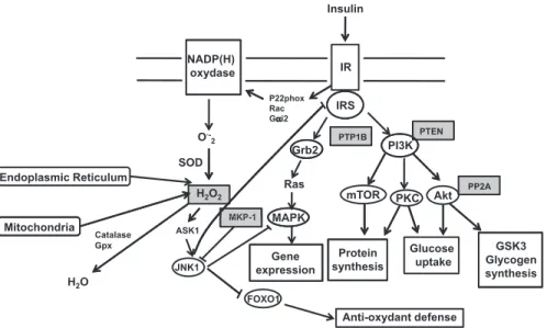
Fig. 1. Scheme of insulin signaling cascade and its regulation by H2O2. Insulin binding to its receptor, a ligand-activated tyrosine kinase, catalyzes the tyrosine autophos-phorylation of the IR and of its cellular IRS. The insulin signal is then transmitted to the different proteins associated with the phospho-tyrosyl side chains of the IRS by their src homology 2 domain. PI3K is linked to the activation of glucose transport, protein synthesis, and gene expression; Grb2 by the Ras pathway is linked to the regulation of gene expression. The steady-state level of tyrosine phosphorylation is regulated by PTPases (e.g., PTP1B), which are directly sensitive to the cysteine oxidation leading to their inhibition. Moreover, other important regulators of this pathway (PTEN, PP2A, MKP-1, and ASK) are sensitive to reactive oxygen species. Insulin binding to insulin receptor induces the activation of NADP(H) oxidase by some pathways not completely identified, but P22phox, Rac, and Gai2 are potential candidates for this signal transduction from insulin receptor to NADP(H) oxidase. The H2O2produced inactivates the proteins sensitive to oxidation (gray squares), which allows insulin signaling. The production of reactive oxygen species by reversible oxidation and inhibition of phosphatases allows insulin signaling. Reactive oxygen species half-lives are short and are transiently produced. However, a higher production of reactive oxygen species from the endoplasmic reticulum or mitochondria after excessive lipid oxidation secondary to overfeeding could lead to the inhibition of antioxidant defense production caused by the inhibition of FOXO1 by JNK1. Moreover, the activation of JNK leads to the inhibition of IRS1. Whether reactive oxygen species promote or inhibit insulin signaling depends on the level and duration of exposure of the inner and outer cellular environment to reactive oxygen species. ASK, apoptosis signal-regulating kinase; FOXO1, Forkhead box O1; Gai2, G protein-a2; Gpx, glutathione peroxidase; Grb2, growth factor receptor-bound protein-2; GSK3, glycogen synthase kinase-3; H2O2, hydrogen peroxide; IR, insulin receptor; IRS, insulin receptor substrate; JNK1, cJun NH2-terminal kinase; MAPK, mitogen-activated protein kinase; MKP-1, tyrosine/serine mitogen-activated protein kinase phosphatase-1; NADP(H), reduced nicotinamide adenine dinucleotide phosphate; O2", superoxide anion; P22phox, flavo-cytochrome b558 subunit; PI3K, phosphoinositide 3-kinase; PKC, protein kinase C; PP2A, serine protein phosphatase-2A; PTEN, phosphatase and tensin homolog; PTP1B, PTP, protein tyrosine phosphatase-1B; PTPases, protein tyrosine phosphatases; SOD, superoxide dismutase.
sensitivity by increasing tyrosine phosphorylation in fructose-fed
rats
[24]
. Also, luteolin, a flavonoid, has been shown to increase
insulin sensitivity by an activation of peroxisome
proliferator-activated receptor-
g
transcriptional activity in 3T3-L1 adipocytes
[25]
and caffeic acid phenethyl ester potently has been found to
stimulate glucose uptake in cultured skeletal muscle cells through
the AMPK pathway
[26]
and stimulate glucose uptake in
insulin-resistant mouse hepatocytes, as has cinnamic acid
[27]
. One of
the phenolic compounds whose effects have been largely studied
on carbohydrate metabolism is unquestionably resveratrol,
a phenolic compound of the stilbene family that, although exerting
antioxidant properties, can interact directly with numerous
metabolic pathways
[28]
. The studies by Lagouge et al.
[29]
and
Baur et al.
[30]
showing how resveratrol improves energy balance,
increases mitochondrial activity, and protects mice against
diet-induced obesity and IR constitute important defending
argu-ments on how this compound may play an important role in the
prevention of metabolic diseases and diabetes. Its different actions
on glucose metabolism are not restricted to its antioxidant capacity
but rather are mediated through sirtuin 1 and thus associated with
gene sequence silencing
[20,31–34]
.
We therefore understand that experimental data are
abun-dantly in favor of the central role of oxidative stress in IR, on the
one hand, and of the protective effect of dietary antioxidants
against these metabolic alterations, on the other hand. However,
intervention studies directly assessing the effects of antioxidants
on glucose metabolism in humans are rare and very few have
used the insulin clamp technique, the gold standard method in
assessing insulin sensitivity. The effects of
a
-lipoic acid and
vitamin C and E supplementation alone or in association with
other antioxidants have been evaluated using this method and
have shown positive effects
[3]
. These positive results must be
weighed against other studies with less favorable conclusions
such as the recently published study by Yfanti et al.
[35]
indi-cating that the administration of antioxidants in the combined
form of vitamin C (500 mg/d) and vitamin E (400 IU/d) during
strenuous endurance training had no effect on the
training-induced increase in insulin sensitivity. These data complement
those of Ristow et al.
[36]
showing that supplementation with
a combination of these same vitamins at high doses (1000 mg/
d and 400 IU/d, respectively) may preclude the
health-promoting effect of exercise on improved insulin sensitivity.
However, these data are in conflict with the finding suggesting
that high levels of vitamin C do not decrease the positive effects
of exercise
[37]
and even decrease the risk of T2D
[38]
.
Regarding polyphenols only, few studies have evaluated their
effects on insulin sensitivity using the insulin clamp. In fact, only
one study showed that cocoa consumption for 2 wk had no
effect on the insulin sensitivity of hypertensive patients
[39]
.
Very recently, the metabolic effects of resveratrol identified thus
far in animals were confirmed in a clinical trial
[40]
. In this work,
the investigators reported that a substantially high dose of 150
mg/d of trans-resveratrol (average daily intake in Europe of
about 0.01–0.45 mg/d
[41]
) taken for 1 mo had favorable effects
on glucose homeostasis in obese subjects by improving their
homeostasis model assessment index, thus indicating favorable
effects on insulin sensitivity. Unfortunately, the investigators
could not determine if these effects resulted in an amelioration
of whole-body insulin sensitivity when using the insulin clamp.
Epidemiologic studies have reported experimental data of
diets rich in antioxidants such as vitamin C
[38]
, vitamin E
[42]
,
a
-tocopherol
[43]
, or
b
-carotene
[43]
exhibiting beneficial effects
on glucose metabolism and on diabetes prevention. Two
meta-analyses examined the association between the intake of fruit,
vegetables, and antioxidants and the risk of T2D. The major
finding of the first study is that the consumption of antioxidants
but not of fruits and vegetables was associated with a 13%
decrease in the risk of T2D, mainly attributed to vitamin E
[44]
,
whereas in the second, the intake of green leafy vegetables was
associated with a 14% decrease of the same risk
[45]
.
The problem is that when reviewing clinical intervention trials
evaluating the effects of antioxidant supplementation, it is hard to
Fig. 2. Environmental and genetic factors favoring oxidative stress and the mechanisms behind reactive oxidative stress-induced insulin resistance and type 2 diabetes mellitus. ROS, reactive oxygen species.
A. Avignon et al. / Nutrition 28 (2012) 715–721 718
pinpoint the positive effects, with all the larger intervention trials
that evaluated the diabetes-preventive potential of antioxidant
supplements consisting of antioxidant vitamins with or without
trace elements reporting negative outcomes
[46,47]
. Moreover,
one study has suggested an increased risk of diabetes in the group
supplemented with
a
-tocopherol and/or
b
-carotene
[48]
.
Thus, although experimental data on cellular and animal
models seem quite clear on the role of oxidative stress in IR and
on the positive effects of dietary antioxidants, human data
show discrepancies. These discrepancies are found when
comparing experimental data with human data, but also within
human data. In this regard, it is interesting to note the results of
tea consumption studies in humans. Several epidemiologic
studies have shown how tea ingestion may be associated with
an increased risk of diabetes. Conversely, other studies found
no such effect, to the point where a meta-analysis suggested
a protective effect of tea (
Table 2
)
[34,40,47,49–59]
. Several
reasons can be given for these disparities. First, free radicals are
necessary for the transduction of certain signals, including that
of insulin, making their excessive neutralization deleterious.
Second, the antioxidant capacity of dietary antioxidants may be
modified by environmental conditions such as pH, the presence
of metal ions, or their concentration. In fact, antioxidants can
become pro-oxidants beyond certain concentrations
[60]
.
Third, digestion metabolism leading to the production of
specific potent metabolites and conjugated derivatives and the
complexity of food matrix synergism may explain some of the
differences found between in vivo and in vitro studies. Fourth,
the efficiency of most natural products and/or diet
supple-ments possessing antioxidant-like actions is not restricted to
their antioxidative capacity, which can further add to the
variability in response, depending on the model studied.
Table 2
Main antioxidants, their natural sources, and recent evidence concerning their effects on glucose metabolism in humans
Antioxidant compounds Type of study Effects on glucose metabolism Polyphenols
Flavonoids
Flavonols: quercetin, myricetin, kampeferol (peas, carrot, broccoli, spinach, cauliflower, apple, plum, apricot, strawberries, tomatoes, black and green teas)
prospective epidemiological study high intake of flavonols and flavones not associated with decrease in T2D, although a modest inverse association with apples and tea intake cannot be ruled out[49]; no effect Flavanols: catechins, epicatechin (cocoa, black
chocolate, black and green teas)
randomized controlled trial epicatechin has no effect on IR[50]; no effect Anthocyanins: cyanidin, delphinidin, luteolinidin
(berries, orange, eggplant, cherries, red grape, red wine)
randomized trial flaxseed supplementation decreases thiobarbituric acid-reactive substances, plasma glucose, and HOMA-IR of overweight, hypertensive subjects with family history of diabetes[51]; positive effect
Isoflavones: genistin, formononetin, coumestrol (Soy, black beans, alfalfa, peanuts)
randomized controlled trial supplement containing combination of antioxidants extracted from fruit, berries, and vegetables has no effect on blood glucose, HbA1c, insulin[52]; no effect
Flavanones: hesperidin, naringenin (orange, grapefruit, lemon, lime, tomato skin)
Flavones: apigenin, luteolin, tangeritin (parsley, celery, sweet pepper)
Phenolic acids
Hydroxybenzoic acid derivatives: gallic acid, ellagic acid (black and green teas, red wine, berries, potatoes)
epidemiologic, cross-sectional study green tea consumption associated with increased prevalence of diabetes mellitus in an Iranian population
[53]; negative effect Hydroxycinnamic acid derivatives: chlorogenic acid,
caffeic acid, hydrocinnamic acid (blueberries, coffee, kiwi fruit, apples, pears, red wine, broccoli, plums, cherries)
epidemiologic, cross-sectional study coffee but not green tea consumption has beneficial effects on glycemic parameters (fasting plasma glucose, HOMA-IR, HOMA-b, plasma HbA1c) in multiethnic Asian population
[54]; positive effect of coffee, no effect of tea
prospective epidemiologic study long-term consumption of oolong tea may be predictive factor for new-onset diabetes in a Japanese population[55]; negative effect
randomized controlled trial green tea extract has no effect on IR[56]; no effect meta-analysis of cohort studies consumption of #4 cups/d of tea has beneficial effect on T2D
prevention[57]; positive effect Stilbenoids
Trans-resveratrol (skin of red grapes, cranberries, blueberries, bilberries)
randomized controlled trial resveratrol supplementation improves HOMA-IR in patients with T2D[34]; positive effect
randomized controlled trial 30 d of high-dose resveratrol supplementation induces favorable metabolic changes in obese humans, including improvement in HOMA-IR[40]; positive effect Vitamin E tocopherol (unheated vegetable oil: wheat germ
oil, palm oil; cereal, almonds, hazelnuts, green vegetables, butter, milk, egg, avocado, oily fish, e.g., tuna)
epidemiologic, cross-sectional study serum vitamin C but not vitamin E is inversely associated with HOMA-IR in an adult population[58]; positive effect Vitamin CL-ascorbic acid (acerola, jujube, broccoli, Brussels
sprouts, lychee)
randomized controlled trial
Lipoic acid (spinach, broccoli) lipoic acid and vitamin E supplementation alone or in combination did not affect lipid profile or insulin sensitivity of patients with T2D[59]; no effect
Carotenoids:b-cryptoxanthin,b-carotene, lutein (alfalfa, carrot, tomato, grapefruit, watermelon)
randomized controlled trial no significant overall effects of vitamin C, vitamin E, and
b-carotene long-term supplementation (10 y) on risk of developing T2D in women at high risk of CVD[47]; no effect CVD, cardiovascular disease; Hb, hemoglobin; HOMA-IR, homeostasis model assessment for insulin resistance; IR, insulin resistance; T2D, type 2 diabetes mellitus
Conclusions
The involvement of ROS in the control of carbohydrate
metabolism seems undeniable. Experimentally, the modulation
of oxidative stress by antioxidants appears to have a positive
outcome, but current intervention studies do not allow the
recommendation of antioxidant supplementation for the sole
purpose of preventing T2D. Most studies, however, have used
supplements in the form of one or two vitamins associated or not
with trace elements, although plants naturally contain a
multi-tude of antioxidants. Among the countless compounds present in
a particular plant food, it is often difficult to identify the one that
plays the critical part; also, the overall total antioxidant capacity
of the diet might be more important than the presence of any
particular food
[2]
. Hence, the ideal antioxidant supplement for
diabetes prevention will certainly be one that will be able to
reproduce as closely as possible the innate combination of
antioxidants found in plant foods even if we cannot exclude the
fact that the beneficial effects of the latter might be synergistic
with other compounds.
References
[1] Bull!o M, Lamuela-Ravent!os R, Salas-Salvad!o J. Mediterranean diet and oxidation: nuts and olive oil as important sources of fat and antioxidants. Curr Top Med Chem 2011;11:1797–810.
[2] Puchau B, Zulet MA, de Ech!avarri AG, Hermsdorff HHM, Mart!ınez JA. Dietary total antioxidant capacity is negatively associated with some metabolic syndrome features in healthy young adults. Nutrition 2010;26:534–41. [3] Bisbal C, Lambert K, Avignon A. Antioxidants and glucose metabolism
disorders. Curr Opin Clin Nutr Metab Care 2010;13:439–46.
[4] Jones DP, Go Y-M. Redox compartmentalization and cellular stress. Diabetes Obes Metab 2010;12(suppl 2):116–25.
[5] Samocha-Bonet D, Heilbronn LK, Lichtenberg D, Campbell LV. Does skeletal muscle oxidative stress initiate insulin resistance in genetically predis-posed individuals? Trends Endocrinol Metab 2010;21:83–8.
[6] Hoehn KL, Salmon AB, Hohnen-Behrens C, Turner N, Hoy AJ, Maghzal GJ, et al. Insulin resistance is a cellular antioxidant defense mechanism. Proc Natl Acad Sci U S A 2009;106:17787–92.
[7] Bonnard C, Durand A, Peyrol S, Chanseaume E, Chauvin M-A, Morio B, et al. Mitochondrial dysfunction results from oxidative stress in the skeletal muscle of diet-induced insulin-resistant mice. J Clin Invest 2008;118:789–800.
[8] Dandona P, Ghanim H, Chaudhuri A, Dhindsa S, Kim SS. Macronutrient intake induces oxidative and inflammatory stress: potential relevance to atherosclerosis and insulin resistance. Exp Mol Med 2010;42:245–53. [9] Anderson EJ, Lustig ME, Boyle KE, Woodlief TL, Kane DA, Lin C-T, et al.
Mitochondrial H2O2 emission and cellular redox state link excess fat intake to insulin resistance in both rodents and humans. J Clin Invest 2009; 119:573–81.
[10] Sies H, Stahl W, Sevanian A. Nutritional, dietary and postprandial oxidative stress. J Nutr 2005;135:969–72.
[11] Bunck MC, Corn!er A, Eliasson B, Heine RJ, Shaginian RM, Wu Y, et al. One-year treatment with exenatide vs. insulin glargine: effects on post-prandial glycemia, lipid profiles, and oxidative stress. Atherosclerosis 2010;212:223–9.
[12] Madec S, Corretti V, Santini E, Ferrannini E, Solini A. Effect of a fatty meal on inflammatory markers in healthy volunteers with a family history of type 2 diabetes. Br J Nutr 2011;106:364–8.
[13] Amini M, Esmaillzadeh A, Shafaeizadeh S, Behrooz J, Zare M. Relationship between major dietary patterns and metabolic syndrome among individ-uals with impaired glucose tolerance. Nutrition 2010;26:986–92. [14] Michel CI, Holley CL, Scruggs BS, Sidhu R, Brookheart RT, Listenberger LL,
et al. Small nucleolar RNAs U32a, U33, and U35a are critical mediators of metabolic stress. Cell Metab 2011;14:33–44.
[15] Yuzefovych L, Wilson G, Rachek L. Different effects of oleate vs. palmitate on mitochondrial function, apoptosis, and insulin signaling in L6 skeletal muscle cells: role of oxidative stress. Am J Physiol Endocrinol Metab 2010;299:E1096–105.
[16] Perez-Martinez P, Garcia-Quintana JM, Yubero-Serrano EM, Tasset-Cuevas I, Tunez I, Garcia-Rios A, et al. Postprandial oxidative stress is modified by dietary fat: evidence from a human intervention study. Clin Sci 2010;119:251–61.
[17] Petersson H, Ris!erus U, McMonagle J, Gulseth HL, Tierney AC, Morange S, et al. Effects of dietary fat modification on oxidative stress and inflam-matory markers in the LIPGENE study. Br J Nutr 2010;104:1357–62.
[18] Andrikopoulos S. Obesity and type 2 diabetes: slow down!dcan metabolic deceleration protect the islet beta cell from excess nutrient-induced damage? Mol Cell Endocrinol 2010;316:140–6.
[19] Raffaella C, Francesca B, Italia F, Marina P, Giovanna L, Susanna I. Alterations in hepatic mitochondrial compartment in a model of obesity and insulin resistance. Obesity (Silver Spring) 2008;16:958–64.
[20] Chuang C-C, Martinez K, Xie G, Kennedy A, Bumrungpert A, Overman A, et al. Quercetin is equally or more effective than resveratrol in attenu-ating tumor necrosis factor-a–mediated inflammation and insulin resistance in primary human adipocytes. Am J Clin Nutr 2010;92: 1511–21.
[21] Scazzocchio B, Var"ı R, Filesi C, D’Archivio M, Santangelo C, Giovannini C, et al. Cyanidin-3-O-b-glucoside and protocatechuic acid exert insulin-like effects by upregulating PPARgactivity in human omental adipocytes. Diabetes 2011;60:2234–44.
[22] Yoshida H, Takamura N, Shuto T, Ogata K, Tokunaga J, Kawai K, et al. The citrus flavonoids hesperetin and naringenin block the lipolytic actions of TNF-alpha in mouse adipocytes. Biochem Biophys Res Commun 2010; 394:728–32.
[23] Zygmunt K, Faubert B, MacNeil J, Tsiani E. Naringenin, a citrus flavonoid, increases muscle cell glucose uptake via AMPK. Biochem Biophys Res Commun 2010;398:178–83.
[24] Kannappan S, Anuradha CV. Naringenin enhances insulin-stimulated tyrosine phosphorylation and improves the cellular actions of insulin in a dietary model of metabolic syndrome. Eur J Nutr 2010;49:101–9. [25] Ding L, Jin D, Chen X. Luteolin enhances insulin sensitivity via activation
of PPARgtranscriptional activity in adipocytes. J Nutr Biochem 2010; 21:941–7.
[26] Eid HM, Vallerand D, Muhammad A, Durst T, Haddad PS, Martineau LC. Structural constraints and the importance of lipophilicity for the mito-chondrial uncoupling activity of naturally occurring caffeic acid esters with potential for the treatment of insulin resistance. Biochem Pharmacol 2010;79:444–54.
[27] Huang D-W, Shen S-C, Wu JS-B. Effects of caffeic acid and cinnamic acid on glucose uptake in insulin-resistant mouse hepatocytes. J Agric Food Chem 2009;57:7687–92.
[28] Szkudelski T, Szkudelska K. Anti-diabetic effects of resveratrol. Ann N Y Acad Sci 2011;1215:34–9.
[29] Lagouge M, Argmann C, Gerhart-Hines Z, Meziane H, Lerin C, Daussin F, et al. Resveratrol improves mitochondrial function and protects against metabolic disease by activating SIRT1 and PGC-1alpha. Cell 2006;127:1109–22. [30] Baur JA, Pearson KJ, Price NL, Jamieson HA, Lerin C, Kalra A, et al.
Resver-atrol improves health and survival of mice on a high-calorie diet. Nature 2006;444:337–42.
[31] de Kreutzenberg SV, Ceolotto G, Papparella I, Bortoluzzi A, Semplicini A, Dalla Man C, et al. Downregulation of the longevity-associated protein sir-tuin 1 in insulin resistance and metabolic syndrome: potential biochemical mechanisms. Diabetes 2010;59:1006–15.
[32] Mercader J, Palou A, Bonet ML. Resveratrol enhances fatty acid oxidation capacity and reduces resistin and retinol-binding protein 4 expression in white adipocytes. J Nutr Biochem 2011;22:828–34.
[33] Vigilanza P, Aquilano K, Baldelli S, Rotilio G, Ciriolo MR. Modulation of intracellular glutathione affects adipogenesis in 3T3-L1 cells. J Cell Physiol 2011;226:2016–24.
[34] Brasny!o P, Moln!ar GA, Moh!as M, Mark!o L, Laczy B, Cseh J, et al. Resveratrol improves insulin sensitivity, reduces oxidative stress and activates the Akt pathway in type 2 diabetic patients. Br J Nutr 2011;106:383–9. [35] Yfanti C, Nielsen AR, #Akerström T, Nielsen S, Rose AJ, Richter EA, et al.
Effect of antioxidant supplementation on insulin sensitivity in response to endurance exercise training. Am J Physiol Endocrinol Metab 2011; 300:E761–70.
[36] Ristow M, Zarse K, Oberbach A, Klöting N, Birringer M, Kiehntopf M, et al. Antioxidants prevent health-promoting effects of physical exercise in humans. Proc Natl Acad Sci U S A 2009;106:8665–70.
[37] Thamer C, Machicao F, Stefan N, Fritsche A, Häring H-U. High baseline vitamin C levels do not prevent a positive outcome of a lifestyle inter-vention. Diabetes Care 2009;32:e112.
[38] Harding A-H, Wareham NJ, Bingham SA, Khaw K, Luben R, Welch A, et al. Plasma vitamin C level, fruit and vegetable consumption, and the risk of new-onset type 2 diabetes mellitus: the European prospective investi-gation of cancerdNorfolk Prospective Study. Arch Intern Med 2008; 168:1493–9.
[39] Muniyappa R, Hall G, Kolodziej TL, Karne RJ, Crandon SK, Quon MJ. Cocoa consumption for 2 wk enhances insulin-mediated vasodilatation without improving blood pressure or insulin resistance in essential hypertension. Am J Clin Nutr 2008;88:1685–96.
[40] Timmers S, Konings E, Bilet L, Houtkooper RH, van de Weijer T, Goossens GH, et al. Calorie restriction-like effects of 30 days of resveratrol supplementation on energy metabolism and metabolic profile in obese humans. Cell Metab 2011;14:612–22.
[41] Edwards JA, Beck M, Riegger C, Bausch J. Safety of resveratrol with exam-ples for high purity, trans-resveratrol, resVida>>. Ann N Y Acad Sci 2011;1215:131–7.
A. Avignon et al. / Nutrition 28 (2012) 715–721 720
[42] Costacou T, Ma B, King IB, Mayer-Davis EJ. Plasma and dietary vitamin E in relation to insulin secretion and sensitivity. Diabetes Obes Metab 2008; 10:223–8.
[43] Arnlöv J, Zethelius B, Ris!erus U, Basu S, Berne C, Vessby B, et al. Serum and dietary beta-carotene and alpha-tocopherol and incidence of type 2 dia-betes mellitus in a community-based study of Swedish men: report from the Uppsala Longitudinal Study of Adult Men (ULSAM) study. Diabetologia 2009;52:97–105.
[44] Carter P, Gray LJ, Troughton J, Khunti K, Davies MJ. Fruit and vegetable intake and incidence of type 2 diabetes mellitus: systematic review and meta-analysis. BMJ 2010;341:c4229.
[45] Hamer M, Chida Y. Intake of fruit, vegetables, and antioxidants and risk of type 2 diabetes: systematic review and meta-analysis. J Hypertens 2007;25:2361–9.
[46] Czernichow S, Vergnaud A-C, Galan P, Arnaud J, Favier A, Faure H, et al. Effects of long-term antioxidant supplementation and association of serum antioxidant concentrations with risk of metabolic syndrome in adults. Am J Clin Nutr 2009;90:329–35.
[47] Song Y, Cook NR, Albert CM, Van Denburgh M, Manson JE. Effects of vita-mins C and E and beta-carotene on the risk of type 2 diabetes in women at high risk of cardiovascular disease: a randomized controlled trial. Am J Clin Nutr 2009;90:429–37.
[48] Kataja-Tuomola M, Sundell JR, Männistö S, Virtanen MJ, Kontto J, Albanes D, et al. Effect of alpha-tocopherol and beta-carotene supplementation on the incidence of type 2 diabetes. Diabetologia 2008;51:47–53.
[49] Song Y, Manson JE, Buring JE, Sesso HD, Liu S. Associations of dietary flavonoids with risk of type 2 diabetes, and markers of insulin resistance and systemic inflammation in women: a prospective study and cross-sectional analysis. J Am Coll Nutr 2005;24:376–84.
[50] Brown AL, Lane J, Coverly J, Stocks J, Jackson S, Stephen A, et al. Effects of dietary supplementation with the green tea polyphenol epigallocatechin-3-gallate on insulin resistance and associated metabolic risk factors: randomized controlled trial. Br J Nutr 2009;101:886–94.
[51] Rhee Y, Brunt A. Flaxseed supplementation improved insulin resistance in obese glucose intolerant people: a randomized crossover design. Nutr J 2011;10:44.
[52] Rytter E, Vessby B, Asg#ard R, Ersson C, Moussavian S, Sjödin A, et al. Supplementation with a combination of antioxidants does not affect gly-caemic control, oxidative stress or inflammation in type 2 diabetes subjects. Free Radic Res 2010;44:1445–53.
[53] Golozar A, Khademi H, Kamangar F, Poutschi H, Islami F, Abnet CC, et al. Diabetes mellitus and its correlates in an Iranian adult population. PLoS ONE 2011;6:e26725.
[54] Rebello SA, Chen CH, Naidoo N, Xu W, Lee J, Chia KS, et al. Coffee and tea consumption in relation to inflammation and basal glucose metabolism in a multi-ethnic Asian population: a cross-sectional study. Nutr J 2011; 10:61.
[55] Hayashino Y, Fukuhara S, Okamura T, Tanaka T, Ueshima H. High oolong tea consumption predicts future risk of diabetes among Japanese male workers: a prospective cohort study. Diabet Med 2011;28:805–10. [56] Hsu C-H, Liao Y-L, Lin S-C, Tsai T-H, Huang C-J, Chou P. Does
supplemen-tation with green tea extract improve insulin resistance in obese type 2 diabetics? A randomized, double-blind, and placebo-controlled clinical trial. Altern Med Rev 2011;16:157–63.
[57] Jing Y, Han G, Hu Y, Bi Y, Li L, Zhu D. Tea consumption and risk of type 2 diabetes: a meta-analysis of cohort studies. J Gen Intern Med 2009;24:557–62. [58] Beydoun MA, Shroff MR, Chen X, Beydoun HA, Wang Y, Zonderman AB.
Serum antioxidant status is associated with metabolic syndrome among U.S. adults in recent national surveys. J Nutr 2011;141:903–13.
[59] de Oliveira AM, Rond!o PHC, Luzia LA, D’Abronzo FH, Illison VK. The effects of lipoic acid anda-tocopherol supplementation on the lipid profile and insulin sensitivity of patients with type 2 diabetes mellitus: a randomized, double-blind, placebo-controlled trial. Diabetes Res Clin Pract 2011;92:253–60. [60] Bouayed J, Bohn T. Exogenous antioxidantsddouble-edged swords in cellular
redox state: health beneficial effects at physiologic doses versus deleterious effects at high doses. Oxidative Med Cell Longevity 2010;3:228–37.