HAL Id: tel-01111143
https://hal.sorbonne-universite.fr/tel-01111143
Submitted on 29 Jan 2015
HAL is a multi-disciplinary open access archive for the deposit and dissemination of sci-entific research documents, whether they are pub-lished or not. The documents may come from teaching and research institutions in France or abroad, or from public or private research centers.
L’archive ouverte pluridisciplinaire HAL, est destinée au dépôt et à la diffusion de documents scientifiques de niveau recherche, publiés ou non, émanant des établissements d’enseignement et de recherche français ou étrangers, des laboratoires publics ou privés.
Approches intégrés des mécanismes moléculaires de la
tolérance au cuivre chez les algues brunes
Andrés Ritter Traub
To cite this version:
Andrés Ritter Traub. Approches intégrés des mécanismes moléculaires de la tolérance au cuivre chez les algues brunes. Biologie moléculaire. Paris 6; Pontificia Universidad Católica de Chile, 2009. Français. �tel-01111143�
Avertissement
Au vu de la législation sur les droits d'auteur, ce travail de thèse demeure la
propriété de son auteur, et toute reproduction de cette oeuvre doit faire l'objet
d'une autorisation de l'auteur. (cf Loi n°92-597; 1/07/1992. Journal Officiel,
2/07/1992)
THESE DE DOCTORAT DE L’UNIVERSITE PIERRE ET MARIE CURIE
Spécialité Biologie Ecole doctorale La logique du vivant (ED223)
Présentée par
M. Andrés Ritter Traub
Pour obtenir le double grade de
DOCTEUR de l’UNIVERSITÉ PIERRE ET MARIE CURIE Et
DOCTOR en Ciencias Biológicas Mención Ecología de la Pontificia Universidad Católica de Chile
Approches intégrées des mécanismes
moléculaires de la tolérance au cuivre chez
les algues brunes
soutenue le 16, janvier 2009 devant le jury composé de :
M. Cyrille Forestier Ingénieur de Recherche-HDR CEA (Rapporteur)
M. Didier Marion Directeur de Recherche INRA (Rapporteur)
M. Christophe Destombe Prof. Université Pierre et Marie Curie (Président) M. Frithjof C. Küpper P.I. Lect. Scottish Association for Marine Science (Examinateur) M. Sylvain Faugeron Prof. Pontifica Universidad Católica de Chile (Examinateur)
M. Philippe Potin Directeur de Recherche C.N.R.S (Directeur)
M. Juan A. Correa Prof. Pontificia Universidad Católica de Chile (Co-Directeur)
M. Thierry Tonon M.d.C. Université Pierre et Marie Curie (Examinateur)
Université Pierre & Marie Curie - Paris 6
Bureau d’accueil, inscription des doctorants et base de données
Esc G, 2ème étage
15 rue de l’école de médecine 75270-PARIS CEDEX 06
Tél. Secrétariat : 01 42 34 68 35 Fax : 01 42 34 68 40 Tél. pour les étudiants de A à EL : 01 42 34 69 54 Tél. pour les étudiants de EM à ME : 01 42 34 68 41 Tél. pour les étudiants de MF à Z : 01 42 34 68 51
A mis Padres A Blanche A mi abuela Lina
Acknowledgments
Acknowledgments
I thank Catherine Boyen for allowing me to carry my research in her laboratory, and for her kind support as well.
I express all my gratitude to the members of my PhD committee (jury et comités de these) for their contribution to the evaluation of my work during all of these years.
I thank the French embassy and the CONICYT for providing me a fellowship for my third year.
This research adventure started over six years ago when Bernard Kloareg introduced me to Philippe. I still remember all the fusing ideas coming out of Philippe’s mind, who was programming a research project for the next 10 years within only a 5- minutes-discussion… At the end of this meeting I didn’t understood much about the funny topics described by Philippe, but at least I understood that I could work with seaweeds in collaboration with a Chilean lab. Six years latter, I still have troubles sometimes understanding your crazy ideas Philippe, but at least the ones that I understood have helped me to enlarge my scientific culture and to influence my research towards a wide view of biological processes. In addition to scientific issues, I also discovered in you a friend with whom I could discuss multiple subjects such as politics, fishing or nature. I thank you Philippe for supporting and believing me for all these years. It has really been a pleasure to have you as a supervisor.
A couple of months following my first meeting with Philippe, I arrived as a tourist from France to live my first experience in Juan’s lab in Santiago. This was in the middle of summer vacations, so no one was working at this time of the year. After a couple of hours waiting for someone to show up, as I was about to leave the lab, I ran into a “chief-looking-person”. This is how I met Juan. Then we both realised that there had been a misunderstanding about the date of my arrival (the first but not the last). Since then I have been his “atypical” Chilean-French or French-Chilean (who knows?) student responsible for more than one of his white hairs… Thank you Juan for your support, understanding and patience. I must underline that I started my PhD without any fellowship, which made of myself a heavy financial burden for both labs. Therefore I must thank Juan and Philippe for all the efforts they deployed to solve this issue and guarantee the best conditions to carry my research work.
The third masterpiece of my PhD is Thierry. I met him during my Master. I noticed right away his funny southern French accent, but also the characteristics of a serious and capable researcher eager to friendly help and advice my work. Thierry had been the “stabilizer” element of my thesis, putting down onto the ground the “megalomaniac” plans we built up with Philippe. On top of that, Thierry had the heavy task to supervise me and my pal PO. I can imagine how many aspirins we made him swallow… I thank you Thierry for your guidance, efforts and patience. With you I have learned how to work in a lab. I also thank you for your friendship and loyalty.
Philippe, Juan and Thierry have helped me in a very complementary manner. I was very lucky to have them and I thank you all for being there in good and less good moments. I must specially thank Sophie Goulitquer, Jean Pierre Salaün, Fanny Gaillard, Sarah Romac, Aaron Man, Jessica Beltrand, Santiago Andrade and Loretto Contreras,
Acknowledgments
who have closely collaborated with me. Our interaction was determining for the good development of my project, providing me interesting and very complementary skills.
A great thanks to the students who provided me with precious work during this project: Martin Ubertini, Francis Lemadec, Carolina Henriquez. Working with you has been a pleasure, and I thank you for your enthusiasm, humour and initiatives. In addition to your generous help, you offered me your friendship, which I really appreciated.
J’ai aussi une pensée particulière pour mes collègues de bureau, PO et Maud. Vous avez été de bien fidèles compagnons de parcours, et je vous en remercie. J’ai tellement de bons souvenirs et anecdotes dans notre bazar de bureau qu’une thèse ne suffirait pas pour les raconter… Merci mon grand PO pour ces bonnes discussions autour de notre bon vieux billard (quand est-ce qu’on met la pâtée à Robert ?), à propos de la pêche « sportive » ou de la chasse (sans palmes). Je n’aurais pas pu avoir de meilleur pote pendant ces années, merci d’avoir été là, tes blagues vont me manquer au labo.
Un grand merci à l’UMR7139 pour toute votre aide, votre généreuse disponibilité et votre bonne ambiance et dynamisme qui ont fortement enrichi mon travail. Un grand merci pour tous les bons moments passés avec vous, votre amitié et générosité.
Enfin un grand merci à tout le personnel de la Station Biologique de Roscoff, grâce à qui j’ai pu réaliser ma thèse dans des conditions idéales. J’ai une pensée particulière pour le personnel du Gulf Stream, merci pour tous ces moments autours de vos bons petits plats qui ont su agrémenter mon travail au quotidien.
Agradezco igualmente la ayuda de todo el departamento de ecología de la PUC, por haberme acogido y ayudado a lo largo de estos tres años.
Je voudrais également remercier ma Blanche pour son grand soutien pendant chaque minute de ces trois années. Si je suis arrivé jusqu’ici c’est en grande partie grâce à toi. Merci d’avoir cru en moi plus que je n’ose croire en moi-même et pour avoir toujours été là dans les bons et les mauvais moments. Merci d’avoir supporté mes horaires « flexibles » du boulot (même si tu n’en comprenais pas toujours la raison…) et mes humeurs changeantes pendant la rédaction. Je profite au passage pour remercier ma chatoune « ploma », qui a été une compagne fidèle (même aux heures avancées de la nuit) pendant la rédaction de ce manuscrit.
Comment pourrais-je oublier ma famille d’adoption, les Magariños-Rey. Merci à vous de m’avoir encouragé à suivre ma voie. Merci de votre soutien et de l’intérêt que vous avez porté à ma thèse. Je souligne que c’est grâce à vous que j’ai découvert la Bretagne et ses algues. Sans vos « stages de découverte et cuisine des algues » (que je recommande vivement aux lecteurs de cette thèse), je n’aurais sûrement pas fait une thèse de phycologie.
En último lugar agradezco especialmente mis padres y familia. Gracias por haber aceptado mis desiciones, auque no siempre hayan estado de acuerdo con ellas. Su apoyo incondicional, me ha dado la confianza necesaria para poder desarrollarme plenamente. Gracias por haber creido en la importancia de mi trabajo. Sin su ejemplo ni apoyo, nunca hubiese podido llegar a ser él que soy. Aunque no este presente con ustedes los llevo siempre en mis pensamientos.
List of abreviations
LIST OF ABREVIATIONS
12-OPDA 12-oxo-phytodienoic acid
12R,13S-diHEPE 12R,13S-dihydroxi-5(Z),8(Z),10(E),14(Z),17(Z)-eicosapentaenoic acid
12R,13S-diHETE 12R,13S-dihydroxi-5(Z),8(Z),10(E),14(Z)-eicosatetraenoic acid
12S-HEPE 12S-hydroxieicosapentaenoic acid
12S-HETE 12S-hydroxieicosatetraenoic acid
13S-HODE 13S-hydroxioctadecadienoic acid
13S-HOTrE 13S-hydroxioctadecatrienoic acid
17H-18-oxo-ETE 18-hydroxi-17-oxo-eicosatetraenoic acid
2-DE Two-dimensional gel electrophoresis
4-HDDE 4-Hydroxy-dodecadienal
4-HHE 4-hydroxy-(E)-2-hexenal
4-HNE 4-hydroxy-(E)-2-nonenal
9S-HETE 9S-hydroxieicosatetraenoic acid
ACN acetonitrile
afc% Percentage of autofluorescent cells
AOC Allene Oxyde Synthase
AOS Allene Oxyde Cyclase
AsA Ascorbic Acid
ATX Antioxidant
CALR Calreticulin
CAT Catalase
CAX Calcium exchangers
CCH Copper chaperone
CCS Copper chaperone for Superoxide dismutase
CDF Cation diffusion facilitator
Chl Chlorophyll
COX Cytochrome C
ECH enoyl-CoA hydratase
EST Expressed sequence Tag
FA Fatty acid
Fcp Fucoxanthine chlorophyll a-c binding protein
FFA Free Fatty Acid
Fv/Fm Photosynthetic yield
GAPD glyceraldehyde-3-phosphate deshydrogenase
GC Gas Chromatography
GOGAT glutamate synthase
GPX Glutathione Peroxidase
GR Glutathione Reductase
GS glutamine synthetase
GSH Reduced Glutathione
List of abreviations
HMA Heavy Metal Associated
HPL Hydroperoxide lyase
HSP Heat Shock Protein
IEF Isoelectric focusing
IPG Immobilized pH Gradient
LC Liquid Chromatography
LC50 Median (50%) lethal concentration
LHC Ligth Harvesting Complex
LOX Lipoxygenase
LOX lipoxygenase
LT Leukotriene
MALDI Matrix Assisted Laser Desorption Ionisation
ManA Mannose-6-phosphate isomerase
MC Metacaspase
MDA Malonaldehyde
MDAR Monodeshydroascorbade reductase
MeJA Methyljasmonate
MS Mass spectrometry
MSR Methionine sulfoxide reductase
MT Metallothionein
NA Nicotianamine
OEC Oxygen Evolving Complex
PAM Pulse AmplitudeModulated Fluorimetry
PC Phytochelatin
PCS Phytochelatin synthase
PG Prostaglandin
PKK phosphoribulokinase
PMF Protein mass fingerprint
PRX Peroxiredoxin
PVP polyvinylpyrrolidone
RC Reaction Center
ROS Reactive Oxygen Species
SAM S-adenosylmethionine synthetase
SHMT serine hydroxymethyl transferase
SMALDO fructose 1,6-bisphosphate aldolase
SOD Superoxide Dismutase
TCA Trichloroacetic acid
TCA Trichloroacetic acid
TFA Trifluoroacetic acid
TKT transketolase
TOF Time of flight
TRX Thioredoxine
Table of contents
TABLE OF CONTENTS
TABLE OF CONTENTS ... 1 Résumé ... 5 1 INTRODUCTION... 13 1.1. GENERAL INTRODUCTION ... 131.1.1. Ecological and physiological features of brown algae (Phaeophyceae) ... 13
1.1.2. General aspects of abiotic stress in seaweeds ... 22
1.1.3. Ecological effects of copper excess in seaweeds: example of mine wasted areas in northern Chile... 24
1.2. TOXIC EFFECTS OF COPPER IN LAND PLANTS AND MACROALGAE ... 27
1.2.1. Inhibition of growth and spore germination... 27
1.2.2. Inhibition of photosynthesis ... 28
1.2.3. Copper-induced oxidative stress ... 29
1.2.4. Effect of copper on enzyme and protein activity ... 31
1.2.5. Other alterations induced by copper excess ... 32
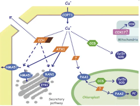
1.3. CELLULAR MECHANISMS OF COPPER HOMEOSTASIS AND AVOIDANCE IN VASCULAR PLANTS ... 33
1.3.1. Copper delivery into cells ... 33
1.3.2. Cellular mechanism for copper avoidance ... 37
1.4. CELLULAR MECHANISMS OF COPPER EXCESS AVOIDANCE IN BROWN ALGAE ... 44
1.4.1. Copper exclusion... 44
1.4.2. Phytochelatins ... 44
1.4.3. Metallothioneins... 45
1.4.4. Physode sequestration of heavy metals and phenolics chelation ... 45
1.4.5. Cell wall and polysaccharide heavy metal binding ... 46
1.4.6. Repair of damaged structures ... 46
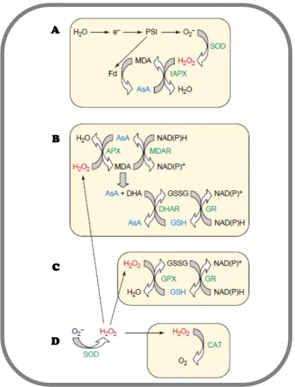
1.5. HEAVY METAL-INDUCED ANTIOXIDANT DEFENSES IN PLANTS AND ALGAE ... 47
1.5.1. ROS scavenging mechanisms ... 47
1.5.2. ROS signal transduction pathways... 50
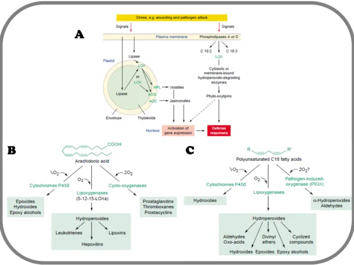
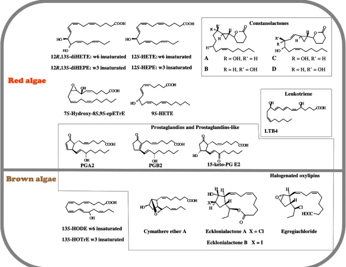
1.6. OXYLIPIN METABOLISM IN MARINE ALGAE ... 52
1.6.1. Lipase mediated free fatty acid liberation in algae ... 55
1.6.2. Oxylipin biosynthesis ... 55
1.7. PROTEOMICS AND TWO DIMENSIONAL ELECTROPHORESIS ... 61
1.7.1. From Genomics to Proteomics ... 61
1.7.2. 2-DE based analysis of metal-stress related proteins in plants and algae ... 64
1.8. OBJECTIVES ... 66
1.8.1. General objectives ... 66
1.8.2. Specific objectives... 66
1.9. REFERENCES... 69
2 COPPER STRESS INDUCES BIOSYNTHESIS OF OCTADECANOID AND EICOSANOID OXYGENATED DERIVATIVES IN THE BROWN ALGAL KELP Laminaria digitata... 89
3 RELEASE OF VOLATILE ALDEHYDES BY THE BROWN ALGAL KELP Laminaria digitata IN RESPONSE TO BOTH BIOTIC AND ABIOTIC STRESSES ... 105
Table of contents
4 TWO-DIMENSIONAL GEL ELECTROPHORESIS ANALYSIS OF BROWN ALGAL
PROTEIN EXTRACTS ... 115
4.1 SUPPLEMENTARY MATERIAL ... 125
5 COPPER STRESS PROTEOMICS HIGHLIGHTS LOCAL ADAPTATION OF TWO STRAINS OF THE MODEL BROWN ALGA Ectocarpus siliculosus ... 131
5.1 Introduction ... 133
5.2 Materials and Methods ... 136
5.2.1 Plant material and cultivation treatments ... 136
5.2.2 Copper toxicity bioassay ... 136
5.2.3 In vivo fluorescence measurements and observations... 137
5.2.4 Protein extraction and 2-DE separation ... 137
5.2.5 Image analysis ... 137
5.2.6 Protein Mass Fingerprints (PMF)... 138
5.2.7 RNA extraction and RT-qPCR analysis... 139
5.2.8 Bromoperoxidase activity assay... 139
5.2.9 Statistical analysis ... 140
5.3 Results ... 141
5.3.1 Copper toxicity tests in strains Es32 and Es524 ... 141
5.3.2 Analyses of the proteins induced by chronic copper stress in Es32... 142
5.3.3 Analyses of the proteins induced by chronic copper stress in Es524... 143
5.3.4 Inter-strain differences in protein expression caused by copper excess... 144
5.3.5 Expression analysis of selected key genes by RT-qPCR under acute and chronic stress conditions ... 144
5.3.6 Bromoperoxidase activity assay... 145
5.4 Discussion ... 146
5.4.1 Common protein expression features between the two isolates exposed to copper 146 5.4.2 Strain-specific responses ... 148
5.4.3 Concluding remarks ... 150
5.5 References ... 152
5.6 FIGURES ... 159
6 ANNOTATION OF THE E. siliculosus GENOME ... 170
6.1 A SURVEY FOR GENES INVOLVED IN COPPER HOMEOSTASIS, DETOXIFICATION AND ANTI-OXIDANT RESPONSES ... 170
6.1.1 INTRODUCTION... 170
6.1.2 METHODS... 171
6.1.3 RESULTS... 172
6.1.4 CONCLUDING REMARKS ... 188
6.2 PRELIMINARY CHARACTERIZATION OF CANDIDATE GENES IDENTIFIED THROUGH E. siliculosus GENOME ANNOTATION ... 191
6.2.1 INTRODUCTION... 191
6.2.2 MATERIALS AND METHODS ... 192
6.2.3 RESULTS AND DISCUSSION ... 195
6.3 REFERENCES... 201
6.4 SUPPLEMENTARY MATERIAL ... 205
7 GENERAL CONCLUSIONS AND PERSPECTIVES ... 215
7.1 Oxylipin metabolism in response to copper excess ... 216
7.2 Proteomic developments and molecular actors of the E. siliculosus tolerance... 219
Table of contents
7.4 New functional approaches in brown algae to identify copper tolerance mechanisms 224
7.5 REFERENCES... 226
8 SUPPLEMENTARY MATERIAL ... 231
8.1 CURRICULUM VITAE ... 231
Résumé
Résumé
Les algues brunes (Phaeophyceae) sont des organismes photosynthétiques macroscopiques
fixés qui représentent une biomasse considérable dans les écosystèmes rocheux des côtes
tempérés et froides de tous les océans en zone intertidale et subtidale. Par conséquent elles
doivent faire face à des agressions constantes d’origine naturelle ou anthropique. Le
déversement des métaux lourds comme le cuivre provenant des activités minières, portuaires
et agricoles constitue une importante source de pollution dans l'environnement marin. De part
son potentiel redox, le cuivre est un micronutriment essentiel pour diverses metalloprotéines,
mais par opposition à fortes doses, ces qualités se transforment en défauts, pouvant causer de
sérieux dommages cellulaires. Parmi ces dommages comptent l'inactivation d'enzymes, la
dépolarisation des membranes et la production de formes activées de l’oxygène. Le Chili
produit plus de 30% du cuivre mondial; en conséquence des grands volumes de déchets
rejetés en mer depuis les années 1930 ont fortement affecté la biodiversité des écosystèmes
côtiers. Dans ces zones, un faible nombre d’espèces arrive à s’établir, parmi lesquelles l’algue
brune Ectocarpus sp. Bien que les mécanismes moléculaires de la tolérance aux métaux
lourds soient bien décrits dans la littérature chez d’autres organismes, très peu d’exemples
concernent les algues brunes. Dans ce contexte, l’objectif de mon travail visait à apporter des
réponses concernant le maintien d’une biodiversité algale très réduite dans les zones
impactées par les rejets des mines de cuivre sur les côtes du nord du Chili par de nouvelles
approches intégratives pour comprendre l’adaptation dans des environnements pollués. Plus
particulièrement, il s’agissait de déterminer les mécanismes biochimiques et moléculaires de
la tolérance au cuivre chez les algues brunes. Pour cela, nous avons choisi comme modèles
Résumé
Laminaria digitata est une espèce d’une grande importance écologique des côtes nord atlantiques. De part la composition de sa paroi, cette algue peut accumuler des quantités
importantes de métaux lourds, cependant peu d’études la décrivent comme tolérante. En
réponse à des stress biotiques cette algue produit des oxylipines dérivées à la fois des
eicosanoides et des octadécanoides qui sont probablement impliquées dans des mécanismes
de défense. L’existence de ce métabolisme rend ce modèle intéressant à considérer pour
l’étude du stress chez les algues brunes.
D’autre part ce projet s’est centré sur le modèle Ectocarpus siliculosus. Cette petite algue
brune filamenteuse a un grand intérêt à la fois pour des raisons techniques et biologiques.
Cette espèce est développée au laboratoire comme modèle de génétique, génomique et
protéomique pour les algues brunes. Son génome a été entièrement séquencé et est en cours
d’annotation actuellement. D’autre part, de nombreux travaux antérieurs décrivent cette
espèce comme metallotolérante. De plus Ectocarpus est une des rares espèces d’algues brunes
à pouvoir se développer dans les zones impactées par les rejets des mines de cuivre sur les
côtes du nord du Chili.
Ce projet de doctorat en co-tutelle s’est développé dans le cadre du Laboratoire
International Associé LIA <DIAMS> (Dispersal and Adaptation in Marine Species ;
responsables Myriam Valero et Juan Correa). L’objectif général du LIA créé en Juin 2004
entre les équipes françaises de la Station Biologique de Roscoff (SBR) et les équipes
chiliennes de la Pontificia Universidad Catolica de Chile (PUCCh) est d’étudier, en relation
avec les changements climatiques et l’activité humaine, les processus qui sont responsables
des changements dans la biodiversité marine côtière au sein des communautés et des
populations. Cette collaboration est fondée sur la complémentarité des approches menées à la
Résumé
génomique fonctionnelle, écologie moléculaire, -incluant l’écologie larvaire-, écologie
chimique des populations et des communautés, biologie de la conservation ; gestion des
ressources côtières, impacts humains et écologie des zones côtières).
Etude de l’implication des oxylipines dans la réponse des L. digitata aux métaux lourds.
Les oxylipines sont des composés produits au cours du métabolisme des acides gras
polyinsaturés par réaction avec des espèces activées de l’oxygène. Chez de nombreux
organismes, ces molécules sont toxiques lorsqu’elles sont synthétisées en trop grande
quantité. Cependant, elles peuvent aussi intervenir dans les mécanismes de signalisation
lorsqu’elles sont présentes en très faible concentration et dans des conditions physiologiques
bien spécifiques. La biosynthèse des oxylipines est précédée par l'activation d'une lipase qui
libère des acides gras libres, qui deviennent substrats pour un certain nombre d’enzymes, dont
des lipoxygénases (LOX); des cyclooxygénases (COX) et des cytochromes P450 (CYP450).
Chez les plantes vasculaires ces métabolites régulent les mécanismes de defense suite à des
stress biotiques et abiotiques et chez les animaux ils contrôlent la réponse inflammatoire.
Tenant compte de ce dernier point, un de nos objectifs a consisté à étudier la possible
implication des oxylipines dans les réponses aux métaux lourds chez les algues brunes.
Au cours de mes travaux, j’ai notamment développé une approche de profilage
métabolique des acides gras oxydés en étroite collaboration avec Sophie Goulitquer (U.BO.,
Brest). Ces résultats m’ont permis d’appréhender la nature des lipoperoxydes produits suite au
stress par le cuivre, mais aussi de leur attribuer des fonctions potentielles notamment dans la
signalisation cellulaire qui contrôle les mécanismes de tolérance au cuivre. Chez les plantes
supérieures, la signalisation par les composés organiques volatiles (COV) induit la mise en
place des mécanismes de défense. Les aldéhydes sont impliqués dans cette signalisation. Ils
Résumé
d’acides gras polyinsaturés et sont libérés après un stress biotique ou abiotique. Nous nous
sommes intéressés à la signature métabolique, relative aux aldéhydes, lors de la réponse de L.
digitata au stress par le cuivre dans un laps de temps réduit (1 heure). Ainsi nous avons démontré pour la première fois chez ces algues qu’un stress par le cuivre engendre l’émission
d’une grande diversité d’aldehydes parmi lesquels comptent des composés comparables à
ceux trouvés chez les plantes vasculaires en C6 et en C9. D’autre part nous avons démontré
qu’une exposition longue de 24h induit la libération des acides gras polyinsaturés en C18 et
C20 qui servent de substrat pour la production des oxylipines à longue chaine. Parmi les
oxylipines synthétisés, nous avons détecté pour la première fois des structures cycliques
complexes telles que le précurseur des jasmonates, le 12-OPDA, ou les dérivés eicosanoides
de type prostaglandines. Enfin, nous avons mis en évidence la production d’un nouveau
composé apparemment spécifique des algues brunes, l’acide
18-Hydroxy,17oxo-eicosatetraénoique (18-H,17-OETE). La synthèse de ces composés s’est accompagnée par
l’induction des gènes codant pour des protéines impliquées dans des mécanismes de
détoxication tels qu’une Glutathion-S-transferase (GST) ou la Heat Shock Protein 70 (HSP70)
entre autres. Ceci suggère fortement l’implication des oxylipines dans le déclenchement des
mécanismes de protection cellulaire.
Etude protéomique différentielle chez Ectocarpus siliculosus
La tolérance au cuivre a été étudiée chez la souche d’E. siliculosus Es524 provenant
d’une zone impactée par des rejets de cuivre au nord du Chili, en la comparant avec la souche
Es32 provenant d’un site non pollué au sud du Perou. Pour ceci, j’ai mené des tests de survie
à différentes concentrations, accompagnés par des analyses physiologiques tels que l’étude
des paramètres photosynthétiques et l’observation par microscopie à epifluorescence de la
Résumé
souche Es524 est remarquablement plus tolérante que la souche Es32. Alors que les
concentrations naturelles de cuivre dans l’eau de mer s’établissent à des concentrations de
l’ordre du 1 µ g/L, Es524 ne montre pas de variations majeures dans son métabolisme jusqu’à
des concentrations de 250 µg/L (Lc50). Au contraire, Es32 est bien plus sensible, montrant des
effets délétères pour des concentrations supérieures à 50 µ g/L (Lc50). Ces premiers résultats
prouvent que la tolérance des souches est corrélée avec l’historique de pollution par le cuivre
des sites d’échantillonnage, ce qui pourrait constituer un signe d’adaptation locale. Dans un
deuxième temps je me suis intéressé d’une part aux mécanismes communs de réponse au
cuivre chez ses deux souches, et également, aux traits qui pourraient expliquer la tolérance
accrue de Es524. Pour ceci, j’ai mené une approche protéomique globale par électrophorèse
bidimensionnelle (2-DE). Cependant, avant de pouvoir mener cette analyse, il a été nécessaire
de mettre au point une méthode d’extraction du protéome soluble, fiable et compatible avec la
technique 2-DE. J’ai évalué les qualités des nombreux protocoles décrits chez d’autres
organismes, cependant aucun n’a donné des résultats satisfaisants. Nous avons donc adapté à
notre modèle biologique une méthode basée sur l’extraction par le phénol. Ce protocole
abouti à des gels d’une grande résolution (800 – 1000 spots) en éliminant les contaminants
pour une faible biomasse de départ. D’ailleurs des tests sur d’autres espèces d’algue brune
donnent des résultats comparables à ceux obtenus chez E. siliculosus, ce qui nous fait penser
que ce protocole peut aussi bénéficier à d’autres espèces d’algues. Suite à cette mise au point
technique, j’ai mené une étude protéomique différentielle des réponses au stress causé par le
cuivre chez Es32 et Es524. Les résultats montrent premièrement des mécanismes communs
de réponse au cuivre chez les deux souches. Ainsi l’accumulation suite au stress des enzymes
intervenant dans des voies métaboliques tels que la β-oxydation ou la voie des pentose
phosphates, nous font penser que la régulation des processus énergétiques est importante chez
Résumé
renouvellement et recyclage protéique, faisant intervenir des protéines tels que des HSP. Ces
résultats confirment ceux obtenus au préalable pour le gène de HSP70 chez L. digitata. De
façon intéressante chez les deux souches, plusieurs enzymes appartenant à la synthèse du
métabolisme du glutathion (GSH) sont induites. Le GSH participe d’une façon déterminante
chez d’autres modèles pour la détoxication des ROS et aussi des métaux. D’une part c’est un
antioxidant notoire, participant aux cycles comme celui du glutathion-ascorbate, et d’autre
part il sert de substrat pour la synthèse des phytochelatines qui est un des principaux moyens
de chélation des métaux lourd chez les plantes vasculaires. Le GSH sert aussi de substrat aux
enzymes de détoxication GSTs, qui sont induites lors d’un stress par le cuivre chez L.
digitata. Enfin la synthèse ou la modification de composés phénoliques pourrait aussi jouer un rôle important dans la détoxication des deux souches.
Dans un deuxième temps j’ai comparé les profils d’expression 2-DE entre des individus
stressés de Es524 et Es32 afin de chercher des protéines qui pourraient être responsables de la
différence de tolérance observée. Parmi les protéines accumulées uniquement chez Es524,
plusieurs enzymes qui confèrent une résistance aux stress abiotiques ont été identifiées
comme par exemple deux hélicases à ARN de la famille des DEAD box et une
bromopéroxydase à vanadium (vBPO). Cette dernière protéine a été proposée comme une
enzyme clé du métabolisme halogéné des algues brunes qui a d’ailleurs été récemment
reconnu comme étant un mécanisme antioxidant unique à ces organismes. Un autre aspect
qui a différentié Es524 par rapport à Es32 est l’accumulation marquée des protéines
structurelles photosynthétiques telles que la composante du complexe de production
d'oxygène OEC33 ou la protéine du complexe collecteur de lumière Fucoxanthine cholophyll
a-c binding protein. La photosynthèse étant une des principales cibles de toxicité chez les
organismes photosynthétiques, ces résultats suggèrent que l’expression de ces protéines
Résumé
En conclusion, mon étude contribue à apporter des réponses sur les bases biochimiques et
moléculaires menant à la tolérance des algues brunes des zones polluées par le cuivre. De
plus, il est fort probable que les mécanismes spécifiques de réponse au stress chez les
individus tolérants soient la résultante d’une forte pression de sélection opérant dans ces sites.
Annotation du génome d’E. siliculosus : étude in silico des l’homéostasie et la tolérance
au cuivre chez Ectocarpus
Le séquençage du génome d’Ectocarpus a été finalisé en 2007. Ce projet a été méné au Centre
National de Séquençage-Génoscope d’Evry par un consortium des laboratoires coordonné par
Mark Cock de l’UMR7139 à la Station Biologique de Roscoff. L’annotation automatisée du
génome a identifie un total de 17920 gènes lesquels ont ensuite été annotés pour une grande
partie manuellement par les membres du consortium. Dans ce cadre, j’ai pu contribuer à
l’annotation du génome en m’intéressant aux gènes impliqués dans l’homéostasie et la
tolérance au cuivre, identifiés par homologie aux autres modèles biologiques. Les résultats
indiquent qu’une grande partie des processus de transport du cuivre sont conservés tels que
des CTR, P-ATPases ou des chaperones du cuivre. Au contraire des facteurs de transcriptions
connus pour agir comme senseurs du taux de cuivre cellulaire n’ont pas été identifiés. D’autre
part, j’ai pu identifier des gènes codant pour des protéines de impliquée dans la chélation
intracellulaire du cuivre telle que des metallothionéines (MT) et une phytochelatine synthase
(PCS). Malgré la présence de ces systèmes conservés chez d’autres organismes, il est fort
probable que les algues brunes utilisent aussi des mécanismes propres d’absorption ou de
chélation. Un exemple pourrait être l’utilisation de leur paroi chargée come un système
d’échange ionique, cependant il est nécessaire de mener des études approfondies avant de
Résumé
Ce travail a contribué à augmenter sensiblement la compréhension des réponses de
défense des algues brunes à des concentrations excessives de cuivre. Cependant, la poursuite
d’approches globales, notamment de transcriptomique, en utilisant une puce à ADN
représentant la quasi-totalité du génome mise au point au laboratoire par S. Dittami et T.
Tonon, n’a pu être réalisée dans le temps imparti pour ce travail. L’intégration des données
d’expression de gènes, des données de protéomique déjà obtenues ou nouvelles et la poursuite
des approches métaboliques initiées permettra d’élargir le spectre de protéines et de voies
métaboliques impliquées dans la détoxication et la régulation des réponses des algues brunes
au stress cuivrique. Les prolongements de mon travail, qui a fortement suggéré l’existence
d’une adaptation locale au cuivre chez E. siliculosus doivent aussi se placer dans un contexte
de génomique des populations en exploitant les marqueurs moléculaires neutres développés
pour la carte génétique d’E. siliculosus dans l’UMR 7139. Ceci pourrait permettre d’utiliser
des approches de cartographie d’associations sur des souches isolées du milieu naturel de
degré de tolérance contrasté, voire de développer des lignées pour des études de type QTLs
(Quantitative Traits Loci). Ces études de génétique quantitative sont susceptibles de révéler
des gènes de tolérance fortement sélectionnés dans ces régions du génome impliquée dans la
tolérance. D’autres, des approches fonctionnelles nouvelles sans a priori telle que la génétique
formelle ou des approches à priori sur des gènes candidats pourront être menées au sein de
l’UMR 7139 par mutagénèse UV de gamètes d’Ectocarpus. La mise au point de cribles
robustes de sélection de mutants sensibles ou résistants au cuivre pourra être menée, de même
qu’une approche de type TILLING sur des gènes comme le gène de la vBPO.
Les éléments de réponse à toutes les questions restées ouvertes à l’issue de ce travail
affineront la connaissance sur les mécanismes de tolérance aux métaux conservés avec les
autres eucaryotes ou spécifiques aux algues brunes qui leur permettent de survivre dans des
Introduction-General Introduction
1 INTRODUCTION
1.1.
GENERAL INTRODUCTION
1.1.1. Ecological and physiological features of brown algae (Phaeophyceae)
Research into the biology of the brown algae has been stimulated by their global
economic and ecological importance. Brown seaweeds (Phaeophyceae) represent an
important resource, with a wide range of uses in the food, cosmetic, and fertilizer industries,
and are attracting increasing attention as a source of active biomolecules [1]. The class of
Phaeophyceae is an important assemblage of organisms that are classified in about 265 genera
with more than 1500 species [2]. They occur primarily in the marine environment where they
appear mainly as an intertidal component. Brown algae belong to the heterokont lineage
which bear one of the evolutionary distinct branches of the tree of life (Fig. 1A) [3].
Heterokont stands for the presence of a flagellate cell (at one point of their life cycle) with
two different flagella. One of these flagella is a long mastigonemate flagellum, directing
forward during swimming, and the other flagella is a shorter smooth flagellum which lacks
the stiff hairs, situated backwards [2,4].
Brown algae are also of interest from a developmental point of view, because they
represent one of the only five eukaryotic lineages that have independently evolved complex
multicellularity (the four others being animals, fungi, green plants, and red algae). In addition,
the alternation between gametophyte and sporophytes (Fig. 2D; 5), which involves sequential
development of two independent complex multicellular organisms, represents a novel
situation compared with to life cycles of model organisms from other groups, such as green
plants and animals, in which the gametophyte generation is usually highly reduced or absent
[5]. The marine environment affects several aspects of photosynthesis in brown algae. When
Introduction-General Introduction
conditions by the build-up of light harvesting complexes that can absorb wider light spectra
than land plants [6]. Chlorophyll c and the caroteonoid fucoxhantin are the main
light-harvesting pigments of Phaeophyceae, which give them their characteristic brown color.
Plastids of green and red algae originate from the primary endosymbiosis of a cyanobacterium
in a eukaryotic host cell, while brown algae derive from a secondary endosymbiosis of a
unicellular red alga in a eukaryotic host cell [7]. As a consequence, the plastid of the green
plants and red algae is surrounded by two membranes, whereas it is surrounded by four
membranes in the brown algae (Fig. 4.).
The brown algal cell wall also represents an original feature of this class. It does not
only provide rigidity, it is also essential for processes such as cell growth, development,
reproduction, host – pathogen interactions, ionic exchange and environmental adaptation
[6,8-11]. The cell wall is composed of a polysaccharide fibrillar skeleton and an amorphous
embedding matrix. The fibrillar skeleton is composed of neutral polysaccharides such as
cellulose, whereas the matrix contains the unique anionic polysaccharides alginates and in
smaller amounts the sulphated polysaccharide fucoidan [9]. In contrast to Chlorophyta,
Phaeophyceae utilize soluble carbon storage polysaccharides such as laminarin and mannitol,
the latter compound also being employed by these organisms as an osmolyte [12]. Around the
nucleus, lie numerous stongly refractile vesicles (physodes), whose contents are formed
within the chloroplast (Fig. 4). These vesicles contain secondary metabolites known as
“phaeophycean tannin” or phlorotannin. These are polymers of phoroglucinol only known in
brown algae. The exact role of these compounds is not fully determined, however it is likely
that they play important roles in biotic stress responses against grazers and in abiotic stress
through UV protection, scavenging of reactive oxygen species (ROS) and heavy metal
chelation [13-16]. Iodide metabolism constitutes also an original feature in brown algae with
Introduction-General Introduction
most effective known living iodine accumulators with tissue concentrations often exceeding
50 mM, which constitutes over 30,000 times the iodine concentration in seawater [18]. In the
past years, major advances have been done in the understanding of the function of iodide in
brown algae. Iodide acts as ROS scavengers in brown algae by its massive efflux upon stress,
which constitutes a unique process [19]. In Laminaria digitata, iodide is highly concentrated
in the apoplasmic subcellular region, which provides an abundant and accessible source of
labile iodine species that can be easily remobilized for potential chemical defense and
antioxidative activities [20].
In addition to its evolutionary distance from other well-studied biological models, few
genomic and genetic data are available within the Phaeophyceae. For this reason, in 2004, a
consortium of laboratories led by the Station Biologique in Roscoff and Genoscope has
initiated a project to sequence the genome of Ectocarpus siliculosus, which is a key step for
the emergence of this species as a model for the brown algae. Therefore, during my PhD.
Introduction-General Introduction
A
B
A
B
Figure 1. Phylogeny of brown algae and Ectocarpales. (A) Position of brown algae within the
eukaryotes (adapted from Baldauf, 2003 [3]). Brown algae belong to the heterokont phylum, which is phylogenetically distant from land plants and the green and red algae. Brown algae are arrowed. (B) Position of the Ectocarpales (in bold) within the brown algae (adapted from Charrier et al. 2008).
1.1.1.1. Laminaria digitata
Laminaria is a reduced genus of 63 species commonly called kelps which includes the largest known seaweeds. This order was recently reviewed by Bartsch et al. [21]. The
ecological significance of kelp is great in most temperate rocky coasts of the world. Due to
the large size of many species, kelp communities make up three dimensional landscapes,
Introduction-General Introduction
ecosystem with a high diversity of organisms, and its productivity is comparable to a
temperate terrestrial forest [22]. In addition, brown seaweeds represent a considerable
biomass which is used for the commercial extraction of alginates, a cell-wall polysaccharide
present in all brown algae but particularly abundant in kelps. The industrial applications of
alginates are numerous, ranging from their use as thickeners in the food industry to medical
applications such as wound dressing and medical capsule materials.
Laminaria digitata (Hudson) J.V. Lamouroux is an intertidal kelp distributed along the north western Atlantic coasts. More specifically, this species is found in the lower intertidal zone;
being therefore rarely exposed to emersion. This means that its physical and chemical
environment is relatively stable. For this reason, L. digitata is relatively sensitive to the
exposure to abiotic stresses compared to algae of the upper intertidal zone [23]. Concidering
its ecological importance, this species represents an important biological model for research
[21]. Laminaria has a heteromorphic and diplohaplontic life cycle with an alternation between
a highly differentiated diploid sporophyte and a microscopic, haploid filamentous
gametophyte (Fig. 2D). Sporophytes bear up to 2 meter long macrothalli which present a
complex parenchymatous structure and exhibit intercalary growth, through the activity of a
meristoderm (Fig. 2C). The whole life cycle of L. digitata can theoretically be carried out in
laboratory conditions (Fig. 2F); however, due to the large size of the sporophytes, its cycle is
usually only partially completed in culture by isolating spores from fertile wild sporophytes,
to give gametophytes and then juvenile sporophytes which are used for experimentation. (Fig
2E). Over the last years, new methods in physiology and ecology have drastically changed the
old perception of kelps [21]. From a genomic point of view, cDNA libraries had been
constructed from sporophytes, gametophytes (Fig. 2D), protoplasts, and elicited sporophytes
of L. digitata, representing 4,000 (about 1,800 putative proteins) expressed sequence tags
Introduction-General Introduction
the molecular physiology of kelps, such as the ones involved in carbon-concentrating
mechanisms, cell wall biosynthesis and stress responses [24,25].
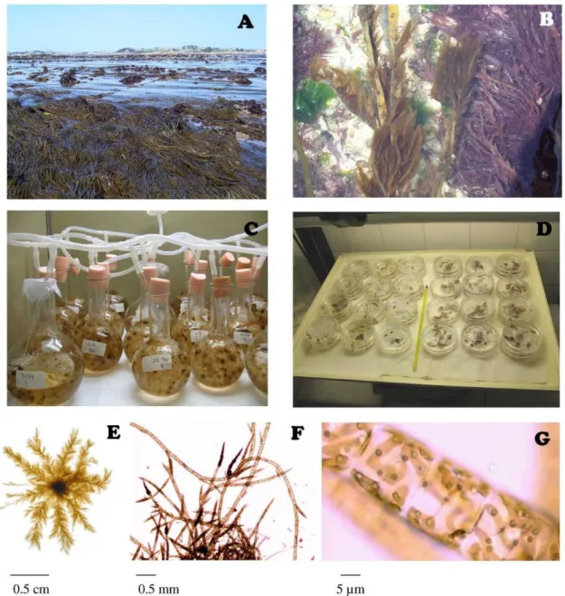
Figure 2. Presentation of Laminaria digitata. (A-B) Underwater Laminaria sp. forest
(pictures A. Ritter). (C) Morphological description. (D) Life cycle. Diploid macroscopic sporophytes produce meiospores in sporangia (sori). Meiospores grow into male or female microscopic gametophytes producing gametes. Gametophytes produce gametes. Fusion of gametes produces a zygote that grows into a diploid sporophyte, completing the sexual cycle. (E) L. digitata juveniles (picture D. Scornet). (F) L. digitata sporophyte cultures (picture P.O. de Franco)
Introduction-General Introduction
1.1.1.2. Ectocarpus siliculosus
Ectocarpus is a genus of filamentous uniseriate marine brown algae that contains 98 species. This latter number may be largely overestimated because of the difficulties to
differentiate some of the species within this genus. Two species are currently recognized, E.
fasciculatus (Harvey) and E. siliculosus (Dillwyn) Lyngbye. They are short-lived annual organisms, which can develop on many inert substrates, or as epiphytes on big seaweeds (Fig.
3 A-B). In the intertidal zone, Ectocarpus members occur from the top of the intertidal zone,
for instance in rocky pools, to the sublittoral zone. E. siliculosus (Fig 3E-F) is a cosmopolitan
alga, present on all coasts of temperate climate zones. This species has the remarkable ability
to develop in hostile environments, and has been described as growing at extremely low
salinities (even in freshwater) or in copper-enriched environments [26-28]. Although the
Ectocarpales have long been considered as the most primitive members of Phaeophyceae,
mainly because of their morphology, molecular systematics has shown that this order belongs
to a group of brown algae that has evolved quite recently (Fig. 1B). In addition, they are
closely related to kelps, which have a large size and complex morphology. E. siliculosus has
been intensively studied since the 19th century, with emphasis on its reproduction and life
history (Fig. 4A-E). The basic plan of the life cycle is diplohaplontic and isomorphic. The
diploid sporophytes produce haploid meiospores in unilocular meiosporangia which become
gametophytes and asexual diploid mitospores in plurilocular sporangia, which then produce
diploid sporophytes. The haploid gametophytes produce haploid gametes in plurilocular
gametangia. Unfused gametes may grow parthenogenetically and form haploid
parthenosporophytes. (Fig. 5) [2,5]. Other major aspects that have been studied include sexual
pheromone signaling, viral infections, cell ultrastructure, photosynthesis, carbon uptake,
gamete recognition and resistance to anti-fouling agents such as copper [5]. Altogether,
Introduction-General Introduction
These include its small size, the fact that the entire life cycle is well known and can be
completed in the laboratory (Fig. 3C-D), its high fertility and rapid growth (the life cycle can
be completed within 2 months) [29]. The genus Ectocarpus is the only brown alga for which
detailed genetic studies have been described, its genome (214 Mbp) has been sequenced and
is currently being annotated (Cock et al 2009). In addition, an international consortium has
been created to establish this species as a genomic and genetic model in Phaeophyceae [5,29].
Figure 3. Macroscopic and microscopic images of E. siliculosus. (A-B) Ectocarpus in the
field (pictures A.R.). (C-D) Cultures of E. siliculosus (pictures A.R.). (E-F) Microscopic view of E. siliculosus (pictures D. Scornet and J. Beltrand).
Introduction-General Introduction
Figure 4. General ultrastructure of a vegetative cell of Ectocarpus siliculosus. The general
ultrastructure of a vegetative cell is similar in both prostrate and erect filaments (Adapted from Charrier et al 2008 [5]). The different compartments of the cell are illustrated (see text for details). Lines represent membranes and define subcellular compartments, except for thylakoids, drawn as a thick black line. Depending on their type and age, the vegetative cell size varies from 10 to 35 µm in length, and 5 to 15 µm in width (under laboratory culture conditions).
Introduction-General Introduction
1.1.2. General aspects of abiotic stress in seaweeds
Marine macroalgae are multicellular benthic photosynthetic eukaryotes. They dominate
coastal intertidal and subtidal areas of temperate, cold and tropical regions. Coastal regions
and especially the intertidal zone, represent a steep gradient of environmental variations over
a small spatial scale, from marine to fully terrestrial conditions (Fig. 6). Under emersion
marine organisms are exposed to different stressors, such as variable temperature, UV
radiation, salinity changes, and they are affected by reduced amount of nutrients [22,23]. In
addition to natural oscillations, seaweeds may also be exposed to several sources of stress
resulting from industrial, urban and agricultural activities [30,31]. As a result, seaweeds
growing along the intertidal zone must be adapted to their environment and tolerate constant
fluctuating abiotic conditions. Most of the stress conditions trigger the production of ROS
[11,32-36], by different processes. Under abiotic stress, ROS synthesis results from altered
photosynthesis and inefficient reparation processes. During biotic stress, a so-called
“oxidative burst” is observed and corresponds to a rapid, transient, and intense ROS
production by enzymatic processes.
All organisms, even within the same species, are not responding in the same way to
changes in environmental conditions. If tolerance is expressed as a differential response to a
stressor, then selection may occur to produce genetically adapted individuals [37]. In
macroalgae, previous studies report some inter-population differences inherited against
environmental stress, such as tolerance to excess copper [38,39]. At the biochemical level,
seaweeds alter their metabolism in various ways to acclimate/adapt to environmental stresses
[22,23], however, the molecular basis of this phenomenon is poorly understood. In contrast,
the molecular events linking the perception of a stress signal to the metabolic responses
leading to tolerance have been intensively investigated recently in land plants, underlining the
Introduction-General Introduction
signaling molecules has been highlighted [42]. ROS produced under abiotic or biotic stress
can trigger a variety of cellular responses [43]. One of them is the activation of fatty acids by
the synthesis of oxygenated polyunsaturated fatty acid derivatives known as oxylipins, which
are know to play a pivotal role during abiotic and biotic stress in plants and metazoans. In
plants, their implication during wounding stress and pathogen and herbivore attacks has been
recognized in the past and is still an active field of research [44,45]. In macroalgae, oxylipins
have been shown to trigger defensive mechanisms upon biotic aggression [46,47]. Today,
new analytical tools developed within the “omics” era open a large panel of possibilities
towards the understanding of the molecular bases leading to the tolerance of seaweeds against
stressful environments.
A
B
Figure 6. Influence of the tidal cycle in Roscoff coastal area. The pictures were taken at high
and low tide the same day. Only 6 hours separate the first and the second picture (pictures P.O. de Franco).
Introduction-General Introduction
1.1.3. Ecological effects of copper excess in seaweeds: example of mine wasted areas in northern Chile
Metals occur naturally, and several of them are essential components of global
ecosystems. In seawater, most metals are bound to organic matter, which decrease their low
bioavailability. The term “heavy metal” designates those metals having densities greater than
5 g/cm3 (in their elemental stage). However, from a biological point of view, this term is
utilized for those metals and metalloids that cause toxic effects on living organisms [48].
Metals such as copper (Cu) and zinc (Zn) are essential micronutrients, whereas others such as
lead (Pb) and mercury (Hg) are not known to have useful biochemical function.
Environmental pollution by metals extended as mining and industrial activities increased in
the late 19th and early 20th centuries. The current worldwide mine production of Cu, Cd, Pb,
and Hg is considerable. These pollutants, ultimately derived from a growing number of
diverse anthropogenic sources (industrial effluents and wastes, urban runoff, sewage, boating
activities, agricultural fungicide runoff, domestic garbage dumps, mining operations), have
progressively affected different ecosystems [15,30]. Copper, in its ionic state (Cu2+), is the
second most toxic metal for living organisms [6]. In seawater, copper concentration vary
widely, fluctuating between 0.5 and 3 µg/L; the most meaningful concentration is the
bioavailable fraction which ranges at 10-7-10-8 µg/L [49].
Copper mining activities are still one of the most significant worldwide sources of this
metal, which is released into the environment due to the large volumes of wastes produced.
Copper mine wastes had severe negative effects on the coasts of England [50], Australia [51]
and Chile [30]. Historically, copper mines of Andes Mountains have been a major economic
input into Chilean budget. In 2007 the mineral production was 1,665 Mt, representing over
US$ 17,000 millions gains (http://www.codelco.cl/la_corporacion/cifras.asp). On the other
Introduction-General Introduction
areas is the Chañaral bay, which received tailings from the mine El Salvador from 1938 to
1975. During this period, more than 150 x106 t of untreated tailings dumped caused severe
beach degradation and total loss in the biodiversity (Fig. 7A-B) [52,53]. From 1976 to 1989
the dumping site was moved to Caleta Palito (26°16’S, 70°41’W), located 10 km northward,
which has received 130x106 t of tailings during the thirteen years. From 1990 to 2005, after
the building of a sedimentation dam inside the land (at 80 Km of the coast), wastewaters have
been channeled from the dam to Caleta Palito at a flow rate of 200-250 L/s, thereby
maintaining severe degradation into biota (Fig. 7C-E) [28,30]. Persistently high copper
concentrations prevent the growth of most algae, allowing only some opportunistic species to
develop, such as the green alga Ulva sp or the brown algae Scytosiphon lomentaria or E.
siliculosus [28,30]. Studies on some of these species have identified their special capability to tolerate metal stress, and the inheritance of this character has been discussed [35,54,55]. In
2005 a study indicated that no significant reduction of total dissolved copper had occurred in
the surroundings of the Chañaral bay since 1994 [28]. In relation to this results, high amonts
of dissolved copper were still observed at the sites of La Lancha and Palito (Fig. 5A) [28].
Despite sutained high copper concentrations, an increase in the species richness has been
observed, accounting for the occurrence of a recovery process. These results were explained
by a top-down effect caused by the absence of carnivores and the development of copper
avoidance strategies. Furthermore, no population genetics studies have been carried out at this
site in order to assess adaptative traits linked to this environment. Heavy metal tolerance was
firmly established in the literature as one of the best examples of adaptation [56-59], and
Ectocarpus could provide an excellent model system of the genetic bases of an adaptative trait in seaweeds.
Introduction-General Introduction
Figure 7. (A) Chañaral area and “study sites”. The discharge point of the copper mine tailing
is indicated with an arrow (Adapted from Medina et al 2005 [28]). (B) Image of the Chañaral bay, the artificial tailings created beach is indicated with an arrow (picture J. Correa). (C) River channeling copper-enriched water from the mine (picture J. Correa). (D) Representative picture of northern Chile intertidal coast (picture L. Contreras); (E) Caleta Palito (Chañaral zone) intertidal rocky shore (picture L. Contreras).
Introduction – Toxic effects of copper in algae and land plants
1.2.
TOXIC EFFECTS OF COPPER IN LAND PLANTS AND
MACROALGAE
Because of its redox properties, copper is essential for biology; however, for this same
reason it also represents a serious threat for cellular processes at high concentrations [60].
Copper homeostasis should therefore be tightly controlled to avoid excess or limitations, and
in this way ensure effective delivery into the active centres of many proteins and enzymes
(Box 1) [61,62]. High concentration of copper causes protein inactivation, oxidative stress
propagation and cell charge modification. All of these effects can lead to severe cell damage
and death. In this chapter, we will focus on the observed physiological effects of copper with
special focus on land plants and macroalgae.
1.2.1. Inhibition of growth and spore germination
Growth decrease and mortality represent the first parameters accounting for the
toxicity of a product. Copper excess as well as other heavy metals largely inhibit elongation
growth of land plants and algae [63,64]. Although several explanations have been proposed,
the exact biochemical process inhibiting growth is not yet well understood. Some authors
consider that copper inhibition of the cell cycle is the basis for the growth inhibition [65,66].
During the past years, ROS have been found responsible for modulating cell cycle and
developmental processes [67,68]. Therefore, the copper-induced ROS production may explain
the modulation of growth upon stress. In marine diatoms, it has been proposed that the high
reactivity of Cu with most amino acids could cause cross linking with hydroxyproline-rich
protein cell wall components, thereby inhibiting normal proliferation [69]. It has also been
hypothesized that Cu-induced growth inhibition in land plants could result from the
involvement of systemic responses triggered by oxylipins [70]. Finally, the ion uptake
Introduction – Toxic effects of copper in algae and land plants
of ionic processes (such as calcium signaling) [64]. Further research needs to be carried out to
determine particular growth inhibition processes.
The toxicity threshold of copper is highly variable in plants and macroalgae. In
addition, the toxicity threshold varies within the tissues and life stages. In non-metallophyte
plants, inhibitory effects arise at concentrations higher than 20 ppm, whereas in sedges the
toxicity threshold is 575 ppm [71]. In extreme cases, such as the metallophyte copper flower
(Haumaniastrum katangense), optimal growth is observed when copper concentrations reach
1000 ppm, although these authors do not specify the bioavailable copper concentration [72].
As in land plants, copper toxicity affects growth and causes mortality of seaweeds. Metal
sensitivity varies among species, however the toxic concentration affecting growth for a wide
range of seaweeds ranges around 12-75 µg/L [49]. In some cases, populations naturally
exposed to high concentrations of copper present differences in their growth rate within the
same species, accounting for local adaptation. The green alga E. compressa harvested from
copper-enriched areas showed faster growth than non-tolerant isolates for concentrations up
to 600 µg/L of copper [55]. Similar patterns of response to copper enrichment have been
reported for copper-tolerant populations of E. siliculosus from England [27]. Macroalgae can
have complex life histories, and different stages have been found to have different tolerance to
copper. In the kelp Lessonia nigrescens, a high sensitivity to copper was demonstrated for
early developmental gametophytic and sporophytic stages, which is not the case for adult
sporophytes [30,73]. Furthermore, these authors have suggested that the absence of L.
nigrescens from copper-polluted areas resulted from the high sensitivity of its early life cycle stages.
1.2.2. Inhibition of photosynthesis
Copper is a potent inhibitor of photosynthesis, both in algae and higher plants. The
carbon-Introduction – Toxic effects of copper in algae and land plants
fixing part, is the most important site of inhibition [61,70,74]. In all studies investigating this,
a much stronger inhibition is observed in photosystem II (PSII) compared to photosystem I
[75,76]. Drastically different types of damages seem to occur, depending on the irradiance
conditions. The so-called shade reaction occurs in the chlorophyll (Chl) molecules of the
Light Harvesting Complex (LHC) II in Chlorophyta and in the homologous Chl a/c complex
in brown algae [61,77]. Substitution of Mg2+ in Chl by Cu2+ results in an impairment of the
correct function of the LHC antenna. At high irradiance, the so-called “sun reactions” cause
direct damage to PSII reaction center [61]. The PS contains several metalloenzyme
components, thus copper toxicity targets are likely to be diverse. Several studies have
concluded that the target of copper is close to the primary photochemical event in PSII either
at the donor or at the acceptor sides [74,78,79]. Moreover, in vivo studies have concluded that
Cu inhibits charge separation in PSII by the insertion of Cu2+ into the phaeophytin of the PSII
reaction center (RC) which would cause a blockage in the electron transfer from chlorophyll
to phaeophytin [77,80]. This Cu-induced blocking of electron flux leads to the production of
ROS [70]. Copper-induced inhibition of photosynthesis can be observed by fluorescence
measurements [81]. This method is currently used today for measuring copper stress in field
and laboratory experiments [38,82,83].
1.2.3. Copper-induced oxidative stress
The term “reactive oxygen species” is employed for those forms of oxygen, sometimes
combined with hydrogen, which are more reactive than the relatively stable molecule O2.
Formation of ROS is a normal process in brown algae and land plants. The fundamental
electron chain reactions, photosynthesis and respiration, are the two main processes by which
ROS are produced. In fact, these oxygen species represent intermediates emerging during the
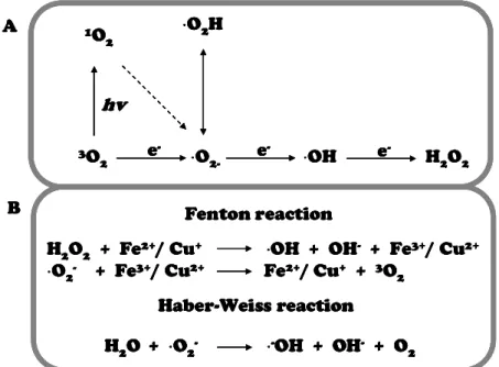
successive reduction of O2 to H2O. ROS formation starts by the excitation of O2 leading to
Introduction – Toxic effects of copper in algae and land plants
formation of superoxide radicals (·O2
-), hydrogen peroxide (H2O2) or hydroxyl radicals (·OH),
respectively (Fig. 8A) [84]. Under normal growth conditions, the production of ROS in cell is
low, thereby scavenged by cellular antioxidant systems [85]. Under uncontrolled stress
conditions, ROS production can overcome antioxidant systems, shifting balance towards ROS
accumulation [42]. As previously exposed, copper inhibition of photosystems results in the
blockage of electron transport which causes active generation of singlet oxygen and
superoxide radicals [70,86]. Singlet oxygen reacts with water to form the highly reactive ·OH
in the so-called Haber-Weiss reaction (Fig. 8B). In addition, redox active transition metals
such as Cu+ and Fe2+ can convert H2O2 to the highly reactive ·OH molecule in a
metal-catalyzed reaction known as the Fenton reaction (Fig. 8B) [87]. The ·OH molecule is one of
the most reactive species known. Because of its ability to initiate radical chain reactions, it is
very likely that this reaction is responsible for irreversible chemical modifications of various
cellular components. Cellular damage by free radicals is observed in proteins, DNA,
carbohydrates, and lipids [60,88]. ROS action on cell membranes results in the natural
metabolic process of peroxidation of polyunsaturated fatty acids (PUFA) in membrane lipids
(especially in chloroplast). This non-enzymatic reaction is initiated mainly by the most
reactive oxygen species, OH and·O2H, which are more lipophilic than the non-protonated
form, ·O2
-, and thus able to penetrate the membranes more easily. Several laboratories and
field examples of the heavy metal induced oxidative stress in plants and algae have been
published [15,83,89,90]. Furthermore, chronic oxidative stress has been linked to the
![Figure 1. Phylogeny of brown algae and Ectocarpales. (A) Position of brown algae within the eukaryotes (adapted from Baldauf, 2003 [3])](https://thumb-eu.123doks.com/thumbv2/123doknet/14690008.561195/26.892.109.709.103.725/figure-phylogeny-brown-ectocarpales-position-eukaryotes-adapted-baldauf.webp)

![Figure 5. Life cycle of Ectocarpus siliculosus . Adapted from Charrier et al . 2008 [5]](https://thumb-eu.123doks.com/thumbv2/123doknet/14690008.561195/31.892.110.707.761.1090/figure-life-cycle-ectocarpus-siliculosus-adapted-charrier-et.webp)
![Figure 10. Summary of potential cellular mechanisms implicated in metal detoxification and tolerance in higher plants; modified from Hall (2002) [128]](https://thumb-eu.123doks.com/thumbv2/123doknet/14690008.561195/53.892.106.804.106.768/summary-potential-cellular-mechanisms-implicated-detoxification-tolerance-modified.webp)
![[PDF] Introduction à la programmation pratique du langage C et C++ | Formation informatique](data:image/gif;base64,R0lGODlhAQABAIAAAP///wAAACH5BAEAAAAALAAAAAABAAEAAAICRAEAOw==)