HAL Id: hal-02329031
https://hal.archives-ouvertes.fr/hal-02329031
Submitted on 26 May 2020
HAL is a multi-disciplinary open access
archive for the deposit and dissemination of
sci-entific research documents, whether they are
pub-lished or not. The documents may come from
teaching and research institutions in France or
abroad, or from public or private research centers.
L’archive ouverte pluridisciplinaire HAL, est
destinée au dépôt et à la diffusion de documents
scientifiques de niveau recherche, publiés ou non,
émanant des établissements d’enseignement et de
recherche français ou étrangers, des laboratoires
publics ou privés.
Distributed under a Creative Commons Attribution| 4.0 International License
Sovanna Tan, Frederic Debellé, Pascal Gamas, Florian Frugier, Mathias Brault
To cite this version:
Sovanna Tan, Frederic Debellé, Pascal Gamas, Florian Frugier, Mathias Brault. Diversification of
cytokinin phosphotransfer signaling genes in Medicago truncatula and other legume genomes. BMC
Genomics, BioMed Central, 2019, 20 (1), pp.373. �10.1186/s12864-019-5724-z�. �hal-02329031�
R E S E A R C H A R T I C L E
Open Access
Diversification of cytokinin phosphotransfer
signaling genes in Medicago truncatula and
other legume genomes
Sovanna Tan
1, Frédéric Debellé
2, Pascal Gamas
2, Florian Frugier
1*and Mathias Brault
1Abstract
Background: Legumes can establish on nitrogen-deprived soils a symbiotic interaction with Rhizobia bacteria, leading to the formation of nitrogen-fixing root nodules. Cytokinin phytohormones are critical for triggering root cortical cell divisions at the onset of nodule initiation. Cytokinin signaling is based on a Two-Component System (TCS) phosphorelay cascade, involving successively Cytokinin-binding Histidine Kinase receptors, phosphorelay proteins shuttling between the cytoplasm and the nucleus, and Type-B Response Regulator (RRB) transcription factors activating the expression of cytokinin primary response genes. Among those, Type-A Response Regulators (RRA) exert a negative feedback on the TCS signaling. To determine whether the legume plant nodulation capacity is linked to specific features of TCS proteins, a genome-wide identification was performed in six legume genomes (Cajanus cajan, pigeonpea; Cicer arietinum, chickpea; Glycine max, soybean; Phaseolus vulgaris, common bean; Lotus japonicus; Medicago truncatula). The diversity of legume TCS proteins was compared to the one found in two non-nodulating species, Arabidopsis thaliana and Vitis vinifera, which are references for functional analyses of TCS components and phylogenetic analyses, respectively.
Results: A striking expansion of non-canonical RRBs was identified, notably leading to the emergence of proteins where the conserved phosphor-accepting aspartate residue is replaced by a glutamate or an asparagine. M. truncatula genome-wide expression datasets additionally revealed that only a limited subset of cytokinin-related TCS genes is highly expressed in different organs, namely MtCHK1/MtCRE1, MtHPT1, and MtRRB3, suggesting that this“core” module potentially acts in most plant organs including nodules.
Conclusions: Further functional analyses are required to determine the relevance of these numerous non-canonical TCS RRBs in symbiotic nodulation, as well as of canonical MtHPT1 and MtRRB3 core signaling elements.
Keywords: Phosphorelay, Cytokinin signaling, Histidine kinase, Response regulator, Legumes, Symbiotic nitrogen-fixing nodulation
Background
Cytokinin plant hormones are involved in numerous as-pects of plant growth and development in relation to their environment. They regulate the balance between cell division and differentiation, and consequently plant growth, but also nutrient uptake and shoot/root meta-bolic relationships, as well as the adaptation toward en-vironmental abiotic or biotic constraints [1–4]. These
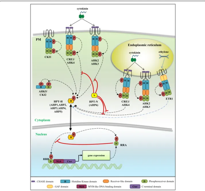
signals are transduced depending on a typical phosphor-elay (or phosphotransfer) Two-Component System (TCS) pathway that was elucidated in the reference plant Arabidopsis thaliana[5, 6]. Cytokinins are perceived by a small family of Histidine Kinase receptors containing a CHASE (Cyclases/Histidine kinases Associated Sensory Extracellular) domain (CHKs, [7–9]). Cytokinin percep-tion induces an autophosphorylapercep-tion of a conserved his-tidine (H) residue in the kinase domain (Fig. 1). The phosphate is thereafter transferred to a conserved aspar-tate (D) located at the C-terminal end of the protein, in the phosphoreceiver domain. These receptors are there-fore termed hybrid receptors [10]. The signal is then
© The Author(s). 2019 Open Access This article is distributed under the terms of the Creative Commons Attribution 4.0 International License (http://creativecommons.org/licenses/by/4.0/), which permits unrestricted use, distribution, and reproduction in any medium, provided you give appropriate credit to the original author(s) and the source, provide a link to the Creative Commons license, and indicate if changes were made. The Creative Commons Public Domain Dedication waiver (http://creativecommons.org/publicdomain/zero/1.0/) applies to the data made available in this article, unless otherwise stated.
* Correspondence:florian.frugier@cnrs.fr
1IPS2 (Institute of Plant Sciences Paris-Saclay), CNRS, Université Paris-Sud, Université Paris-Diderot, INRA, Université d’Evry, Université Paris-Saclay, Rue de Noetzlin, 91190 Gif-sur-Yvette, France
translocated into the nucleus, through the transfer of the phosphate group on a Histidine PhosphoTransfer pro-tein (HPT) shuttling between the cytosol and the nu-cleus [11]. The phosphate is finally transmitted to type-B Response Regulators (RRBs), which are transcription
factors that trigger the transcriptional activation of cyto-kinin primary response genes.
Features of CHK, HPT and Response Regulator (RR) proteins involved in cytokinin phosphorelay signaling have been well characterized in A. thaliana [3, 12].
Fig. 1 A model for the cytokinin Two Component System (TCS) phosphorelay signaling. In Arabidopsis thaliana, cytokinins (CKs) are perceived by histidine-kinase receptors (HK) containing two (for CRE1/AHK4) or three (for AHK2 and AHK3) transmembrane domains. CKs interact with the CHASE (Cyclases/Histidine kinases Associated Sensory Extracellular) domain of CHK receptors, inducing an autophosphorylation of a conserved histidine (H) residue. The phosphate is then transferred to a conserved aspartate (D) residue in the phosphoreceiver domain of the HKs, and to a conserved H residue of an Histidine Phosphotransfer protein (HPT-H). HPT-H proteins shuttle between the cytoplasm and the nucleus where they can transfer the phosphate group on a conserved D residue of Type-B Response Regulators (RRBs). This leads to an activation of RRBs, acting as transcription factors thanks to their Myb-like DNA binding (Myb) and transactivation domains, regulating the expression of CK-responsive genes such as Type-A Response Regulators (RRA). RRAs act as negative regulators of the CK signaling likely by competing with RRBs for phosphotransfer activation. In the HPT-N AHP6 variant, an asparagine (N) substitution of the conserved H leads to an inhibitory role on the CK signaling. In addition, CKI1, CKI2/AHK5 and the ethylene receptor ETR1 contain all domains defining an active hybrid Histidine Kinase receptor, and can interact with HPT-H, suggesting a potential function as modulators of cytokinin signaling
CRE1 (Cytokinin Response 1, also named AHK4, Arabi-dopsis Histidine Kinase 4), was the first CHK identified following a loss-of-function genetic screen designed to search for mutants impaired in cytokinin responses [7]. Whole-genome sequencing allowed the identification of two other A. thaliana CHKs, AHK2 and AHK3 [7, 8]. CHKs specifically bind bioactive cytokinins thanks to their CHASE domain that is delimited by transmem-brane domains [13, 14]. The three AHKs additionally contain an authentic histidine kinase domain displaying N, G1, F and G2 motifs required for the histidine kinase activity [12]. A phosphoreceiver domain is present at the C-terminal end of the proteins, containing the conserved D required for the phosphotransfer. Finally, a receiver-like domain is found between the kinase domain and the phosphoreceiver domain in all three AHKs.
Two classes of HPTs have been defined in A. thaliana. The first class corresponds to HPTs harboring a con-served H involved in phosphate acceptance (HPT-H, five genes in A. thaliana), and which are therefore able to transduce the phosphorelay initiated from CHKs to-wards nuclear RRBs. They are for this reason positive regulators of cytokinin signal transduction pathways [15]. In the second HPT class, the conserved H is replaced by an asparagine (N) (HPT-N, one gene in A. thaliana: AHP6), a residue not able to bind phosphate and therefore to mediate phosphotransfer from CHKs to RRBs. Consist-ently, AHP6 acts as negative regulator of cytokinin signal-ing notably dursignal-ing protoxylem formation [10].
RRs involved in cytokinin signaling are divided into two groups depending both on their structure and on their transcriptional regulation by cytokinins. All RRs have a phosphoreceiver domain structurally close to that of CHKs, with a conserved D required for the phospho-transfer. Type-A RRs (or RRAs) contain only a phos-phoreceiver domain and their expression is rapidly induced by cytokinins, making these genes markers of the activation of the cytokinin primary response [16]. Genetic analyses have demonstrated that RRAs function as negative regulators of cytokinin signaling [17] (Fig.1). Type-B RRs (or RRBs) have in addition a Myb-like DNA-binding domain, and a C-terminal transactivation domain [18]. Both RRAs and RRBs are nuclear proteins [5,18,19] and RRBs function as transcription factors dir-ectly controlling the expression of RRA genes [19–22]. In contrast to RRAs, RRB gene expression is generally not regulated by cytokinins [23]. The induction of RRAs is proposed to lead to a negative feedback competition with RRBs for accepting phosphate groups from the HPTs on the conserved D residue of their phosphoreceiver domain [17]. The RRB C-terminal transactivation domain is rich in proline (P) and glutamine (G), and its deletion impairs the ability of RRBs to promote transcriptional activation [18, 24]. In contrast, the deletion of the N-terminal
phosphoreceiver domain or the replacement of the con-served D by a glutamate (E) phosphomimic residue leads to a constitutive activation of RRBs. This indicates that the phosphoreceiver domain negatively regulates RRB transcriptional activity and that this inhibitory activity can be relieved by the phosphorylation of the conserved D residue [18,25,26].
Other TCS elements not directly linked to cytokinin signaling exist in plants, and some of them were shown to interfere with the phosphorelay cascades activated by cytokinins. CKI1 was identified in an activation tagging genetic screen in A. thaliana, and its ectopic expression induced typical cytokinin responses even in the absence of exogenous cytokinins [27]. CKI1 is an authentic histi-dine kinase with all required features to function in a phosphorelay cascade but that does not contain a CHASE domain, and that is therefore not able to bind cytokinins [28]. When expressed in protoplasts, CKI1 could nevertheless constitutively activate cytokinin phos-phorelay cascades, indicating that CKI1 may interfere with cytokinin signalling pathways [5,29] by interacting with and phosphorylating AHPs [30, 31]. CKI1 regulates A. thalianafemale gametogenesis and vascular tissue de-velopment [15,29,32], and was recently proposed to be a potential link between light and cytokinin responses to control plant development [33]. The CKI2/AHK5 gene was identified in the same genetic screen as CKI1, and may similarly interfere with cytokinin signalling as its overexpression in A. thaliana calli induces cytokinin re-sponses [27]. As CKI1, CKI2/AHK5 has authentic histi-dine kinase and phosphoreceiver domains but no transmembrane and CHASE domains [34]. CKI2/AHK5 is proposed to regulate abiotic and biotic responses in A. thalianabut no link with cytokinins has yet been estab-lished [34,35].
Other TCS elements are involved in the perception of signals different than cytokinin, such as the A. thaliana AHK1 osmosensor comprising all features of an active phosphotransfer protein [36] and ethylene receptors which do not all display hallmarks of authentic histidine kinases. Indeed, several ethylene receptor proteins (ETR1, EIN4 and ETR2 in Arabidopsis; [37]) comprise from the N- to the C-terminus three transmembrane domains corre-sponding to the ethylene-binding domain, a GAF (cGMP-specific phosphodiesterases, Adenylyl cyclases and FhlA) domain likely involved in protein-protein interac-tions, a non-canonical histidine kinase domain (except A. thalianaETR1 which has a canonical histidine kinase do-main) and a phosphoreceiver domain. Other ethylene re-ceptors (ERS1 and ERS2 in Arabidopsis) lack both a histidine kinase and a phosphoreceiver domain and are therefore not able to interact with HPT proteins in a phos-phorelay cascade [30]. The unique Arabidopsis ethylene re-ceptor able to function as an authentic histidine kinase
receptor, ETR1, was indeed reported to physically interact with the HPT protein AHP1 and to positively regulate the ARR2 type-B RR depending on a phosphorelay cascade [30,38–40]. However, as the etr1 mutant can be comple-mented with a kinase-dead ETR1 gene, it was concluded that the histidine kinase activity was not essential for ethylene signaling [37,41]. A crosstalk between cytokinin and ethylene signaling may however occur through phos-phorelay signaling [38,42]. Furthermore, RRCs represent a third class of RRs beside RRAs and RRBs. RRCs contain a unique receiver domain harboring the conserved D re-quired for phosphotransfer as in RRBs, but their se-quences are phylogenetically more related to HK receiver domains than to RRA receiver domains [43]. In addition, in contrast to RRAs, RRC gene expression is not induced in response to cytokinins. Overexpression of the Arabi-dopsis RRC ARR22 results in a phenotype similar to the wolCRE1/AHK4 mutant [43]. However, it is not yet clear whether RRCs could inhibit cytokinin signaling as RRAs do. Finally, the fourth and last group of RR proteins are “clock-related RRs” containing a receiver domain where the D phospho-acceptor residue is replaced by an E, and an additional C-terminal CCT domain (for CONSTANS, CONSTANS-LIKE, and TOC1) that is involved in protein-protein interactions [44]. Such clock-RRs are in-volved in the control of circadian rhythms, explaining their name, and no direct interaction with the TCS cytoki-nin signaling has been established.
Symbiotic nodule formation results from a molecular dia-log between legume roots and rhizobia. Roots release spe-cific flavonoids, which activate the production of Nodulation factors (or Nod factors) by rhizobia. The Nod factors, once perceived in the root epidermis, trigger a genetic program leading to bacterial infection and nodule organogenesis. Medicago truncatula forms indeterminate-type growing nodules, with a persistent apical meristem allowing for a continuous (indeterminate) growth [45,46]. Consequently, a metabolically active nodule comprises an apico-basal devel-opmental gradient, consisting in an apical zone I corre-sponding to the meristem, followed by a plant and bacteria cell differentiation zone (zone II), and a metabolically active nitrogen-fixation zone (III) [47]. In some other legumes, such as Lotus japonicus, the nodule organogenesis is deter-minate as the meristem is not maintained, leading to the for-mation of round-shaped nodules. The organogenesis of both determinate and indeterminate nodules however highly relies on the activation of a cytokinin phosphorelay signaling pathway [48, 49]. Indeed, a gain of function mutation in a specific L. japonicus CHK most closely related to Arabidop-sis AHK4/CRE1, LHK1 (Lotus Histidine Kinase 1), is neces-sary and sufficient to lead to spontaneous nodule formation in the absence of rhizobia [50], while loss-of-function mu-tants of LHK1 or MtCRE1 in M. truncatula are impaired in nodule formation [51–55]. Several RRB and RRA genes have
been linked to nodulation based on their expression profiles [51,56–60]. Furthermore, silencing of a subset of RRA genes (MtRR4, MtRR5, MtRR9 and MtRR11) in M. truncatula roots decreases nodule formation [56].
In this study, to determine whether the nodulation capacity of legume plants may be linked to a specific subset of TCS proteins, we performed a genome-wide analysis of the M. truncatula genome in order to identify genes encoding putative TCS phosphorelay components associated to cytokinin signaling or potentially interfer-ing with this pathway. We additionally proposed a uni-fied nomenclature for M. truncatula accordingly to guidelines proposed in [61]. The identified TCS genes were then compared to the ones found in other legume genomes, namely Cicer arietinum (chickpea) forming in-determinate nodules as M. truncatula, and Glycine max (soybean), Lotus japonicus, Cajanus cajan (pigeonpea), and Phaseolus vulgaris (common bean) forming deter-minate nodules [62–66]. In addition, we included A. thaliana and Vitis vinifera as reference dicot genomes because most functional analyses of TCS genes were performed in Arabidopsis and no recent Whole Genome Duplication (WGD) occurred in V. vinifera [67]. Finally, extensive expression datasets available in M. truncatula and corresponding to different organs [68], nodule zones [69] and the early response to Nod factors in the root epidermis [60] were used to identify a subset of cytoki-nin signaling genes mostly linked to nodulation and therefore anticipated to act in this symbiotic interaction.
Results
A constrained expansion of the CHK family proteins
M. truncatula has one AHK4/CRE1 homolog (CHK1/ CRE1), one AHK2 homolog (CHK4) and two AHK3 ho-mologs (CHK2 and CHK3; [55]; Fig. 2a). An analysis of gene duplications indicated that the two AHK3 homo-logs, CHK2 and CHK3, result from block duplication (Fig. 3). An additional M. truncatula CHK with a trun-cated C-terminus region (Medtr2g067240.1, CHK5) was identified in this study, containing a CHASE domain delimited by two transmembrane domains associated to a partial histidine kinase domain and neither a phos-phoreceiver nor a receiver-like domain (Table 1). This truncated CHK protein is most closely related to AtAHK4/CRE1 (Fig.2b). Despite the WGD at the origin of the Fabaceae family, M. truncatula has therefore a single additional gene encoding a full length canonical CHK compared to V. vinifera, as well as A. thaliana (Table 1). Similarly in the four other legume genomes studied, a CHK gene was retrieved in each AtCHK clade and only one additional CHK gene was detected com-pared to V. vinifera and A. thaliana (Additional file 1). This retained duplicated CHK gene is in the AHK3 clade for C. arietinum, as for M. truncatula, whereas it is in
the AHK4/CRE1 clade for C. cajan, L. japonicus, and P. vulgaris(Additional file2). In the soybean lineage, a more recent WGD occurred 13 Ma ago (Mya) in addition to the WGD that is common to all papilionoid legumes and which occurred about 58 Mya [70, 71]. As expected, two CHKs are retrieved in each clade (Additional files 1,2) while an additional duplication occurred and has been retained in the AHK4 clade, indicating a cytokinin-receptor diversifica-tion as for C. cajan, L. japonicus, and P. vulgaris. Overall, these analyses suggest that the AHK4 duplication has been retained in a common ancestor of these four legumes and lost in M. truncatula and C. arietinum forming indetermin-ate nodules. Conversely, the AHK3 duplication has been
conserved in a common ancestor of M. truncatula and C. arietinum. By contrast, the truncated CHK-like gene that is uniquely found in M. truncatula could be the result of a re-cent gene duplication, frequently observed in the M. trun-catulagenome [72].
In M. truncatula, CHK1/CRE1 is the most highly expressed CHK gene in the different organs (Fig.2c). All genuine CHK genes are expressed in roots and nodules. CHK1/CRE1 is upregulated in response to Nod factors in the root epidermis, in contrast to other MtCHK genes that are expressed but not strongly regulated (Fig. 2c; [60]). Considering the M. truncatula AHK3 homolog pair, CHK2 shows a weaker expression than CHK3 in
A
B
C
Fig. 2 Histidine kinases in Medicago truncatula. a Number of genes encoding HKs found in the genome of A. thaliana, M. truncatula and V. vinifera. b Phylogenetic tree of HKs, based on full-length protein sequences from A. thaliana, M. truncatula and V. vinifera. Protein sequences were aligned with the Muscle algorithm and the phylogenic tree was built with the Seaview software package. Numbers indicate the probability for each branch. Proteins labelled with a blue dot are non-canonical HKs. c Heat map of the expression pattern of M. truncatula genes encoding HK proteins. Data were retrieved from the M. truncatula Gene Expression Atlas Affymetrix microarray database (MtGEA; [68]) for various organs (left panel), from [69] for roots, nodules and nodule zones (middle panels) and from [60] for root epidermis expression and response to Nod factors (NF, right panel). Meristematic zone (ZI), distal and proximal differentiation/rhizobial infection zones (ZIId and ZIIp), inter-zone (IZ) and nitrogen fixation zone (ZIII). The red/white color scale indicates log2 expression values for each heat map, which were normalized independently, with highest expression as red and lowest expression as white. A median was used as the central value and black boxes indicate that no probe was available on the microarray
the different organs analyzed (Figs.2and3). The expres-sion of the CHK5 CHK-like gene is as well weak in the different organs analyzed (Fig.2c; [60, 68, 69]). The five CHK genes are expressed in the different nodule zones, redundantly in the apical meristem except CHK5.
To determine if the CHK genes loss following WGD is specific of this HK subset, we also analyzed the diversifi-cation of HKs involved in ethylene perception. Com-pared to V. vinifera and A. thaliana, the M. truncatula genome contains two and one additional ethylene recep-tor genes, respectively. In M. truncatula, both ETR2 and EIN4 genes are duplicated whereas in A. thaliana only ETR2 is duplicated, leading to the emergence of the ERS2 variant that lacks a receiver domain (Table 1; Fig.2b). A similar distribution of HK ethylene receptors is observed in the other five legume genomes analyzed (Additional file 3). As for CHKs, soybean has twice as many ethylene receptor genes compared to other legume genomes, consistently with its recent WGD. Overall, these analyses revealed that similarly to CHKs, most of the ethylene-related HK genes were not retained in the different legume genomes analyzed, indicating that this feature is not specific for CK perception.
Finally, regarding HKs that may interfere with cytoki-nin TCS phosphorelay signaling, AHK1, CKI1 and CKI2/ AHK5 genes exist in two copies in M. truncatula (re-spectively named MtHK1–2, MtHK3–4 and MtHK5–6 following [61]) and other genomes analyzed, in accord-ance with the legume WGD, except for CKI1 in G. max and P. vulgaris (Fig. 2b; Additional files 2, 4, 5, 6). For
CKI2/AHK5, a third gene (MtHK7) exists specifically in M. truncatula but is predicted to encode a truncated protein with neither a complete histidine domain nor a phosphoreceiver domain. This third gene could result from local gene duplication since MtHK6 and MtHK7 have close locations on chromosome 1 (Fig. 3). Among these HKs, only MtHK1 in the AtAHK1 clade, the ca-nonical MtHK6 gene and the non-caca-nonical MtHK7 gene in the CKI2/AHK5 clade, are expressed in roots and/or nodules (Fig.2c).
Within HPTs, only HPT1 is strongly expressed in different M. truncatula organs
V. vinifera and A. thaliana genomes contain respectively eight and six genes encoding HPT proteins while M. trunca-tula has 10 genes (Table 1; Fig. 4a). A similar number of genes (five or six) encoding HPT-H phosphoproteins is re-trieved in these three genomes, whereas two HPT-N genes were identified in M. truncatula vs one in A. thaliana and V. vinifera(Table1, Fig.4a). Besides, V. vinifera and M. trun-catulahave additional genes encoding non-canonical HPT proteins, respectively one and two, where the conserved H is replaced by an arginine (R) for M. truncatula and an iso-leucine (I) for V. vinifera (Table1, Additional file7). These non-canonical HPT proteins are grouped in a specific clade of the HPT protein phylogenetic tree (collectively named HPT-X; Fig. 4b) and also clustered on the M. truncatula chromosome 2 (Fig. 3). In the other legume genomes, a similar number of HPT-H and HPT-N genes were identified as in M. truncatula (Additional file 7). Additional
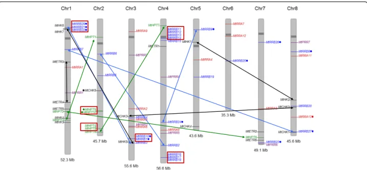
Fig. 3 Chromosomal distribution of M. truncatula genes encoding TCS elements. Histidine kinase (HK, CHK and ETR) genes are indicated in black,
phosphotransfer protein (HPT) genes in green, RRA genes in red, RRB genes in blue. Position of each gene was plotted on M. truncatula chromosomes using the Phenogram software. Gene names labelled with a dot encode proteins with non-canonical features. Tandem duplicated genes are shown with red boxes while genes resulting from block duplications are connected with black, green, blue and red arrows for CHK, HPT, RRB and RRA genes respectively
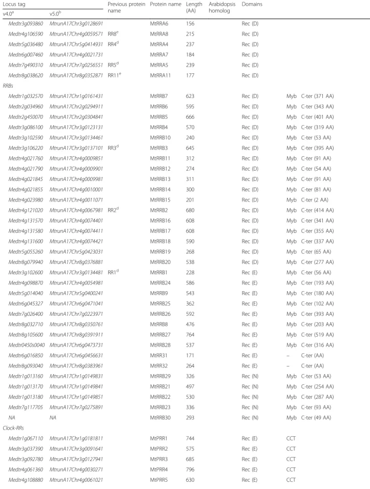
Table 1 List of Two-Component-System associated proteins found in the genome of M. truncatula
Locus tag Previous protein name
Protein name Length (AA) Arabidopsis homolog Domains v4.0a v5.0b Histidine kinases
Medtr5g097410 MtrunA17Chr5g0447641 MtCHK4c 1270 AHK2 CHASE His (H) HATPase_c (N, G1, F, G2)
Rec (D) Medtr3g085130 MtrunA17Chr3g0122391 MtCHK3c 1035 AHK3 CHASE His (H) HATPase_c
(N, G1, F, G2) Rec (D) Medtr8g080770 MtrunA17Chr8g0377481 MtCHK2c 971 AHK3 CHASE His (H) HATPase_c
(N, G1, F, G2) Rec (D) Medtr8g106150 MtrunA17Chr8g0392301 CRE1 MtCHK1c 1004 AHK4 CHASE His (H) HATPase_c
(N, G1, F, G2) Rec (D) Medtr2g067240 MtrunA17Chr2g0311191 MtCHK5 256 AHK4-like CHASE His (H) – – Medtr5g022470 MtrunA17Chr5g0405701 MtHK1 1201 AHK1 His (H) HATPase_c
(N, G1, F, G2) Rec (D) Medtr8g075340 MtrunA17Chr8g0371601 MtHK2 1174 AHK1 His (H) HATPase_c
(N, G1, F, G2) Rec (D) Medtr1g090850 MtrunA17Chr1g0197591 MtHK3 1051 CKI1 His (H) HATPase_c
(N, G1, F, G2) Rec (D) Medtr1g087140 MtrunA17Chr1g0194741 MtHK4 1103 CKI1 His (H) HATPase_c
(N, G1, F, G2) Rec (D) Medtr3g105590 MtrunA17Chr3g0136601 MtHK5 950 CKI2 / AHK5 His (H) HATPase_c
(N, G1, F, G2) Rec (D) Medtr1g013360 MtrunA17Chr1g0150511 MtHK6 1013 CKI2 / AHK5 His (H) HATPase_c
(N, G1, F, G2) Rec (D) Medtr1g014670 MtrunA17Chr1g0150531 MtHK7 390 CKI2 / AHK5 His (H) – – Medtr4g031150 MtrunA17Chr4g0013831 MtETR1 791 ETR1 EBD
(D,Y, I1,P, I2, C, H)
GAF His (H) HATPase_c (N, G1, F, G2)
Rec (D) Medtr7g109150 MtrunA17Chr7g0269631 MtETR2 636 ERS1 EBD
(D,Y, I1,P, I2, C, H)
GAF His (H) HATPase_c (N, G1, F, G2) – Medtr1g044210 MtrunA17Chr1g0168161 MtETR3 761 ETR2 EBD
(D,Y, I1,P, I2, C, H)
GAF His (−) HATPase_c (−, −, −, −)
Rec (D) Medtr1g073840 MtrunA17Chr1g0186431 MtETR4 760 ETR2 EBD
(D,Y, I1,P, I2, C, H)
GAF His (−) HATPase_c (−, G1, −, −)
Rec (D) Medtr1g079790 MtrunA17Chr1g0190021 MtETR5 763 EIN4 EBD
(D,Y, I1,P, I2, C, H)
GAF His (H) HATPase_c (−, G1, −, G2)
Rec (D) Medtr7g116330 MtrunA17Chr7g0274831 MtETR6 766 EIN4 EBD
(D,Y, I1,P, I2, C, H)
GAF His (H) HATPase_c (−, G1, −, G2) Rec (D) Phosphorelay-proteins Medtr1g082290 MtrunA17Chr1g0191351 MtHPT2d 152 Hpt (H) Medtr1g089130 MtrunA17Chr1g0196361 MtHPT3 149 Hpt (H) Medtr2g020770 MtrunA17Chr2g0286781 MtHPT1d 148 Hpt (H) Medtr2g100880 MtrunA17Chr2g0331011 MtHPT4 150 Hpt (H) Medtr2g100900 MtrunA17Chr2g0331021 MtHPT5 150 Hpt (H) Medtr7g114020 MtrunA17Chr7g0273321 MtHPT8 147 Hpt (H) Medtr4g010160 MtrunA17Chr4g0003251 MtHPT7 169 Hpt (N) Medtr2g103870 MtrunA17Chr2g0333131 MtHPT6 158 Hpt (N) Medtr2g085155 NA MtHPT9 151 Hpt (R) Medtr2g086010 NA MtHPT10 151 Hpt (R) RRAs
Medtr1g049100 MtrunA17Chr1g0170741 MtRRA1 198 Rec (D) Medtr3g015490 MtrunA17Chr3g0082861 RR9e MtRRA9 164 Rec (D) Medtr3g078613 MtrunA17Chr3g0118211 MtRRA2 201 Rec (D) Medtr3g088630 MtrunA17Chr3g0124861 MtRRA3 235 Rec (D)
Table 1 List of Two-Component-System associated proteins found in the genome of M. truncatula (Continued)
Locus tag Previous protein name
Protein name Length (AA)
Arabidopsis homolog
Domains v4.0a v5.0b
Medtr3g093860 MtrunA17Chr3g0128691 MtRRA6 156 Rec (D) Medtr4g106590 MtrunA17Chr4g0059571 RR8e MtRRA8 215 Rec (D) Medtr5g036480 MtrunA17Chr5g0414931 RR4d MtRRA4 237 Rec (D) Medtr6g007460 MtrunA17Chr4g0021731 MtRRA7 184 Rec (D) Medtr7g490310 MtrunA17Chr7g0256551 RR5d MtRRA5 239 Rec (D) Medtr8g038620 MtrunA17Chr8g0352871 RR11e MtRRA11 177 Rec (D) RRBs
Medtr1g032570 MtrunA17Chr1g0161431 MtRRB7 623 Rec (D) Myb C-ter (371 AA) Medtr2g034960 MtrunA17Chr2g0294911 MtRRB6 595 Rec (D) Myb C-ter (343 AA) Medtr2g450070 MtrunA17Chr2g0304841 MtRRB5 666 Rec (D) Myb C-ter (401 AA) Medtr3g086100 MtrunA17Chr3g0123131 MtRRB4 570 Rec (D) Myb C-ter (319 AA) Medtr3g102590 MtrunA17Chr3g0134461 MtRRB10 240 Rec (D) Myb C-ter (53 AA) Medtr3g106220 MtrunA17Chr3g0137101 RR3d MtRRB3 645 Rec (D) Myb C-ter (395 AA) Medtr4g021760 MtrunA17Chr4g0009851 MtRRB11 312 Rec (D) Myb C-ter (91 AA) Medtr4g021790 MtrunA17Chr4g0009901 MtRRB12 274 Rec (D) Myb C-ter (54 AA) Medtr4g021845 MtrunA17Chr4g0009981 MtRRB13 311 Rec (D) Myb C-ter (91 AA) Medtr4g021855 MtrunA17Chr4g0010001 MtRRB14 300 Rec (D) Myb C-ter (81 AA) Medtr4g023980 MtrunA17Chr4g0011071 MtRRB15 201 Rec (D) Myb C-ter (2 AA) Medtr4g121020 MtrunA17Chr4g0067981 RR2d MtRRB2 680 Rec (D) Myb C-ter (414 AA) Medtr4g131570 MtrunA17Chr4g0074401 MtRRB16 608 Rec (D) Myb C-ter (341 AA) Medtr4g131580 MtrunA17Chr4g0074411 MtRRB17 608 Rec (D) Myb C-ter (355 AA) Medtr4g131600 MtrunA17Chr4g0074421 MtRRB18 590 Rec (D) Myb C-ter (337 AA) Medtr5g055260 MtrunA17Chr5g0423031 MtRRB19 268 Rec (D) Myb C-ter (65 AA) Medtr8g079940 MtrunA17Chr8g0376881 MtRRB20 538 Rec (D) Myb C-ter (277 AA) Medtr3g102600 MtrunA17Chr3g0134481 RR1d MtRRB1 228 Rec (E) Myb C-ter (56 AA) Medtr4g098870 MtrunA17Chr4g0054981 MtRRB24 586 Rec (E) Myb C-ter (193 AA) Medtr5g014040 MtrunA17Chr5g0400241 MtRRB9 543 Rec (E) Myb C-ter (180 AA) Medtr6g045327 MtrunA17Chr6g0471041 MtRRB25 362 Rec (E) Myb C-ter (102 AA) Medtr7g026400 MtrunA17Chr7g0223971 MtRRB26 592 Rec (E) Myb C-ter (393 AA) Medtr8g032710 MtrunA17Chr8g0350761 MtRRB8 476 Rec (E) Myb C-ter (203 AA) Medtr8g105600 MtrunA17Chr8g0391911 MtRRB27 764 Rec (E) Myb C-ter (519 AA) Medtr0450s0040 MtrunA17Chr6g0473731 MtRRB28 537 Rec (E) Myb C-ter (316 AA) Medtr6g016850 MtrunA17Chr6g0456631 MtRR31 171 Rec (E) – C-ter (AA) Medtr8g093040 MtrunA17Chr8g0383961 MtRR32 264 Rec (E) – C-ter (AA) Medtr1g013160 MtrunA17Chr1g0149831 MtRRB29 326 Rec (N) Myb C-ter (53 AA) Medtr1g013170 MtrunA17Chr1g0149841 MtRRB21 497 Rec (N) Myb C-ter (254 AA) Medtr1g013180 MtrunA17Chr1g0149851 MtRRB22 530 Rec (N) Myb C-ter (287 AA) Medtr7g117705 MtrunA17Chr7g0275891 MtRRB23 336 Rec (N) Myb C-ter (93 AA) NA NA MtRRB30 293 Rec (N) Myb C-ter (49 AA) Clock-RRs
Medtr1g067110 MtrunA17Chr1g0181811 MtPRR1 744 Rec (E) CCT Medtr3g037390 MtrunA17Chr3g0091641 MtPRR2 575 Rec (E) CCT Medtr3g092780 MtrunA17Chr3g0127941 MtPRR3 685 Rec (E) CCT Medtr4g061360 MtrunA17Chr4g0030271 MtPRR4 796 Rec (E) CCT Medtr4g108880 MtrunA17Chr4g0061021 MtPRR5 630 Rec (E) CCT
non-canonical HPTs were also retrieved: HPT-R variants in C. cajan, G. max and P. vulgaris; and two HPT-L variants in G. max and one in L. japonicus, respectively (Add-itional files7,8). At the predicted phosphoacceptor position (H77 in MtHPT3) in the 78 HPTs identified in this study, H (65%) or N (19%) residues are found in 84% of HTPs (Add-itional file9A). Regarding H positions different than the pre-dicted phosphoacceptor site, only 2 to 53% contains a H or a N residue within the 78 HPT proteins analyzed. This sug-gests that the rate of substitution of the H involved in phos-photransfer is reduced compared to other H residues.
Among the 10 M. truncatula HPT genes, MtHPT1 has the highest expression in all plant organs studied, in-cluding nodules where the expression is maximal in the meristematic zone I and the distal part of differenti-ation/rhizobial infection zone II (Fig. 4c). MtHPT1 ex-pression is also induced by NFs in the root epidermis. MtHPT3, 4 and 5 are expressed in leaves and flowers, MtHPT3 and 8 in roots and nodules, even though their expression is not regulated by NFs in the root epidermis (Fig.4c). Genes encoding non-canonical HPT-N (MtHPT6 and MtHPT7) and HPT-R (MtHPT9 and MtHPT10) are weakly expressed whatever the organ considered (Fig.4c) potentially because of an expression pattern limited to a small number of cells.
Expansion of non-canonical RRBs in legume genomes
The M. truncatula genome contains 32 predicted pro-teins grouping with A. thaliana and V. vinifera RRBs (Table 1, Fig. 5a, c), i.e. about three times more than V. viniferaand two times more than A. thaliana. Seventeen of them encode authentic RRBs (i.e. with a phosphore-ceiver domain containing a conserved phosphoacceptor D residue, a DNA-binding domain, and a transactivation domain) vs 10 in V. vinifera and 11 in A. thaliana. The remaining 15 M. truncatula RRBs are non-canonical, the conserved D being replaced by E or N in most cases (Table1). Seven MtRRBs seem to have a transactivation domain shorter than 100 residues, vs 200–500 residues in authentic RRBs (Table1). V. vinifera and A. thaliana genomes encode respectively only one and three non-canonical RRB genes (with the D replaced by either an E, N or Q residue; Additional file10), indicating that there has been comparatively a strong expansion of non-canonical RRBs in M. truncatula. This expansion likely results from tandem duplications since these
proteins are clustered in the phylogenetic tree in clades where V. vinifera or A. thaliana RRB proteins are absent (Fig.5c), and most of them are also physically clustered in four blocks on M. truncatula chromosomes 1, 3 and 4 (Fig.3). Block duplications are in addition observed, corre-sponding to four pairs of genes: MtRRB3/MtRRB29, MtRRB7/MtRRB27, MtRRB2/MtRRB6, MtRRB1/MtRRB10 (Fig. 3). In two of these block-duplicated pairs, one of the paralogs has lost the conserved D residue required for phosphotransfer (Figs.3and5; Table1). Two M. truncatula RRs (MtRR31 and MtRR32) grouping with authentic RRBs consist of a single receiver domain with neither a DNA binding domain nor a trans-activation domain. These two proteins therefore resemble RRA proteins (see below) but in contrast to authentic RRA they have an E instead of the conserved D residue associated to the phosphotransfer. The other legume genomes analyzed have roughly a similar number of authentic RRBs as V. vinifera and A. thaliana, but the number of non-canonical RRB genes is also in-creased, while this number remains similar also in G. max despite its additional WGD (Additional files10,11). Inter-estingly, in all legume genomes analyzed, non-canonical RRBs have conserved D to E or N substitutions. We ana-lyzed in the 138 RRBs identified in this study the substitu-tion rates for different D posisubstitu-tions within or outside the predicted phosphoacceptor site (D64 in MtRRB3) (Additional file 9B). In 95% of the phosphoacceptor sites, the position was occupied by a D (64%), an E (25%) or an N (6%), (Additional file9B), while at D positions outside of this site (eg D192 in MtRRB3) 30% of the 138 RRBs had a residue different than D, E, or N. This suggests that, as for HPT proteins, the predicted phosphoacceptor site has a re-duced substitution rate.
Expression of 27 of the 32 M. truncatula RRB genes was detected in the transcriptomic datasets analyzed, including 14 (out of 18) canonical and 13 (out of 14) non-canonical RRBs, in different plant organs includ-ing nodules (Fig. 5d). Three RRB genes, one non-canonical D-to-E RRB (MtRRB1) and two canon-ical (MtRRB2 and MtRRB3), show the highest expres-sion level in roots and nodules. The expresexpres-sion of other non-canonical RRB genes can be detected in roots and nodules, corresponding to three D-to-E RRBs and the one truncated RRB lacking the Myb domain (Fig. 5d). Beside MtRRB2 and MtRRB3, most other authentic RRBs are expressed in different
Table 1 List of Two-Component-System associated proteins found in the genome of M. truncatula (Continued)
Locus tag Previous protein name
Protein name Length (AA)
Arabidopsis homolog
Domains v4.0a v5.0b
Medtr7g118260 MtrunA17Chr7g0276361 MtPRR6 559 Rec (E) CCT Medtr8g024260 MtrunA17Chr8g0345901 MtPRR7 585 Rec (E) CCT
For each gene, the current protein name, as well as a previously published name when available, the protein length, the A. thaliana most closely related protein, and the conserved protein domains are listed.a[73],b[74],c[55],d[51] ande[56]. NA: Not Annotated
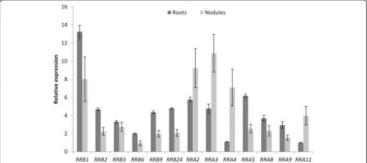
organs and notably in roots and in the different nod-ule zones. The expression level in roots and nodnod-ules of MtRRB genes independently tested by real-time RT-PCR revealed similar results as transcriptomic datasets (Fig. 6). Considering the origin of these genes, tandem duplicated genes are weakly expressed with the exception of MtRRB1 and MtRRB10 (Figs. 3
and 5d), whereas for block-duplicated genes, in each of the three pairs identified, one of the paralogs shows a weaker expression than the other duplicated gene (Figs. 3 and 5d).
A constrained expansion and structure conservation of the RRA family
The M. truncatula genome contains 10 genes encoding RRA proteins, similarly to A. thaliana and V. vinifera genomes that contain respectively 10 and 11 RRA genes (Table 1; Fig. 5b). Among the six legume genomes studied here, six genes encoding potential RRC proteins were found in G. max (Additional file 12). The M. truncatula genome contains seven genes encoding clock-RRs, i.e. two more genes that V. vinifera and A. thaliana (Additional file 13). All RRA and clock-RR
A
B
C
Fig. 4 HPT proteins in Medicago truncatula. a Number of genes encoding HPT-H (blue bars), HPT-N (red bars) and non-canonical HPTs (green bars) found in the genome of A. thaliana, M. truncatula and V. vinifera. b Phylogenetic tree of HPTs, based on full-length proteins from A. thaliana, M. truncatula and V. vinifera. Protein sequences were aligned with the Muscle algorithm and the phylogenic tree was built with the Seaview software package. Numbers indicate the probability for each branch. The tree was rooted on the HPT Ostta_34,527 from Ostreococcus tauri [75]. Blue dots indicate non-canonical HPTs. c Heat map of the expression pattern of M. truncatula genes encoding HPT proteins. Data were retrieved from the M. truncatula Gene Expression Atlas Affymetrix microarray database (MtGEA; [68]) for various organs (left panel), from [69] for roots, nodules and nodule zones (middle panels) and from [60] for root epidermis expression and response to Nod factors (NF, right panel). Meristematic zone (ZI), distal and proximal differentiation/rhizobial infection zones (ZIId and ZIIp), inter-zone (IZ) and fixation zone (ZIII). Dashes indicate that there is no probe available on the Affymetrix M. truncatula chip [68]. The red/white color scale indicates log2 expression values for each heat map, which were normalized independently, with highest expression as red and lowest expression as white. A median was used as the central value and black boxes indicate that no probe was available on the microarray
A
B
C
D
genes are expressed in most organs and in all nodule zones (Fig.5d).
All RRA proteins have a canonical structure and display the conserved D required to act in a phosphorelay cas-cade. Among all these genes, only MtRRA2 and MtRRA8 result from block duplication (Figs.3and 5d). Other leg-ume genomes analyzed also contains between 8 and 14 RRA genes while G. max has 20 genes due to its specific WGD, all being authentic RRAs (Additional files 11, 14). Thus, in contrast to RRBs, the ancestral legume WGD was not followed by an expansion of RRA genes, and the additional soybean WGD was not followed by a global loss of RRAs.
Expression of RRA genes is detected in all organs ana-lyzed. MtRRA2/3/4/8/11 transcripts are more abundant in nodules than in roots whereas MtRRA5 shows an op-posite expression pattern (Fig. 5d). In nodules, the
expression of different RRA genes is detected in the dif-ferent zones, MtRRA4 being the most expressed, mainly in the differentiation/rhizobial infection zone II. The ex-pression level in roots and nodules of MtRRA genes in-dependently tested by real-time RT-PCR overall revealed similar results as transcriptomic datasets (Fig. 6). A sub-set of RRA genes (MtRRA2/5/8/9/11) is expressed in the root epidermis and induced by Nod factors consistently with cytokinin signaling pathways being active in the epidermis (Fig.5d; [56,59, 60]). In contrast, MtRRA4 is not regulated by NFs in the root epidermis, suggesting that it may be more related to nodule organogenesis in the root cortex as previously proposed (Fig. 5d; [53]). We finally searched in the promoter of all these M. trun-catula RRA genes the number of “AGATHY” cytokinin responsive cis-elements (Additional file 15) proposed in A. thalianato be directly regulated by RRB transcription
(See figure on previous page.)
Fig. 5 Type-A and Type-B Response Regulators in Medicago truncatula. a-b Number of genes encoding RRBs (a) and RRAs (b), respectively, identified in the genome of A. thaliana, M. truncatula and V. vinifera. c Phylogenetic tree of RRs, based on full-length proteins from A. thaliana, M. truncatula and V. vinifera. Protein sequences were aligned with the Muscle algorithm and the phylogenic tree was built with the Seaview software package. Numbers indicate the probability for each branch. The tree was rooted on the ARR22 from A. thaliana [75]. Proteins labelled with a blue dot are non-canonical RRs. d Heat map of the expression pattern of M. truncatula genes encoding RR proteins. Data were retrieved from the M. truncatula Gene Expression Atlas Affymetrix microarray database (MtGEA; [68]) for various organs (right panels), from [69] for roots, nodules and nodule zones (middle panels), and from [60] for root epidermis expression and response to Nod factors (NF, right panels). Meristematic zone (ZI), distal and proximal differentiation/ rhizobial infection zones (ZIId and ZIIp), inter-zone (IZ) and fixation zone (ZIII). Dashes indicate that there is no probe available on the Affymetrix M. truncatula chip [68]. The red/white color scale indicates log2 expression values for each heat map, which were normalized independently, with highest expression as red and lowest expression as white. A median was used as the central value and black boxes indicate that no probe was available on the microarray
Fig. 6 Expression of selected RRB and RRA cytokinin signaling related genes in roots and nodules. Real-time RT-PCR analysis of RRB and RRA encoding gene expression in non-inoculated roots or in nodules (8 days after inoculation with S. meliloti), selected based on their detectable expression and/or their differential expression level in roots versus nodules in transcriptomic datasets. The value of the lowliest expressed gene in non-inoculated roots (MtRRA11) was arbitrarily set to 1 as a calibrator. Data were normalized using the mean expression of two reference genes, and error bars represent Standard Deviations from one representative biological replicate out of two. RRB, B Response Regulator; RRA, Type-A Response Regulator
factors, and therefore cytokinin signaling [76]. Between 6 to 21 AGATHY motifs per 2.5 kb of promoter regions were identified; this number was however neither strictly correlated to the strength of gene expression in roots or during nodulation, nor in relation to root and nodule ex-pression clusters identified using a hierarchical cluster-ing approach (Additional file16).
Discussion
A function for non-canonical TCS variants
The most striking characteristic of the legume cytokinin signaling gene families is an expansion of TCS proteins with non-canonical features, as compared to V. vinifera and A. thaliana. This is especially obvious for MtRRBs for which almost half of the genes encode non-canonical tran-scription factors. About one third of these non-canonical RRBs show a detectable expression in the conditions ana-lyzed. The expression of the remaining genes may take place in other conditions or be restricted to a few cells, making its detection difficult, or alternatively may be dis-appearing because of pseudogenization [77]. A recent study of TCS in various plants but not legumes revealed that among TCS families expansion mostly occurs in the RR gene family, in agreement with our results [78]. An ex-pansion of non-canonical RRBs was however not reported, even in more detailed studies focused on rice and poplar genomes [79, 80]. Further dedicated studies would be needed to definitively establish whether this variant en-richment is legume-specific or not.
Considering all non-canonical RRB and HPT proteins identified within the six selected legume genomes, a striking observation is that the conserved residue required for the phosphotransfer (a D for RRBs, or an H for HPTs) is mostly replaced by a residue a priori unable to participate in the phosphotransfer but restricted to a few amino acids. This substitution might relate to a functional diversification of these proteins: indeed, the conserved D-to-E substitution frequently observed in legumes at the RRB predicted phos-phoreceiver site has been shown to maintain RRB transcrip-tion factors in a constitutive active state [5, 26, 81]. In contrast, the H-to-N substitution identified initially in the Arabidopsis AHP6 protein at the phosphoacceptor site im-pedes its activation by phosphotransfer [10]. Specific func-tional variants may have then arisen in legume genomes following WGD, block, and/or tandem gene duplication events. In addition to the loss of conserved residues required for phosphotransfer regulation, D for RRBs or H for HPT proteins, most of these atypical duplicated genes display a very weak or narrow expression pattern. This is especially noticeable for block-duplicated genes where one of the two paralog shows a strong expression pattern while the second can be almost not expressed. Indeed, beside cases where one gene is retained while the second duplicated gene is lost or pseudogenized, an alternative fate is that both genes
remain functional, either with a shared function or with a neofunctionalization [70]. In A. thaliana for example, single, double and triple mutants affecting the canonical RRBs pro-teins ARR1, ARR10 and ARR12 showed a progressive in-crease in the number of deregulated target genes, indicating that gene duplication increases both the diversity of target genes and the robustness of their regulation [82]. In the case of the AHP6 non-canonical HPT (HPT-N), the phospho-transfer capacity is lost leading to an opposite function than canonical HPTs as a negative regulator of cytokinin phos-photransfer signaling [10]. Specific expansion of a subset of expressed non-canonical D-to-N or D-to-E RRBs in differ-ent legume genomes suggests that some of these RRBs may have acquired new functions, either as inhibitors of the phosphotransfer, or as phosphotransfer-independent tran-scription factors that may or may not be linked to cytokinin signaling. Interestingly in Arabidopsis, the APRR2 protein is similar to authentic RRBs due to its Myb-like DNA-binding and phosphoreceiver domains, but cannot be regulated by a phosphorelay cascade since the conserved D is replaced by a E. By interacting with the calmodulin protein CML9, APRR2 seems to be involved in responses to abiotic stress and ABA signaling more than in a cytokinin-signaling pathway [83]. In tomato, an APRR2 ortholog was proposed to participate in the control fruit ripening [84], another physiological process for which a cytokinin regulation is usually not re-ported as critical.
The roles of such non-canonical RRs are not yet eluci-dated in legumes. MtRRB1 is a non-canonical RRB highly expressed in M. truncatula roots and nodules ([51]; this study). In contrast to authentic RRBs, MtRRB1 is predicted to be constitutively activated because of the D-to-E substitution in the predicted phosphoreceiver site. MtRRB1 can bind promoters of early nodulation genes such as NSP2, as well as of cytokinin primary tar-get genes such as RRA4, but no nodulation phenotype was reported upon silencing by RNAi or overexpression [21]. MtRRB1 overexpression in A. thaliana roots how-ever increased root length [21], a phenotype opposite to the one expected for an authentic RRB acting as a posi-tive regulator of cytokinin signaling, and which might suggest a negative role in this signaling pathway. As RRBs that have lost the predicted phosphoacceptor D residue are expected to be unable to be regulated by phosphotransfer, these non-canonical proteins may be activated by an alternative mode of regulation, as re-ported for APRR2 in Arabidopsis [83], e.g. by a binding to calmodulin, S / T phosphorylation, ubiquitination or other post-translational regulatory modifications.
Cytokinin signaling and symbiotic nodulation: a main core signaling recruited from existing pathways?
One objective of analyzing proteins related to cytokinin signaling in legumes was to define which subsets of
proteins could be linked specifically to the nitrogen-fix-ing symbiotic capacity of these plants, and to determine whether differences correlating with the ability to form determinate or indeterminate nodules could be identi-fied in TCS gene families. No specific feature was highlighted concerning the ability of legumes to form in-determinate- or in-determinate-type, except the structur-ation of the CHK family. This perceived correlstructur-ation may be however linked to the close relationships between the genomes analyzed, and additional phylogenetic analyses based on more diverse high-quality legume genomes, when available, would be needed to more convincingly address this issue. In addition, it remains to be tested whether differences may exist in upstream events linked to cytokinin metabolism, and/or to downstream RRB target gene regulation. The independent expression data-sets analyzed in this study revealed that in each TCS protein family, a few members are more strongly expressed in nodules than others, leading to define a core symbiotic nodule cytokinin signaling module, notably highlighted by a hierarchical clustering focused on tran-scriptomic datasets from roots and nodules, consisting of the MtCRE1/MtCHK1 receptor, the MtHPT1 phospho-transfer protein, and the MtRRB3 transcription factor, while more variation in expression levels was observed for RRAs. The functional relevance of this core pathway re-mains to be evaluated, even though it is already estab-lished in different legumes that, at early symbiotic stages, the most expressed cytokinin receptor (MtCHK1/CRE1, LjLHK1, AhHK1 in Arachis hypogea, or AeHK1 in Aeschynomene evenia) gene is also the most functionally relevant for nodulation [51–55, 85, 86]. Noteworthy, the MtCHK1(MtCRE1)/MtHPT1/MtRRB3 cytokinin signaling core is also the most highly expressed in the different M. truncatula organs analyzed, indicating that this is not a nodule-specific cytokinin signaling module. Considering the different nodule zones defined in M. truncatula indeter-minate nodules, no clear-cut sub-specialization of cytokinin signaling protein family members could be identified for CHKs, HPTs and RRBs, with notably MtCRE1/MtCHK1, MtHPT1 and MtRRB3 being expressed in all different zones. Therefore, the proposed “core cytokinin signaling module” may regulate processes as diverse as the mainten-ance of the nodule apical meristem, cell differentiation and infection by symbiotic rhizobia bacteria, and nitrogen fix-ation, as suggested for MtCRE1 [55]. Finally, the expression pattern of RRA genes shows more variation within the dif-ferent nodule zones, with MtRRA3, MtRRA4, MtRRA6 and MtRRA7mostly expressed in the nodule apex (zones I and II), while MtRRA3 and MtRRA6 are in addition expressed in the nitrogen-fixing zone (III). Strikingly, the hierarchical clustering did not reveal any cluster associating CHK/HPT/ RRBgenes with RRA genes, which all grouped in separated clusters. This diversity of RRA expression patterns may
reflect that various mechanisms modulate cytokinin signal-ing dependsignal-ing on organs and even nodule zones, likely de-pending on other regulatory signals.
Finally, regarding HKs that can potentially modulate TCS cytokinin signaling in Arabidopsis [42], expression data reveal that all ethylene receptors (MtETR1–6), but also the osmosensor MtHK1 and the two CKI2 homo-logs MtHK6–7 have at least partially overlapping expres-sion patterns with CHK and HPT genes in the different organs analyzed, including the different nodule zones. This suggests that these histidine kinases receptors could indeed interfere with cytokinin signaling phosphorelay as already proposed in Arabidopsis. Cytokinin and ethyl-ene hormones are indeed both known to participate in the control of nodule initiation [49, 87]. Each of these two hormones can influence positively the accumulation and/or the response of the other [57, 88]. At the mo-lecular level however, the ethylene-cytokinin crosstalk remains poorly described in symbiotic nodulation, and among other mechanisms, one can speculate that an interaction between the two hormones may exist at the TCS phosphorelay cascade level.
Conclusions
In this study, we have identified all genes encoding pro-teins predicted to participate in or interfere with cytoki-nin phosphorelay signaling, and proposed for the M. truncatula genome a unified nomenclature accordingly to guidelines proposed in [9]. A MtCHK1(MtCRE1)/ MtHPT1/MtRRB3 typical cytokinin signaling core has been defined, which is the most highly expressed in the different M. truncatula organs analyzed including sym-biotic nodules. Whereas following the ancestral WGD associated to the papilionoid subfamily of legumes, M. truncatula and all other legumes analyzed have main-tained a number of CHK, HPT and RRA genes similar as in V. vinifera and A. thaliana reference genomes, indi-cating a high selection after WGDs, the RRB gene family was systematically expanded. More strikingly, this in-volved an increase of TCS proteins with non-canonical features, with almost half of MtRRBs encoding non-canonical transcription factors from which one third show a detectable expression in the conditions an-alyzed. Further work is needed to evaluate the function-ality of these variants as well as their occurrence in non-legume genomes.
Methods
Material, plant growth conditions and treatments
The Medicago truncatula Jemalong A17 genotype was used in this study. Seeds were scarified by immersion in pure sulfuric acid for 3 min, rinsed six times with water, and sterilized for 20 min in Chlorofix (8.25 mg/L. Bayrol, France). After three washes with sterilized water, seeds
were sown on 1% agar plates, and stratified for 3 days at 4 °C in the dark. Germination was triggered by an over-night incubation at 24 °C in the dark. Germinated seeds were grown in vitro on a Fahraeus medium without ni-trogen [89] with 1.5% bacto-agar (Gibco) in a growth chamber (16 h light at 150μE intensity, 24 °C, 60% rela-tive air humidity), and the Sinorhizobium meliloti Sm1021 strain was used to nodulate plants. Bacteria were grown overnight at 30 °C on a Yeast Extract Broth (YEB) medium. Roots were inoculated for 1 h with a bacterial suspension (OD600nm= 0.05), collected and
im-mediately frozen in liquid nitrogen for RNA extraction.
Sequence identification, analysis and classification
To identify all TCS proteins in the different genomes se-lected, BlastP searches (e-value cut-off of 1.0) were per-formed using as queries, as suggested by [61], the receiver domain of ARR6 (At5g62920.1) for the identifi-cation of RR proteins, the histidine kinase domain of AHK4/CRE1 (At2g01830.2) for the identification of HK proteins and the HPT domain of AHP1 (At3g21510.1) for the identification of HPT proteins against the pro-teomes of various papilionoid legume genomes available in the Legume Information System database (LIS,
https://legumeinfo.org/): M. truncatula genotype A17 (JCVI Mt4.0v1), G. max (Wm82.a2.v1), C. arietinum (CDC Frontier, v1.0), C. cajan (v1.0), P. vulgaris (v1.0), and L. japonicus (v3). As the Brassicaceae lineage of A. thaliana was subjected to two additional and successive WGDs during lineage diversification [67], we also included the Vitis vinifera genome (v1.0) that did not undergo such additional WGDs [67]. All protein sequences are listed in Additional files 19, 20, 21. Proteins identified by BlastP search were then classified into the different TCS protein families depending on their domain composition. Protein domain composition of each protein was determined by a Hidden Markov Model (HMM; HMMER 3.0 [90]; e-value cut-off of 1e− 10) search against the Pfam domain database (http://pfam.xfam.org/; [91]). The domain composition of each TCS protein family is given in Additional file17. For each protein, the identification of residues involved in histidine-aspartate phosphotransfer (H and/or D) was ob-tained after protein sequence alignment with a reference Arabidopsis protein sequence for which the position of these amino acids was previously functionally documented (At2g01830.1_AHK4/CRE1 for HKs, At3g21510.1_AHP1 for HPTs, At3g16857.2_ARR1 for RRBs, At5g62920.1_ARR6 for RRAs;www.arabidopsis.org).
The chromosomal distribution of all genes identified in the M. truncatula genome was established using the Phenogram software (http://visualization.ritchielab.psu. edu/phenograms/plot). Tandem and block duplicated genes were identified using the WGMapping whole gen-ome mapping tool of the PLAZA 3.0 online database
(https://bioinformatics.psb.ugent.be/plaza/versions/ plaza_v3_dicots/; [92]).
Phylogenetic and promoter analyses
Sequences were analyzed using Seaview (ver. 4.4.0; [93]) driving Muscle, GBlocks and PhyML. Full-length protein sequence alignments were generated with Muscle [94] and optimized with Gblocks [95]. Phylogenetic relation-ships were analyzed with a maximum likelihood ap-proach. The tree was built with PhyML [96] using the LG substitution model [97] and four substitution rate categories. Support for each node was gained by ap-proximate likelihood ratio tests (aLRT SH-like [96]). Phylogenetic trees were rooted with an Ostreococcus tauri HPT sequence (ID: 34527; https://genome.jgi.doe. gov) for HPT proteins and A. thaliana ARR22 (At3g04280) for RRs [75].
Promoter sequences (2.5 kb upstream the start codon) from all M. truncatula RRA encoding genes were retrieved from the M. truncatula genotype A17 genome (JCVI Mt4.0v1). The AGATHY cis-element motif, predicted to be bound by A. thaliana RRBs by [76] was searched in these promoters using the PlantPan 2.0 software ( http://plant-pan2.itps.ncku.edu.tw/promoter.php; [98].
Expression data
Transcriptomic data were retrieved, using the M. trunca-tulaGenome Database v4.0 (MtGD; http://www.medica-gogenome.org/) IDs, on the M. truncatula Gene Expression Atlas (MtGEA) Affymetrix microarray data-base for the different plant organs ([68]; https://mtgea. noble.org/v3/), and on the Symbimics expression database (https://iant.toulouse.inra.fr/symbimics/) for RNAseq datasets from [65] for nodule zones and from [57] for the response to Nod factors in the root epidermis. All these experiments have been performed in the same genotype (Jemalong A17). Heat maps were built using conditional formatting in Excel (Microsoft) with a color scale from red (strongest expression) to white (weakest expression).
Hierarchical clustering of gene expression datasets re-trieved from [60, 69] was performed using the MeV soft-ware (http://mev.tm4.org/), and the tree was build using Euclidean distances and an average linkage clustering.
For real-time RT-PCR analyses, total RNAs were ex-tracted from frozen roots or nodules (8 days post- S. meli-lotiinoculation, or dpi) using the RNeasy plant mini kit (Qiagen,http://www.qiagen.com/). The first-strand cDNA was synthesized from 1μg of total RNAs using the Super-script II first strand synthesis kit (Invitrogen,http://www. thermofisher.com/). Primer design was performed using the OligoPerfect™ Designer software ( https://www.thermo- fisher.com/fr/fr/home/life-science/oligonucleotides- primers-probes-genes/custom-dna-oligos/oligo-design-tools/oligoperfect.html). Primer combinations showing a
minimum amplification efficiency of 90% were retained (Additional file18), and real-time RT-PCR reactions were performed using the Light Cycler Fast Start DNA Master SYBR Green I kit on a Light Cycler 480 apparatus accord-ing to manufacturer’s instructions (Roche). Cyclaccord-ing condi-tions were as follows: 95 °C for 10 min, and then 40 cycles at 95 °C for 15 s, 60 °C for 15 s, and 72°Cfor 15 s. PCR amplification specificity was verified using a dissociation curve. MtRBP1 and MtACTIN11 were previously selected as reference genes using the Genorm software (https:// genorm.cmgg.be/).
Additional files
Additional file 1:List of putative cytokinin receptors in the genome of Arabidopsis thaliana, Vitis vinifera and all studied legumes. For each chromosomal locus, the TCS protein name, as well as a previously published name when available, the protein length, the A. thaliana most closely related protein, and the conserved domains are listed.a[12];b [99];c[94];d[54];e[55]. (XLS 37 kb)
Additional file 2:Histidine kinases in Arabidopsis thaliana, Cajanus cajan, Cicer arietinum, Glycine max, Lotus japonicus, Medicago truncatula, Phaseolus vulgaris, Vitis vinifera. Phylogenetic tree of HKs based on full-length protein sequences from the seven-studied genomes. Protein se-quences were aligned with the Muscle algorithm and the phylogenic tree was built with the Seaview software package. Numbers indicate the prob-ability for each branch. (PDF 44 kb)
Additional file 3:List of putative ethylene receptors in the genome of Arabidopsis thaliana, Vitis vinifera and all studied legumes. For each chromosomal locus, the TCS protein name, as well as a previously published name when available, the protein length, the A. thaliana most closely related protein, and the conserved domains are listed.a[12];b
[99];c[94]. (XLS 42 kb)
Additional file 4:List of putative AHK1 proteins in the genome of Arabidopsis thaliana, Vitis vinifera and all studied legumes. For each chromosomal locus, the TCS protein name, as well as a previously published name when available, the protein length, the A. thaliana most closely related protein, and the conserved domains are listed.a[12];b [100]. (XLS 35 kb)
Additional file 5:List of putative CKI1 proteins in the genome of Arabidopsis thaliana, Vitis vinifera and all studied legumes. For each chromosomal locus, the TCS protein name, as well as a previously published name when available, the protein length, the A. thaliana most closely related protein, and the conserved domains are listed.a[12];b
[99];c[100]. (XLS 32 kb)
Additional file 6:List of putative CKI2 proteins in the genome of Arabidopsis thaliana, Vitis vinifera and all studied legumes. For each chromosomal locus, the TCS protein name, as well as a previously published name when available, the protein length, the A. thaliana most closely related protein, and the conserved domains are listed.a[12];b [99];c[100]. (XLS 33 kb)
Additional file 7:List of putative HPT proteins in the genome of Arabidopsis thaliana, Vitis vinifera and all studied legumes. For each chromosomal locus, the TCS protein name, as well as a previously published name when available, the protein length, the A. thaliana most closely related protein, and the conserved domains are listed.a[12];b
[51];c[99];d[100]. (XLS 45 kb)
Additional file 8:Histidine Phosphotransfer proteins in Arabidopsis thaliana, Cajanus cajan, Cicer arietinum, Glycine max, Lotus japonicus, Medicago truncatula, Phaseolus vulgaris, Vitis vinifera. Phylogenetic tree of HPTs based on full-length proteins from the seven-studied genomes. Pro-tein sequences were aligned with the Muscle algorithm and the phylo-genic tree was built with the Seaview software package. Numbers
indicate the probability for each branch. The tree was rooted on the HPT Ostta_34527 from Ostreococcus tauri [75]. (PDF 42 kb)
Additional file 9:Amino-acid substitution type and rate of the predicted H or D phosphoacceptor residue in HPT or RRB proteins. A. For the 78 legume HPT proteins identified, residue substitutions were analyzed, using MtHPT3 as a reference, at the H phosphoacceptor site (H77) and at the other H residues. B. For the 138 RRB proteins identified, residue substitutions were analyzed, using MtRRB3 as a reference, at the D phosphoacceptor site (D64) and at all other D residues. In both cases, D/N and D/E substitutions were analyzed separately whereas all other possible residue substitutions (“others”) were grouped together. (PDF 345 kb)
Additional file 10:List of putative RRBs in the genome of Arabidopsis thaliana, Vitis vinifera and all studied legumes. For each chromosomal locus, the TCS protein name, as well as a previously published name when available, the protein length, the A. thaliana most closely related protein, and the conserved domains are listed.a[12];b[51];c[99];d[100]. (XLS 53 kb)
Additional file 11:Phylogenetic tree of Response Regulators in Arabidopsis thaliana, Cajanus cajan, Cicer arietinum, Glycine max, Lotus japonicus, Medicago truncatula, Phaseolus vulgaris, Vitis vinifera. Phylogenetic tree of RRs based on full-length proteins from the seven-studied genomes. Protein sequences were aligned with the Muscle algo-rithm and the phylogenic tree was built with the Seaview software pack-age. Numbers indicate the probability for each branch. The tree was rooted on the ARR22 from A. thaliana [75]. (PDF 60 kb)
Additional file 12:List of putative RRCs in the genome of Arabidopsis thaliana, Vitis vinifera and all studied legumes. For each chromosomal locus, the TCS protein name, as well as a previously published name when available, the protein length, the A. thaliana most closely related protein, and the conserved domains are listed.a[12];b[100]. (XLS 32 kb)
Additional file 13:List of putative Clock-RRs in the genome of Arabi-dopsis thaliana, Vitis vinifera and all studied legumes. For each chromosomal locus, the TCS protein name, as well as a previously published name when available, the protein length, the A. thaliana most closely related protein, and the conserved domains are listed.a [12];b[100]. (XLS 34 kb)
Additional file 14:List of putative RRAs in the genome of Arabidopsis thaliana, Vitis vinifera and all studied legumes. For each chromosomal locus, the TCS protein name, as well as a previously published name when available, the protein length, the A. thaliana most closely related protein, and the conserved domains are listed.a[12];b[51];c[99];d[100]; e[56]. (XLS 38 kb)
Additional file 15:Identification of predicted cytokinin response cis-elements in the promoter of M. truncatula RRA genes. Promoter sequences (2.5 kb upstream the start codon) from all M. truncatula RRA encoding genes were retrieved from the M. truncatula genome, and the number of the predicted AGATHY A. thaliana RRB binding motif was retrieved using the PlantPan 2.0 software. H stands for A/C/T and Y for C/T. (PDF 36 kb)
Additional file 16:Hierarchical clustering of the expression in roots and nodules of Medicago truncatula genes related to the Two Component System (TCS) signaling. Selected M. truncatula genome-wide expression datasets were used, corresponding to the Symbimics RNAseq database for roots, nodules and nodule zones [69], and for the root epidermis after a Nod Factors (NF) treatment [60]. Log2 ex-pression values (deseq), normalized as described in the previously cited articles, were used for all TCS signaling genes identified in the M. truncatula genome to construct with the MeV software a heat-map based on Euclidean distances and average linkage clustering. The color scale ranges from red (no expression) to blue (strongest expression). A color code was additionally used for gene names cor-responding to the different TCS protein families: in black, CHKs (CHASE domain containing Histidine Kinases); in green, HPTs (Histi-dine PhosphoTranfert proteins); in blue, RRBs (Type-B Response Regu-lators); and in red, RRAs (Type-A Response Regulators). Non-canonical proteins are labelled with a blue dot, and the bracket indicates the core cytokinin signaling identified in the study. Nodule zones were defined as in [69]: ZI, meristematic zone; ZIId and ZIIp, distal and