HAL Id: inserm-00419345
https://www.hal.inserm.fr/inserm-00419345
Submitted on 23 Sep 2009
HAL is a multi-disciplinary open access
archive for the deposit and dissemination of
sci-entific research documents, whether they are
pub-lished or not. The documents may come from
teaching and research institutions in France or
abroad, or from public or private research centers.
L’archive ouverte pluridisciplinaire HAL, est
destinée au dépôt et à la diffusion de documents
scientifiques de niveau recherche, publiés ou non,
émanant des établissements d’enseignement et de
recherche français ou étrangers, des laboratoires
publics ou privés.
viral antigens from influenza-virus exposed cells.
Gabrielle Lui, Olivier Manches, Juliette Angel, Jean-Paul Molens, Laurence
Chaperot, Joël Plumas
To cite this version:
Gabrielle Lui, Olivier Manches, Juliette Angel, Jean-Paul Molens, Laurence Chaperot, et al..
Plas-macytoid dendritic cells capture and cross-present viral antigens from influenza-virus exposed cells..
PLoS ONE, Public Library of Science, 2009, 4 (9), pp.e7111. �10.1371/journal.pone.0007111�.
�inserm-00419345�
Plasmacytoid Dendritic Cells Capture and Cross-Present
Viral Antigens from Influenza-Virus Exposed Cells
Gabrielle Lui1,2,3, Olivier Manches1,2,3, Juliette Angel1,2,3, Jean-Paul Molens1,2,3, Laurence Chaperot1,2,3.*, Joe¨l Plumas1,2,3.
1 Universite´ Joseph Fourier, Grenoble, France, 2 Inserm, U823, Immunobiologie et Immunothe´rapie des Cancers, Grenoble, France, 3 EFS Rhoˆne-Alpes, Laboratoire R&D, Grenoble, France
Abstract
Among the different subsets of dendritic cells (DC), plasmacytoid dendritic cells (PDC) play a unique role in secreting large amounts of type I interferons upon viral stimulation, but their efficiency as antigen-presenting cells has not been completely characterized. We show here, by flow cytometry, with human primary blood PDC and with a PDC cell line, that PDC display poor endocytic capacity for soluble or cellular antigens when compared to monocyte-derived myeloid DC. However, immature PDC efficiently take up cellular material from live influenza-exposed cells, subsequently mature and cross-present viral antigens very efficiently to specific CD8+ T cells. Therefore, during viral infection PDC not only secrete immunomodulatory cytokines, but also recognize infected cells and function as antigen cross-presenting cells to trigger the anti-viral immune response.
Citation: Lui G, Manches O, Angel J, Molens J-P, Chaperot L, et al. (2009) Plasmacytoid Dendritic Cells Capture and Cross-Present Viral Antigens from Influenza-Virus Exposed Cells. PLoS ONE 4(9): e7111. doi:10.1371/journal.pone.0007111
Editor: Mauricio Martins Rodrigues, Federal University of Sa˜o Paulo, Brazil
Received May 27, 2009; Accepted August 20, 2009; Published September 22, 2009
Copyright: ß 2009 Lui et al. This is an open-access article distributed under the terms of the Creative Commons Attribution License, which permits unrestricted use, distribution, and reproduction in any medium, provided the original author and source are credited.
Funding: This work was supported in part by grants 4498 and 4798 from ARC (Association pour la Recherche sur le Cancer), grant 2003.26 from EFS (Etablissement Francais du Sang), and by the INCA (Institut National du Cancer), Canceropole 2004-05. GL and OM were recipients for grants from La ligue contre le Cancer - comites Haute-Savoie et Isere. The funders had no role in study design, data collection and analysis, decision to publish, or preparation of the manuscript.
Competing Interests: The authors have declared that no competing interests exist. * E-mail: laurence.chaperot@efs.sante.fr
.These authors contributed equally to this work.
Introduction
Antigen presenting cells (APC) play a crucial role in the triggering and control of immune responses by their capacity to take up and process antigens, and to efficiently activate naive or memory T cells. Among APC, plasmacytoid dendritic cells (PDC) constitute a special subset of dendritic cells (DC) [1,2], which were originally described as the main producers of type I interferons in viral infections [3,4] and initiate immune responses.
Human PDC express the intracellular pattern-recognition receptors TLR7 and TLR9, allowing them to recognize viral or bacterial genetic material. Indeed, single-stranded RNAs were recently identified as natural ligands for TLR7/TLR8 [5,6], whereas TLR9 recognizes unmethylated CpG oligonucleotides in endosomes or lysosomes after their internalization[7,8]. Upon TLR ligation by natural or artificial TLR agonists, activated PDC secrete high amounts of IFNa, participating in the initiation of immune activation. Moreover, they rapidly become cytotoxic by acquiring TRAIL expression, so they can kill target cells expressing DR4 or DR5[9], receptors which are up-regulated upon viral infections[10]. Activated PDC are also involved in the selective recruitment of effector cells for both innate and adaptive immune responses[11].
PDC are able to differentiate into mature dendritic cells, when cultured with IL-3 and CD40-L [12], or upon viral stimulation [13]. Based on the initial stimulus, PDC stimulate allogeneic naı¨ve CD4+ T lymphocytes and induce their differentiation in either
Th2 [14], Th1 [15], IFNc/IL10 producing T cells [16], or IL-10 producing T regulatory cells[17]. However it remains unclear whether they can prime a naı¨ve T cell response [18,19], although they can clearly restimulate anti-viral effector/memory CD4+ and CD8+ T lymphocytes by direct presentation [20,21].
Besides direct priming, cross-priming is the mechanism of stimulation of CD8+ T cells by APC presenting exogenously-derived antigens; this is particularly important when viruses do not infect APC. Cross-presentation involves different steps; i) endocy-tosis of antigens, ii) their degradation and processing of the generated peptides onto MHC class I molecules, and iii) the expression of costimulatory molecules on APC surface. It has been suggested that human PDC, compared to myeloid DC, have a poor capacity to capture soluble or particulate antigen [12,22]. PDC may enhance myeloid DC-mediated cross-presentation by means of IFNa secretion by PDC, which might favor this mechanism[23], but PDC’s own ability to cross-present to CD8+ T cells is still a controversial and unresolved matter[24]. Indeed, the scarcity of PDC in peripheral blood mononuclear cells (0,5%)[25] and the difficulty to generate them in large numbers in vitro [26] render their study difficult. We have recently described a leukemic counterpart of PDC (LPDC for leukemic PDC) [27], which displays similar phenotypic and functional features to primary PDC [28]. A cell line has been generated from fresh LPDC, as described previously [9], and by using this cell line as a model, as well as primary PDC, we investigated endocytosis of soluble and cellular antigens by PDC. Our study aimed also at
characterizing the ability of PDC to cross-present antigens taken up from whole cells during virus infection. Our data further demonstrate the prominent role PDC play in viral infections.
Materials and Methods Reagents and phenotypes
The medium used for cell culture was RPMI 1640 Glutamax (GibcoBRL, Cergy-Pontoise, France) supplemented with 1 mM sodium pyruvate, 20mg/ml Gentamycin, non-essential amino acids (referred to as complete medium), and 10% heat inactivated Fetal Calf Serum (FCS, Gibco).
Immunophenotyping was performed by flow cytometry on a FACScan (Becton Dickinson, Mountain View, CA) using direct or indirect labelling. The following FITC-, PE- or biotin- conjugated antibodies, as well as PC5-conjugated streptavidin, were pur-chased from Immunotech (Marseille, France): CD1a (clone BL6), CD4 (13B8.2), CD11b (Bear1), CD11c (BU15), CD18 (7B4), CD19 (J4.119), CD20 (B9E9), CD32 (2 E 1), CD36 (FA6-152), CD40 (mAb89), CD64 (22), CD80 (MAB104), CD83 (HB15A),
HLA-ABC (B9.12.1), DR (B8.12.2), CD16 (3G8), CD86
(HA5.2B7), MR (3.29.B1.10). Antibodies against CD1c (M241) were purchased from Ancell, BDCA-2 (AC144), and BDCA-4 (AD5-17F6) from Miltenyi Biotech (Bergisch Gladbach, Ger-many), avb3 (LM609) and avb5 (P1F6) from Chemicon International (Temecula CA), CD1d (CD1d42) from Becton Dickinson, DC-SIGN (120507) from (R&D system, Lille, France), and CD123 (9F5) from Pharmigen (San Diego CA). The anti-mouse FITC-conjugated antibodies CD91 (A2MRa-2) was purchased from Dako (Glosgrup, Denmark).
Cell morphology was analyzed by microscopy on cytospins of cell suspension after staining with May-Gru¨nwald Giemsa (Kit Ral 555). IFNa was measured by ELISA (PBL biomedical laboratories, Piscataway, NJ)
Cells
The cell line GEN3 was generated from leukemic PDC [27], as described for the GEN2.2 cell line [9]. Tumor cells from the blood of the patient GEN were seeded on irradiated adherent mouse stromal cells (MS-5) in 10% FCS complete RPMI medium, without any cytokine. This cell line has been growing for months in our laboratory.
B lymphocytes were purified from healthy donors blood (B1 and B2) or from spleen biopsies obtained from a patient with idiopathic thrombocytopenic purpura (B3), by standard negative immuno-magnetic selection with dynabeads (Dynal, Oslo, Norway). Purified B lymphocyte suspension contained more than 95% B cells, and were cryopreserved in liquid nitrogen and thawed immediately before use. B cells were chosen as a model as they represent a suitable target for influenza virus in vitro [29].
Myeloid dendritic cells (MoDC) were generated from mono-cytes of HLA-A2*0201 healthy donors as described [30]. Monocytes were purified from fresh blood by Rosette Sep isolation kit (Stem Cell Technologies, Meylan, France) and cultured for six days in complete medium supplemented with 10% of FCS, 500 U/ml GM-CSF (Leucomax, Schering-Plough, France) and 10 ng/ml IL-4 (Tebu Bio, Le Perray-en-Yvelines, France). At the end of the culture, MoDC were 100% CD1a+, CD14- and CD83-.
We isolated primary PDC from blood of healthy donors by using the positive selection BDCA-4 or BDCA-2 separator kit (Miltenyi Biotech), following the instructions of the manufacturer. The purity of enriched cell suspensions, was at least .90%, as assessed by FITC-conjugated BDCA-2 or BDCA-4, and
PE-conjugated CD123. The immunophenotyping of PDC was done either on purified PDC, or in PBMC after gating on the BDCA-2 positive population.
Endocytosis experiments
GEN3, primary PDC, or MoDC (0.56106cells/ml) were incubated with 1 mg/ml Lucifer Yellow (LY, for liquid phase uptake) (Sigma Aldrich, Saint Quentin Fallavier, France), 0.1 mg/ ml FITC-conjugated Dextran (Dex, for carbohydrate endocytosis) (MW 40,000-Molecular probes), and 0.1 mg/ml FITC-conjugated Ovalbumin (OVA, for protein endocytosis) (Molecular probes) for 2 hours at 37uC or at 4uC. After two washes in ice-cold 2% FCS HBSS (Hank’s Balanced Salt Solution, GibcoBRL), cells were analyzed by flow cytometry. Mean fluorescence intensity (MFI) values were calculated by subtracting MFI of cells incubated with the tracers at 4uC, from the MFI at 37uC.
PKH 26 staining and phagocytosis assays
For phagocytosis assays, B cells (406106cells/ml) were stained with the lipophilic dye PKH26 (2mM, (Sigma) (l emission = 576 nm)) following the indications of the manufacturer. After staining, B cells were then prepared as described in a previous work [31], in order to obtain apoptotic (c-irradiated cells,137Cs source, 45 Gy), necrotic (heated for 45 min at 56uC), and Rituximab-opsonized cells (1mg/ml).
For treatment with virus, B cells were incubated overnight with formaldehyde-inactivated influenza virus (strain A, New Caledo-nia/20/99 IVR116 (H1N1), Aventis Pasteur, Val de Rueil, France)(corresponding to 137 ng/ml hemagglutinin) in 10% FCS complete RPMI supplemented with 10 ng/ml IL-4 (Tebu Bio), and washed twice before use. These cells are further referred to as ‘‘flu-B cells’’. A non-replicating virus was used throughout this study, in order to avoid the production of new virus that could interfere with cross-presentation experiments by directly infecting the antigen presenting cells. This virus is able to enter into target cells, since the nucleoprotein of the virus can be detected into the cytoplasm of B cells after incubation with the virus [29]. The non-infectious status of the virus was verified on eggs (data not shown). In inhibition experiments, EDTA (2 mM), or cytochalasin D (10mM) were added. For GEN3 maturation, CpG 2336 (12,5mg/ ml) (Coley Pharma, Ottawa Canada) was added for 24 h before addition of B cells.
For endocytosis assessment, APC (0.56106cells/ml) were co-incubated for 2 hours at 37uC or 4uC with PKH26-stained B cells (0.756106cells/ml). Endocytosis was stopped and conjugates were dissociated by adding EDTA (2 mM) on ice. APC were then stained: GEN3 with FITC-conjugated CD4 and CD36, MoDC with conjugated CD11b, and primary PDC with FITC-conjugated BDCA-2 and CD36. Simultaneously, B cells were stained with PECy-5-conjugated CD19 and CD20. After washing, PKH26 fluorescence was analyzed by flow cytometry gating on FITC-labeled APC. CD19 and CD20 positive APC were excluded from the analysis as they had likely formed conjugates with B cells but had not necessarily internalized cellular material.
Evaluation of PDC maturation and IFNa secretion after endocytosis
GEN3 or purified BDCA4+ PDC were incubated or not with influenza virus for 24 hours, and cells were stained with FITC- or PE-conjugated CD40, CD80, CCR7 or HLA I mAb. To check the maturation and secretion of IFNa by PDC after contact with flu-B cells, B cells were exposed or not to virus for 18 hours, washed, and then incubated with GEN3 or purified BDCA4+ PDC. After a
24-hour co-culture, supernatant were collected, and their IFNa content was assessed by ELISA. Cells were stained with CD40, CD80, CCR7 mAb, and maturation was accessed by flow cytometry gating on GEN3 cells (according to their FSC/SSC profile) or on PDC (labeled with BDCA2 plus BDCA4 biotinylated mAb, and PeCy5-streptavidin).
Cross-presentation assay
We assessed cross-presentation by GEN3, and primary PDC by measuring IFNc released by influenza-specific T cells. Influenza-specific T cells were generated in one week-culture of purified autologous CD8+ T lymphocytes activated by influenza-treated and irradiated (30 Gy) HLA-A2+ MoDC [32,33]. These influenza-specific cell lines contained 15 to 30% tetramer HLA-A2/Flu58-66positive specific T cells; their specificity was checked
using irrelevant and Flu-peptide pulsed T2 cells.
In the cross-presentation assay, allogeneic HLA-A2neg B cells were treated with the virus during 18 hours and then extensively washed. In round-bottomed plates, in quadruplicates, 104GEN3 or primary PDC (HLA-A2pos) were co-incubated with 56103 irradiated (30Gy) B cells, treated or not with influenza virus, for 4 hours to allow endocytosis. 56105influenza-specific T cells were then added, and after 48 hours, culture supernatants were recovered and their IFNc content was measured using Cytokine Bead Array kit (Becton Dickinson, Le Pont de Claix, France) following the manufacturer’s instructions.
Results
GEN3 is a PDC cell line derived from leukemic PDC
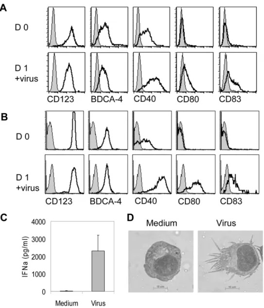
GEN3 cell line was generated from leukemic plasmacytoid dendritic cells [27,34]according to the method previously de-scribed[9]. They highly expressed CD123, BDCA-4, and CD40, whereas were negative for CD80 and CD83 (Figure 1A). Incubation with formaldehyde-inactivated influenza virus for 24 hours led to their maturation: GEN3 cells up-regulated CD40, CD80, and CD83 (Figure 1A), similarly to purified primary PDC (Figure 1B). GEN3 also secreted IFN-a (Figure 1C), and acquired a dendritic cell morphology (Figure 1D).
PDC express few endocytic receptors
We then evaluated the surface expression of molecules involved in antigen capture (Figure 2). Primary PDC and GEN3 did not express the Fc receptors CD16 nor CD64, whereas a low expression of CD32 was detected. A weak expression of the C-type lectin mannose receptor (MR) was detected on GEN3, but not on primary PDC, whereas both primary PDC and GEN3 were negative for DC-SIGN and Langerin (data not shown). The b2 integrin CD18, a subunit of the complement receptors CR3 (CD11b/CD18) and CR4 (CD11c/CD18) was highly expressed by both primary PDC and GEN3, but CD11c and CD11b were hardly detectable. CD91 and calreticulin are involved in the uptake of apoptotic cells that are opsonized either with complement, collectins (SP-A and SP-D) or with heat shock proteins (review [35]). A low expression of CD91 was found on a small subset of primary PDC but not on GEN3. avb3 and avb5 are two integrins implicated in the clearance of apoptotic cells by DC and macrophages [32,36], in association with CD36. Both GEN3 and primary PDC displayed a moderate expression of avb5 and a sharp expression of CD36. avb3 was present only on GEN3 cells. As a matter of comparison, the same phenotype was also performed on monocyte-derived dendritic cells, and we found that except for CD64, all the molecules studied were expressed on MoDC, and such a wide range of endocytic receptors may favor
their ability to capture antigens. Conversely, PDC and GEN displayed a very restricted set of endocytic receptors.
Endocytic capacity of PDC is weak
We assessed the ability of GEN3, primary PDC, and MoDC, to endocytose soluble antigens by using Lucifer Yellow (LY), FITC-conjugated Ovalbumin (OVA), and Dextran (Dex) (Figure 3A). Both GEN3 and primary PDC engulfed LY and OVA with the same efficiency, but less efficiently than MoDC (about ten-fold higher tracer capture was observed with MoDC compared to PDC). By contrast, primary PDC and GEN3 cells did not take up Dextran, whereas MoDC efficiently captured this potential ligand for mannose receptor.
We then evaluated the ability of PDC to take up cellular material from apoptotic, necrotic, opsonized and non-treated cells, in comparison to MoDC. To this end, we used a method that allows the detection of endocytosis with a high sensitivity and distinguishes between endocytosed or conjugated cells [29]. In these experiments, endocytosis was measured by flow cytometry using PKH26-labeled B-cells as target cells and appropriate FITC-conjugated mAbs to identify APC (Figure 3B). Conjugates were excluded using an additional labeling with PC5-conjugated CD19 and CD20. After 2 hours, at least half of the MoDC acquired cellular material from B cells, whatever the B cell treatment. By contrast, GEN3 remained PKH26neg, whatever the condition, suggesting their inability to capture opsonized or dead cells, or to perform ‘‘nibbling’’ of cells.
PDC capture cellular material from influenza-treated cells
Because PDC are involved in anti-viral immune responses, we sought to determine whether GEN3 cells capture cellular antigens from virus-infected cells. We used B cells incubated with formalde-hyde-inactivated non-replicating influenza virus as a model of virus-containing cells, henceforth referred to as ‘‘flu-B cells’’. As shown in Figure 4A, cellular material from flu-B cells was efficiently taken up by GEN3 (46 and 67% of GEN3 were PKH26pos in the two representative experiments shown). As expected, non-treated B cells were not captured. Interestingly, primary purified BDCA4+ PDC were also able to capture cellular material from flu-B cells (25 and 48% of PDC were PKH26posin the two representative experiments shown) (Figure 4B). So, PDC seems to specifically take up cell-derived material from virus-containing surrounding cells. Microscopic observation of cell fluorescence showed that GEN3 took up small membrane-containing fragments from virus-treated cells (Figure 4C). This uptake of cellular material from flu-B cells required actin polymerization and divalent cations, as demonstrated by inhibition of uptake with cytochalasin D and EDTA, respectively (Figure 4D). This endocytosis was not due to the capture of PKH26posvesicles such as exosomes or other kinds of membrane shed particles. Indeed, when GEN3 were incubated for two hours with the supernatant of 18-hour flu-B cells culture, they remained PKH26neg(Figure 4A).
To test whether cell maturation influenced this particular endocytic capacity of PDC, we assessed capture after activation of GEN3 cells with a TLR9 agonist. Figure 4E shows that in the presence of CpG A (2336), the capacity of GEN3 cells to take up cellular material from flu-B cells was inhibited, i.e. the percentage of PKH26posGEN3 decreased from 45% (for immature GEN3) to
less than 10% (for mature GEN3).
Endocytosis of flu-B cells activates PDC and induces cross-presentation of viral antigens
We next analyzed the consequences of flu-B cells capture on PDC maturation. When GEN3 were directly treated with the PDC Cross-Present Viral Ag
inactivated virus, they upregulated CD40, CD80, CCR7 (Figure 5A, upper panels), and HLA-I expression (data not shown). Interestingly, after endocytosis of flu-B cells, GEN3 were found activated to a similar extent (Figure 5A, lower panels). Primary purified BDCA4+ PDC also matured after endocytosis of flu-B-cells, upregulating CD40 and CD80 up to intensity comparable to that observed after direct contact with the virus. (Figure 5A, right panels). Therefore, PDC became activated following the capture of cellular material from virus-treated cells. Since the supernatant of flu-B cells did not induce the maturation of PDC (data not shown) the involvement of free viral particles was excluded. We also found significant IFNa secretion by GEN3 and primary PDC after endocytosis of flu-B cells (Figure 5B).
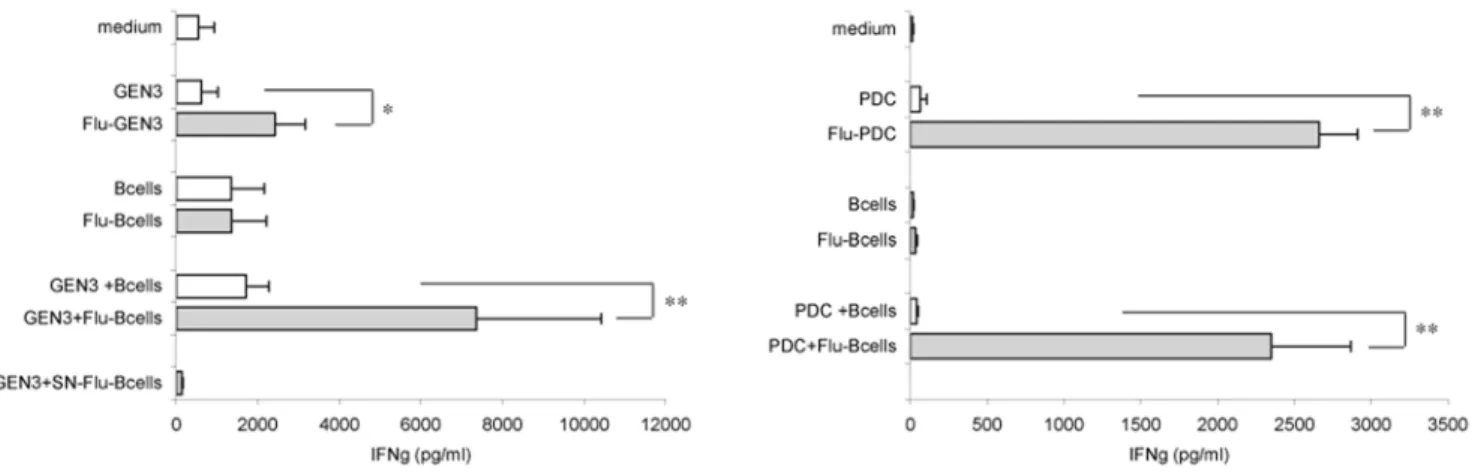
We next analyzed the ability of PDC to activate influenza-specific T cells. The activation of HLA-A2posinfluenza-specific CD8+ T cells by both kinds of PDC was assessed by measuring IFNc release by T cells. As expected, when virus-treated GEN3 (Figure 6A) or primary
purified BDCA2+ PDC (Figure 6B) were used, IFNc secretion was observed, confirming the ability of PDC to perform direct presentation of viral antigens. Cross-presentation of viral antigens by PDC was then examined by incubating GEN3 or primary PDC (both HLA-A2pos) with B cells or flu-B cells (HLA-A2neg), prior to addition of T cells. Strikingly, GEN3 (Figure 6A) as well as primary PDC (Figure 6B) pulsed with flu-B cells induced a highly significant increase of IFNc secretion by T cells, when compared to non-treated B cells (p,0,005, student t test). This increase was not due to direct T cell activation by flu-B cells, as flu-B cells alone did not induce any IFNc increase. Moreover, T-cell activation driven by GEN3 pulsed with the supernatant of 48h-virus-treated B cell culture was found negative, ruling out the involvement of free viral particles. These data demonstrate that PDC capture viral antigens from virus-exposed B cells and subsequently cross-present these antigens to specific CD8+ T cells. This highly significant result indicates a striking specific role of PDC in activation of T cell responses in the context of viral infection. Figure 1. Characterization of the GEN3 plasmacytoid dendritic cell line. The LPDC-derived cell line GEN3 or primary purified PDC were incubated for 18 hours in the presence of formaldehyde-inactivated influenza virus. GEN3 cells (A) or primary PDC (B) were stained with indicated mAb (open curves) or isotype control (filled curves) at day 0 (upper panels) and day 1 (lower panels). At day 1 production of IFNa by GEN3 was measured by ELISA (C) (mean6SD of four experiments), and cell morphology was examined after May-Gru¨ndwald Giemsa staining of cell cytospins (D). Data are representative of 3 independent experiments.
Discussion
PDC are involved early in innate phases of viral immunity through the secretion of large amounts of IFNa[1,2], and TRAIL-mediated cytotoxicity[9]. Previous studies have clearly established that, upon exposure to viruses, PDC mature and acquire costimulatory molecules (reviews[1,2]), so that infected PDC become able to present viral antigens to CD8+ T cells[20,37]. These data suggested the involvement of PDC in the initiation of adaptive immunity in the context of viruses that directly infect PDC. Here, we evaluated the capacity of PDC to perform cross-presentation, i.e. to acquire, process, and present
exogenously-derived antigens on HLA class I molecules to activate specific CD8+ T cells[38]. The physiological essential function of cross-presentation in viral infections has been demonstrated in the context of immune responses to viruses that do not infect APC[39]. Recent studies in human reported contradictory data regarding the capacity of PDC to cross-present antigens[37,40].
We took advantage here of a cell line derived from leukemic PDC (LPDC[27,28]), GEN3, as a model to study the uptake of antigens by PDC, and their capacity for cross-presentation. According to previous studies [22,41], PDC and GEN3 displayed a weak capacity to capture dextran, which correlates with the absence of mannose receptor and DC-SIGN expression, whereas Figure 2. Assessment of the expression of receptors involved in antigen capture. Monocyte-derived dendritic cells (MoDC), GEN3 cells, and primary blood BDCA2+ PDC were stained with indicated mAb (open curves) or isotype control (filled curves). Data are representative of two independent experiments.
doi:10.1371/journal.pone.0007111.g002
they took up the liquid phase tracer Lucifer Yellow, and a soluble protein such as ovalbumin, although less efficiently when compared to MoDC. We have shown that GEN3 expressed quite the same pattern of endocytic receptors as primary PDC, the most striking difference being the expression of avb3 on GEN3 but not on primary PDC. On both cells, we found a weak expression of CD32 (as previously described [42]), and a faint expression of CD11b and CD11c. However, CD32 did not allow the capture of Rituximab-opsonized cells by GEN3 albeit it was shown to mediate the capture of immune complexes containing DNA in the
context of human lupus [43], or KLH after vaccination in melanoma patients[44]. According to previous work[45], PDC did not take up cellular material from live, apoptotic, or necrotic B cells even though they expressed CD36 and low levels of avb5, two receptors involved in the capture of dying cells. This was in contrast with MoDC, which efficiently endocytosed B-cell derived material from all the different sources of B cells. This phenomenon may be due to the expression of more diverse endocytic receptors on MoDC allowing them to capture apoptotic or necrotic cells (CD91, CD36, avb3, avb5), opsonized cells (CD16 or CD32), or Figure 3. Measurement of soluble or cellular antigens uptake by flow cytometry. (A) Endocytosis of soluble tracers. MoDC, purified BDCA-4+ primary PDC or GEN3 cells were incubated for 2 hours at 4uC (filled curves) or 37uC (open curves) with Ovalbumin, Lucifer yellow and Dextran. Mean fluorescence intensity (MFI) values are indicated. Results are representative of two experiments. (B) Uptake of cellular material. PKH26-stained B cells were induced into apoptosis, necrosis, or were opsonized with CD20 mAb Rituximab or left untreated. They were then incubated with MoDC or GEN3 for 2 hours and stained as described in materials and methods section. PKH26 fluorescence, measuring cellular material uptake, was analyzed gating on MoDC (CD11bpos) or GEN3 (CD4posCD36pos) not involved in cell conjugates (CD19negCD20neg). Percentages of positive cells at 37uC (open
curves), relative to incubation at 4uC (filled curves) are indicated. These data are representative of two independent experiments. doi:10.1371/journal.pone.0007111.g003
to perform the so-called ‘‘nibbling’’ of small vesicles from live cells [46–48]. Altogether, MoDC are undoubtedly more efficient than PDC at endocytosing cells.
Interestingly, our results show an efficient transfer of cellular material from influenza-treated B cells (flu-B cells) to PDC, in an actin and Ca2+dependent manner. We documented this uptake by fluorescence microscopy, and found that most PDC contained fragments of virus-treated cells. The functional consequences of this endocytosis by PDC were then examined. Upon exposure to flu-B cells of GEN3 or primary PDC secreted IFNa and matured,
as evidenced by acquisition of costimulatory molecules. The up-regulation of CCR7 suggests that after the encounter with infected cells in periphery, and capture of cell-derived antigenic material, PDC could traffic to T cell zones in the lymph nodes and present acquired antigens. Furthermore, our study provides further evidence that following uptake and maturation, PDC are able to cross-present viral antigens captured from virus-exposed cells to specific T cells. The first demonstration of the ability of PDC to cross-present viral antigens taken from infected cells was recently performed by Hoeffel at Al[49], in a model where PDC cross-Figure 4. PDC internalized cellular material from influenza-treated B cells. (A and B) measurement of influenza-treated B cells uptake by flow cytometry. B cells (B1 and B2) were stained with PKH26, exposed (lower panels) or not (upper panels) to virus for 18 hours, extensively washed and then incubated with purified PDC or GEN3 for 2 hours at 4uC (filled curves) or 37uC (open curves). PKH26 fluorescence was evaluated as in Figure 3, gating on primary PDC and GEN3 not involved in conjugates with B cells. As control, GEN3 were also incubated following the same procedure for two hours with the supernatant of 18-hour flu-B cell culture (SN-B2). (C) Fluorescence microscopy imaging of B cell material by GEN3. PKH26+ flu-B cells appear yellow and GEN3 were stained in green with FITC-conjugated CD4 and CD36. (D) Membrane capture is Ca2+and
actin-dependent. Cellular uptake was measured as described in Fig. 3A, at 4uC (open curves) or 37uC (filled curves) in the presence or absence of EDTA or cytochalasin D (CDD). (E) Maturation down-regulated cell capture by PDC. GEN3 were matured in presence of CpG for 24 h (lower panel) or not (upper panel) and incubated with B cells exposed (open curves) or not (filled curves) to virus for 18 hours.
doi:10.1371/journal.pone.0007111.g004
presented to specific T cells antigens taken from apoptotic HIV-infected cells. This mechanism reveals a highly relevant role for PDC in eliciting anti-viral immune responses. Indeed, during a natural infection, DC are not necessarily the primary infected cells, and admittedly virally infected non-APC are unable to stimulate CD8+ T cell immunity. Moreover, many viruses such as HIV, measles virus or HCMV infect DC, and have evolved mechanisms to subvert DC function, by disturbing their maturation or ability to secrete cytokines[50–52]. Here, by their ability to engulf viral antigens from virus-containing cells without being themselves infected, PDC could acquire and present viral antigens, avoiding virus-induced subversion of their functions.
Our results show that after capture, viral-antigens are processed and presented into the MHC class I pathway. How do PDC detect the infection, and acquire cell-derived fragments? Among flu-B cells, some died in vitro, however, in our hands PDC engulfed neither necrotic nor apoptotic cells (our data and [45]). Therefore, recognition of influenza-treated living cells by PDC likely did not rely on dead cell capture, contrary to the model recently described in MoDC by Frleta et Al [53]. Flu-B cells expressed viral hemagglutinin on the B cell surface (data not shown), a molecule that could allow PDC recognition of virus-treated cells. A potential receptor is NKp44, which recognizes influenza hemagglutinin[54], but it is expressed on PDC only after activation[55]. Since mature Figure 5. Activation of PDC upon exposure to flu-B cells. (A) In upper panels, GEN3 or purified PDC were incubated (open curves) or not (filled curves) with influenza virus for 24 hours, as positive control of their maturation, and cells were stained with indicated mAb. In lower panels, B cells were exposed (open curves) or not (filled curves) to virus for 18 hours, washed, and then incubated with GEN3 or purified PDC. After a 24-hour co-culture, cells were stained with indicated mAb, and maturation was assessed by flow cytometry after gating on GEN3 cells (according to their FSC/SSC profile) or on BDCA2posBDCA4posPDC. Data are representative of two independent experiments. (B) GEN3 or purified PDC were incubated with or without influenza virus, B cells or Flu-B cells for 24 hours, and then IFNa levels in supernatants were measured by ELISA. Data represent mean6SD of 2 experiments performed in duplicates.
GEN3 lost their endocytic capacity, the receptor involved is likely down regulated during PDC maturation. Further studies are required to determine whether the recognition of infected cells by PDC relies on virus- or endogenous cell-derived signals, and what kind of receptor on PDC is involved.
The efficiency with which viral antigens from flu-B cells were cross-presented may be due to the high efficiency of their uptake by PDC. It is also possible that IFNa secreted by PDC in association with other unknown factors may enhance the cross-presentation of internalized antigens[23], through a still undeter-mined mechanism. IFN-b, not measured in our study, could also be involved, as this pro-inflammatory cytokine secreted by various kinds of infected cells has been described to prime DC, enhancing their activation in response to influenza virus[56]. Alternatively, engagement of TLR (likely TLR7, expressed by PDC[57] and involved in influenza virus recognition[5]) on PDC may influence phagosome maturation in PDC [58], and favor the generation of viral peptides for cross-presentation. Whatever the mechanism, cross-presentation of viral antigens was at least as efficient as direct presentation by PDC exposed to influenza virus.
In conclusion, our study identifies a role for PDC as a central professional APC during viral infections, by being extremely sensitive not only to direct viral exposure, but also to infection of bystander cells. We suggest that PDC play a major role in the control of viral infection through IFNa secretion and TRAIL expression during the innate phase of immune response, and by the further activation CD8+ T cell adaptive immunity through cross-presentation of viral derived antigens captured from infected cells. Thus, the role of PDC as a major link between innate and adaptive immunity is further emphasized.
Acknowledgments
We thank Christophe Caux for critical review of the manuscript, and Davor Frleta for proofreading.
Author Contributions
Conceived and designed the experiments: LC JP. Performed the experiments: GL OM JA. Analyzed the data: GL LC JP. Contributed reagents/materials/analysis tools: JPM. Wrote the paper: GL LC JP.
References
1. Liu YJ (2005) IPC: Professional Type 1 Interferon-Producing Cells and Plasmacytoid Dendritic Cell Precursors. Annu Rev Immunol 23: 275–306. 2. Colonna M, Trinchieri G, Liu YJ (2004) Plasmacytoid dendritic cells in
immunity. Nat Immunol 5: 1219–1226.
3. Siegal FP, Kadowaki N, Shodell M, Fitzgerald Bocarsly PA, Shah K, et al. (1999) The nature of the principal type 1 interferon-producing cells in human blood. Science 284: 1835–1837.
4. Cella M, Jarrossay D, Facchetti F, Alebardi O, Nakajima H, et al. (1999) Plasmacytoid monocytes migrate to inflamed lymph nodes and produce large amounts of type I interferon. Nat Med 5: 919–923.
5. Diebold SS, Kaisho T, Hemmi H, Akira S, Reis ESC (2004) Innate antiviral responses by means of TLR7-mediated recognition of single-stranded RNA. Science 303: 1529–1531.
6. Heil F, Hemmi H, Hochrein H, Ampenberger F, Kirschning C, et al. (2004) Species-specific recognition of single-stranded RNA via toll-like receptor 7 and 8. Science 303: 1526–1529.
7. Latz E, Schoenemeyer A, Visintin A, Fitzgerald KA, Monks BG, et al. (2004) TLR9 signals after translocating from the ER to CpG DNA in the lysosome. Nat Immunol.
8. Guiducci C, Ott G, Chan JH, Damon E, Calacsan C, et al. (2006) Properties regulating the nature of the plasmacytoid dendritic cell response to Toll-like receptor 9 activation. J Exp Med 203: 1999–2008.
9. Chaperot l, Blum A, Manches O, Lui G, Angel J, et al. (2006) Virus or TLR agonists induce TRAIL-mediated cytotoxicy of plasmacytoid dendritic cells. J Immunol 176: 248–255.
10. Kotelkin A, Prikhod’ko EA, Cohen JI, Collins PL, Bukreyev A (2003) Respiratory syncytial virus infection sensitizes cells to apoptosis mediated by tumor necrosis factor-related apoptosis-inducing ligand. J Virol 77: 9156–9172. 11. Bendriss-Vermare N, Burg S, Kanzler H, Chaperot L, Duhen T, et al. (2005) Virus overrides the propensity of human CD40L-activated plasmacytoid dendritic cells to produce Th2 mediators through synergistic induction of IFN-{gamma} and Th1 chemokine production. J Leukoc Biol 78: 954–966. 12. Grouard G, Rissoan MC, Filgueira L, Durand I, Banchereau J, et al. (1997) The
enigmatic plasmacytoid T cells develop into dendritic cells with interleukin (IL)-3 and CD40-ligand. J Exp Med 185: 1101–1111.
13. Kadowaki N, Antonenko S, Lau JY, Liu YJ (2000) Natural interferon alpha/ beta-producing cells link innate and adaptive immunity. J Exp Med 192: 219–226.
14. Rissoan MC, Soumelis V, Kadowaki N, Grouard G, Briere F, et al. (1999) Reciprocal control of T helper cell and dendritic cell differentiation. Science 283: 1183–1186.
15. Cella M, Facchetti F, Lanzavecchia A, Colonna M (2000) Plasmacytoid dendritic cells activated by influenza virus and CD40L drive a potent TH1 polarization. Nat Immunol 1: 305–310.
Figure 6. PDC cross-present viral antigens derived from flu-B cells to influenza-specific CD8+ T lymphocytes. HLA-A2neg
B cells exposed (filled bars) or not (open bars) to inactivated influenza virus for 18 hours were washed and incubated with HLA-A2posAPC (either GEN3 (A) or
PDC (B)), for four hours, followed by the addition of influenza-specific CD8+ T lymphocytes (recognizing flu58-66influenza matrix peptide in the
context of HLA-A2 molecules). After a 2-day co-culture, T cell activation was assessed by measuring the IFNc content of the culture supernatants. In control wells, PDC were directly treated or not with the virus, or were omitted (medium). (A) Values are mean+ SD of six independent experiments, except for the last control bar (SN-Flu-B cells), mean of two experiments, where Flu-B cells were washed extensively, and further incubated for 24 hours to recover the supernatant which was subsequently added to influenza-specific T cells. (B) Values are representative of two experiments and shown the mean+ SD of quadruplicate wells. * P,0.05, ** P,0.005 by Student’s t test.
doi:10.1371/journal.pone.0007111.g006
16. Kawamura K, Kadowaki N, Kitawaki T, Uchiyama T (2006) Virus-stimulated plasmacytoid dendritic cells induce CD4+ cytotoxic regulatory T cells. Blood 107: 1031–1038.
17. Ito T, Yang M, Wang YH, Lande R, Gregorio J, et al. (2007) Plasmacytoid dendritic cells prime IL-10-producing T regulatory cells by inducible costimulator ligand. J Exp Med 204: 105–115.
18. Krug A, Veeraswamy R, Pekosz A, Kanagawa O, Unanue ER, et al. (2003) Interferon-producing cells fail to induce proliferation of naive T cells but can promote expansion and T helper 1 differentiation of antigen-experienced unpolarized T cells. J Exp Med 197: 899–906.
19. Salio M, Palmowski MJ, Atzberger A, Hermans IF, Cerundolo V (2004) CpG-matured Murine Plasmacytoid Dendritic Cells Are Capable of In Vivo Priming of Functional CD8 T Cell Responses to Endogenous but Not Exogenous Antigens. J Exp Med 199: 567–579.
20. Fonteneau JF, Gilliet M, Larsson M, Dasilva I, Munz C, et al. (2003) Activation of influenza virus-specific CD4+ and CD8+ T cells: a new role for plasmacytoid dendritic cells in adaptive immunity. Blood 2: 2.
21. Schlecht G, Garcia S, Escriou N, Freitas AA, Leclerc C, et al. (2004) Murine plasmacytoid dendritic cells induce effector/memory CD8+ T-cell responses in vivo after viral stimulation. Blood 104: 1808–1815.
22. Robinson SP, Patterson S, English N, Davies D, Knight SC, et al. (1999) Human peripheral blood contains two distinct lineages of dendritic cells. Eur J Immunol 29: 2769–2778.
23. Le Bon A, Etchart N, Rossmann C, Ashton M, Hou S, et al. (2003) Cross-priming of CD8+ T cells stimulated by virus-induced type I interferon. Nat Immunol 4: 1009–1015.
24. Villadangos JA, Young L (2008) Antigen-presentation properties of plasmacytoid dendritic cells. Immunity 29: 352–361.
25. Olweus J, BitMansour A, Warnke R, Thompson PA, Carballido J, et al. (1997) Dendritic cell ontogeny: a human dendritic cell lineage of myeloid origin. Proc Natl Acad Sci U S A 94: 12551–12556.
26. Blom B, Ho S, Antonenko S, Liu YJ (2000) Generation of interferon alpha-producing predendritic cell (Pre-DC)2 from human CD34(+) hematopoietic stem cells. J Exp Med 192: 1785–1796.
27. Chaperot L, Bendriss N, Manches O, Gressin R, Maynadie M, et al. (2001) Identification of a leukemic counterpart of the plasmacytoid dendritic cells. Blood 97: 3210–3217.
28. Chaperot L, Perrot I, Jacob M-C, Blanchard D, Salaun V, et al. (2004) Leukemic plasmacytoid dendritic cells share phenotypic and functional features with their normal counterparts. Eur J Immunol 34: 418–426.
29. Manches O, Lui G, Molens JP, Sotto JJ, Chaperot L, et al. (2008) Whole lymphoma B cells allow efficient cross-presentation of antigens by dendritic cells. Cytotherapy 10: 642–649.
30. Sallusto F, Lanzavecchia A (1994) Efficient presentation of soluble antigen by cultured human dendritic cells is maintained by granulocyte/macrophage colony stimulating factor plus interleukin 4 and downregulated by tumor necrosis factor alpha. J Exp Med 179: 1109–1118.
31. Lui G, Manches O, Chaperot L, Ducrot T, Molens JP, et al. (2004) Preparation of purified lymphoma cells suitable for therapy. Cytotherapy 6: 235–243. 32. Albert ML, Pearce SFA, Francisco LM, Sauter B, Roy P, et al. (1998) Immature
dendritic cells phagocytose apoptotic cells via avb5 and CD36 and cross present antigens to cytotoxic T lymphocytes. J Exp Med 188: 1359–1368.
33. Albert ML, Sauter B, Bhardwaj N (1998) Dendritic cells acquire antigen from apoptotic cells and induce class I restricted CTLs. Nature 392: 86–89. 34. Jacob M-C, Chaperot L, Mossuz P, Feuillard J, Valensi F, et al. (2003) CD4+
CD56+ lineage negative (lin-) leukemia/lymphoma: a new entity developed from malignant early plasmacytoid dendritic cells (pDC). Hematologica 88: 941–955. 35. Henson PM, Bratton DL, Fadok VA (2001) Apoptotic cell removal. Curr Biol
11: R795–805.
36. Savill J, Dransfield I, Hogg N, Haslett C (1990) Vitronectin receptor-mediated phagocytosis of cells undergoing apoptosis. Nature 343: 170–173.
37. Di Pucchio T, Chatterjee B, Smed-Sorensen A, Clayton S, Palazzo A, et al. (2008) Direct proteasome-independent cross-presentation of viral antigen by plasmacytoid dendritic cells on major histocompatibility complex class I. Nat Immunol 9: 551–557.
38. Heath WR, Belz GT, Behrens GM, Smith CM, Forehan SP, et al. (2004) Cross-presentation, dendritic cell subsets, and the generation of immunity to cellular antigens. Immunol Rev 199: 9–26.
39. Sigal LJ, Crotty S, Andino R, Rock K (1999) Cytotoxic T-cell immunity to virus-infected non-hematopoietic cells requires presentation of exogenous antigen. Nature 398: 77–80.
40. Schnurr M, Chen Q, Shin A, Chen W, Toy T, et al. (2005) Tumor antigen processing and presentation depend critically on dendritic cell type and the mode of antigen delivery. Blood 105: 2465–2472.
41. Dzionek A, Fuchs A, Schmidt P, Cremer S, Zysk M, et al. (2000) BDCA-2, BDCA-3, and BDCA-4: three markers for distinct subsets of dendritic cells in human peripheral blood. J Immunol 165: 6037–6046.
42. Bave U, Magnusson M, Eloranta ML, Perers A, Alm GV, et al. (2003) Fc gamma RIIa is expressed on natural IFN-alpha-producing cells (plasmacytoid dendritic cells) and is required for the IFN-alpha production induced by apoptotic cells combined with lupus IgG. J Immunol 171: 3296–3302. 43. Means TK, Latz E, Hayashi F, Murali MR, Golenbock DT, et al. (2005)
Human lupus autoantibody-DNA complexes activate DCs through cooperation of CD32 and TLR9. J Clin Invest 115: 407–417.
44. Benitez-Ribas D, Adema GJ, Winkels G, Klasen IS, Punt CJ, et al. (2006) Plasmacytoid dendritic cells of melanoma patients present exogenous proteins to CD4(+) T cells after FcgammaRII-mediated uptake. J Exp Med 203: 1629–1635.
45. Dalgaard J, Beckstrom KJ, Jahnsen FL, Brinchmann JE (2005) Differential capability for phagocytosis of apoptotic and necrotic leukemia cells by human peripheral blood dendritic cell subsets. J Leukoc Biol.
46. Harshyne LA, Watkins SC, Gambotto A, Barratt-Boyes SM (2001) Dendritic cells acquire antigens from live cells for cross-presentation to CTL. J Immunol 166: 3717–3723.
47. Harshyne LA, Zimmer MI, Watkins SC, Barratt-Boyes SM (2003) A role for class A scavenger receptor in dendritic cell nibbling from live cells. J Immunol 170: 2302–2309.
48. Heath WR, Carbone FR (2001) Cross-presentation in viral immunity and self-tolerance. Nat Rev Immunol 1: 126–134.
49. Hoeffel G, Ripoche AC, Matheoud D, Nascimbeni M, Escriou N, et al. (2007) Antigen crosspresentation by human plasmacytoid dendritic cells. Immunity 27: 481–492.
50. Barron MA, Blyveis N, Palmer BE, MaWhinney S, Wilson CC (2003) Influence of plasma viremia on defects in number and immunophenotype of blood dendritic cell subsets in human immunodeficiency virus 1-infected individuals. J Infect Dis 187: 26–37.
51. Servet-Delprat C, Vidalain PO, Bausinger H, Manie S, Le Deist F, et al. (2000) Measles virus induces abnormal differentiation of CD40 ligand-activated human dendritic cells. J Immunol 164: 1753–1760.
52. Moutaftsi M, Mehl AM, Borysiewicz LK, Tabi Z (2002) Human cytomegalo-virus inhibits maturation and impairs function of monocyte-derived dendritic cells. Blood 99: 2913–2921.
53. Frleta D, Yu CI, Klechevsky E, Flamar AL, Zurawski G, et al. (2009) Influenza virus and poly(I:C) inhibit MHC class I-restricted presentation of cell-associated antigens derived from infected dead cells captured by human dendritic cells. J Immunol 182: 2766–2776.
54. Arnon TI, Lev M, Katz G, Chernobrov Y, Porgador A, et al. (2001) Recognition of viral hemagglutinins by NKp44 but not by NKp30. Eur J Immunol 31: 2680–2689.
55. Fuchs A, Cella M, Kondo T, Colonna M (2005) Paradoxic inhibition of human natural interferon-producing cells by the activating receptor NKp44. Blood 106: 2076–2082.
56. Phipps-Yonas H, Seto J, Sealfon SC, Moran TM, Fernandez-Sesma A (2008) Interferon-beta pretreatment of conventional and plasmacytoid human dendritic cells enhances their activation by influenza virus. PLoS Pathog 4: e1000193. 57. Jarrossay D, Napolitani G, Colonna M, Sallusto F, Lanzavecchia A (2001)
Specialization and complementarity in microbial molecule recognition by human myeloid and plasmacytoid dendritic cells. Eur J Immunol 31: 3388–3393.
58. Blander JM, Medzhitov R (2004) Regulation of phagosome maturation by signals from toll-like receptors. Science 304: 1014–1018.