HAL Id: inserm-00143972
https://www.hal.inserm.fr/inserm-00143972
Submitted on 30 Apr 2007HAL is a multi-disciplinary open access
archive for the deposit and dissemination of sci-entific research documents, whether they are pub-lished or not. The documents may come from teaching and research institutions in France or abroad, or from public or private research centers.
L’archive ouverte pluridisciplinaire HAL, est destinée au dépôt et à la diffusion de documents scientifiques de niveau recherche, publiés ou non, émanant des établissements d’enseignement et de recherche français ou étrangers, des laboratoires publics ou privés.
Differential regulation of RANTES and IL-8 expression
in lung adenocarcinoma cells.
Corinne Henriquet, Claire Gougat, Audrey Combes, Gwendal Lazennec, Marc
Mathieu
To cite this version:
Corinne Henriquet, Claire Gougat, Audrey Combes, Gwendal Lazennec, Marc Mathieu. Differential regulation of RANTES and IL-8 expression in lung adenocarcinoma cells.. Lung Cancer, Elsevier, 2007, 56 (2), pp.167-74. �10.1016/j.lungcan.2006.12.003�. �inserm-00143972�
1
Differential regulation of RANTES and IL-8 expression in lung
adenocarcinoma cells
Corinne Henriquet a , Claire Gougat a , Audrey Combes a , Gwendal Lazennec b , Marc Mathieu a, * aInserm, U454 – IFR3, Montpellier, F-34295, France
b
Inserm, U540 – IFR3, Montpellier, F-34090, France. *To whom correspondence should be addressed: Inserm U454
Hôpital Arnaud de Villeneuve 34295 Montpellier Cedex 5 France
Tel: +33 467 41 52 13 Fax: +33 467 63 28 55
E-mail: mathieu@montp.inserm.fr.
Running title: RANTES and IL-8 expression in lung adenocarcinoma cells
HAL author manuscript inserm-00143972, version 1
HAL author manuscript
Summary
In lung adenocarcinoma, expression of Regulated upon Activation, Normal T cell Expressed and presumably Secreted (RANTES) is a predictor of survival while that of interleukin (IL)-8 is associated with a poor prognosis. In several models, tumorigenesis is abolished by RANTES, while it is facilitated by IL-8. We studied the regulation of RANTES and IL-8 expression in A549 lung adenocarcinoma cells. The effects of tumor necrosis factor (TNF)-α and regulators of protein kinases C (PKC)α/β were tested because these have been shown to modulate cancer development and progression. TNF-α stimulated expression of both chemokines, while the PKCα/β activator 12-O-tetradecanoyl-phorbol-13-acetate (TPA) induced only expression of IL-8 and inhibited TNF-α-induced RANTES expression. The PKCα/β inhibitor Gö 6976 increased TNF-α-induced RANTES production and prevented its down-regulation by TPA. In contrast, it decreased TNF-α or TPA-induced IL-8 release. The differential regulation of RANTES and IL-8 expression was further analyzed. Site-directed mutagenesis indicated that regulation of RANTES promoter activity required two nuclear factor (NF)-κB response elements but not its activator protein (AP)-1 binding sites. An AP-1 and a NF-κB recognition sites were necessary for full induction of IL-8 promoter activity by TNF-α and TPA. Moreover, electrophoretic mobility shift assays demonstrated that NF-κB response elements from the RANTES promoter were of lower affinity than that from the IL-8 promoter. Immunoblotting experiments showed that TPA was more potent than TNF-α to induce in a PKCα/β dependent manner the p44/p42 mitogen-activated protein kinases signaling cascade which controls AP-1 activity. Conversely, TPA inhibited TNF-α-induced NF-κB signaling and was a weak activator of this pathway. Thus, TPA did not sufficiently activate NF-κB to increase transcription through the low affinity NF-κB binding sites on RANTES promoter and its inhibitory effect on TNF-α-induced NF-κB signaling resulted in a reduced transcription rate. On IL-8 promoter, increased transcription through the high affinity NF-κB binding site occurred even with poorly activated NF-κB and the functional AP-1 response element compensated any loss of transcription rate. These data provide a mechanistic insight into the differential regulation of IL-8 and RANTES expression by PKCα/β in lung adenocarcinoma cells.
Key Words: Adenocarcinoma; Bronchiolo-Alveolar; Chemokines; Interleukin-8; NF-kappaB; Protein Kinase C; RANTES; Transcription Factor AP-1.
3
Introduction
Lung cancer is the most common cause of cancer-related deaths in industrialized nations and its incidence is on a constant increase [1]. Lung adenocarcinomas represent 30% of all lung cancers [2]. The variability of survival within a given stage and the poor efficiency of current therapies have led to the search for tumor-produced factors that may serve as new prognostic factors or therapeutic targets. In this line, Moran and colleagues have used a global gene expression profiling approach and found that the chemokine Regulated upon Activation, Normal T cell Expressed and presumably Secreted (RANTES) is a predictor of survival in stage I lung adenocarcinoma [3]. RANTES could therefore be used as a prognostic factor to further stratify patients with this disease and is a potential therapeutic target. Indeed, tumor cells that are genetically engineered to secrete RANTES, loose their tumorigenicity in a mouse sarcoma model [4]. Similarly, in a murine lymphoma model, gene transfer of RANTES into established tumors provokes complete tumor regression in 50% of treated mice [5]. These therapeutic effects are the result of a RANTES-induced anti-tumor immune response [4, 5]. In addition, RANTES seems to act in an autocrine manner to decrease tumor growth since abrogation of cell surface expression of the RANTES receptor CCR5 enhances proliferation of human breast tumor cells injected in mice [6]. However, the role of RANTES in breast cancer is debated [6-10].
In contrast to RANTES, interleukin (IL)-8 expression in non-small-cell lung cancer, including adenocarcinoma, is of poor prognosis [11]. This chemokine was found to be produced by cancer cells and to facilitate tumorigenesis, probably through its angiogenic activity. Xenograft experiments in mice have shown that IL-8 increases growth of lung [12], prostate [13] bladder [14] and breast cancer [15], as well as melanoma [16]. One exception is ovarian cancer, in which IL-8 was found in contrary to attenuate tumor growth [17].
We examined herein the effects of tumor necrosis factor (TNF)-α and regulators of protein kinases C (PKC)α/β on the expression of RANTES and IL-8 in A549 lung adenocarcinoma cells. TNF-α is a major mediator of inflammation, which can be produced by tumor-infiltrating macrophages [18]. It has paradoxical roles in the evolution and treatment of malignant disease [19]. PKCα/β were previously shown to affect tumor progression and malignant phenotype [20, 21]. We show that the PKCα/β activator 12-O-tetradecanoyl-phorbol-13-acetate (TPA) antagonized TNF-α-induced RANTES expression, while both stimuli increased IL-8 expression. Treatment with the PKCα/β inhibitor Gö 6976 prevented TPA from down-regulating RANTES expression. This compound also increased TNF-α-induced RANTES production and diminished TPA or TNF-α-TNF-α-induced IL-8 release. Further analysis showed that the NF-κB signaling pathway is down-regulated through PKCα/β and provided insight into the mechanism underlying the differential regulation of RANTES and IL-8 in A549 cells.
Materials and Methods
MaterialsTPA and human recombinant TNF-α were purchased from Sigma and BD Pharmingen, respectively. Gö 6976 was purchased from Calbiochem.
Plasmids
Luciferase reporter constructs carrying intact or mutated 1.4 kb human RANTES promoter sequence were obtained from Hiroyuki Moriuchi (Nagasaki University School of Medicine, Nagasaki, Japan). The promoter sequence was point mutated at response elements for NF-κB
and AP-1 [22]. An additional RANTES promoter construct mutated on two NF-κB response elements was purchased from Top Gene Technologies. Promoter of the human IL-8 gene (1.5 kb) was point mutated at AP-1 and NF-κB binding sites as previously described [23]. The cytomegalovirus (CMV)gal plasmid consists of the CMV early promoter driving the β-galactosidase gene. The plasmid pCCS26 contains a ribosomal protein S26 cDNA and was obtained from Philippe Fort (Centre de Recherches de Biochimie Macromoléculaire, Montpellier, France) [24].
Cell maintenance
The A549 human lung adenocarcinoma cell line was obtained from the American Type Culture Collection (ATCC CCL-185) and maintained in Ham's F12/DMEM medium containing 10% heat-inactivated FCS v/v, 100 units/ml penicillin, 100 µg/ml streptomycin and 2 mM glutamine.
Transient transfection and reporter gene assays
100 000 cells per well were serum deprived overnight, transfected with 60 ng of luciferase construct and 25 ng of CMVβ-gal, and stimulated as indicated in the figure legends. Transfection, luciferase and β-galactosidase assays were performed as described previously [25]. Luciferase activity was divided by β-galactosidase activity to normalize values for variations in transfection efficiency.
Western Blot
One million cells per well were serum deprived overnight, stimulated as indicated in the figure legends and lysed in 10 mM Tris-HCl, pH 7.4, 50 mM NaCl, 30 mM sodium pyrophosphate, 1% Triton X-100 v/v, 1mM EDTA, 1 mM EGTA, 20 mM β-glycerophosphate, 0.1 mM sodium orthovanadate, 50 nM okadaic acid, 1 mM dithiothreitol and a protease inhibitor cocktail (Roche). After 5 min on ice, cell extracts were sonicated four times for 5 s, centrifuged at 20 000 g for 10 min at 4°C and analyzed by Western blotting on nitrocellulose membranes. Antibodies used were anti-p44/p42 mitogen-activated protein kinases (MAPK) (9102; Cell signaling Technology), anti-p44/p42 MAPK phosphorylated on Thr202 and Tyr204 (9101; Cell signaling Technology), anti-IκBα (sc-371; Santa Cruz Biotechnology), anti-p65 (sc-109; Santa Cruz Biotechnology), and anti-p65 phosphorylated on Ser536 (#3031; Cell signaling Technology). After incubation with peroxidase-conjugated secondary antibodies, immunoblots were developed in 0.2 mM coumaric acid, 1.25 mM 3-aminophthalhydrazide, and 0.009% hydrogen peroxide mixture and exposed to a cooled charge-coupled device camera (Kodak).
RANTES and IL-8 immunoassays
100 000 cells per well were serum deprived overnight. These were then stimulated as indicated in the figure legends. Concentrations of RANTES and IL-8 in supernatants were determined by ELISA (R&D Systems and Diaclone Elipair, respectively).
Northern blot analysis
Total RNA was isolated with RNA PLUS (Quantum Biotechnologies). Northern Blot was performed according to standard protocols with 32
P-labeled probes. Specific mRNAs were detected with a Storm phosphorimager (Amersham Biosciences). Ribosomal protein S26 mRNA served as an internal control because it is found at an invariable amount in mammalian cell lines [24].
5
Electrophoretic mobility shift assay (EMSA)
Nuclear extracts were prepared as described [26]. Double-stranded DNA was labelled with [α-32
P]CTP (Amersham) using Klenow DNA polymerase (Invitrogen). The following oligonucleotides (MWG Biotech) were used:
1- 5’-GGGGAAACTCCCCTTAGGGGATGCCCCT-3’ 2- 5’-GGAGGGGCATCCCCTAAGGGGAGTTTCC-3’ 3- 5’-GGGGAATTTCCCGAGGGGAATTTCCCAG-3’ 4- 5’-GGCTGGGAAATTCCCCTCGGGAAATTCC-3’
Annealing of oligonucleotides 1-2 and 3-4 formed 2xNF-κBRE RANTES and 2xNF-κBRE IL-8, respectively. Sequences corresponding to the putative NF-κB response elements are underlined. Ten µg of nuclear extract in EMSA buffer (50 mM Tris pH 8, 12 mM MgCl2, 2
µg poly (dI-dC), 1 mM EDTA, 2 mM dithiothreitol, 20% glycerol v/v, 100 µM orthovanadate and 50 nM okadaic acid) containing a protease inhibitor cocktail (Roche) were incubated with labelled probe and competitors for 30 min at 25°C. Samples were subjected to 6% w/v native PAGE and fixed in the gel for 20 min in 10% acetic acid v/v, 45% methanol v/v. The gel was dried before autoradiography with a phosphorimager.
Statistical analysis
Differences between groups were assessed using one-way analysis of variance with appropriate post hoc testing. Statistical significance was set up at p < 0.05.
Results
Differential regulation of RANTES and IL-8 expression
Expression of RANTES and IL-8 in A549 lung adenocarcinoma cells was initially analyzed at the RNA level by Northern blotting (Fig. 1A). RANTES mRNA was not detectable in non-stimulated cells and TPA treated cells. TNF-α induced an increase in RANTES mRNA steady state level that was partially inhibited after co-treatment with TPA. In contrast, the amount of IL-8 mRNA was increased by both stimuli. Similar results were obtained by quantifying the amount of the corresponding chemokines in cell supernatants. RANTES release was induced by TNF-α (p < 0.001) but not by TPA, and the latter inhibited TNF-α-induced RANTES release by 46% (p < 0.001) (Fig. 1B). In contrast, IL-8 production was increased following treatment with either stimulus (p < 0.001) (Fig. 1C). To examine the involvement of PKCα/β in the regulation of RANTES and IL-8 expression, the cells were pretreated with Gö 6976. This compound prevented the inhibitory effect of TPA on RANTES release (p < 0.001) (Fig. 1B). Gö 6976 also increased TNF-α-induced RANTES production (p < 0.01) (Fig. 1B). In contrast, this compound inhibited TPA-induced IL-8 production (p < 0.05) (Fig. 1C). However, when TNF-α was used alone or in combination with TPA to stimulate IL-8 expression, an inhibitory effect of Gö 6976 was observed but was not statistically significant (Fig. 1C).
Regulation of RANTES and IL-8 promoter activity after site-directed mutagenesis
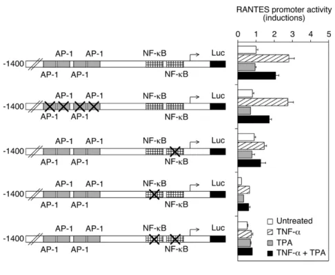
To evaluate the role of AP-1 and NF-κB in the regulation of RANTES expression, a series of point mutated RANTES promoter constructs were analyzed by a reporter gene assay (Fig. 2A). The RANTES 1.4 kb promoter region contains four AP-1 binding sites and two NF-κB response elements. TNF-α-induced intact promoter activity was partially repressed by TPA. Mutation of one or the other NF-κB recognition site reduced overall promoter activity, but some regulation by TNF-α and TPA still occurred. The promoter became almost unresponsive when both NF-κB recognition sequences were mutated. In contrast, mutation of the AP-1 response elements did not affect promoter activity in any conditions. The same approach was used to study the involvement of AP-1 and NF-κB in the regulation of IL-8 expression (Fig. 2B). The IL-8 1.5 kb promoter region displays one AP-1 binding site and one NF-κB recognition sequence. Mutation of either the AP-1 or NF-κB response element dramatically reduced induction of IL-8 promoter activity by either TNF-α or TPA. The promoter became unresponsive when both sequences were mutated.
Regulation of p44/p42 mitogen-activated protein kinases phosphorylation by TNF-α, TPA and Gö 6976
We next analyzed the effect of TNF-α, TPA and Gö 6976 on p44/p42 mitogen-activated protein kinases (MAPK) signaling cascade. PKC activation is known to lead to phosphorylation of p44 and p42 MAPK, which in turn phosphorylate Elk-1 transcription factor, thereby increasing c-fos transcription and thus AP-1 activity (reviewed in [27]). Western blot analyses show an increase in phosphorylation status of both MAPK upon stimulation with TPA and, to a lesser extent, with TNF-α (Fig. 3). Basal phosphorylation of p44/p42 MAPK as well as that induced by TNF-α or TPA was inhibited by pretreatment with Gö 6976. However, Gö 6976 did not diminish phosphorylation induced by co-treatment with TNF-α and TPA.
7
Regulation of NF-κB signaling pathway by TNF-α, TPA and Gö 6976
To monitor activation of NF-κB, the level and phosphorylation status of its p65 subunit and of its inhibitor IκBα were determined by Western blotting. IκBα is phosphorylated by IκB kinases and undergo proteolytic degradation. In addition, these kinases phosphorylate p65, thereby increasing its transactivating capacity [28]. Time-course analyses show that phosphorylation of these proteins was more rapid and complete with TNF-α than with TPA. Upon TNF-α stimulation, phosphorylation of both proteins was clearly apparent at 2 min. The amount of phosphorylated p65 reached a maximum at 5 min and diminished progressively afterwards, but remained higher than in untreated cells for at least 2 h (Fig. 4A and data not shown). At 5 min, phosphorylated IκBα was predominant over IκBα and began to be degraded. Both forms of IκBα disappeared by 10 min, but were later resynthesized. With TPA, the amount of phosphorylated p65 was very low compared to that obtained after TNF-α treatment, while phosphorylation and degradation of IκBα was delayed and incomplete (Fig. 4B). Thus, as compared to TNF-α, TPA is a much weaker activator of NF-κB in A549 cells. Pretreating the cells with TPA for as short as 5 min prevented TNF-α from phosphorylating efficiently p65 and IκBα (Fig. 5A). This inhibitory effect of TPA was clearly weakened in the presence of Gö 6976 (Fig. 5B). The latter compound also slightly increased the amount phosphorylated IκBα after TNF-α treatment (Fig. 5B).
Affinity of NF-κB response elements from the RANTES and IL-8 promoters
As shown in Figure 2B, TPA induced IL-8 promoter activity via the AP-1 binding site but also through the NF-κB response element, even though it is a weak activator of the NF-κB signaling pathway. In contrast, TPA was unable to induce significantly RANTES promoter activity. Analysis of NF-κB recognition sequences reveals that those on RANTES promoter (GGAAACTCC and GGGATGCCC) diverge by one base pair from consensus (GGAA
/CTN T
/CCC) [29], while that on IL-8 promoter (GGAATTTCC) is identical to
consensus, suggesting that these may have different binding affinities. To compare their relative affinity, competitive binding followed by EMSA was performed. The two NF-κB binding sites present in tandem on RANTES promoter (2xNF-κBRE RANTES) and a tandem repeat of the NF-κB response element from the IL-8 promoter (2xNF-κBRE IL-8) were used as competitors. Nuclear extract of TNF-α-stimulated cells was incubated with 32-P labelled
κBRE IL-8 in the presence of increasing amount of competitor. Data show that 2xNF-κBRE IL-8 is a better competitor, and hence of higher affinity than 2xNF-2xNF-κBRE RANTES (Fig. 6). Supershift experiments indicated that the complex observed in TNF-α-stimulated cells contained the p65 subunit of NF-κB (data not shown).
Discussion
Analysis of lung adenocarcinoma clinical series indicate that IL-8 expression is associated with a poor prognosis while that of RANTES is a predictor of survival [3, 11]. In several models using xenografts of tumor cells in mice, it was found that IL-8 facilitates disease progression while RANTES reduces tumor growth (cf. Introduction for details and references). Thus, IL-8 and RANTES are potential therapeutic targets in lung adenocarcinoma. We found herein that RANTES and IL-8 expression is differentially regulated through PKCα/β in A549 lung adenocarcinoma cells. The mechanism underlying this differential regulation was investigated.
Firstly, regulation of RANTES promoter activity required intact NF-κB binding sites while the four putative AP-1 recognition sites were dispensable as shown by reporter gene assays (Fig. 2A). Others have shown that these AP-1 response elements contributed modestly to the induction of RANTES promoter activity in T lymphocytes [22]. Thus, these sites appear to be non functional or weakly active, although two of them are identical to the AP-1 consensus recognition sequence [22, 29]. Possibly, lack of responsiveness may be dictated by the surrounding sequences.
Secondly, we show that TPA was a weak activator of the NF-κB signaling pathway and, in fact, inhibited the strong induction of this pathway by TNF-α (Figs. 4 and 5). Conversely, TPA was more potent than TNF-α to induce in a PKCα/β dependent manner the p44/p42 MAPK signaling cascade, which is known to control AP-1 activity (Fig. 3). The precise mechanism by which TPA weakly activates NF-κB signaling pathway and simultaneously prevents its strong activation by TNF-α remains to be determined. Nevertheless, our results indicate that the negative effect on NF-κB activation went through PKCα/β and targeted directly or indirectly IκB kinases. Indeed, a decreased phosphorylation of IκBα and of p65 at Ser536 is indicative of a down-regulation of these kinases [30].
Thirdly, regulation of IL-8 promoter activity involved strong cooperation between an AP-1 recognition sequence and a high affinity NF-κB binding site while that of RANTES required only two low affinity NF-κB response elements (Figs. 2 and 6).
These data provide an explanation as to why TPA stimulated expression of IL-8 but not that of RANTES. These also explain why TNF-α-induced expression of RANTES was inhibited by TPA and restored by Gö 6976. TPA did not inhibit TNF-α-induced IL-8 expression probably because its inhibitory effect on TNF-α-induced NF-κB activity was compensated by the activation of AP-1. In addition, the high affinity NF-κB binding site on IL-8 promoter conferred a significant response even after weak activation of NF-κB by TPA (Fig. 2B). Of note, the PKCα/β inhibitor Gö 6976 increased TNF-α-induced RANTES production and prevented its down-regulation by TPA, while it diminished TNF-α or TPA induced IL-8 release (Fig. 1). Gö 6976 has been described to be very effective at sensitizing breast cancer cells to toxicity of DNA-damaging agents [31]. It was also found to promote formation of cell junctions and to inhibit invasion of urinary bladder carcinoma cells [32]. Together, these data support the notion that Gö 6976 may be of therapeutic value in the treatment of cancer, including lung adenocarcinoma. Of note, a PKCα antisense oligonucleotide did not enhance survival and other efficacy measures in patients with advanced non-small cell lung cancer [33]. It is tempting to speculate that this approach was not efficient because it did not target PKCβ as well. Indeed, PKCβ has apparently similar function to PKCα in cancer cells; it has been shown to account for increased invasion [34, 35] and proliferation rate [36]. In addition, inhibiting PKCα/β activity may prove efficacy only at early stage of the disease and/or in a subtype of non-small cell lung cancer, such as lung adenocarcinoma. Finally, we observed
9
IL-8 production was not statistically significant when cells were stimulated with TNF-α alone or concomitantly with TPA. Hence, this compound may reach efficacy only in combination therapy that includes other anti-angiogenics.
Acknowledgments: The authors thank Philippe Fort and Hiroyuki Moriuchi for providing
plasmids, and Patrick Atger for reprographic services.
11
Conflict of interest statement
None declared
References
[1] Garfinkel A. Cancer statistics and trends. In: Holleb AI, Fink DJ, Murphy GP (eds.), American Cancer Society Textbook of Clinical Oncology. Atlanta: American Cancer Society, 1991:2-9.
[2] Mountain CF. Lung cancer staging classification. Clin Chest Med 1993;14:43-53. [3] Moran CJ, Arenberg DA, Huang CC, Giordano TJ, Thomas DG, Misek DE, et al.
RANTES expression is a predictor of survival in stage I lung adenocarcinoma. Clin Cancer Res 2002;8:3803-12.
[4] Mule JJ, Custer M, Averbook B, Yang JC, Weber JS, Goeddel DV, et al. RANTES secretion by gene-modified tumor cells results in loss of tumorigenicity in vivo: role of immune cell subpopulations. Hum Gene Ther 1996;7:1545-53.
[5] Kutubuddin M, Federoff HJ, Challita-Eid PM, Halterman M, Day B, Atkinson M, et al. Eradication of pre-established lymphoma using herpes simplex virus amplicon vectors. Blood 1999;93:643-54.
[6] Manes S, Mira E, Colomer R, Montero S, Real LM, Gomez-Mouton C, et al. CCR5 expression influences the progression of human breast cancer in a p53-dependent manner. J Exp Med 2003;198:1381-9.
[7] Luboshits G, Shina S, Kaplan O, Engelberg S, Nass D, Lifshitz-Mercer B, et al. Elevated expression of the CC chemokine regulated on activation, normal T cell expressed and secreted (RANTES) in advanced breast carcinoma. Cancer Res 1999;59:4681-7.
[8] Niwa Y, Akamatsu H, Niwa H, Sumi H, Ozaki Y, Abe A. Correlation of tissue and plasma RANTES levels with disease course in patients with breast or cervical cancer. Clin Cancer Res 2001;7:285-9.
[9] Adler EP, Lemken CA, Katchen NS, Kurt RA. A dual role for tumor-derived chemokine RANTES (CCL5). Immunol Lett 2003;90:187-94.
[10] Robinson SC, Scott KA, Wilson JL, Thompson RG, Proudfoot AE, Balkwill FR. A chemokine receptor antagonist inhibits experimental breast tumor growth. Cancer Res 2003;63:8360-5.
[11] Yuan A, Yang PC, Yu CJ, Chen WJ, Lin FY, Kuo SH, et al. Interleukin-8 messenger ribonucleic acid expression correlates with tumor progression, tumor angiogenesis, patient survival, and timing of relapse in non-small-cell lung cancer. Am J Respir Crit Care Med 2000;162:1957-63.
[12] Arenberg DA, Kunkel SL, Polverini PJ, Glass M, Burdick MD, Strieter RM. Inhibition of interleukin-8 reduces tumorigenesis of human non-small cell lung cancer in SCID mice. J Clin Invest 1996;97:2792-802.
[13] Moore BB, Arenberg DA, Stoy K, Morgan T, Addison CL, Morris SB, et al. Distinct CXC chemokines mediate tumorigenicity of prostate cancer cells. Am J Pathol 1999;154:1503-12.
[14] Inoue K, Slaton JW, Kim SJ, Perrotte P, Eve BY, Bar-Eli M, et al. Interleukin 8 expression regulates tumorigenicity and metastasis in human bladder cancer. Cancer Res 2000;60:2290-9.
[15] Salcedo R, Martins-Green M, Gertz B, Oppenheim JJ, Murphy WJ. Combined administration of antibodies to human interleukin 8 and epidermal growth factor receptor results in increased antimetastatic effects on human breast carcinoma xenografts. Clin Cancer Res 2002;8:2655-65.
[16] Huang S, Mills L, Mian B, Tellez C, McCarty M, Yang XD, et al. Fully humanized neutralizing antibodies to interleukin-8 (ABX-IL8) inhibit angiogenesis, tumor
13
[17] Lee LF, Hellendall RP, Wang Y, Haskill JS, Mukaida N, Matsushima K, et al. IL-8 reduced tumorigenicity of human ovarian cancer in vivo due to neutrophil infiltration. J Immunol 2000;164:2769-75.
[18] Balkwill F. Cancer and the chemokine network. Nat Rev Cancer 2004;4:540-50. [19] Mocellin S, Rossi CR, Pilati P, Nitti D. Tumor necrosis factor, cancer and anticancer
therapy. Cytokine Growth Factor Rev 2005;16:35-53.
[20] Mackay HJ, Twelves CJ. Protein kinase C: a target for anticancer drugs? Endocr Relat Cancer 2003;10:389-96.
[21] Koivunen J, Aaltonen V, Peltonen J. Protein kinase C (PKC) family in cancer progression. Cancer Lett 2006;235:1-10.
[22] Moriuchi H, Moriuchi M, Fauci AS. Nuclear factor-kappa B potently up-regulates the promoter activity of RANTES, a chemokine that blocks HIV infection. J Immunol 1997;158:3483-91.
[23] Freund A, Jolivel V, Durand S, Kersual N, Chalbos D, Chavey C, et al. Mechanisms underlying differential expression of interleukin-8 in breast cancer cells. Oncogene 2004;23:6105-14.
[24] Vincent S, Marty L, Fort P. S26 ribosomal protein RNA: an invariant control for gene regulation experiments in eucaryotic cells and tissues. Nucleic Acids Res 1993;21:1498.
[25] Mathieu M, Gougat C, Jaffuel D, Danielsen M, Godard P, Bousquet J, et al. The glucocorticoid receptor gene as a candidate for gene therapy in asthma. Gene Ther 1999;6:245-52.
[26] Stein B, Rahmsdorf HJ, Steffen A, Litfin M, Herrlich P. UV-induced DNA damage is an intermediate step in UV-induced expression of human immunodeficiency virus type 1, collagenase, c-fos, and metallothionein. Mol Cell Biol 1989;9:5169-81.
[27] Pearson G, Robinson F, Beers Gibson T, Xu BE, Karandikar M, Berman K, et al. Mitogen-activated protein (MAP) kinase pathways: regulation and physiological functions. Endocr Rev 2001;22:153-83.
[28] Sizemore N, Lerner N, Dombrowski N, Sakurai H, Stark GR. Distinct roles of the Ikappa B kinase alpha and beta subunits in liberating nuclear factor kappa B (NF-kappa B) from I(NF-kappa B and in phosphorylating the p65 subunit of NF-(NF-kappa B. J Biol Chem 2002;277:3863-9.
[29] Faisst S, Meyer S. Compilation of vertebrate-encoded transcription factors. Nucleic Acids Res 1992;20:3-26.
[30] Vermeulen L, De Wilde G, Notebaert S, Vanden Berghe W, Haegeman G. Regulation of the transcriptional activity of the nuclear factor-kappaB p65 subunit. Biochem Pharmacol 2002;64:963-70.
[31] Kohn EA, Yoo CJ, Eastman A. The protein kinase C inhibitor Go6976 is a potent inhibitor of DNA damage-induced S and G2 cell cycle checkpoints. Cancer Res 2003;63:31-5.
[32] Koivunen J, Aaltonen V, Koskela S, Lehenkari P, Laato M, Peltonen J. Protein kinase C alpha/beta inhibitor Go6976 promotes formation of cell junctions and inhibits invasion of urinary bladder carcinoma cells. Cancer Res 2004;64:5693-701.
[33] Paz-Ares L, Douillard JY, Koralewski P, Manegold C, Smit EF, Reyes JM, et al. Phase III study of gemcitabine and cisplatin with or without aprinocarsen, a protein kinase C-alpha antisense oligonucleotide, in patients with advanced-stage non-small-cell lung cancer. J Clin Oncol 2006;24:1428-34.
[34] Schwartz GK, Jiang J, Kelsen D, Albino AP. Protein kinase C: a novel target for inhibiting gastric cancer cell invasion. J Natl Cancer Inst 1993;85:402-7.
[35] Zhang J, Anastasiadis PZ, Liu Y, Thompson EA, Fields AP. Protein kinase C (PKC) betaII induces cell invasion through a Ras/Mek-, PKC iota/Rac 1-dependent signaling pathway. J Biol Chem 2004;279:22118-23.
[36] Murray NR, Davidson LA, Chapkin RS, Clay Gustafson W, Schattenberg DG, Fields AP. Overexpression of protein kinase C betaII induces colonic hyperproliferation and increased sensitivity to colon carcinogenesis. J Cell Biol 1999;145:699-711.
15
Figure legends
Fig. 1. Regulation of RANTES and IL-8 expression. (A) A549 cells were serum deprived
overnight and stimulated for 4 h with TNF-α (10 ng/ml) and TPA (100 ng/ml) as indicated. Total RNA was analyzed by Northern blotting. The ribosomal protein S26 mRNA served as loading control. Approximate size of transcript was 1.9 kb for RANTES, 2.2 kb for IL-8, and 0.7 kb for S26. (B and C) A549 cells were pretreated with Gö 6976 (1 µM) for 1 h and stimulated with TNF-α (10 ng/ml) and TPA (100 ng/ml) for 20 h as indicated. Concentrations of RANTES (B) and IL-8 (C) in supernatants were determined. Data (n = 6) are plotted as means ± SE.
Fig. 2. Regulation of RANTES and IL-8 promoter activity after site-directed mutagenesis. (A) A549 cells were transfected with intact or point mutated RANTES
promoter-luciferase gene construct as indicated, and CMVβ-gal. These were treated for 4 h with TNF-α (10 ng/ml) or TPA (100 ng/ml) or the combination of both compounds, and lysed to determine relative luciferase activities after normalization for β-galactosidase activity. Data (n = 6) are shown as fold inductions over basal activity and are plotted as means + SE. (B)
A549 cells were transfected with intact or mutated IL-8 promoter-luciferase gene construct as indicated, and CMVβ-gal. These were then treated as in (A). Data (n = 6) are shown as fold
+
17
Fig. 3. Regulation of p44/42 MAPK signaling. A549 cells were pretreated with Gö 6976 (1
µM) for 60 min and exposed to TNF-α (10 ng/ml) and TPA (100 ng/ml) for 10 min as indicated. Cell extracts were analyzed by Western blotting for their content in total p44/42 and phosphorylated p44/42 (phospho-p44/42).
Fig. 4. Time-course analyses of NF-κB signaling. (A) A549 cells were exposed to TNF-α
(10 ng/ml) for the indicated time. Cell extracts were analyzed by Western blotting for their content in p65, phosphorylated p65 (phospho-p65), IκBα and phosphorylated IκBα (phospho-IκBα). (B) A549 cells were exposed to TNF-α (10 ng/ml) or TPA (100 ng/ml) for the indicated time. Cell extracts were analyzed as in (A).
19
Fig. 5. Regulation of TNF-α-induced NF-κB signaling by PKCα/β. (A) A549 cells were
pretreated with TPA (100 ng/ml) for the indicated time and further exposed for 5 min to TNF-α (10 ng/ml). Cell extracts were analyzed by Western blotting for their content in p65, phosphorylated p65 (phospho-p65), IκBα and phosphorylated IκBα (phospho-IκBα). (B) A549 cells were pretreated with Gö 6976 (1 µM) and TPA (100 ng/ml) for 60 min and 10 min, respectively. These were then treated with TNF-α (10 ng/ml) for 5 min. Cell extracts were analyzed as in (A).
Fig. 6. Relative affinity of NF-κB response elements from the IL-8 and RANTES promoters. Nuclear proteins were extracted from A549 cells treated for 1 h with medium
alone (first lane) or 10 ng/ml of TNF-α (+). EMSA was performed using as labelled probe a tandem repeat of the NF-κB response element from the IL-8 promoter (2xNF-κBRE IL-8; 15 nM) and as competitors 2xNF-κBRE IL-8 or the two NF-κB binding sites present in tandem on RANTES promoter (2xNF-κBRE RANTES) at increasing concentrations.