LPS induces GFAT2 expression to promote O-GlcNAcylation and
1attenuate inflammation in macrophages
23 4
Running title: GFAT2 is a FoxO1-dependent LPS-inducible gene 5
6
Hasanain AL-MUKH, Léa BAUDOIN, Abdelouhab BOUABOUD, José-Luis SANCHEZ-7
SALGADO, Nabih MARAQA, Mostafa KHAIR, Patrick PAGESY, Georges BISMUTH, 8
Florence NIEDERGANG and Tarik ISSAD 9
10
Université de Paris, Institut Cochin, CNRS, INSERM, F-75014 Paris, France 11
Address correspondence to: Tarik Issad, Institut Cochin, Department of Endocrinology, 12
Metabolism and Diabetes, 24 rue du Faubourg Saint-Jacques, 75014 Paris FRANCE. Tel : + 33 1 13 44 41 25 67; E-mail: tarik.issad@inserm.fr 14 15 16
Abstract 1
O-GlcNAc glycosylation is a reversible post-translational modification that regulates the 2
activity of intracellular proteins according to glucose availability and its metabolism through 3
the hexosamine biosynthesis pathway (HBP). This modification has been involved in the 4
regulation of various immune cell types, including macrophages. However, little is known 5
concerning the mechanisms that regulate protein O-GlcNAcylation level in these cells. In the 6
present work, we demonstrate that LPS treatment induces a marked increase in protein O-7
GlcNAcylation in RAW264.7 cells, bone-marrow-derived and peritoneal mouse 8
macrophages, as well as human monocyte-derived macrophages. Targeted deletion of OGT in 9
macrophages resulted in an increased effect of LPS on NOS2 expression and cytokine 10
production, suggesting that O-GlcNAcylation may restrain inflammatory processes induced 11
by LPS. The effect of LPS on protein O-GlcNAcylation in macrophages was associated with 12
an increased expression and activity of glutamine fructose 6-phosphate amido-transferase 13
(GFAT), the enzyme that catalyzes the rate-limiting step of the HBP. More specifically, we 14
observed that LPS potently stimulated GFAT2 isoform mRNA and protein expression. 15
Genetic or pharmacological inhibition of FoxO1 impaired LPS effect on GFAT2 expression, 16
suggesting a FoxO1-dependent mechanism. We conclude that GFAT2 should be considered as 17
a new LPS-inducible gene involved in regulation of protein O-GlcNAcylation, which permits 18
to limit exacerbated inflammation upon macrophage activation. 19
20 21
Key points: 22
- LPS stimulates GFAT2 expression in macrophages via a FoxO1-dependent mechanism 23
- LPS-induced GFAT activity promotes a general increase in protein O-GlcNAcylation 24
- Protein O-GlcNAcylation limits excessive LPS-triggered inflammation in macrophages 25
26 27
Introduction 1
O-GlcNAcylation is a post-translational modification that regulates the activity of cytosolic, 2
nuclear and mitochondrial proteins. This modification is controlled by only two enzymes: 3
OGT, that adds N-Acetylglucosamine (GlcNAc) on serine or threonine residues, and OGA, 4
that removes it. O-GlcNAcylation, which regulates a wide array of biological processes (1), 5
depends on the availability of glucose and its metabolism through the hexosamine 6
biosynthesis pathway (Fig. 1A). This modification has been involved in the modulation of 7
various signalling pathways, and perturbations of O-GlcNAcylation participate in several 8
important human pathologies (2-4). Moreover, a number of studies indicated a link between 9
O-GlcNAcylation and inflammatory conditions, including diabetes, auto-immune diseases 10
and cancer (5-11). Moreover, several lines of evidence have suggested a role for O-11
GlcNAcylation in immune cell signalling. Indeed, O-GlcNAcylation was first discovered at 12
the surface of lymphocytes (12) and dynamic changes upon lymphocyte activation were 13
described at the beginning of the nineties (13). More recently, several studies indicated that 14
protein O-GlcNAcylation plays major roles in the immune system. In T cells, TCR (T-cell 15
receptor) activation results in global elevation of protein O-GlcNAcylation (14, 15), increased 16
O-GlcNAcylation of several transcription factors (16-19) and accumulation of OGT at the 17
immunological synapse (14). Moreover, in B lymphocytes, activation of the B-cell receptor 18
(BCR) induces O-GlcNAcylation of several signalling molecules involved in BCR signalling 19
(20, 21). Overall, these studies strongly support a major role in O-GlcNAcylation in adaptive 20
immunity. However, the molecular mechanisms by which immune cell activation modulates 21
protein O-GlcNAcylation remain elusive. 22
In macrophages, contradictory results have been obtained concerning the effect of LPS on 23
protein O-GlcNAcylation and its role in inflammatory processes. Whereas some studies 24
indicated that LPS induced a general increase in protein O-GlcNAcylation (22, 23), others 25
found that LPS decreases protein O-GlcNAcylation in macrophages (24, 25). Moreover, 26
whereas several lines of evidence indicate that increased O-GlcNAcylation of signaling 27
proteins in the NFkB pathway potentiates inflammatory processes (26-29), other studies 28
indicate that increased O-GlcNAcylation may inhibit pro-inflammatory signals (24, 30). More 29
recently, O-GlcNAcylation of the signalling adaptor MAVS in macrophages has been 30
involved in antiviral signalling response (31), whereas increased RIPK3 O-GlcNAcylation 31
was shown to regulate necroptosis signalling upon LPS stimulation (25). Thus, whereas 32
numerous evidences suggest that modulation of O-GlcNAcylation signalling constitutes an 33
important facet of innate immune response, the mechanisms by which macrophage activation 1
regulates O-GlcNAc, and its consequences on inflammatory processes, remain to be firmly 2
established. 3
In the present work, using different macrophage cell models, we clearly demonstrated that 4
LPS markedly stimulates protein O-GlcNAcylation in macrophages through increased 5
expression and activity of the rate-limiting enzyme of the hexosamine biosynthetic pathway, 6
glutamine fructose-6-phosphate amidotransferase (GFAT). More specifically, we showed that 7
whereas resting macrophages express mainly the GFAT1 isoform, upon LPS stimulation, a 8
marked increase in GFAT2 expression was observed, revealing GFAT2 as a new LPS-9
inducible gene in macrophages that promotes a general increase in protein O-GlcNAcylation. 10
In macrophages with conditional OGT deletion, lack of LPS-induced O-GlcNAyclation was 11
associated with an increased in the production of pro-inflammatory molecules, suggesting that 12
GFAT2 induction may be part of a regulatory loop that may limit inflammation and/or permit 13
its resolution after TLR4 activation. 14
15 16
Materiel and Methods 1
Chemicals and antibodies 2
Thiamet G (3aR,5R,6S,7R,7aR)-2-(ethylamino)-3a,6,7,7a-tetrahydro-5-(hydroxymethyl)-5H-3
Pyrano.thiazole-6,7-diol) and LPS (Lipopolysaccharides) from Salmonella enterica serotype 4
typhimurium were from Sigma-Aldrich (Saint Quentin Fallavier France). FoxO1 inhibitor 5
AS1842856 was from Calbiochem. PUGNAc (O-(2-acetamido-2-deoxy-d-6
glucopyranosylidene)-amino-N-phenylcarbamate) was from Toronto Research Chemicals, 7
Inc. (North York, ON, Canada). Anti-GFAT2 antibody (EPR 19095) and Anti-FOXO1A antibody 8
(Chip Grade ab39670) were from Abcam; Anti-GFAT1 (H-49), Anti-NOS2 (N-20), Anti-GAPDH 9
(O411), Anti-Tubulin (TU-02) Anti-UBF (F9) and HRP-conjugated anti-rabbit antibodies were from 10
Santa-Cruz Biotechnolology; Anti-FOXO1 antibody for western-blotting (L27) was from Cell 11
Signaling Technology; GlcNAc Transferase antibody (O 6264) was from Sigma; Anti-O-12
GlcNAcase antibody (NBP2-32233) was from Novus Biologicals, Anti-O-GlcNAc antibody (RL2) 13
was from Thermo Fisher; Anti-Clathrin antibody (610500) was from BD Transduction Laboratories 14
and HRP-conjugated anti-mouse antibody was from Jackson ImmunoResearch, Laboratories. 15
16
cDNA constructs 17
The cDNA coding for nuclear, cytosolic and plasma membrane FRET O-GlcNAc biosensors 18
(32) were generously provided by Prof. L.K. Mahal (University of Texas, Austin, USA). 19
BRET biosensors were developed based on these FRET constructs by replacing the CFP by a 20
Rluc8 sequence (33). 21
Human (-801>>>0) and mouse GFAT2 (-501>>>0) putative promoter sequences were 22
amplified by PCR and cloned in a firefly luciferase plasmid (pGL4-20, Promega). FoxO1 23
binding sites on GFAT2 promoter were identified using Regulatory Sequence Analysis Tools 24
(RSAT) web server (http://rsat.ulb.ac.be/rsat/). Mutagenesis of Foxo1 binding sites on the 25
mouse GFAT2 reporter gene was performed using mutagenesis kit (QuikChangeII XL-Agilent 26
Technologies). 27
Constitutively active FOXO1-TM (mutated on the 3 Akt phoshorylation sites) has been 28
described previously (34). 29
30
Cell culture and transfection 31
RAW264.7 murine macrophage cells were maintained in media constituted of RPMI 1640-32
glutamax medium supplemented with 10% fetal calf serum (FCS), 50µM β-mercaptoethanol, 33
1 mM sodium pyruvate, 10 mM HEPES and 2 mM L-Glutamine (GIBCO). 34
Plasmid transfections were performed by cell electroporation using the Electrobuffer kit (Cell 1
projects Ltd). For each transfection, cells grown to sub-confluence in a 100 mm plate were 2
transfected with 15 µg of plasmid DNA. Cells were electroporated at 250 V, 900 µF in 0.4 cm 3
cuvettes (Biorad) using the Gene Pulser X electroporation system (Bio-Rad Laboratories). 4
After electroporation, cells were immediately re-suspended in culture medium, transferred into 5
96-well White Optiplates previously coated with polylysine (Perkin Elmer) and then cultured 6
for 18 hours at 37°C and 5% CO2 before treatments. 7
HEK 293-T cells were cultured in DMEM and transfected with Fugene as described 8
previously (34). 9
10
Chromatin immunoprecipitation (ChIP): 11
RAW264.7 cells (1x10⁷ cells) were prepared for ChIP assay using HighCell# ChIP kit protein 12
A (Diagenode). After cross-linking with formaldehyde, DNA was sonicated into 200-300bp 13
fragments, and protein-DNA complexes were immunoprecipitated using either FoxO1-ChIP 14
grade antibody (Abcam, ab39670) or control rabbit IgG. Protein-DNA crosslinks were 15
reversed by heating, and precipitated DNA was quantified by real-time PCR using primers 16
(Suppl. Table 1) that amplify either Foxo1 binding site 1 or 2 present in the 500bp upstream 17
of the transcription start site of the mouse GFAT2 gene. 18
19
BRET experiments 20
Transfected cells were treated with LPS (100ng/mL) and/or Thiamet G (10µM) for 24h. 21
BRET experiments were then performed exactly as described previously (35, 36) using the 22
Infinite F200 Pro microplate analyser (Tecan). Briefly, cells were pre-incubated for 5 min in 23
PBS in the presence of 5µM coelenterazine. Each measurement corresponded to the signal 24
emitted by the whole population of cells present in a well. BRET signal was expressed in 25
milliBRET Unit (mBU). The BRET unit has been defined previously as the ratio 530 nm/485 26
nm obtained in cells expressing both luciferase and YFP, corrected by the ratio 530 nm/485 27
nm obtained under the same experimental conditions in cells expressing only luciferase (37, 28 38). 29 30 Animals 31
8 to 12 weeks-old C57BL/6J male mice were used. To study the function of Foxo1 in 32
macrophages, we crossed mice carrying two floxed Foxo1 alleles kindly provided by Prof. 33
Hedricks, Univ. of California, San Diego, USA) with LysM-Cre transgenic mice (kindly 34
provided by Carol Peyssonnaux, Institut Cochin, Paris) in which Cre is specifically expressed 1
in the myelomonocytic cell lineage (LysMCre-Foxo1/Foxo1 knock-out (KO) mice, thereafter 2
refered to as Foxo1-KO mice). To study the role of O-GlcNAcylation on pro-inflammatory 3
effects of LPS, mice with OGT deletion in the myelomonocytic cell lineage were generated by 4
crossing mice carrying two floxed OGT alleles (obtained from Jackson laboratories) with 5
LysM-Cre transgenic mice (LysMCre-OGT knock-out (KO) mice, thereafter referred to as 6
OGT-KO mice). All mice were housed in the Institut Cochin animal facility. All experiments 7
were performed in accordance with accepted standards of animal care, as established in the 8
Institut National de la Santé et de la Recherche Médicale (INSERM) and the Centre National 9
de la Recherche Scientifique (CNRS) guidelines and were approved by the national ethical 10
committee (APAFIS N°A751402). 11
12
Preparation of bone-marrow-derived and peritoneal macrophages from mice. 13
Mice were sacrificed by CO2 asphyxiation. Bone marrow-derived macrophages (BMDM) 14
were prepared from bone marrow cells flushed from femurs and tibias (39). Cells were seeded 15
in sterile Petri dishes at the concentration of 106/ml in RPMI medium supplemented with 10% 16
FCS, 100µg/ml gentamycin, 10mM HEPES, 1mM sodium pyruvate, 50µM β-mercaptoethanol 17
and 20ng/mL mouse MCSF (Miltenyi Biotec). Cells were differentiated during 6 days, then 18
washed to eliminate non-adherent cells, and macrophages were detached in cold PBS -/- and 19
seeded in 6 well-plates at 106 cells/ml in complete media. Cells were treated the day after. 20
For peritoneal macrophages collection, after mice sacrifice, the peritoneum was infused with 21
10mL of cold RPMI. Cells were seeded in 6 well-plates at 106cells/ml in complete media and 22
allowed to adhere overnight. Non-adherent cells were then washed away and macrophages 23
treated with LPS. 24
In some experiments, mice were injected intraperitoneally with LPS (0.6 mg/kg). 6h after 25
injection, mice were sacrificed and peritoneal cells were harvested as described above and 26
plated in a 6 well-plate at 37 °C with 5% CO2. After 2h, non-adherent cells were washed 27
away, and adherent cells were lysed for analysis by western blot. 28
To evaluate the effect of LPS on cytokine production in vivo in OGT-KO mice, LPS was 29
injected intraperitoneally, mice were sacrificed 6h after injection and blood was collected by 30
cardiac puncture. Blood was centrifuged at 1500 rpm for 10 min at 4°C and the serum was 31
collected and frozen at -80°C for subsequent determination of cytokine concentrations using 32
MSD kit. 33
Preparation of Human monocytes derived macrophages 1
Human primary macrophages were isolated from blood of healthy donors (Etablissement 2
Français du Sang, Ile-de-France, Site Trinité, Agreement number INSERM-EFS:18/EFS/030) 3
by density gradient sedimentation on Ficoll (GE Healthcare), followed by negative selection 4
on magnetic beads (Stem cells, Cat n°19059) and adhesion on plastic at 37°C during 2 h. Cells 5
were then cultured in the presence of complete culture medium (RPMI 1640 supplemented 6
with 10 % FCS (Eurobio), 100 mg/ml streptomycin/penicillin and 2 mM L-glutamine (Gibco) 7
containing 10 ng/ml rhM-CSF (R&D systems) during 4-5 days (40). 8
9
RNA extraction, reverse transcription and qPCR 10
Macrophages cultured in 6-well plates were lysed in Trizol reagent (Life Technologies). RNA 11
was isolated and reverse transcribed (41). Levels of the cDNA of interest were measured by 12
qPCR (LightCycler FastStart DNA Master SYBR Green 1 kit; Roche Diagnostics). The 13
absence of genomic DNA contamination was verified by treating RNA samples in parallel 14
without reverse transcriptase and controlling for the absence of amplification by qPCR. Gene 15
expression was normalized over cyclophilin and HPRT (Hypoxanthine-guanine 16
phosphoribosyltransferase) RNA levels. The sequences of the primers used for qPCR are 17
given in Supplemental Table 1. 18
19
OGA enzymatic assay. 20
Control and LPS-treated RAW264.7 cells were lysed in extraction buffer containing 50mM 21
Tris, 137 mM NaCl, 1% triton, 10% glycerol, phosphatases (50 mM NaF, 10 mM di-sodium 22
β-glycerophosphate, 1 mM Na 3VO4) and proteases (AEBSF, leupeptine, antipaine aprotinine 23
and pepstatine, 1µg/ml each) inhibitors. OGA activity was measured using 4-24
methylumbellifery-N-acetylb-D-glucosamine (MU-GlcNAc, Sigma), which is converted into 25
fluorescent 4-methylumbelliferon upon hydrolysis by OGA and other hexosaminidases (42). 26
4-methylumbelliferon fluorescence was measured at 448 nm after excitation at 362 nm. 27
Fluorescent measurements were performed after 30 min and 60 min of incubation at 37 °C, to 28
ensure that the determination was performed during the linear phase of the reaction. To 29
specifically determine OGA activity versus other hexosaminidase present in the lysate, all 30
reactions were performed both in absence and presence of 100 µM Thiamet G (a specific 31
OGA inhibitor). The difference between the fluorescent signals obtained in absence (activity 32
of OGA plus other hexosaminidases) and presence of Thiamet G (activity of the other 33
hexosaminidases) reflected the amount of 4-methylumbelliferon produced by OGA (9). 34
OGT enzymatic assay. 1
Control and LPS-treated RAW264.7 cells were lysed in extraction buffer containing 50mM 2
Tris, 137 mM NaCl, 1% triton, 10% glycerol, phosphatases (50 mM NaF, 10 mM di-sodium 3
β-glycerophosphate, 1 mM Na 3VO4) and proteases (AEBSF, leupeptine, antipaine aprotinine 4
and pepstatine, 1µg/ml each) inhibitors. Immunoprecipitation of OGT was performed as 5
described below. 400 µg of proteins were incubated with 1.5 µg of anti-OGT antibody 6
(Sigma-Aldrich) for 2h at 4°C. Precipitation was performed by incubating 25µL equilibrated 7
protein G-sepharose beads (GE Healthcare) for 30 min at 4°C. After 3 washes, the 8
precipitated proteins were submitted to an additional wash in OGT assay buffer containing 50 9
mM Tris-HCl and 12.5 mM MgCl2, pH7.5. OGT assay was then performed on protein-G 10
sepharose bound OGT (33) using the bioluminescent UDP-GloTM glycosyltransferase assay 11
(Promega) exactly as described in the manufacturer instructions (43). 12
13
GFAT enzymatic assay. 14
Glutamine fructose-6-phosphate amidotransferase enzymatic activity was measured as 15
described previously (44). Control and LPS-treated RAW264.7 cells, human monocyte-16
derived macrophages or mouse bone-marrow-derived macrophages were lysed in extraction 17
buffer containing 50mM Tris, 137 mM NaCl, 1% triton, 10% glycerol, phosphatases (50 mM 18
NaF, 10 mM di-sodium β-glycerophosphate, 1 mM Na 3VO4) and proteases (AEBSF, 19
leupeptine, antipaine aprotinine and pepstatine, 1µg/ml each) inhibitors. 15-30 µg of proteins 20
were incubated in a buffer containing 10 mM fructose 6-phosphate, 6 mM glutamine, 0.3 mM 21
3-acetylpyridine adenine dinucleotide (APAD), 50 mM KC1, 100 mM KH2PO4 (pH 7.5), 1 22
mM dithiothreitol and 30 unit/ml of glutamate dehydrogenase. After incubation for 30 min at 23
37°C, the change in absorbance due to reduction of APAD to APADH was monitored 24
spectrophotometrically at 365 nm using TECAN™ infinite M1000 Pro Microplate Reader. 25
Western blotting 26
Cells were lysed with buffer containing 50mM Tris–HCl (pH 8), 137mM NaCl, 10% (v/v) 27
glycerol, 1% (v/v) triton, 50mM NaF, 10mM di-sodium β-glycerophosphate, 1mM Na 3VO4, 28
protease inhibitors (1µg/ml pepstatin, antipain, leupeptin, aprotinin and AEBSF), 29
supplemented with 10µM PUGNAc in order to preserve the GlcNAcylation state of proteins 30
during the extraction procedure. Proteins were then analysed by SDS-PAGE followed by 31
western-blotting as described previously (45). Clathrin was used as a loading control, because 32
its molecular weight (180 kDa) is far away from the 50-70 kDa region where many proteins 1
of interest are found (46, 47).
2
In some experiments, O-GlcNAcylated proteins were precipitated on wheat germ lectin as 3
described previously (48, 49). Briefly, cell lysates (400 µg of proteins) were incubated for 2 4
hours at 4° C on a rotating wheel with 20 µl of agarose beads coupled to wheat germ lectin 5
(which binds the N-Acetyl-Glucosamine pattern). At the end of the incubation, the beads were 6
pelleted by centrifugation at 3000 g for 2 min. The supernatant was removed and the pellet 7
was washed 3 times in extracting buffer. Twenty µL of Laemmli sample buffer were added to 8
the beads, and the samples were boiled at 95°C for 5 minutes and then submitted to western-9
blotting as described previously (48, 49). 10
Western-blots were revealed by chemoluminescence (Thermo Scientific) and visualized using 11
a Fusion FX7-Vilber Lourmat camera. The signals obtained were then quantified using Image 12 J software. 13 14 Statistical analysis 15
Statistical analyses were performed using PRISM software. Comparison between groups were 16
performed using Student's t test, or ANOVA followed by Dunnett’s or Tukey’s post-test as 17
indicated in the figure legends. 18
Results 1
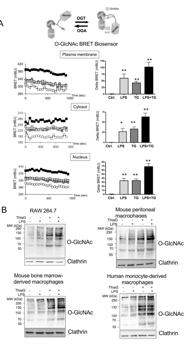
LPS stimulation markedly increased protein O-GlcNAcylation in macrophages 2
Sub-cellular relocalisation of OGT in different cell compartments has been observed upon 3
stimulation of membrane receptors (50, 51), resulting in compartment specific changes in O-4
GlcNAcylation activity. In order to evaluate whether LPS stimulation affects protein O-5
GlcNAcylation in different cellular compartments in macrophages, we used BRET-based O-6
GlcNAc biosensors (33). These biosensors are composed of Rluc8 luciferase fused to a lectin 7
domain (GafD), a known OGT substrate peptide derived from casein kinase II, followed by 8
the Venus variant of the yellow fluorescent protein (Fig. 1B). Upon O-GlcNAcylation, the 9
casein kinase peptide binds to the lectin, resulting into a conformational change detected as an 10
increased BRET signal. These biosensors were fused to addressing sequences for targeting to 11
the internal face of the plasma membrane (using Lyn myristoylation/palmitoylation 12
sequence), the cytosol (using the HIV-1 Rev protein nuclear exclusion sequence) or the 13
nucleus (using the SV40 nuclear localisation sequence) (32). 14
RAW264.7 cells transfected with these biosensors were incubated for 24h in presence of 15
LPS, Thiamet G (an inhibitor of OGA) or both. We observed that LPS treatment increased 16
BRET signal with all three biosensors (Fig. 1B), indicating that LPS stimulation promotes a 17
general rather than compartment-specific increase in O-GlcNAcylation in RAW264.7 18
macrophages. Interestingly, the effects of LPS and Thiamet G were additive, suggesting that 19
the effect of LPS on O-GlcNAcylation is independent of OGA activity. 20
LPS-induced O-GlcNAcylation of proteins, both in absence and presence of Thiamet G, was 21
further demonstrated by western-blotting using anti-O-GlcNAc antibody in RAW264.7 cells, 22
but also in mouse bone marrow-derived and peritoneal primary macrophages, as well as in 23
human monocyte-derived macrophages (Fig. 1C). Therefore, increased O-GlcNAcylation 24
upon LPS stimulation also appears to occur in primary macrophages from different origins 25
and species, suggesting a general mechanism elicited by TLR4 activation. 26
GFAT2 is a LPS-inducible gene in macrophages 27
We further explored, using RAW264.7 macrophages, the mechanism involved in this LPS-28
induced GlcNAcylation. As shown in Figure 2, LPS-induced increase in protein O-29
GlcNAcylation (Fig. 2A) was not associated with any detectable change in OGT and OGA 30
mRNA or protein expression (Fig. 2B and C), suggesting that it was not mediated by 31
regulation of the expression level of O-GlcNAc cycling enzymes. GFAT, the enzyme that 32
catalyzes the rate-limiting step of the hexosamine biosynthesis pathway (Fig.1A), exists as 33
two isoforms (GFAT1 and GFAT2), encoded by two separate genes (also denominated GFPT1 34
and GFPT2, respectively) that are differentially expressed in a cell-type specific manner. We 1
observed that in the basal state, only GFAT1 protein was highly expressed in macrophages, 2
whereas GFAT2 protein expression was barely detectable (Fig. 2B). GFAT1 protein 3
expression was moderately increased (1.3 fold) by LPS treatment, whereas LPS induced a 4
major increase (7 fold) in GFAT2 protein expression (Fig. 2B). In agreement with these 5
results, Figure 2C shows that LPS increased the expression of GFAT1 mRNA by 2 to 3-fold, 6
and markedly induced (by 10 to 15-fold) the expression of GFAT2 mRNA, suggesting a 7
regulation of GFAT1 and 2 at the transcriptional level. Enzymatic assays indicated that 8
whereas LPS treatment had no detectable effect on OGT or OGA activities, it significantly 9
increased the activity of GFAT in macrophages (Fig. 2D). These results suggest that LPS-10
induced increase in the expression of GFAT, and more specifically GFAT2, translates into an 11
increase in the activity of the rate-limiting step of the hexosamine biosynthesis pathway. 12
Time-course experiments indicated that maximal induction of GFAT2 mRNA was observed 13
3h after LPS stimulation, whereas GFAT1 mRNA expression was barely modified at early 14
time points (Suppl. Fig. S1A), revealing that GFAT2 is an early TLR4-target gene. However, 15
despite marked stimulation of GFAT2 mRNA at early time-points, GFAT2 protein expression 16
was barely increased after 6h of LPS treatment, and robust stimulation was only detected after 17
24h of treatment with LPS (Suppl. Fig. S1B). Interestingly, GFAT activity was also barely 18
increased after 6h of treatment, and closely followed the increase in GFAT2 protein 19
expression at time 24h (Suppl. Fig. S1C). In agreement with this, LPS-induced increase in 20
protein O-GlcNAcylation could be detected at 6h, but became statistically significant only at 21
time 24h (Suppl. Fig. S1D). 22
Increased GFAT2 protein and mRNA expression was also observed in human monocyte-23
derived macrophages (Fig. 3A), mouse bone marrow-derived macrophages (Fig. 3B), as well 24
as well as peritoneal macrophages (Fig. 3C). In agreement with these results, GFAT 25
enzymatic activity was also increased upon LPS stimulation in human and mouse primary 26
macrophages (Suppl. Fig. S2A). Moreover, intraperitoneal injection of LPS in mice also 27
induced a modest increase in GFAT1 and a marked increase in GFAT2 protein expression in 28
peritoneal cells, indicating that this response was also operative in vivo (Suppl. Fig. S2B). 29
Altogether, our results suggest that LPS induces an increase in protein O-GlcNAcylation 30
through stimulation of the expression of the rate-limiting enzyme of the hexosamine 31
biosynthesis pathway. 32
Impaired O-GlcNAcylation promotes pro-inflammatory response in macrophages 33
To evaluate the role of LPS-induced O-GlcNAcylation in macrophages, we isolated 34
peritoneal macrophages from mice with conditional deletion of OGT in the myeloid lineage. 1
As expected, OGT expression and LPS-induced O-GlcNAcylation were markedly impaired in 2
OGT-KO cells (Fig. 4A), although residual protein O-GlcNAcylation was sometimes 3
observed, probably reflecting incomplete deletion of OGT by LysM-Cre in resident peritoneal 4
macrophages. We noticed that basal GFAT1 and both basal and LPS-induced GFAT2 5
expression were increased in OGT-KO macrophages, suggesting a compensatory response to 6
the low level of O-GlcNAcylation in these cells. Interestingly, OGT deletion resulted in an 7
increase in LPS-induced NOS2 expression. Moreover, we observed an increased production 8
of IFNg and IL1b in the culture medium of OGT-KO macrophages. These results suggest that 9
increased O-GlcNAcylation upon LPS stimulation may have a counter-regulatory effect that 10
restrains excessive cytokines production by macrophages (Fig. 4B). In agreement with this 11
notion, increases in pro-inflammatory cytokines IFN g, IL1b and TNFa were also observed in 12
vivo in the serum of OGT-KO mice intraperitoneally injected with LPS (Fig. 4C). We also 13
evaluated time-dependency of cytokine production by measuring IL1b and IFNg in the culture 14
medium of control and OGT-KO macrophages 6h and 24h after LPS stimulation (Suppl. Fig. 15
S2C). These experiments indicated that after 6 hours of treatment, LPS-induced increase in 16
IL1b secretion was higher in culture medium of OGT-KO macrophages than wild-type 17
macrophages. IFN g secretion also appeared to be higher at 6 hours, although the difference 18
was not statistically significant at this time point (Suppl. Fig. S2C). 19
These results suggest that increased O-GlcNAcylation upon LPS stimuation participates in 20
the counter-regulation of proinflammatory signalling in macrophages. Induction of GFAT2 21
expression may therefore constitute a new mechanism to limit excessive inflammation upon 22
LPS stimulation. However, it cannot be excluded that the lack of basal O-GlcNAcylation in 23
OGT-KO macrophages, independently of LPS-induced O-GlcNAcylation, may also 24
participate in the observed alterations on gene expression and cytokine production, for 25
instance by perturbing the expression or activity of signaling intermediates or transcription 26
factors that are involved in the regulation LPS pro-inflammatory effects. 27
GFAT2 expression is dependent on FoxO1 transcription factor in macrophages 28
The transcription factor FoxO1 has been shown previously to display dual regulatory 29
effects on inflammatory signals in myeloid cells, with either pro- (52-55) or anti-30
inflammatory (56-59) effects, depending on the pathophysiological context. Analysis of 31
mouse and human GFAT2 putative promoters revealed canonical FoxO1 recognition sites 32
within the 500bp and 800pb regions upstream of the transcription start site of the mouse and 33
human genes, respectively (Suppl. Fig. S3A). We inserted these upstream sequences in a 1
luciferase reporter gene plasmid. Transfection in HEK293 cells with this plasmid revealed that 2
the activities of both human and mouse reporter genes were markedly increased by co-3
transfection with a constitutively active form of FOXO1, FOXO1-TM (Fig. 5A). 4
The mouse 500bp promoter contains two FoxO1 recognition sites (referred to as Site1 and 5
Site 2 on Fig. 5B and Suppl Fig. S3A). To further demonstrate the contribution of these sites, 6
we mutated either one or both FoxO1 binding sites and measured the activity of the mutated 7
promoters in the luciferase assay. 8
As shown in Fig. 5B, FOXO1-TM effect was markedly reduced by mutation of Site 1 and 9
totally abolished when both Site 1 and Site 2 were mutated. This suggests that both sites 10
might be important for regulation of GFAT2 expression by FoxO1. Binding of Foxo1 to Site 1 11
and Site 2 was further demonstrated on the endogenous GFAT2 promoter in RAW264.7 cells, 12
in chromatin immunoprecipitation experiments using a FoxO1-specific antibody (Fig. 5C). 13
These results suggested that FoxO1 may be involved in the regulation of GFAT2 14
expression in macrophages. To evaluate the potential involvement of FoxO1 in the regulation 15
of GFAT2 expression, we used a small molecule specific inhibitor of FoxO1 activity, the 16
AS1842856 drug (60). We observed, both in RAW264.7 cells (Fig. 6A) and human monocyte-17
derived macrophages (Fig. 6B), that inhibition of FoxO1 by this compound markedly 18
impaired the effect of LPS on induction of GFAT2 mRNA and protein expression. The effect 19
of AS1842856 on GFAT1 mRNA and protein expression was quite variable. In HMDM, 20
AS1842856 had no significant effect on GFAT1 mRNA or protein expression. In RAW264.7 21
cells, whereas AS1842856 appeared to reduce LPS effect on GFAT1 mRNA expression, it had 22
no significant effect on GFAT1 protein expression. In agreement with a major role of GFAT2 23
in LPS-induced GFAT activity, we observed that basal GFAT activity was not affected by 24
AS1842856 treatment, whereas inhibition of LPS-induced GFAT2 expression by this 25
compound was associated with a marked inhibition of LPS-induced GFAT activity (Suppl. 26
Fig. S3B). 27
To further demonstrate the role of Foxo1 in LPS-induced GFAT2 expression, we also 28
evaluated the effect of LPS on peritoneal macrophages from mice with conditional Foxo1 29
deletion in the myeloid lineage. We observed that LPS-induced GFAT2 protein and mRNA 30
expression was markedly impaired in Foxo1 KO macrophages (Fig. 6C). Again, higher 31
variability was observed concerning GFAT1 expression, but Foxo1 invalidation appeared to 32
have no significant effect on GFAT1 mRNA or protein expression. Inhibition of GFAT2 33
expression was associated with inhibition of LPS effect on protein O-GlcNAcylation. These 34
results demonstrate that Foxo1 is necessary for induction of GFAT2 by LPS and support the 1
notion that increased GFAT2 mediates LPS-induced increase in protein O-GlcNAcylation in 2
macrophages. 3
Discussion 1
Several lines of evidence have suggested a role for O-GlcNAcylation in the regulation of 2
inflammatory processes in macrophages (61). However, contradictory results have been 3
obtained concerning the effect of TLR4 activation on protein O-GlcNAcylation. Indeed, 4
depending on the experimental setting, both increases (22, 23) and decreases (24, 25) in the 5
general O-GlcNAcylation profile were observed upon LPS stimulation. 6
In the present work, we showed, using two different methodological approaches (BRET-7
based assay (Fig. 1B) and western-blotting on crude cell lysate as well as WGL-bound 8
fraction (Fig. 1C and 2A)) that LPS induced a major increase in global protein O-9
GlcNAcylation in RAW264.7 macrophages. The use of plasma membrane-, cytosol- or 10
nucleus- targeted BRET GlcNAc biosensors indicated that the LPS-induced increased in O-11
GlcNAcylation was not restricted to a specific cell compartment. Increased protein O-12
GlcNAcylation upon LPS treatment was also confirmed in primary mice and human 13
macrophages, indicating that our observation was not a cell line specific effect. The reason 14
underlying the discrepancies between different laboratories are unknown at the present time. 15
However, it is possible technical differences in the extraction procedure may affect the 16
detection of O-GlcNAc by western-blotting. For instance, whereas investigators generally add 17
proteases and phosphatases inhibitors in their cell lysis buffer, they do not mention the use of 18
any hexosaminidase inhibitor to prevent loss of O-GlcNAc during the extraction procedure. 19
Given that O-GlcNAc is a very dynamic and labile modification, that can be rapidly 20
hydrolyzed upon cellular damage or during protein isolation (62), it is quite possible that loss 21
of O-GlcNAc might occur during the sample preparation. In contrast, we always included 22
PUGNAc at a concentration of 10µM in our extraction buffer, in order to preserve O-23
GlcNAcylation state of proteins obtained in cells after LPS treatment. In addition, the 24
confirmation of our western-blotting results by an independent technique based on the use of 25
a BRET biosensor, which monitors O-GlcNAcylation changes in intact living cells without 26
any processing of cellular proteins, strongly argues in in favour of an LPS-induced general 27
increase in O-GlcNAcylation in macrophages. 28
Interestingly, the effect of LPS on protein O-GlcNAcylation were additive to those of a 29
maximally inhibitory concentration of the OGA inhibitor, Thiamet G. This suggested that 30
LPS-induced O-GlcNAcylation was not mediated by regulation of OGA activity. In 31
agreement with this notion, using a fluorogenic substrate, we observed that OGA activity was 32
similar in cell extracts from control and LPS stimulated RAW264.7 macrophages (Fig.2 D). 33
Moreover, using a luminescent assay (33, 43), we found that LPS treatment had no detectable 34
effect on OGT activity in cell extracts from RAW264.7 cells (Fig. 2D). Therefore, LPS-1
induced O-GlcNAcylation does not appear to be mediated by regulation of O-GlcNAc cycling 2
enzymes. 3
One of the most important finding of our study is that LPS treatment resulted in an 4
increased expression and activity of GFAT (Fig. 2). GFAT is the enzyme that catalyses the 5
rate limiting step of the hexosamine biosynthesis pathway, which eventually leads to the 6
production of UDP-GlcNAc, the substrate used by OGT for protein O-GlcNAcylation. GFAT 7
exists in two isoforms, GFAT1 and GFAT2, encoded by two different genes (63, 64). Although 8
little data are available concerning differential roles of these enzymes, these two isoforms 9
present different tissue distribution, with GFAT1 mRNA being predominantly expressed in 10
pancreas, placenta and testis, whereas GFAT2 mRNA were found throughout the central 11
nervous system (64). Whereas some differences in the regulation of their catalytic activities 12
by cAMP-induced phosphorylation has been described (65), very little is known about 13
differential regulation of GFAT1 versus GFAT2 expression in different cell types. Most 14
surprisingly, to our knowledge, only one study evaluated in macrophages the expression of 15
GFAT in a mouse macrophage cell line (ANA-1) (66). These authors reported that GFAT1 16
was constitutively expressed in these cells and they indicated (as data not shown) that no 17
effect of LPS or IFNg on its expression was observed. However, GFAT2 expression was not 18
evaluated in this study. Interestingly, we found that GFAT1 mRNA and protein were indeed 19
expressed at significant levels in resting macrophages, whereas GFAT2 expression was barely 20
detectable. LPS stimulated both GFAT1 and GFAT2 expression, although the stimulatory 21
effect was much higher for GFAT2, whereas the effect of LPS on GFAT1 expression was 22
comparatively much lower and quite variable, depending on the experimental conditions. 23
LPS-induced increase in O-GlcNAcylation is likely to be at least in part mediated by the 24
induction of the expression of the GFAT2 isoform in macrophages. Indeed, several 25
observations argue in favor of a predominant role of GFAT2 in LPS effect on protein O-26
GlcNAcylation. First, measurement of GFAT activity 6 and 24 h after LPS stimulation 27
indicated that LPS-induced GFAT activity tightly correlated with LPS-induced GFAT2 28
protein expression level (Suppl. Fig. S1B and C). Second, the inhibition of FoxO1 in 29
RAW264.7 cells markedly impaired GFAT2 expression, whereas it had no significant effect 30
on GFAT1 expression. Accordingly, basal GFAT activity was not affected by AS1842856, 31
whereas LPS-induced activity was markedly impaired upon inhibition of LPS-induced 32
GFAT2 expression by this compound (Fig. 6A and Suppl. Fig. S3B). Third, in Foxo1 KO 33
macrophages, GFAT1 protein expression was not significantly affected, whereas LPS-34
induced GFAT2 expression was markedly impaired. This was accompanied by blunted LPS-1
induced protein O-GlcNAcylation, while basal protein O-GlcNAcylation remained essentially 2
unaffected (Fig. 6C).Together, these observations strongly suggest that that while GFAT1 is 3
the enzyme that control HBP under basal condition and thereby permit protein O-4
GlcNAcylation in the basal state, GFAT2 induction permit to increase the activity of the 5
pathway and mediate LPS-induced protein O-GlcNAcylation. 6
In addition to GFAT, it cannot be excluded that other enzymatic steps involved in glucose 7
metabolism in the HBP pathway may be affected by LPS treatment, contributing to increased 8
in protein O-GlcNAcylation. However, even if LPS stimulated the expression or activity of 9
other enzymes, the resulting increase in the flux through the HBP pathway will eventually be 10
conditioned by the activity of the rate-limiting enzyme GFAT, and therefore, by LPS effect on 11
GFAT2 expression. 12
It is also possible that part of the increase in protein O-GlcNAcylation is mediated by 13
subtle changes in OGT expression that were not detected in our studies. Indeed, we noticed on 14
several occasions a small increase in OGT mRNA or protein level. Although these changes 15
generally did not reach significance, we cannot exclude that modification of OGT expression 16
may also participate in LPS-induced protein O-GlcNAcylation. 17
Our work clearly demonstrates that GFAT2 is an early LPS-inducible gene in 18
macrophages. Although the mechanism by which LPS stimulates GFAT2 expression remains 19
elusive, we provide strong evidence for a role of FoxO1 in this process. Indeed, using gene 20
reporter as well as chromatin immunoprecipitation assays, we demonstrated the presence of 21
FoxO1 binding sites on GFAT2 putative promoter. Moreover, pharmacological inhibition or 22
genetic deletion of FoxO1 in macrophages markedly impaired LPS-induced GFAT2 23
expression, confirming the involvement of FoxO1 in this regulation. However, the exact 24
mechanism by which LPS stimulates FoxO1 activity in these cells remains to be explored. 25
FoxO1 activity is known to be controlled by regulation of its nucleo-cytoplasmic localization 26
(67). In cell fractionation experiments, we did not detect any significant change in FoxO1 27
subcellular localization (Suppl Fig. S3C), suggesting that other mechanisms must be 28
involved. FoxO1 activity can also be controlled independently of any change in its nuclear 29
localization through various post-translational modifications (68) as well as interaction with 30
numerous binding partners (69). Clearly, elucidation of the mechanism by which LPS induces 31
FoxO1 activity to stimulate GFAT2 expression deserves further investigations. 32
Previous studies have shown that O-GlcNAcylation can either promote inflammation or 33
reduces it, according to the cellular context and type of insult. Thus, whereas O-34
GlcNAcylation has pro-inflammatory effects in situations of chronic hyperglycaemia, it 1
appears to be protective in acute stress conditions, such as ischemia-reperfusion injury in the 2
heart (61). We observed that impaired O-GlcNAcylation in OGT-KO macrophages resulted in 3
marked increase in NOS2 expression and pro-inflammatory cytokines production, suggesting 4
that the O-GlcNAc tone may exert a break on inflammatory processes. Therefore, the rapid 5
induction of GFAT2 expression may constitute a protective mechanism to limit exacerbated 6
inflammation upon LPS stimulation. 7
In summary, we have shown that LPS stimulation promotes a general increase in protein O-8
GlcNAcylation in macrophages. This effect is at least in part mediated by increased 9
expression and activity of the rate-limiting enzyme of the hexosamine biosynthesis pathway, 10
GFAT, with the GFAT2 isoform being the most responsive to LPS activation. Indeed, while 11
GFAT1 may control the activity of the HBP in the basal state, our work revealed that GFAT2 12
is a new TLR4-inducible gene in macrophages, permitting a rapid adaptive response to 13
environmental changes. 14
Acknowledgments 1
We are very grateful to Prof. L.K. Mahal for the cDNA coding for the FRET O-GlcNAc 2
biosensors. We also thank Laura Francese for her help in some of the BMDM experiments. 3
This work was performed within the Département Hospitalo-Universitaire AUToimmune and 4
HORmonal diseases. 5
6 7
References 1
1. Hart, G. W., M. P. Housley, and C. Slawson. 2007. Cycling of O-linked beta-N-2
acetylglucosamine on nucleocytoplasmic proteins. Nature 446:1017-1022. 3
2. Issad, T., E. Masson, and P. Pagesy. 2010. O-GlcNAc modification, insulin signaling and 4
diabetic complications. Diabetes Metab 36:423-435. 5
3. Fardini, Y., V. Dehennaut, T. Lefebvre, and T. Issad. 2013. O-GlcNAcylation: A New Cancer 6
Hallmark? Front Endocrinol (Lausanne) 4:99. 7
4. Hart, G. W. 2019. Nutrient regulation of signaling and transcription. J Biol Chem 294:2211-8
2231. 9
5. Ma, Z., D. J. Vocadlo, and K. Vosseller. 2013. Hyper-O-GlcNAcylation Is Anti-apoptotic and 10
Maintains Constitutive NF-kappaB Activity in Pancreatic Cancer Cells. J Biol Chem 288:15121-11
15130. 12
6. Yang, Y. R., D. H. Kim, Y. K. Seo, D. Park, H. J. Jang, S. Y. Choi, Y. H. Lee, G. H. Lee, K. Nakajima, 13
N. Taniguchi, J. M. Kim, E. J. Choi, H. Y. Moon, I. S. Kim, J. H. Choi, H. Lee, S. H. Ryu, L. Cocco, 14
and P. G. Suh. 2015. Elevated O-GlcNAcylation promotes colonic inflammation and 15
tumorigenesis by modulating NF-kappaB signaling. Oncotarget 6:12529-12542. 16
7. Liu, R., X. Ma, L. Chen, Y. Yang, Y. Zeng, J. Gao, W. Jiang, F. Zhang, D. Li, B. Han, R. Han, R. Qiu, 17
W. Huang, Y. Wang, and J. Hao. 2017. MicroRNA-15b Suppresses Th17 Differentiation and Is 18
Associated with Pathogenesis of Multiple Sclerosis by Targeting O-GlcNAc Transferase. J 19
Immunol 198:2626-2639. 20
8. Machacek, M., H. Saunders, Z. Zhang, E. P. Tan, J. Li, T. Li, M. T. Villar, A. Artigues, T. Lydic, G. 21
Cork, C. Slawson, and P. E. Fields. 2019. Elevated O-GlcNAcylation enhances pro-22
inflammatory Th17 function by altering the intracellular lipid microenvironment. J Biol Chem. 23
9. Pagesy, P., C. Tachet, A. Mostefa-Kara, E. Larger, and T. Issad. 2018. Increased OGA 24
expression and activity in leukocytes from patients with diabetes: correlation with 25
inflammation markers Exp Clin Endocrinol Diabetes doi: 10.1055/a-0596-7337. 26
10. Filhoulaud, G., F. Benhamed, P. Pagesy, C. Bonner, Y. Fardini, A. Ilias, J. Movassat, A. F. 27
Burnol, S. Guilmeau, J. Kerr-Conte, F. Pattou, T. Issad, and C. Postic. 2019. O-GlcNacylation 28
Links TxNIP to Inflammasome Activation in Pancreatic beta Cells. Front Endocrinol (Lausanne) 29
10:291. 30
11. Szymura, S. J., J. P. Zaemes, D. F. Allison, S. H. Clift, J. M. D'Innocenzi, L. G. Gray, B. D. 31
McKenna, B. B. Morris, S. Bekiranov, R. D. LeGallo, D. R. Jones, and M. W. Mayo. 2019. NF-32
kappaB upregulates glutamine-fructose-6-phosphate transaminase 2 to promote migration 33
in non-small cell lung cancer. Cell Commun Signal 17:24. 34
12. Torres, C. R., and G. W. Hart. 1984. Topography and polypeptide distribution of terminal N-35
acetylglucosamine residues on the surfaces of intact lymphocytes. Evidence for O-linked 36
GlcNAc. J Biol Chem 259:3308-3317. 37
13. Kearse, K. P., and G. W. Hart. 1991. Topology of O-linked N-acetylglucosamine in murine 38
lymphocytes. Arch Biochem Biophys 290:543-548. 39
14. Lund, P. J., J. E. Elias, and M. M. Davis. 2016. Global Analysis of O-GlcNAc Glycoproteins in 40
Activated Human T Cells. J Immunol 197:3086-3098. 41
15. Swamy, M., S. Pathak, K. M. Grzes, S. Damerow, L. V. Sinclair, D. M. van Aalten, and D. A. 42
Cantrell. 2016. Glucose and glutamine fuel protein O-GlcNAcylation to control T cell self-43
renewal and malignancy. Nat Immunol 17:712-720. 44
16. Ramakrishnan, P., P. M. Clark, D. E. Mason, E. C. Peters, L. C. Hsieh-Wilson, and D. Baltimore. 45
2013. Activation of the transcriptional function of the NF-kappaB protein c-Rel by O-GlcNAc 46
glycosylation. Sci Signal 6:ra75. 47
17. Juang, Y. T., E. E. Solomou, B. Rellahan, and G. C. Tsokos. 2002. Phosphorylation and O-linked 48
glycosylation of Elf-1 leads to its translocation to the nucleus and binding to the promoter of 49
the TCR zeta-chain. J Immunol 168:2865-2871. 50
18. Liu, B., O. C. Salgado, S. Singh, K. L. Hippen, J. C. Maynard, A. L. Burlingame, L. E. Ball, B. R. 1
Blazar, M. A. Farrar, K. A. Hogquist, and H. B. Ruan. 2019. The lineage stability and 2
suppressive program of regulatory T cells require protein O-GlcNAcylation. Nat Commun 3
10:354. 4
19. Golks, A., T. T. Tran, J. F. Goetschy, and D. Guerini. 2007. Requirement for O-linked N-5
acetylglucosaminyltransferase in lymphocytes activation. Embo J 26:4368-4379. 6
20. Wu, J. L., H. Y. Wu, D. Y. Tsai, M. F. Chiang, Y. J. Chen, S. Gao, C. C. Lin, C. H. Lin, K. H. Khoo, Y. 7
J. Chen, and K. I. Lin. 2015. Temporal regulation of Lsp1 O-GlcNAcylation and 8
phosphorylation during apoptosis of activated B cells. Nat Commun 7:12526. 9
21. Wu, J. L., M. F. Chiang, P. H. Hsu, D. Y. Tsai, K. H. Hung, Y. H. Wang, T. Angata, and K. I. Lin. 10
2017. O-GlcNAcylation is required for B cell homeostasis and antibody responses. Nat 11
Commun 8:1854. 12
22. Ryu, I. H., and S. I. Do. 2011. Denitrosylation of S-nitrosylated OGT is triggered in LPS-13
stimulated innate immune response. Biochem Biophys Res Commun 408:52-57. 14
23. Hwang, J. S., M. Y. Kwon, K. H. Kim, Y. Lee, I. K. Lyoo, J. E. Kim, E. S. Oh, and I. O. Han. 2017. 15
Lipopolysaccharide (LPS)-stimulated iNOS Induction Is Increased by Glucosamine under 16
Normal Glucose Conditions but Is Inhibited by Glucosamine under High Glucose Conditions in 17
Macrophage Cells. J Biol Chem 292:1724-1736. 18
24. Hwang, S. Y., J. S. Hwang, S. Y. Kim, and I. O. Han. 2013. O-GlcNAc transferase inhibits LPS-19
mediated expression of inducible nitric oxide synthase through an increased interaction with 20
mSin3A in RAW264.7 cells. Am J Physiol Cell Physiol 305:C601-608. 21
25. Li, X., W. Gong, H. Wang, T. Li, K. S. Attri, R. E. Lewis, A. C. Kalil, F. Bhinderwala, R. Powers, G. 22
Yin, L. E. Herring, J. M. Asara, Y. L. Lei, X. Yang, D. A. Rodriguez, M. Yang, D. R. Green, P. K. 23
Singh, and H. Wen. 2019. O-GlcNAc Transferase Suppresses Inflammation and Necroptosis by 24
Targeting Receptor-Interacting Serine/Threonine-Protein Kinase 3. Immunity 50:576-590 25
e576. 26
26. Yang, W. H., S. Y. Park, H. W. Nam, H. Kim do, J. G. Kang, E. S. Kang, Y. S. Kim, H. C. Lee, K. S. 27
Kim, and J. W. Cho. 2008. NFkappaB activation is associated with its O-GlcNAcylation state 28
under hyperglycemic conditions. Proc Natl Acad Sci U S A 105:17345-17350. 29
27. Allison, D. F., J. J. Wamsley, M. Kumar, D. Li, L. G. Gray, G. W. Hart, D. R. Jones, and M. W. 30
Mayo. 2012. Modification of RelA by O-linked N-acetylglucosamine links glucose metabolism 31
to NF-kappaB acetylation and transcription. Proc Natl Acad Sci U S A 109:16888-16893. 32
28. Pathak, S., V. S. Borodkin, O. Albarbarawi, D. G. Campbell, A. Ibrahim, and D. M. van Aalten. 33
2012. O-GlcNAcylation of TAB1 modulates TAK1-mediated cytokine release. Embo J 31:1394-34
1404. 35
29. Kawauchi, K., K. Araki, K. Tobiume, and N. Tanaka. 2009. Loss of p53 enhances catalytic 36
activity of IKKbeta through O-linked beta-N-acetyl glucosamine modification. Proc Natl Acad 37
Sci U S A 106:3431-3436. 38
30. Xing, D., K. Gong, W. Feng, S. E. Nozell, Y. F. Chen, J. C. Chatham, and S. Oparil. 2011. O-39
GlcNAc modification of NFkappaB p65 inhibits TNF-alpha-induced inflammatory mediator 40
expression in rat aortic smooth muscle cells. PLoS One 6:e24021. 41
31. Li, T., X. Li, K. S. Attri, C. Liu, L. Li, L. E. Herring, J. M. Asara, Y. L. Lei, P. K. Singh, C. Gao, and H. 42
Wen. 2018. O-GlcNAc Transferase Links Glucose Metabolism to MAVS-Mediated Antiviral 43
Innate Immunity. Cell Host Microbe 24:791-803 e796. 44
32. Carrillo, L. D., J. A. Froemming, and L. K. Mahal. 2011. Targeted in vivo O-GlcNAc sensors 45
reveal discrete compartment-specific dynamics during signal transduction. J Biol Chem 46
286:6650-6658. 47
33. Groussaud, D., M. Khair, A. I. Tollenaere, L. Waast, M. S. Kuo, M. Mangeney, C. Martella, Y. 48
Fardini, S. Coste, M. Souidi, L. Benit, C. Pique, and T. Issad. 2017. Hijacking of the O-49
GlcNAcZYME complex by the HTLV-1 Tax oncoprotein facilitates viral transcription. PLoS 50
Pathog 13:e1006518. 51
34. Kuo, M., V. Zilberfarb, N. Gangneux, N. Christeff, and T. Issad. 2008. O-glycosylation of FoxO1 1
increases its transcriptional activity towards the glucose 6-phosphatase gene. FEBS Lett 2
582:829-834. 3
35. Lacasa, D., N. Boute, and T. Issad. 2005. Interaction of the insulin receptor with the receptor-4
like protein tyrosine phosphatases PTPalpha and PTPepsilon in living cells. Mol Pharmacol 5
67:1206-1213. 6
36. Blanquart, C., J. Achi, and T. Issad. 2008. Characterization of IRA/IRB hybrid insulin receptors 7
using bioluminescence resonance energy transfer. Biochem Pharmacol 76:873-883. 8
37. Issad, T., N. Boute, and K. Pernet. 2002. A homogenous assay to monitor the activity of the 9
insulin receptor using Bioluminescence Resonance Energy Transfer. Biochem Pharmacol 10
64:813-817. 11
38. Nouaille, S., C. Blanquart, V. Zilberfarb, N. Boute, D. Perdereau, J. Roix, A. F. Burnol, and T. 12
Issad. 2006. Interaction with Grb14 results in site-specific regulation of tyrosine 13
phosphorylation of the insulin receptor. EMBO Rep 7:512-518. 14
39. Niedergang, F., J. C. Sirard, C. T. Blanc, and J. P. Kraehenbuhl. 2000. Entry and survival of 15
Salmonella typhimurium in dendritic cells and presentation of recombinant antigens do not 16
require macrophage-specific virulence factors. Proc Natl Acad Sci U S A 97:14650-14655. 17
40. Mazzolini, J., F. Herit, J. Bouchet, A. Benmerah, S. Benichou, and F. Niedergang. 2010. 18
Inhibition of phagocytosis in HIV-1-infected macrophages relies on Nef-dependent alteration 19
of focal delivery of recycling compartments. Blood 115:4226-4236. 20
41. Strobel, A., K. Siquier, V. Zilberfarb, A. D. Strosberg, and T. Issad. 1999. Effect of 21
thiazolidinediones on expression of UCP2 and adipocyte markers in human PAZ6 adipocytes. 22
Diabetologia 42:527-533. 23
42. Kim, E. J., D. O. Kang, D. C. Love, and J. A. Hanover. 2006. Enzymatic characterization of O-24
GlcNAcase isoforms using a fluorogenic GlcNAc substrate. Carbohydr Res 341:971-982. 25
43. Rodriguez, A. C., S. H. Yu, B. Li, H. Zegzouti, and J. J. Kohler. 2015. Enhanced transfer of a 26
photocross-linking N-acetylglucosamine (GlcNAc) analog by an O-GlcNAc transferase mutant 27
with converted substrate specificity. J Biol Chem 290:22638-22648. 28
44. Marshall, S., V. Bacote, and R. R. Traxinger. 1991. Discovery of a metabolic pathway 29
mediating glucose-induced desensitization of the glucose transport system. Role of 30
hexosamine biosynthesis in the induction of insulin resistance. J Biol Chem 266:4706-4712. 31
45. Liu, J. F., T. Issad, E. Chevet, D. Ledoux, J. Courty, J. P. Caruelle, D. Barritault, M. Crepin, and 32
B. Bertin. 1998. Fibroblast growth factor-2 has opposite effects on human breast cancer 33
MCF-7 cell growth depending on the activation level of the mitogen-activated protein kinase 34
pathway. Eur J Biochem 258:271-276. 35
46. Braun, V., V. Fraisier, G. Raposo, I. Hurbain, J. B. Sibarita, P. Chavrier, T. Galli, and F. 36
Niedergang. 2004. TI-VAMP/VAMP7 is required for optimal phagocytosis of opsonised 37
particles in macrophages. Embo J 23:4166-4176. 38
47. Dumas, A., G. Le-Bury, F. Marie-Anais, F. Herit, J. Mazzolini, T. Guilbert, P. Bourdoncle, D. G. 39
Russell, S. Benichou, A. Zahraoui, and F. Niedergang. 2015. The HIV-1 protein Vpr impairs 40
phagosome maturation by controlling microtubule-dependent trafficking. J Cell Biol 211:359-41
372. 42
48. Issad, T., M. Combettes, and P. Ferre. 1995. Isoproterenol inhibits insulin-stimulated tyrosine 43
phosphorylation of the insulin receptor without increasing its serine/threonine 44
phosphorylation. Eur J Biochem 234:108-115. 45
49. Fardini, Y., E. Masson, O. Boudah, R. Ben Jouira, C. Cosson, C. Pierre-Eugene, M. S. Kuo, and 46
T. Issad. 2014. O-GlcNAcylation of FoxO1 in pancreatic beta cells promotes Akt inhibition 47
through an IGFBP1-mediated autocrine mechanism. Faseb J 28:1010-1021. 48
50. Yang, X., P. P. Ongusaha, P. D. Miles, J. C. Havstad, F. Zhang, W. V. So, J. E. Kudlow, R. H. 49
Michell, J. M. Olefsky, S. J. Field, and R. M. Evans. 2008. Phosphoinositide signalling links O-50
GlcNAc transferase to insulin resistance. Nature 451:964-969. 51
51. Perez-Cervera, Y., V. Dehennaut, M. Aquino Gil, K. Guedri, C. J. Solorzano Mata, S. Olivier-Van 1
Stichelen, J. C. Michalski, F. Foulquier, and T. Lefebvre. 2013. Insulin signaling controls the 2
expression of O-GlcNAc transferase and its interaction with lipid microdomains. Faseb J 3
27:3478-3486. 4
52. Su, D., G. M. Coudriet, D. Hyun Kim, Y. Lu, G. Perdomo, S. Qu, S. Slusher, H. M. Tse, J. 5
Piganelli, N. Giannoukakis, J. Zhang, and H. H. Dong. 2009. FoxO1 links insulin resistance to 6
proinflammatory cytokine IL-1beta production in macrophages. Diabetes 58:2624-2633. 7
53. Fan, W., H. Morinaga, J. J. Kim, E. Bae, N. J. Spann, S. Heinz, C. K. Glass, and J. M. Olefsky. 8
2010. FoxO1 regulates Tlr4 inflammatory pathway signalling in macrophages. Embo J 9
29:4223-4236. 10
54. Becker, T., G. Loch, M. Beyer, I. Zinke, A. C. Aschenbrenner, P. Carrera, T. Inhester, J. L. 11
Schultze, and M. Hoch. 2010. FOXO-dependent regulation of innate immune homeostasis. 12
Nature 463:369-373. 13
55. Seiler, F., J. Hellberg, P. M. Lepper, A. Kamyschnikow, C. Herr, M. Bischoff, F. Langer, H. J. 14
Schafers, F. Lammert, M. D. Menger, R. Bals, and C. Beisswenger. 2013. FOXO transcription 15
factors regulate innate immune mechanisms in respiratory epithelial cells. J Immunol 16
190:1603-1613. 17
56. Baumgartl, J., S. Baudler, M. Scherner, V. Babaev, L. Makowski, J. Suttles, M. McDuffie, K. 18
Tobe, T. Kadowaki, S. Fazio, C. R. Kahn, G. S. Hotamisligil, W. Krone, M. Linton, and J. C. 19
Bruning. 2006. Myeloid lineage cell-restricted insulin resistance protects apolipoproteinE-20
deficient mice against atherosclerosis. Cell Metab 3:247-256. 21
57. Senokuchi, T., C. P. Liang, T. A. Seimon, S. Han, M. Matsumoto, A. S. Banks, J. H. Paik, R. A. 22
DePinho, D. Accili, I. Tabas, and A. R. Tall. 2008. Forkhead transcription factors (FoxOs) 23
promote apoptosis of insulin-resistant macrophages during cholesterol-induced endoplasmic 24
reticulum stress. Diabetes 57:2967-2976. 25
58. Hwang, J. W., S. Rajendrasozhan, H. Yao, S. Chung, I. K. Sundar, H. L. Huyck, G. S. Pryhuber, V. 26
L. Kinnula, and I. Rahman. 2011. FOXO3 deficiency leads to increased susceptibility to 27
cigarette smoke-induced inflammation, airspace enlargement, and chronic obstructive 28
pulmonary disease. J Immunol 187:987-998. 29
59. Tsuchiya, K., M. Westerterp, A. J. Murphy, V. Subramanian, A. W. Ferrante, Jr., A. R. Tall, and 30
D. Accili. 2013. Expanded granulocyte/monocyte compartment in myeloid-specific triple 31
FoxO knockout increases oxidative stress and accelerates atherosclerosis in mice. Circ Res 32
112:992-1003. 33
60. Roux, A., H. Leroy, B. De Muylder, L. Bracq, S. Oussous, I. Dusanter-Fourt, G. Chougui, R. 34
Tacine, C. Randriamampita, D. Desjardins, R. Le Grand, F. Bouillaud, S. Benichou, F. 35
Margottin-Goguet, R. Cheynier, G. Bismuth, and M. Mangeney. 2019. FOXO1 transcription 36
factor plays a key role in T cell-HIV-1 interaction. PLoS Pathog 15:e1007669. 37
61. Baudoin, L., and T. Issad. 2015. O-GlcNAcylation and Inflammation: A Vast Territory to 38
Explore. Front Endocrinol (Lausanne) 5:235. 39
62. Hart, G. W., C. Slawson, G. Ramirez-Correa, and O. Lagerlof. 2011. Cross talk between O-40
GlcNAcylation and phosphorylation: roles in signaling, transcription, and chronic disease. 41
Annu Rev Biochem 80:825-858. 42
63. Sayeski, P. P., A. J. Paterson, and J. E. Kudlow. 1994. The murine glutamine:fructose-6-43
phosphate amidotransferase-encoding cDNA sequence. Gene 140:289-290. 44
64. Oki, T., K. Yamazaki, J. Kuromitsu, M. Okada, and I. Tanaka. 1999. cDNA cloning and mapping 45
of a novel subtype of glutamine:fructose-6-phosphate amidotransferase (GFAT2) in human 46
and mouse. Genomics 57:227-234. 47
65. Hu, Y., L. Riesland, A. J. Paterson, and J. E. Kudlow. 2004. Phosphorylation of mouse 48
glutamine-fructose-6-phosphate amidotransferase 2 (GFAT2) by cAMP-dependent protein 49
kinase increases the enzyme activity. J Biol Chem 279:29988-29993. 50
66. Manzari, B., J. E. Kudlow, P. Fardin, E. Merello, C. Ottaviano, M. Puppo, A. Eva, and L. Varesio. 1
2007. Induction of macrophage glutamine: fructose-6-phosphate amidotransferase 2
expression by hypoxia and by picolinic acid. Int J Immunopathol Pharmacol 20:47-58. 3
67. Barthel, A., D. Schmoll, and T. G. Unterman. 2005. FoxO proteins in insulin action and 4
metabolism. Trends Endocrinol Metab 16:183-189. 5
68. Wang, Z., T. Yu, and P. Huang. 2016. Post-translational modifications of FOXO family proteins 6
(Review). Molecular medicine reports 14:4931-4941. 7
69. van der Vos, K. E., and P. J. Coffer. 2008. FOXO-binding partners: it takes two to tango. 8 Oncogene 27:2289-2299. 9 10 11 12 13
Figure legends 1
Figure 1: LPS induces O-GlcNAcylation in macrophages 2
(A) The hexosamine biosynthetic pathway (HBP) flux controls O-GlcNAcylation of 3
intracellular proteins. This dynamic and reversible post-translational modification regulates 4
the activity, the localization and/or the stability of proteins, according to the rate of glucose 5
entering the HBP. Fructose-6-phosphate is converted to glucosamine-6-phosphate by the 6
glutamine:fructose-6-phosphate amidotransferase (GFAT), the rate limiting enzyme of the 7
pathway. After a subset of reactions, UDP-N-acetylglucosamine (UDP-GlcNAc) is generated 8
and used by the O-GlcNAc transferase (OGT) as a substrate to add GlcNAc on serine or 9
threonine residues of target proteins. O-GlcNAc moiety is removed from O-GlcNAc-modified 10
proteins by the O-GlcNAcase (OGA). Thiamet G is a highly selective inhibitor of OGA. 11
(B) RAW264.7 cells were transfected with plasma membrane, cytosol or nucleus-targeted 12
BRET biosensors. 18h after transfection, medium was changed and cells were cultured for an 13
additional 24h in presence of LPS (100ng/mL), Thiamet G (TG, 10µM) or both. BRET signal 14
was then measured every 45 seconds during 15 min. In each experiment, the mean of 20 15
repeated BRET measurements in a given experimental condition was taken as the BRET 16
value obtained in this experimental condition. Left panels show the signals obtained during 17
typical BRET experiments with each biosensor (white circles, control; grey squares, LPS; 18
grey circle, Thiamet G; black squares, LPS+ Thiamet G). Right panels correspond to the 19
increase BRET above basal induced by LPS, Thiamet G or LPS+Thiamet G (delta BRET 20
expressed in milliBRET Units) and are the means ± SEM of 8 independent BRET 21
experiments. Statistical analysis was performed using ANOVA followed by Dunnett’s post-22
test (*, **: p<0.05, p<0.01, respectively, when compared to the control condition). 23
(C) RAW264.7 cells, bone marrow-derived and peritoneal primary mouse macrophages, and 24
human monocyte-derived macrophages were cultured during 24h in absence or presence of 25
LPS (100ng/mL), Thiamet G (10µM) or both. Proteins were extracted and analysed by 26
western-blotting using an anti-O-GlcNAc antibody (RL2). Membranes were then re-probed 27
with anti-clathrin antibody to control for protein loading in each well. Each blot is 28
representative of 4 independent experiments. 29
30
Figure 2: Effect of LPS on OGT, OGA and GFAT expression and activity in RAW264.7 31
cells. 32
RAW264.7 cells were cultured during 24h in absence or presence of LPS (100ng/mL) and 33
lysed for protein and RNA extraction. (A) O-GlcNAcylated proteins were precipitated on 34