HAL Id: inserm-00715837
https://www.hal.inserm.fr/inserm-00715837
Submitted on 9 Jul 2012
HAL is a multi-disciplinary open access
archive for the deposit and dissemination of
sci-entific research documents, whether they are
pub-lished or not. The documents may come from
teaching and research institutions in France or
abroad, or from public or private research centers.
L’archive ouverte pluridisciplinaire HAL, est
destinée au dépôt et à la diffusion de documents
scientifiques de niveau recherche, publiés ou non,
émanant des établissements d’enseignement et de
recherche français ou étrangers, des laboratoires
publics ou privés.
HCV-related factors but not antiretrovirals.
Valrie Martinez, Thi Ta, Zahra Mokhtari, Marguerite Guiguet, Patrick
Miailhes, Marc-Antoine Valantin, Frderic Charlotte, Philippe Bertheau,
Jean-Michel Molina, Christine Katlama, et al.
To cite this version:
Valrie Martinez, Thi Ta, Zahra Mokhtari, Marguerite Guiguet, Patrick Miailhes, et al.. Hepatic
steatosis in HIV-HCV coinfected patients receiving antiretroviral therapy is associated with
HCV-related factors but not antiretrovirals.. BMC Research Notes, BioMed Central, 2012, 5 (1), pp.180.
�10.1186/1756-0500-5-180�. �inserm-00715837�
R E S E A R C H A R T I C L E
Open Access
Hepatic steatosis in HIV-HCV coinfected patients
receiving antiretroviral therapy is associated with
HCV-related factors but not antiretrovirals
Valérie Martinez
1,2*, Thi Dieu Ngan TA
3†, Zahra Mokhtari
4†, Marguerite Guiguet
5, Patrick Miailhes
6,
Marc-Antoine Valantin
2,5, Fréderic Charlotte
7, Philippe Bertheau
8, Jean-Michel Molina
9, Christine Katlama
2,5and
Eric Caumes
2,5Abstract
Background: In HIV and hepatitis C virus (HCV) coinfected patients, the role of antiretroviral therapy (ART) on hepatic steatosis (HS) remains controversial.
Methods: HIV/HCV coinfected patients receiving ART and previously untreated for HCV who underwent a liver biopsy were included. Cumulative duration of exposure to each antiretroviral was recorded up to liver biopsy date. Logistic regression analyses evaluated factors associated with steatosis and its severity.
Results: 184 patients were included: median age 41 years, 84% male, 89% Caucasian, 61% with a past history of intravenous drug use. HCV genotypes were 1 (55%), 2 (6%), 3 (26%), and 4 (13%). Median HCV-RNA was 6.18 log10
IU/ml. HIV-RNA was undetectable (<400 copies/ml) in 67% of patients. Median CD4 count was 321/mm3. All patients had been exposed to nucleoside reverse transcriptase inhibitors (median cumulative exposure 56 months); 126 received protease inhibitors (23 months), and 79 non-nucleoside reverse transcriptase inhibitors (16 months). HS was observed in 102 patients (55%): 41% grade 1; 5% grade 2, and 9% grade 3. In multivariate analysis, HCV
genotype 3 and HCV viral load were moderately associated with mild steatosis but strongly with grade 2-3 steatosis. After adjustment for the period of biopsy, no association was detected between HS and exposure to any
antiretroviral class or drug, or duration of ART globally or comparing genotype 3 to others.
Conclusions: Among our ART-treated HIV-HCV cohort predominantly infected with genotype 1, 55% of patients had HS which was associated with HCV-related factors, but not ART class or duration of exposure.
Keywords: HIV, HCV, steatosis, antiretroviral drugs, genotype 3
Introduction
As a result of shared transmission routes, hepatitis C virus (HCV) infection is common in patients infected with human immunodeficiency virus (HIV) [1-3]. In the USA and Western Europe, at least 30% of HIV-infected patients are also infected with HCV [1-4]. The immunosuppression
induced by HIV accelerates the natural history of HCV-related liver disease and the progression of chronic hepa-titis C to cirrhosis and end-stage hepatic disease [5-10].
The introduction of highly active antiretroviral therapy has been associated with a dramatic decline in the morbidity and mortality related to specific HIV complications, whereas that related to liver disease has increased significantly in coinfected patients [1,6,11-13]. The relative increase in mor-bidity and mortality due to liver disease in the HIV popula-tion is a composite of accelerated liver disease progression in HCV patients and extended survival of these individuals due to the benefit of antiretroviral therapy (ART).
Hepatic steatosis (HS), defined by the accumulation of lipid droplets in hepatocytes, is present in 24-75% of HIV
* Correspondence:valerie.martinez@abc.aphp.fr
†
Equal contributors
1
Service de Médecine Interne et Immunologie Clinique, Assistance Publique-Hôpitaux de Paris, INSERM UMR_S 996, Université Paris Sud, Hôpital Antoine Béclère, 157, rue de la Porte de Trivaux, 92141, Clamart, France
2Service des Maladies Infectieuses et Tropicales, Hôpital Pitié-Salpêtrière,
Université Pierre et Marie Curie, APHP, 45/83 Boulevard de l’Hôpital, 75013, Paris, France
Full list of author information is available at the end of the article
© 2012 Martinez et al.; licensee BioMed Central Ltd. This is an Open Access article distributed under the terms of the Creative Commons Attribution License (http://creativecommons.org/licenses/by/2.0), which permits unrestricted use, distribution, and reproduction in any medium, provided the original work is properly cited.
Martinez et al. BMC Research Notes 2012, 5:180 http://www.biomedcentral.com/1756-0500/5/180
and HCV coinfected patients [12,14-23]. Some factors con-tributing to the development of HS in the general popula-tion, such as visceral obesity, alcohol consumppopula-tion, hypertriglyceridemia, hypertension and diabetes mellitus remain hugely discrepant during coinfection [24-28]. In HIV and HCV coinfected patients, HS may occur as a re-sult of the HIV infection or as a consequence of concomi-tant HCV infection, as well as metabolic factors such diabetes, obesity or antiretroviral drugs which could in-duce metabolic syndrome, lipodystrophy or lactic acidosis due to mitochondrial damage [12,14,15,18-20,22,29-32]. Nevertheless, the role of ART, particularly stavudine ex-posure, remains controversial. Moreover, in some studies, HS appears to be more common and severe in coinfected than in HCV-monoinfected patients [16,22] and influence by the viral genotype [16,22]. Borghi et al., showed previ-ously that HIV related steatosis increase in genotype 3 patients and a putative role of ART in patients infected by HCV genotype other than 3 [23]. Other factors besides immunesuppression account for faster progression to ESLD in coinfected patients (i.e. HIV hepatocyte infection, drug liver toxicity).
To assess the prevalence and risk factors of HS, par-ticularly characteristics associated with severity, we reviewed the epidemiological, clinical and biological data of HIV-HCV coinfected patients receiving ART, before HCV therapy and at the time of liver biopsy. Moreover, we compared the effect of ART according to the genotype.
Patients and Methods Patients
For this study, HIV-HCV coinfected patients were retro-spectively screened in histopathology databases of two hospitals. All HIV-infected patients with detectable HCV RNA load (qualitative or quantitative detection), receiving ART but naive of HCV-specific therapy and who under-went a liver biopsy between January 1995 and January 2008, were included. When repeated liver biopsies were performed, only data associated with the first one was studied.
Patients were excluded if they were positive for hepa-titis B surface antigen, had a negative plasma HCV RNA load, or other chronic liver diseases, such as autoimmune hepatitis, hemochromatosis, Wilson’s disease or alpha-1 antitrypsin deficiency. All patients have been tested for all of these parameters and the patients included were negative.
At the day of liver biopsy, the following variables were assessed: age, gender, ethnicity, alcohol (reported by phys-ician in the medical report, but no data about the quantity consumed daily), intravenous drug abuse, duration of docu-mented HIV and HCV infections, risk factors for viral transmission, CD4 cell count, HIV and HCV plasma levels,
HCV genotype, fasting glycemia, total cholesterolemia and triglyceridemia, alanine aminotransferase (ALT), aspartate aminotransferase (AST), gammaglutamyl transferase (GGT) and alkaline phosphatase. Measure of insulinemia, not per-formed in clinical practice, was not available in this retro-spective study, nor measure of weight, body mass index and waist circumference which were not recorded in the medical report. Metabolic parameters were also recorded for each patient on a fasting state. For the pur-poses of this study, metabolic syndrome was defined as triglycerides >1.7 mmol/l and glycemia ≥5.6 mmol/l as recommended by the International Diabetes Federation 2005 and diabetes as a fasting glycemia > 7 mmol/l and excluded waist circumference [33].
HCV RNA detection was performed using a signal amp-lification nucleic acid probe assay (bDNA 3.0, Bayer diag-nostics, Tarrytown NY) and was expressed in KIU/ml. All biological data were assessed directly to the data system of the different laboratories of the 2 hospitals. All of the para-meters collected were routinely performed in HIV-HCV patients who underwent a liver biopsy and were assessed on the day of biopsy or during the week before.
History of antiretroviral therapy was assessed in the medical report and from our database (Nadis softwareW). The cumulative duration of exposure to each drug and class of drugs was recorded for each patient up to the date of liver biopsy. As patients could have received more than one ART regimen, and because of the large diversity of regimens available in France, it would be im-possible to select specific drug combinations for analysis. Therefore, we hypothesized that a relationship between a combination of drugs and severity of HS would be evi-dent by studying each component of the combination.
Histologic evaluation
Percutaneous or transjugular liver biopsy specimens were fixed in formalin and embedded in paraffin. Minimal size was 10 mm and contained at least 6 portal spaces, excepted if cirrhosis. Sections 4 μm thick were stained with hematoxylin and eosin, with picrosirius stain for collagen and Perls’ stain for iron. All liver biopsy speci-mens were evaluated by two experienced pathologists, one in each hospital (F.C. or P.B.).
The grade of activity and the stage of fibrosis were evalu-ated according to the METAVIR scale [34]. Necroinflam-matory activity was graded A0 (none), A1 (mild), A2 (moderate) or A3 (high). The degree of portal and septal fi-brosis was assessed as F0 (none), F1 (portal fifi-brosis with-out septa), F2 (portal fibrosis with a few septa), F3 (portal fibrosis with numerous septa) or F4 (cirrhosis). HS was evaluated and graded as proposed by Brunt et al. [33]: grade 0, none; grade 1, steatosis involving <33% of hepato-cytes; grade 2, 33-66% and grade 3, >66%. HS was defined as mild (grade 1) or severe (grade 2-3).
Statistical analysis
Median and interquartile ranges (IQRs) described continu-ous variables. Comparisons between patients found with and without steatosis were performed using Kruskal-Wallis or Wilcoxon tests for quantitative variables, and χ2 test or Fisher’s exact test for qualitative variables. Logistic regression analyses were used to identify determinants of liver steatosis, and polytomous logistic regression analyses evaluated factors associated with the presence of mild or severe steatosis.
Exposure to ART was studied after adjustment for the period of biopsy (1995-1998, 1999-2001, 2002-2008). The periods were chosen at the time of the statistical analysis according to the introduction of drugs such as abacavir in 1999 and tenofovir in 2002 to show an im-pact of the use of more “metabolic friendly” drugs. Ex-posure to ART and cumulative exEx-posure duration (per 1 year increased) were evaluated globally for all patients and according to genotype: genotype 3 compared to others.
Variables with p < 0.15 in univariate analyses were included in the final model. Analyses were processed with the use of SAS software (SAS Institute, Cary, North Carolina, USA).
Results
Study population
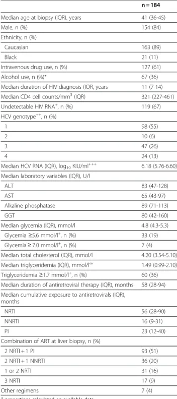
Between January 1995 and January 2008, 250 HIV-HCV coinfected patients who underwent a liver biopsy and fulfilled all inclusion criteria were evaluated. Sixty-six patients were not analyzed for the following reasons cor-responding to exclusion criteria: hepatitis B coinfection (n = 28), loss of medical records (n = 8) or no ART at the time of biopsy (n = 30). Therefore, 184 patients were included. Demographic and biological characteristics are summarized in Table 1.
No correlation was observed between the duration of HIV infection and CD4 cell count in this cohort of ART-treated patients. HIV plasma viral load was undetectable (<400 copies/ml) in 119 out of the 178 (67%) evaluable patients and median HIV viral load on average was 3.93 log10copies/ml for the remaining 33% patients. The
me-dian HCV viral load was 6.18 log10 IU/ml (IQR
5.76-6.60) in the 148 (80%) patients with quantificative HCV values. The other patients (n = 36) had only a positive HCV RNA without quantification.
All patients had been exposed to a nucleoside reverse transcriptase inhibitor (NRTI), including zidovudine (n = 144), lamivudine (n = 161), stavudine (n = 114), zalcita-bine (n = 22), didanosine (n = 105), tenofovir (n = 24) and abacavir (n = 24); 126 patients had received protease inhibi-tors (PI), including ritonavir as boosted-PI (n = 59), indina-vir (n = 68), nelfinaindina-vir (n = 47), saquinaindina-vir (n = 16), lopinaindina-vir (n = 17) and atazanavir (n = 8); and 79 patients had received
Table 1 Characteristics of HIV-HCV coinfected patients treated with antiretroviral therapy at the time of liver biopsy (parameters collected the day of the liver biopsy or during the week before)
n = 184 Median age at biopsy (IQR), years 41 (36-45) Male, n (%) 154 (84) Ethnicity, n (%)
Caucasian 163 (89)
Black 21 (11)
Intravenous drug use, n (%) 127 (61) Alcohol use, n (%)* 67 (36) Median duration of HIV diagnosis (IQR, years 11 (7-14) Median CD4 cell counts/mm3(IQR) 321 (227-461) Undetectable HIV RNA+, n (%) 119 (67)
HCV genotype++, n (%)
1 98 (55)
2 10 (6)
3 47 (26)
4 24 (13)
Median HCV RNA (IQR), log10KIU/ml+++ 6.18 (5.76-6.60)
Median laboratory variables (IQR), U/l
ALT 83 (47-128)
AST 65 (43-97)
Alkaline phosphatase 89 (71-113) GGT 80 (42-160) Median glycemia (IQR), mmol/l 4.8 (4.3-5.3)
Glycemia ≥5.6 mmol/l+, n (%) 33 (19) Glycemia ≥ 7.0 mmol/l+, n (%) 7 (4)
Median total cholesterol (IQR), mmol/l 4.20 (3.54-5.10) Median triglyceridemia (IQR), mmol/l°° 1.49 (0.99-2.10) Triglyceridemia ≥1.7 mmol/l+, n (%) 60 (36)
Median duration of antiretroviral therapy (IQR), months 58 (28-94) Median cumulative exposure to antiretrovirals (IQR),
months
NRTI 56 (28-90) NNRTI 16 (9-31)
PI 23 (12-40)
Combination of ART at liver biopsy, n (%)
2 NRTI + 1 PI 93 (51) 2 NRTI + 1 NNRTI 36 (20) 1 or 2 NRTI 31 (16) 3 NRTI 17 (9) Other regimens 7 (4) +
proportions calculated on available data.
+Data missing for 6 patients;++Data missing for 5 patients;+++Data missing
for 36 patients; ° Data missing for 11 patients; °° Data missing for 18 patients.
*Alcohol: no quantification was available.
Abbreviations: ALT, alanine transaminase; AST, aspartate aminotransferase; GGT,
gamma-glutamyl transpeptidase; IQR, interquartile range; NNRTI, non-nucleoside reverse transcriptase inhibitor; NRTI, non-nucleoside reverse transcriptase inhibitor; PI, protease inhibitor.
Martinez et al. BMC Research Notes 2012, 5:180 Page 3 of 10 http://www.biomedcentral.com/1756-0500/5/180
non-nucleoside reverse transcriptase inhibitors (NNRTI), including efavirenz (n = 51) and nevirapine (n = 44).
Histologic findings
Overall, 102 (55%) out of the 184 patients had HS. Steatosis was grade 1 in 76 (41%) patients, grade 2 in 10 (5%), and 3 in 16 (9%). Macrovesicular fatty changes were observed in 56 patients (55%), microvesicular in 10 (10%), and mixed form in 36 (35%). Fibrosis was present in 163 patients (89%) with METAVIR F1 score in 66 (36%), F2 in 53 (29%), F3 in 39 (21%) and F4 in 4 (2%). Necroinflammatory activ-ity was detected in 164 out of the 184 (89%) patients with A1 in 109 (59%), A2 in 50 (27%) and A3 in 5 (3%).
Factors associated with hepatic steatosis
Comparison of parameters in patients with or without HS are presented in Table 2. In univariate analysis, intravenous drug use (p = 0.05) and HCV genotype 3 (p = 0.005) were strongly linked with HS. The median HCV viral load was
significantly higher in patients with HS compared with those without (p = 0.003). HS was also associated with increased levels of ALT, AST and decreased levels of serum total cholesterol, but not with triglyceridemia nor available parameters of metabolic syndrome (only 7 patients have a glycemia >7 mmol/l with mild steatosis for 6 patients and 1 had no steatosis) (Table 2).
Also, there was no association with CD4 cell count or an undetectable HIV viral load. Similar results were found in patients with mild versus severe steatosis.
HS was also associated with fibrosis (Figure 1). Thus, the frequency of HS increased with the stages of fibrosis, with 76%, 54%, 32%, and 28% of patients without steato-sis having fibrosteato-sis scores of F0, F1, F2, and F3–F4, re-spectively (Cochran-Armitage trend test, p < 0.0001). However, the severity of steatosis was not different among patients presenting fibrosis scoring F1, F2, or F3–F4. No association was observed between HS and necroinflammatory activity.
Table 2 Comparison of various parameters in patients with mild (<33% of hepatocytes affected) or severe (>33% of hepatocytes affected) steatosis and those without steatosis (univariate analysis)
No steatosis (n = 82) Steatosis (n = 102) Mild steatosis (n = 76) Severe steatosis (n = 26) pa pb pc Median age (IQR), yrs 40 (36-45) 41 (37-45) 41 (36-45) 41 (37-48) 0.38 0.66 0.81 Male gender, n (%) 66 (80) 88 (86) 67 (88) 21 (81) 0.32 0.39 0.34 IVDU, n (%) 50 (62) 77 (76) 54 (71) 23 (88) 0.05 0.03 0.11 Alcohol use, n (%) 29 (35) 38 (37) 29 (38) 9 (35) 0.88 0.92 0.82 Median duration of HIV (IQR), yrs 10 (5-14) 12 (8-14) 12 (8-15) 11 (7-13) 0.17 0.23 0.30 Median CD4 cell count (IQR),
/mm3 346 (228-560) 308 (227-423) 305 (227-435) 313 (191-398) 0.10 0.24 0.67
HIV RNA < 400 copies/ml, n (%) 58 (72) 61 (63) 46 (62) 15 (65) 0.26 0.45 1.0 HCV genotype 3, n (%) 13 (16) 34 (35) 18 (25) 16 (64) 0.005 <.0001 <.0001 Median HCV RNA (IQR), logIU/ml 5.97 (5.65-6.50) 6.30 (6.00-6.72) 6.22 (5.80-6.64) 6.69 (6.27-7.06) 0.003 0.0003 0.004 Median ALT (IQR), U/l 67 (40-119) 87 (54-130) 83 (51-121) 113 (84-132) 0.04 0.01 0.03 Median AST (IQR), U/l 56 (37-78) 77.5 (50-104) 70 (48-102) 78 (59-105) 0.001 0.003 0.33 Median alkaline phosphase (IQR),
U/l
90 (70-115) 89 (69-112) 90 (72-113) 80 (65-95) 0.46 0.27 0.15 Median GGT (IQR), U/l 71 (41-139) 87 (43-196) 92 (44-198) 73 (30-180) 0.09 0.09 0.19 Median glycemia (IQR), mmol/l 4.80 (4.30-5.45) 4.92 (4.30-5.22) 4.80 (4.30-5.20) 4.90 (4.35-5.26) 0.79 0.96 0.97 Glycemia ≥5.6 mmol/l, n (%) 16 (21) 17 (17) 12 (16) 5 (21) 0.56 0.77 0.76 Glycemia ≥7.0 mmol/l, n (%) 1 (1) 6 (6) 6 (8) 0 (0) 0.14 0.08 0.33 Median total cholesterol (IQR),
mmol/l
4.40 (3.73-5.33) 4.00 (3.30-4.79) 4.01 (3.30-5.03) 3.72 (3.06-4.37) 0.01 0.02 0.30 Median triglycerides (IQR),
mmol/l
1.47 (0.97-2.08) 1.51 (0.99-2.20) 1.53 (1.04-2.24) 1.23 (0.90-1.74) 0.86 0.51 0.28 Triglycerides ≥1.7 mmol/l, n (%) 30 (40) 30 (33) 23 (33) 7 (29) 0.33 0.56 0.80 Fibrosis F3-F4, n (%) 12 (15) 31 (30) 25 (33) 6 (23) 0.01 <0.001 1.0 Metavir score activity A2/A3,
n (%)
21 (26) 34 (33 ) 29 (38 ) 5 (19 ) 0.28 0.11 0.09
paComparison of patients with versus without steatosis (Kruskal-Wallis test for quantitative variables, χ2test or Fisher's exact test for qualitative variables).
pbComparison of patients with mild or severe steatosis versus without steatosis (Kruskal-Wallis test for quantitative variables, χ2test for qualitative variables).
pcComparison of patients with mild versus severe steatosis (Kruskal-Wallis/Wilcoxon test for quantitative variables, χ2test or Fisher's exact test for qualitative
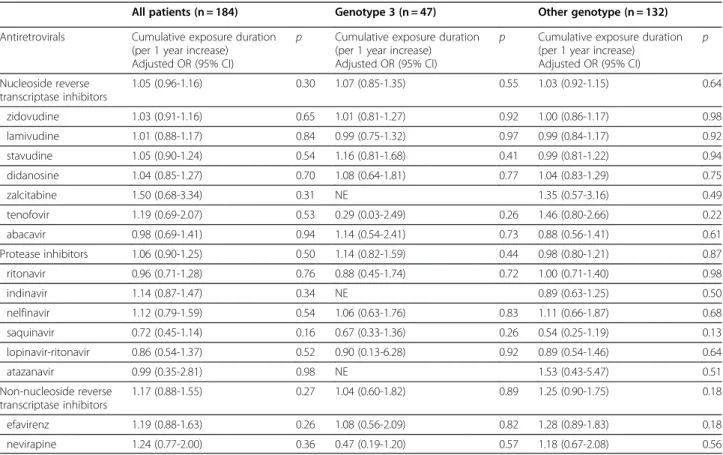
After adjustment for the period of biopsy, neither the type of ART nor the duration of exposure to a specific antiretroviral agent or class of antiretroviral was related to HS (Table 3). The same results were observed in
separate analyses among patients presenting genotype 3, or another genotype (Tables 4 and 5).
In the multivariate analysis, only two independent fac-tors remained associated with an increased risk of HS: HCV genotype 3 (odds ratio [OR], 2.6; 95% confidence intervals [CI], 1.1-6.3), and HCV RNA load (OR, 2.2 per 1 log higher; 95% CI, 1.2-3.8). Using a multivariate poly-tomous logistic regression model, HCV genotype 3 and HCV RNA load were moderately associated with mild steatosis and strongly associated with severe steatosis (Table 6).
Discussion
Hepatic steatosis has emerged as a major comorbidity in HIV-HCV coinfected patients [13,35]. In this retrospect-ive observational study of 184 HIV-HCV coinfected patients ART-treated but untreated for HCV at the time of liver biopsy, HS was present in about half patients (55%), which is similar to rates of 24-75% found in other studies of HIV-HCV coinfected and HCV monoinfected patients [12,14-23,36]. In multivariate analysis, HS and its severity were only significantly associated with HCV genotype 3 and HCV viral load. Neither the type of ART, nor their prolonged duration of exposure with a median of near five years were related to steatosis. Moreover,
0 20 40 60 80 100 % F0 F1 F2 F3−F4 Fibrosis None Mild Severe Steatosis
Figure 1 Distribution of steatosis according to fibrosis index in 184 HIV-HCV coinfected patients treated with antiretroviral therapy. Steatosis is defined as mild (<33% of hepatocytes affected) or severe (>33% of hepatocytes affected).
Table 3 The effects on steatosis of exposure to specific antiretroviral medication in 184 HIV-HCV coinfected patients
Antiretrovirals n Exposure to ARV Adjusted OR° (95% CI)
p Cumulative exposure duration (per 1 year increase)
Adjusted OR (95% CI) p Nucleoside reverse transcriptase inhibitors 184 NE 1.05 (0.96–1.16) 0.30 zidovudine 144 1.11 (0.54-2.30) 0.77 1.03 (0.91-1.16) 0.65 lamivudine 161 1.10 (0.44-2.77) 0.82 1.01 (0.88-1.17) 0.84 stavudine 114 0.96 (0.51-1.78) 0.88 1.05 (0.90-1.24) 0.54 didanosine 105 1.20 (0.66-2.18) 0.57 1.04 (0.85-1.27) 0.70 zalcitabine 22 1.68 (0.60-4.72) 0.33 1.50 (0.68-3.34) 0.31 tenofovir 24 0.87 (0.33-2.27) 0.78 1.19 (0.69-2.07) 0.53 abacavir 24 0.55 (0.22-1.39) 0.20 0.98 (0.69-1.41) 0.94 Protease inhibitors 126 1.15 (0.59-2.24) 0.67 1.06 (0.90-1.25) 0.50 ritonavir 55 0.90 (0.46-1.75) 0.75 0.96 (0.71-1.28) 0.76 indinavir 68 0.94 (0.51-1.74) 0.84 1.14 (0.87-1.47) 0.34 nelfinavir 47 0.96 (0.48-1.89) 0.91 1.12 (0.79-1.59) 0.54 saquinavir 31 0.91 (0.41-2.00) 0.81 0.72 (0.45-1.14) 0.16 lopinavir-ritonavir 17 0.55 (0.19-1.63) 0.28 0.86 (0.54-1.37) 0.52 atazanavir 8 0.76 (0.17-3.43) 0.72 0.99 (0.35-2.81) 0.98 Non-nucleoside reverse transcriptase inhibitors 79 0.93 (0.50-1.74) 0.81 1.17 (0.88-1.55) 0.27 efavirenz 51 1.34 (0.67-2.69) 0.40 1.19 (0.88-1.63) 0.26 nevirapine 44 0.67 (0.33-1.35) 0.26 1.24 (0.77-2.00) 0.36
° Adjusted for period of liver biopsy.
Abbreviations: OR, odds ratio; CI, confidence intervals; NE, not estimable.
Martinez et al. BMC Research Notes 2012, 5:180 Page 5 of 10 http://www.biomedcentral.com/1756-0500/5/180
ART have no differential effect on occurrence of HS according to the genotype 3 compared to others.
We confirmed previously data of higher HS associated with genotype 3 [23]. Here, the rate of severe HS (14%) was higher in our study compared with 2-9% found in US studies in HIV-HCV coinfected individuals [12], but was similar with rates found in other studies [37,38], in particu-lar with those conducted in France [39,40]. Several reasons may explain these discrepancies. Whereas most of our patients, and those included in the study of Bauerle et al. [37], were Caucasian and carrying HCV genotype 3, other studies have included a high proportion of Africo-Ameri-can patients (47-94%) and patients infected with HCV genotype 1 [38-45]. It is well-known that HCV-infected Black people have a lower prevalence of HS than Cauca-sian [41,45-47], probably related to lower visceral adipose tissue [16,22].
HS is a frequent histological finding in patients with chronic hepatitis C virus infection, particularly among those infected with genotype 3 strain [24,48,49]. The preva-lence of 26% of genotype 3 in our study was similar to the prevalence of 18% reported in the French HepaVIH cohort [50]. It has been postulated that genotype 1 is associated with “metabolic” steatosis rather than “viral” steatosis developed through a direct cytopathologic effect observed especially in genotype 3 infected patients [51-56]. When we examined the factors impacting the level of steatosis on
ART patients, genotype 3, and high HCV viral load were two independent factors associated with HS in accordance with previous studies [15,17,19,20]. Both factors moderately increased the risk of mild steatosis, but were strongly asso-ciated with severe steatosis (grade 2 or 3).
Other factors such as greater age [18], higher body mass index (BMI) [12,15-18,20,22,38-40,42,44] [23], hypergly-cemia [12], lower cholesterolemia [17], and presence of lipodystrophy [17] have also been found to be independ-ently associated with steatosis in coinfected patients. In our study, patients with metabolic syndrome were not more likely to present HS. However, only seven patients had diabetes, and this limited sample size could have pre-vent to study this risk factor. Moreover, as expected, ex-posure to PIs and stavudine, were associated with elevated triglycerides (p = 0.08 and p < 0.0001, respectively), but this metabolic abnormality was not a risk factors of HS in our ART-experienced population. According to the lack of data about HOMA scoring, weight and BMI, we probably missed the impact of metabolic steatosis in genotype other than 3 [23,41]. Other limitation of our retrospective study is lack of information about alcohol consumption. Never-theless, as described by Machado et al., metabolic syn-drome and alcohol were not associated to HS. BMI was considered an increasing risk factor but with small magni-tude and diabetes as a possible risk factor with no data for HOMA scoring [16,22]. Moreover, the higher percentage
Table 4 The effects on steatosis of exposure to specific antiretroviral medication in 184 HIV-HCV coinfected patients according to the genotype
All patients (n = 184) Genotype 3 (n = 47) Other genotype (n = 132) Antiretrovirals n Adjusted OR° (95% CI) p Adjusted OR° (95% CI) p Adjusted OR° (95% CI) p
NRTI 184 NE NE NE zidovudine 144 1.11 (0.54–2.30) 0.77 0.91 (0.22-3.82) 0.90 1.42 (0.58-3.49) 0.45 lamivudine 161 1.10 (0.44–2.77) 0.82 0.50 (0.05-5.00) 0.56 1.29 (0.45-3.74) 0.64 stavudine 114 0.96 (0.51–1.78) 0.88 0.58 (0.14-0.47) 0.46 1.05 (0.51-2.17) 0.89 didanosine 105 1.20 (0.66–2.18) 0.57 1.02 (0.27-3.93) 0.97 1.17 (0.58-2.37) 0.66 zalcitabine 22 1.68 (0.60–4.72) 0.33 NE 1.29 (0.41-4.06) 0.66 tenofovir 24 0.87 (0.33–2.27) 0.78 0.15 (0.01-1.80) 0.13 1.44 (0.49-4.22) 0.51 abacavir 24 0.55 (0.22–1.39) 0.20 0.40 (0.07-2.40) 0.32 0.58 (0.18-1.84) 0.35 PI 126 1.15 (0.59–2.24) 0.67 0.19 -0.02-1.76) 0.15 1.46 (0.67-3.15) 0.34 ritonavir 55 0.90 (0.46–1.75) 0.75 0.54 (0.13-2.28) 0.40 1.04 (0.48-2.27) 0.91 indinavir 68 0.94 (0.51–1.74) 0.84 1.13 (0.29-4.41) 0.86 0.81 (0.39-1.68) 0.57 nelfinavir 47 0.96 (0.48–1.89) 0.91 0.37 (0.09-1.56) 0.18 1.19 (0.54-2.66) 0.66 saquinavir 31 0.91 (0.41–2.00) 0.81 0.41 (0.10-1.72) 0.22 0.94 (0.34-2.61) 0.91 lopinavir-ritonavir 17 0.55 (0.19–1.63) 0.28 0.45 (0.02-8.11) 0.59 0.67 (0.20-2.19) 0.50 atazanavir 8 0.76 (0.17–3.43) 0.72 NE 1.70 (0.31-9.24) 0.54 NNRTI 79 0.93 (0.50–1.74) 0.81 0.33 (0.08-1.27) 0.11 1.38 (0.65-2.95) 0.40 efavirenz 51 1.34 (0.67–2.69) 0.40 0.74 (0.17-3.27) 0.69 1.77 (0.79-4.00) 0.16 nevirapine 44 0.67 (0.33–1.35) 0.26 0.34 (0.08-1.38) 0.13 0.80 (0.35-1.83) 0.60
° Adjusted for period of liver biopsy.
of genotype 3 reflect the association between HS and HCV viral parameters.
Many antiretroviral drugs have been associated with hep-atic damage [57-59]. It has been suggested that steatosis due to an accumulation of fatty acids in the hepatocytes could be a consequence of mitochondrial dysfunction, sec-ondary to drugs or viruses inducing oxidative stress [60]. The effect of the drug class and drugs within classes on HS in HIV-HCV coinfected patients remain unclear. Previously, some studies reported no significant association between HS and ART as in our study [12,15,16,18,41]. When focus-ing on the use of stavudine, a medication closely linked with HS and lipodystrophy syndrome resulting from mitochon-drial damage, Sulkowski et al. [45] found that stavudine ex-posure was a risk factor for steatosis like in the study of Borghi et al., whereas no such association was found for this
drug as well as the D-drug group of antiretrovirals (didano-sine, zalcitabine) in other studies [23,38,41,42,61,62]. Re-cently, in a meta-analysis of the risk factors associated with HS in HIV-HCV patients, Machado et al. failed to find any association with antiretrovirals of any class and HS as well as in our study [16,22]. Moreover, Woreta et al., showed in a study including a majority of Black patients (87%) with 94% genotype 1, the lack of association with antiretroviral drugs with a median cumulative drug exposure similar to ours [41]. Despite its relatively small sample size, our study had a statistical power of 80% to detect an increased risk of steatosis of 3 for the antiretroviral drugs used less fre-quently such as abacavir, tenofovir and lopinavir. We could hypothesize that discrepancies with other studies were linked to the differential prevalence of Caucasion subjects, of metabolic characteristics and frequency of genotype 1
Table 6 Results from multivariate polytomous logistic regression analyses of factors associated with mild (<33% of hepatocytes affected ) and severe (>33% of hepatocytes affected) steatosis in 184 HIV-HCV coinfected patients treated with antiretroviral therapy
Mild steatosis AOR (95%CI) p Severe steatosis AOR (95%CI) p HCV genotype 3 1.76 (0.72-4.30) 0.22 19.51 (4.49-84.73) <0.0001 HCV RNA (per 1 log10 increase) 1.80 (1.02-3.16) 0.04 14.86 (3.79-58.22) 0.0001
Abbreviations: AOR, adjusted odds ratio; CI, confidence intervals.
Table 5 The effects on steatosis of cumulative exposure duration to specific antiretroviral medication in 184 HIV-HCV coinfected patients according to the genotype
All patients (n = 184) Genotype 3 (n = 47) Other genotype (n = 132) Antiretrovirals Cumulative exposure duration
(per 1 year increase) Adjusted OR (95% CI)
p Cumulative exposure duration (per 1 year increase) Adjusted OR (95% CI)
p Cumulative exposure duration (per 1 year increase) Adjusted OR (95% CI) p Nucleoside reverse transcriptase inhibitors 1.05 (0.96-1.16) 0.30 1.07 (0.85-1.35) 0.55 1.03 (0.92-1.15) 0.64 zidovudine 1.03 (0.91-1.16) 0.65 1.01 (0.81-1.27) 0.92 1.00 (0.86-1.17) 0.98 lamivudine 1.01 (0.88-1.17) 0.84 0.99 (0.75-1.32) 0.97 0.99 (0.84-1.17) 0.92 stavudine 1.05 (0.90-1.24) 0.54 1.16 (0.81-1.68) 0.41 0.99 (0.81-1.22) 0.94 didanosine 1.04 (0.85-1.27) 0.70 1.08 (0.64-1.81) 0.77 1.04 (0.83-1.29) 0.75 zalcitabine 1.50 (0.68-3.34) 0.31 NE 1.35 (0.57-3.16) 0.49 tenofovir 1.19 (0.69-2.07) 0.53 0.29 (0.03-2.49) 0.26 1.46 (0.80-2.66) 0.22 abacavir 0.98 (0.69-1.41) 0.94 1.14 (0.54-2.41) 0.73 0.88 (0.56-1.41) 0.61 Protease inhibitors 1.06 (0.90-1.25) 0.50 1.14 (0.82-1.59) 0.44 0.98 (0.80-1.21) 0.87 ritonavir 0.96 (0.71-1.28) 0.76 0.88 (0.45-1.74) 0.72 1.00 (0.71-1.40) 0.98 indinavir 1.14 (0.87-1.47) 0.34 NE 0.89 (0.63-1.25) 0.50 nelfinavir 1.12 (0.79-1.59) 0.54 1.06 (0.63-1.76) 0.83 1.11 (0.66-1.87) 0.68 saquinavir 0.72 (0.45-1.14) 0.16 0.67 (0.33-1.36) 0.26 0.54 (0.25-1.19) 0.13 lopinavir-ritonavir 0.86 (0.54-1.37) 0.52 0.90 (0.13-6.28) 0.92 0.89 (0.54-1.46) 0.64 atazanavir 0.99 (0.35-2.81) 0.98 NE 1.53 (0.43-5.47) 0.51 Non-nucleoside reverse transcriptase inhibitors 1.17 (0.88-1.55) 0.27 1.04 (0.60-1.82) 0.89 1.25 (0.90-1.75) 0.18 efavirenz 1.19 (0.88-1.63) 0.26 1.08 (0.56-2.09) 0.82 1.28 (0.89-1.83) 0.18 nevirapine 1.24 (0.77-2.00) 0.36 0.47 (0.19-1.20) 0.57 1.18 (0.67-2.08) 0.56
° Adjusted for period of liver biopsy.
Abbreviations: OR, odds ratio; CI, confidence intervals; NE, not estimable.
Martinez et al. BMC Research Notes 2012, 5:180 Page 7 of 10 http://www.biomedcentral.com/1756-0500/5/180
and 3. Borghi et al. evocated a putative role of ART in the occurrence of HS in patients infected with genotypes other than 3 [23] but in our study, the impact of ART on HS was not different between genotype 3 and the others.
In conclusion, for our Caucasian cohort predomin-antly infected with genotype-HCV 1, hepatic steatosis in HIV-HCV coinfected patients receiving antiretroviral therapy is associated with HCV-related factors particu-larly in genotype 3 patients but not antiretrovirals. Nevertheless, as showed in our study, ART seems play a minor role in HS since the choose and use of more "metabolically friendly" antiretroviral drugs. Overall, we found only viral parameters, HCV genotype 3, and HCV RNA value, which were strongly associated with HS, particularly a severe steatosis. Among HIV-HCV co-infected patients receiving ART and who had never been treated for HCV, neither the type of drugs nor the dur-ation of exposure was related to HS whatever the genotype.
Abbreviations
HIV: human immunodeficiency virus; HCV: hepatitis C virus; ART: antiretroviral therapy; ALT: alanine aminotransferase; AST: aspartate aminotransferase; GGT: gammaglutamyl transferase; IQR: interquartile range; NRTI: nucleoside reverse transcriptase inhibitor; PI: protease inhibitor; NNRTI: non-nucleoside reverse transcriptase inhibitor; OR: odds ratio; CI: confidence intervals; IVDU: intravenous drug use; AOR: adjusted odds ratio; NE: not estimable.
Competing interests
The authors declare that they have no competing interests. Acknowledgements
English language assistance for the preparation of this manuscript was provided by Andrea Bothwell of inScience Communications. Author details
1Service de Médecine Interne et Immunologie Clinique, Assistance
Publique-Hôpitaux de Paris, INSERM UMR_S 996, Université Paris Sud, Hôpital Antoine Béclère, 157, rue de la Porte de Trivaux, 92141, Clamart, France.2Service des
Maladies Infectieuses et Tropicales, Hôpital Pitié-Salpêtrière, Université Pierre et Marie Curie, APHP, 45/83 Boulevard de l’Hôpital, 75013, Paris, France.
3Departement of Infectious Disease, Hanoi Medical University, 01, Ton That
Tung Street, Hanoi, Vietnam.4Departement of Internal Medicine, Hospital National Iranian oil company, Hafez Avenue, Tehran, Iran.5INSERM U943,
UPMC Univ Paris 06, UMR S943, Paris, F-75013, France.6Service de Maladies Infectieuses et Tropicales, 103 Grande-Rue de la Croix-Rousse, Hôpital de la Croix-Rousse, Hospices Civils de Lyon, 69317 Lyon cedex 04, Paris, France.
7Service d’Anatomopathologie, Hôpital Pitié-Salpêtrière, Université Pierre et
Marie Curie, APHP, 45/83 Boulevard de l’Hôpital, 75013, Paris, France.8Service
d’Anatomopathologie, Hôpital Saint-Louis, Université Denis Diderot, APHP, 1, avenue Claude Vellefaux, 75010, Paris, France.9Service des Maladies
Infectieuses et Tropicales, Hôpital Saint-Louis, Université Denis Diderot, APHP, 1, avenue Claude Vellefaux, 75010, Paris, France.
Authors' contributions
VM conceived the study, collected data of patients, participated in its design and coordination and drafted the manuscript. TDNT and ZM collected the data and helped to draft the manuscript. MG made the statistical analysis and helped to draft the manuscript. PM and MAV helped to draft the manuscript. FC and PB made the histological analysis of liver biopsy. CK and EC participated in the design of the study and helped to draft the manuscript. All authors read and approved the final manuscript.
Financial disclosure
The authors have no commercial links or other associations that might pose a conflict of interest (e.g. pharmaceutical stock ownership, consultancy, advisory board membership, relevant patents, or research funding) relevant to this study.
Statement naming sources of financial support (including grant numbers)
None
Received: 24 October 2011 Accepted: 10 April 2012 Published: 10 April 2012
References
1. Martin-Carbonero L, Benhamou Y, Puoti M, Berenguer J, Mallolas J, Quereda C, Arizcorreta A, Gonzalez A, Rockstroh J, Asensi V et al: Incidence and predictors of severe liver fibrosis in human immunodeficiency virus-infected patients with chronic hepatitis C: a European collaborative study. Clin Infect Dis 2004, 38(1):128–133. Epub 2003 Dec 2008.
2. Mehta SH, Thomas DL, Torbenson M, Brinkley S, Mirel L, Chaisson RE, Moore RD, Sulkowski MS: The effect of antiretroviral therapy on liver disease among adults with HIV and hepatitis C coinfection. Hepatology 2005, 41(1):123–131. 3. Qurishi N, Kreuzberg C, Luchters G, Effenberger W, Kupfer B, Sauerbruch T,
Rockstroh JK, Spengler U: Effect of antiretroviral therapy on liver-related mortality in patients with HIV and hepatitis C virus coinfection. Lancet 2003, 362(9397):1708–1713.
4. Lauer GM, Walker BD: Hepatitis C virus infection. N Engl J Med 2001, 345 (1):41–52.
5. Graham CS, Baden LR, Yu E, Mrus JM, Carnie J, Heeren T, Koziel MJ: Influence of human immunodeficiency virus infection on the course of hepatitis C virus infection: a meta-analysis. Clin Infect Dis 2001, 33(4):562–569.
6. Macias J, Castellano V, Merchante N, Palacios RB, Mira JA, Saez C, Garcia-Garcia JA, Lozano F, Gomez-Mateos JM, Pineda JA: Effect of antiretroviral drugs on liver fibrosis in HIV-infected patients with chronic hepatitis C: harmful impact of nevirapine. AIDS 2004, 18(5):767–774.
7. Qurishi N, Kreuzberg C, Luchters G, Effenberger W, Kupfer B, Sauerbruch T, Rockstroh JK, Spengler U: Effect of antiretroviral therapy on liver-related mortality in patients with HIV and hepatitis C virus coinfection. Lancet 2003, 362(9397):1708–1713.
8. Mohsen AH, Easterbrook PJ, Taylor C, Portmann B, Kulasegaram R, Murad S, Wiselka M, Norris S: Impact of human immunodeficiency virus (HIV) infection on the progression of liver fibrosis in hepatitis C virus infected patients. Gut 2003, 52(7):1035–1040.
9. Soriano V, Martin-Carbonero L, Garcia-Samaniego J, Puoti M: Mortality due to chronic viral liver disease among patients infected with human immunodeficiency virus. Clin Infect Dis 2001, 33(10):1793–1795. 10. Thein HH, Yi Q, Dore GJ, Krahn MD: Natural history of hepatitis C virus
infection in HIV-infected individuals and the impact of HIV in the era of highly active antiretroviral therapy: a meta-analysis. AIDS 2008, 22 (15):1979–1991.
11. Bica I, McGovern B, Dhar R, Stone D, McGowan K, Scheib R, Snydman DR: Increasing mortality due to end-stage liver disease in patients with human immunodeficiency virus infection. Clin Infect Dis 2001, 32(3):492–497. 12. Sulkowski MS, Mehta SH, Torbenson M, Afdhal NH, Mirel L, Moore RD,
Thomas DL: Hepatic steatosis and antiretroviral drug use among adults coinfected with HIV and hepatitis C virus. AIDS 2005, 19(6):585–592. 13. Pol S, Lebray P, Vallet-Pichard A: HIV infection and hepatic enzyme
abnormalities: intricacies of the pathogenic mechanisms. Clin Infect Dis 2004, 38(Suppl 2):S65–S72.
14. Sterling RK, Contos MJ, Smith PG, Stravitz RT, Luketic VA, Fuchs M, Shiffman ML, Sanyal AJ: Steatohepatitis: Risk factors and impact on disease severity in human immunodeficiency virus/hepatitis C virus coinfection. Hepatology 2008, 47(4):1118–1127.
15. Bani-Sadr F, Carrat F, Bedossa P, Piroth L, Cacoub P, Perronne C, Degott C, Pol S: Hepatic steatosis in HIV-HCV coinfected patients: analysis of risk factors. AIDS 2006, 20(4):525–531.
16. Gaslightwala I, Bini EJ: Impact of human immunodeficiency virus infection on the prevalence and severity of steatosis in patients with chronic hepatitis C virus infection. J Hepatol 2006, 44(6):1026–1032.
17. Marks KM, Petrovic LM, Talal AH, Murray MP, Gulick RM, Glesby MJ: Histological findings and clinical characteristics associated with hepatic steatosis in patients coinfected with HIV and hepatitis C virus. J Infect Dis 2005, 192(11):1943–1949. Epub 2005 Nov 1942.
18. Monto A, Dove LM, Bostrom A, Kakar S, Tien PC, Wright TL: Hepatic steatosis in HIV/hepatitis C coinfection: prevalence and significance compared with hepatitis C monoinfection. Hepatology 2005, 42(2):310–316.
19. McGovern BH, Ditelberg JS, Taylor LE, Gandhi RT, Christopoulos KA, Chapman S, Schwartzapfel B, Rindler E, Fiorino AM, Zaman MT, et al: Hepatic steatosis is associated with fibrosis, nucleoside analogue use, and hepatitis C virus genotype 3 infection in HIV-seropositive patients. Clin Infect Dis 2006, 43(3):365–372.
20. Neau D, Winnock M, Castera L, Bail BL, Loko MA, Geraut L, Dupon M, Ragnaud JM, Lacoste D, Lafon ME, et al: Prevalence of and factors associated with hepatic steatosis in patients coinfected with hepatitis C virus and HIV: Agence Nationale pour la Recherche contre le SIDA et les hepatites virales CO3 Aquitaine Cohort. J Acquir Immune Defic Syndr 2007, 45(2):168–173. 21. Sanchez-Conde M, Berenguer J, Miralles P, Alvarez F, Carlos Lopez J, Cosin J,
Pilar C, Ramirez M, Gutierrez I, Alvarez E: Liver biopsy findings for HIV-infected patients with chronic hepatitis C and persistently normal levels of alanine aminotransferase. Clin Infect Dis 2006, 43(5):640–644. 22. Castera L, Loko MA, Le Bail B, Coffie P, De Ledinghen V, Trimoulet P,
Winnock M, Dabis F, Neau D: Hepatic steatosis in HIV-HCV coinfected patients in France: comparison with HCV monoinfected patients matched for body mass index and HCV genotype. Aliment Pharmacol Ther 2007, 26(11–12):1489–1498.
23. Borghi V, Puoti M, Mussini C, Bellelli S, Angeletti C, Sabbatini F, Prati F, Cossarizza A, Esposito R: HIV coinfection and antiretroviral therapy enhances liver steatosis in patients with hepatitis C, but only in those infected by HCV genotype other than 3. Antivir Ther 2008, 13(8):1057–1065.
24. Monto A, Alonzo J, Watson JJ, Grunfeld C, Wright TL: Steatosis in chronic hepatitis C: relative contributions of obesity, diabetes mellitus, and alcohol. Hepatology 2002, 36(3):729–736.
25. Sanyal AJ: Review article: non-alcoholic fatty liver disease and hepatitis C–risk factors and clinical implications. Aliment Pharmacol Ther 2005, 22 (Suppl 2):48–51.
26. Hourigan LF, Macdonald GA, Purdie D, Whitehall VH, Shorthouse C, Clouston A, Powell EE: Fibrosis in chronic hepatitis C correlates significantly with body mass index and steatosis. Hepatology 1999, 29(4):1215–1219. 27. Hu KQ, Kyulo NL, Esrailian E, Thompson K, Chase R, Hillebrand DJ, Runyon
BA: Overweight and obesity, hepatic steatosis, and progression of chronic hepatitis C: a retrospective study on a large cohort of patients in the United States. J Hepatol 2004, 40(1):147–154.
28. Sanyal AJ, Contos MJ, Sterling RK, Luketic VA, Shiffman ML, Stravitz RT, Mills AS: Nonalcoholic fatty liver disease in patients with hepatitis C is associated with features of the metabolic syndrome. Am J Gastroenterol 2003, 98(9):2064–2071.
29. Marks KM, Petrovic LM, Talal AH, Murray MP, Gulick RM, Glesby MJ: Histological findings and clinical characteristics associated with hepatic steatosis in patients coinfected with HIV and hepatitis C virus. J Infect Dis 2005, 192(11):1943–1949.
30. Brinkman K: Editorial response: hyperlactatemia and hepatic steatosis as features of mitochondrial toxicity of nucleoside analogue reverse transcriptase inhibitors. Clin Infect Dis 2000, 31(1):167–169.
31. Balasubramanyam A, Sekhar RV, Jahoor F, Jones PH, Pownall HJ: Pathophysiology of dyslipidemia and increased cardiovascular risk in HIV lipodystrophy: a model of 'systemic steatosis'. Curr Opin Lipidol 2004, 15(1):59–67. 32. Ogedegbe AE, Thomas DL, Diehl AM: Hyperlactataemia syndromes
associated with HIV therapy. Lancet Infect Dis 2003, 3(6):329–337. 33. Brunt EM, Janney CG, Di Bisceglie AM, Neuschwander-Tetri BA, Bacon BR:
Nonalcoholic steatohepatitis: a proposal for grading and staging the histological lesions. Am J Gastroenterol 1999, 94(9):2467–2474. 34. The French METAVIR Cooperative Study Group: Intraobserver and
interobserver variations in liver biopsy interpretation in patients with chronic hepatitis C. Hepatology 1994, 20(1 Pt 1):15–20.
35. Tien PC, Grunfeld C: The fatty liver in AIDS. Semin Gastrointest Dis 2002, 13 (1):47–54.
36. Bauerle J, Miquel R, Laguno M, Mallolas J, Murillas J, Gatell JM, Walker UA: Hepatic steatosis with stavudine in HIV/hepatitis C virus co-infection. AIDS 2005, 19(13):1441–1442.
37. Bauerle J, Miquel R, Laguno M, Mallolas J, Murillas J, Gatell JM, Walker UA: Hepatic steatosis with stavudine in HIV/hepatitis C virus co-infection. AIDS 2005, 19(13):1441–1442.
38. Gaslightwala I, Bini EJ: Impact of human immunodeficiency virus infection on the prevalence and severity of steatosis in patients with chronic hepatitis C virus infection. J Hepatol 2006, 44(6):1026–1032.
39. Bani-Sadr F, Carrat F, Bedossa P, Piroth L, Cacoub P, Perronne C, Degott C, Pol S: Hepatic steatosis in HIV-HCV coinfected patients: analysis of risk factors. AIDS 2006, 20(4):525–531.
40. Castera L, Loko MA, Le Bail B, Coffie P, De Ledinghen V, Trimoulet P, Winnock M, Dabis F, Neau D: Hepatic steatosis in HIV-HCV coinfected patients in France: comparison with HCV monoinfected patients matched for body mass index and HCV genotype. Aliment Pharmacol Ther 2007, 26(11–12):1489–1498.
41. Woreta TA, Sutcliffe CG, Mehta SH, Brown TT, Higgins Y, Thomas DL, Torbenson MS, Moore RD, Sulkowski MS: Incidence and risk factors for steatosis progression in adults coinfected with HIV and hepatitis C virus. Gastroenterology 2011, 140(3):809–817.
42. Marks KM, Petrovic LM, Talal AH, Murray MP, Gulick RM, Glesby MJ: Histological findings and clinical characteristics associated with hepatic steatosis in patients coinfected with HIV and hepatitis C virus. J Infect Dis 2005, 192(11):1943–1949. Epub 2005 Nov 1942.
43. Monto A, Dove LM, Bostrom A, Kakar S, Tien PC, Wright TL: Hepatic steatosis in HIV/hepatitis C coinfection: prevalence and significance compared with hepatitis C monoinfection. Hepatology 2005, 42(2):310–316.
44. Neau D, Winnock M, Castera L, Bail BL, Loko MA, Geraut L, Dupon M, Ragnaud JM, Lacoste D, Lafon ME, et al: Prevalence of and factors associated with hepatic steatosis in patients coinfected with hepatitis C virus and HIV: Agence Nationale pour la Recherche contre le SIDA et les hepatites virales CO3 Aquitaine Cohort. J Acquir Immune Defic Syndr 2007, 45(2):168–173.
45. Sulkowski MS, Mehta SH, Torbenson M, Afdhal NH, Mirel L, Moore RD, Thomas DL: Hepatic steatosis and antiretroviral drug use among adults coinfected with HIV and hepatitis C virus. AIDS 2005, 19(6):585–592. 46. Browning JD, Szczepaniak LS, Dobbins R, Nuremberg P, Horton JD, Cohen JC,
Grundy SM, Hobbs HH: Prevalence of hepatic steatosis in an urban population in the United States: impact of ethnicity. Hepatology 2004, 40(6):1387–1395. 47. Martin-Carbonero L, Soriano V: Interplay between hepatitis C, liver
steatosis and antiretroviral therapy in HIV-infected patients. AIDS 2005, 19 (6):621–623.
48. Castera L, Hezode C, Roudot-Thoraval F, Lonjon I, Zafrani ES, Pawlotsky JM, Dhumeaux D: Effect of antiviral treatment on evolution of liver steatosis in patients with chronic hepatitis C: indirect evidence of a role of hepatitis C virus genotype 3 in steatosis. Gut 2004, 53(3):420–424. 49. Kumar D, Farrell GC, Fung C, George J: Hepatitis C virus genotype 3 is
cytopathic to hepatocytes: Reversal of hepatic steatosis after sustained therapeutic response. Hepatology 2002, 36:(5)1266–1272.
50. Loko MA, Salmon D, Carrieri P, Winnock M, Mora M, Merchadou L, Gillet S, Pambrun E, Delaune J, Valantin MA, et al: The French national prospective cohort of patients co-infected with HIV and HCV (ANRS CO13 HEPAVIH): early findings, 2006–2010. BMC Infect Dis 2010, 10:303.
51. Adinolfi LE, Gambardella M, Andreana A, Tripodi MF, Utili R, Ruggiero G: Steatosis accelerates the progression of liver damage of chronic hepatitis C patients and correlates with specific HCV genotype and visceral obesity. Hepatology 2001, 33(6):1358–1364.
52. Castera L, Chouteau P, Hezode C, Zafrani ES, Dhumeaux D, Pawlotsky JM: Hepatitis C virus-induced hepatocellular steatosis. Am J Gastroenterol 2005, 100(3):711–715.
53. Clement S, Negro F: Hepatitis C virus: the viral way to fatty liver. J Hepatol 2007, 46(6):985–987.
54. Hezode C, Roudot-Thoraval F, Zafrani ES, Dhumeaux D, Pawlotsky JM: Different mechanisms of steatosis in hepatitis C virus genotypes 1 and 3 infections. J Viral Hepat 2004, 11(5):455–458.
55. Patton HM, Patel K, Behling C, Bylund D, Blatt LM, Vallee M, Heaton S, Conrad A, Pockros PJ, McHutchison JG: The impact of steatosis on disease progression and early and sustained treatment response in chronic hepatitis C patients. J Hepatol 2004, 40(3):484–490.
56. Rubbia-Brandt L, Quadri R, Abid K, Giostra E, Male PJ, Mentha G, Spahr L, Zarski JP, Borisch B, Hadengue A, et al: Hepatocyte steatosis is a cytopathic effect of hepatitis C virus genotype 3. J Hepatol 2000, 33(1):106–115. Martinez et al. BMC Research Notes 2012, 5:180 Page 9 of 10 http://www.biomedcentral.com/1756-0500/5/180
57. Fortgang IS, Belitsos PC, Chaisson RE, Moore RD: Hepatomegaly and steatosis in HIV-infected patients receiving nucleoside analog antiretroviral therapy. Am J Gastroenterol 1995, 90(9):1433–1436. 58. Sulkowski MS, Mehta SH, Chaisson RE, Thomas DL, Moore RD: Hepatotoxicity
associated with protease inhibitor-based antiretroviral regimens with or without concurrent ritonavir. AIDS 2004, 18(17):2277–2284.
59. Y B: Systemic overview of HAART- associated liver enzyme elevations in patients infected with HIV and co-infected with HCV. In: 13 th Conference on Retroviruses and Opportunistic Infections 2006. Denver, CO, US; 2006. 60. Day CP, James OF: Steatohepatitis: a tale of two "hits"?. Gastroenterology
1998, 114(4):842–845.
61. Bauerle J, Laguno M, Mauss S, Mallolas J, Murillas J, Miquel R, Schmutz G, Setzer B, Gatell JM, Walker UA: Mitochondrial DNA depletion in liver tissue of patients infected with hepatitis C virus: contributing effect of HIV infection?. HIV Med 2005, 6(2):135–139.
62. Monto A, Alonzo J, Watson JJ, Grunfeld C, Wright TL: Steatosis in chronic hepatitis C: relative contributions of obesity, diabetes mellitus, and alcohol. Hepatology 2002, 36(3):729–736.
doi:10.1186/1756-0500-5-180
Cite this article as: Martinez et al.: Hepatic steatosis in HIV-HCV coinfected patients receiving antiretroviral therapy is associated with HCV-related factors but not antiretrovirals. BMC Research Notes 2012 5:180.
Submit your next manuscript to BioMed Central and take full advantage of:
• Convenient online submission • Thorough peer review
• No space constraints or color figure charges • Immediate publication on acceptance
• Inclusion in PubMed, CAS, Scopus and Google Scholar • Research which is freely available for redistribution
Submit your manuscript at www.biomedcentral.com/submit