HAL Id: inserm-00702711
https://www.hal.inserm.fr/inserm-00702711
Submitted on 31 May 2012
HAL is a multi-disciplinary open access
archive for the deposit and dissemination of
sci-entific research documents, whether they are
pub-lished or not. The documents may come from
teaching and research institutions in France or
L’archive ouverte pluridisciplinaire HAL, est
destinée au dépôt et à la diffusion de documents
scientifiques de niveau recherche, publiés ou non,
émanant des établissements d’enseignement et de
recherche français ou étrangers, des laboratoires
Insights from a new tool for meiotic induction in fission
yeast.
Pei-Yun Wu
To cite this version:
Pei-Yun Wu. Insights from a new tool for meiotic induction in fission yeast.. Cell Cycle, Taylor &
Francis, 2012, 11 (11), pp.2050. �10.4161/cc.20537�. �inserm-00702711�
© 2012 Landes Bioscience.
Do not distribute.
Cell Cycle 11:11, 1-6; June 1, 2012; © 2012 Landes Bioscience CeLL CyCLe news & views
neurons can live for more than 80 years in the human brain; however, in some cases, they are also vulnerable and can die as a result of various factors. since neurons cannot be replaced once they are lost, it is crucial to keep them healthy, and, therefore, to under-stand the mechanisms of their survival and death is important. neurons are terminally differentiated cells, quiescent in their cell cycle activities. if mitotic activity is activated ectopi-cally, neurons undergo cell death with incom-plete DnA replication after reentering the cell cycle. Thus, cell cycle activation is a cause of neuronal cell death, which is associated with many neurodegenerative diseases, including Alzheimer.1
Cyclin-dependent kinases (Cdks) are key players in the promotion of cell cycle progres-sion in proliferating cells. Cdk5 is the atypical member of the Cdk family, and it plays a role in many neuronal activities unrelated to the cell cycle.2 Although Cdk5 displays kinase activity
similar to cell cycle Cdks, it has a distinct sub-cellular localization. Unlike the localization of the cell cycle Cdks in the nucleus, the majority of Cdk5 activity is associated with membranes in the cytoplasm. Membrane binding is con-ferred by a myristoylated Cdk5 activator, p35. it is well documented that Cdk5 is deregulated by calpain-mediated cleavage of p35 to p25, a C-terminal Cdk5 activator fragment without the myristoylation site that translocates into the nucleus, leading to neuronal cell death. Membrane anchoring of active Cdk5 is a sur-vival strategy for neurons.
The question of how Cdk5 causes neuronal cell death has been intensively investigated. One proposed hypothesis suggests that cell cycle is activated through Cdk5 phosphoryla-tion of nuclear proteins. neurons reentering
Cell Cycle News & Views
Cdk5-induced neuronal cell death: The activation of the conventional
Rb-E2F G1 pathway in post-mitotic neurons
Comment on: Futatsugu A, et al. Cell Cycle 2012; 11:1603–10; PMID:22456337;
http://dx.doi.org/10.4161/cc.20009
Shin-ichi Hisanaga* and Akiko Asada; Tokyo Metropolitan University; Hachioji, Japan; *Email: hisanaga-shinichi@tmu.ac.jp; http://dx.doi.org/10.4161/cc.20536
the cell cycle should accompany expression of cell cycle proteins. Considering expression of both Retinoblastoma (Rb) and e2Fs pro-teins in neurons, the Rb-e2Fs pathway is the most likely Cdk5 target. e2Fs are transcription factors controlling G1 cell cycle progression, which is regulated by Rb. Hypophosphorylated Rb binds to and inhibits e2F activity, and when Rb is phosphorylated by Cdk4/6, it releases e2Fs to activate transcription activity. Although phosphorylation of Rb by Cdk5 is reported in vitro,3,4 it was not known whether
this cascade functions in vivo. Futatsugi et al. have shown that Cdk5 is a major kinase that phosphorylates Rb at sites phosphorylated by Cdk4/6 using Cdk5−/− mouse brains.5 They also
indicate that Cdk5-p25 stimulates e2F1 tran-scription activity, and although they detect the presence of Cdk5 activity and phosphory-lated Rb in the nucleus of neurons, the levels are insufficient for e2F activation in normal mouse brains. They propose that the pathway is mobilized when deregulated Cdk5 activity is increased in the nucleus. This is the first report that describes upregulation of e2F1 transcription activity by Rb phosphorylation, the conventional G1 progression mechanism in Cdk5-dependent neuron cell death.
There are also two other mechanisms involved in Cdk5 regulation of e2Fs that are worth noting. Kim et al. showed that Cdk5-p25 binds to and inhibits HDAC1 by phosphoryla-tion, resulting in increased expression of cell cycle proteins including e2F1, which may fur-ther stimulate G1 activity.6 Zhang et al. reported
that Cdk5-p35 binds e2F1 to inhibit dimer formation with DP1 in the nucleus of neurons in a kinase-independent manner.7 when cell
cycle activity is initiated, Cdk5 is exported from the nucleus, resulting in e2Fs that create the
transcriptionally active complex with DP1. it was recently reported that nuclear transport of Cdk5 is regulated by the p27 Cdk inhibitor8
or Cdk5 activity itself.9 Therefore, there are at
least three different molecular mechanisms involved in aberrant neuronal activation of e2Fs, which may be used differently according to death paradigms of neurons. Further, it is known that e2F1 can upregulate expression of proteins involved in apoptosis, although it is likely that e2F activation upregulates expres-sion of cell cycle proteins as described above. Considering the potential therapeutics for Cdk5-induced neurodegenerative diseases, it is important to determine which death-inducing signals activate which e2F activation pathways and which of cell cycle or apoptotic proteins the activated e2F upregulates.
References
1. Herrup K, et al. nat Rev neurosci 2007; 8:368-78; PMiD:17453017; http://dx.doi.org/10.1038/ nrn2124.
2. Hisanaga s, et al. J neurochem 2011; 115:1309-21; http://dx.doi.org/10.1111/j.1471-4159.2010.07050.x.
3. Lee Ky, et al. J Biol Chem 1997; 272:5622-6; PMiD:9038171; http://dx.doi.org/10.1074/ jbc.272.9.5622.
4. Hamdane M, et al. J Cell sci 2005; 118:1291-8; PMiD:15741232; http://dx.doi.org/10.1242/ jcs.01724.
5. Futatsugu A, et al. Cell Cycle 2012; 11:1603-10; PMiD:22456337; http://dx.doi.org/10.4161/ cc.20009.
6. Kim D, et al. neuron 2008; 60:803-17; PMiD:19081376; http://dx.doi.org/10.1016/j.neu-ron.2008.10.015.
7. Zhang J, et al. J neurosci 2010; 30:5219-28; PMiD:20392944; http://dx.doi.org/10.1523/ JneUROsCi.5628-09.2010.
8. Zhang J, et al. J Biol Chem 2010; 285:14052-61; PMiD:20189989; http://dx.doi.org/10.1074/jbc. M109.068262.
9. Asada A, et al. J Cell sci 2012; in Press; PMiD:22467861; http://dx.doi.org/10.1242/ jcs.100503; http://dx.doi.org/10.1242/jcs.100503.
© 2012 Landes Bioscience.
Do not distribute.
CeLL CyCLe news & views CeLL CyCLe news & views
Meiosis is the differentiation pathway by which diploid cells produce haploid progeny for sexual reproduction. This specialized process involves one round of DnA replication, exchange of genetic material through recombination and haploidization achieved by two consecu-tive nuclear divisions without an intervening s phase. Meiosis is a tightly regulated process, and the major steps in meiotic progression, including the duplication, recombination and segregation of the genome, are conserved among eukaryotes. The molecular mechanisms underlying these events have been the subject of extensive and fruitful investigations, in par-ticular in two model systems, the budding yeast Saccharomyces cerevisiae and the fission yeast Schizosaccharomyces pombe. in a previous issue of Cell Cycle, Guerra-Moreno and colleagues presented a novel method for the synchronous induction of meiosis in fission yeast and used this approach to uncover new aspects of the meiosis-specific transcriptional program.
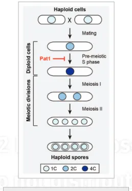
in fission yeast, meiosis involves a physi-ological transition from vegetative growth, and initiation of the process requires inputs from a number of pathways, such as nutrient sensing and response to stress. Upon nitrogen depriva-tion, haploid yeast cells mate to form diploids, which then undergo meiosis and produce four haploid spores (Fig. 1). A highly synchronous meiosis can be induced in S. pombe using a temperature-sensitive allele of the Pat1 kinase (pat1–114),1 a key negative regulator of the
meiotic cycle. This system has been a powerful research tool, allowing for important discoveries in meiosis-specific RnA splicing, gene expres-sion, recombination and chromosome segre-gation. However, one of its major drawbacks is the requirement for a temperature shift, which has been shown to cause stress responses and changes in cellular physiology, notably in gene expression.2 Moreover, this precludes further
dissection of the process through combination with the many temperature-sensitive muta-tions that have been isolated in fission yeast,
for the synchronous induction of meiosis in S. pombe.3 Taking a chemical genetic strategy that
has been used to regulate the activity of various families of kinases,4 they have engineered a
mutation of Pat1 that renders it sensitive to spe-cific inhibition by an ATP analog. inactivation of the kinase activity of this allele, pat1.L95G, after nitrogen deprivation resulted in synchronous entry into pre-meiotic s phase, progression through both meiotic divisions and formation of four haploid spores. The authors further validated this system by confirming the execu-tion of characteristic splicing events that occur during meiosis.
in addition to establishing a promising new tool for the investigation of the
mecha-the transcriptional program of meiosis in fis-sion yeast, in which hundreds of genes are expressed in successive waves.5 They verified
that this pattern is generally preserved in their new system, with one surprising exception: although a number of transcripts associated with stress response were previously shown to be activated upon meiotic induction, Guerra-Moreno and colleagues found a striking dif-ference in the transcriptional profiles of these genes using the pat1.L95G allele, demonstrat-ing that they are, in fact, only activated at later stages in the process. This indicates that the initial characterization of their expression pat-tern was significantly affected by the tempera-ture shift used in previous experiments and suggests that it will be critical to re-evaluate particular aspects of the meiotic program of transcription. second, the authors observed a delay in the timing of s phase entry in pat1.L95G compared with pat1–114, hinting at potential new roles for Pat1 independent of its kinase activity and identifying new directions for future research.
The results obtained using this induction system demonstrate its potential to contribute to our understanding of meiosis. The value and utility of such a method is further underscored by a second paper in this issue from Cipak et al., who have generated a different analog-sensitive allele of pat1 that also achieves a highly synchronous meiosis.6 These
comple-mentary studies provide new insights into meiotic progression and highlight the power of this approach.
References
1. iino y, et al. Proc natl Acad sci UsA 1985; 82:2447-51; PMiD:16593556; http://dx.doi.org/10.1073/ pnas.82.8.2447.
2. Chen D, et al. Mol Biol Cell 2003; 14:214-29; PMiD:12529438; http://dx.doi.org/10.1091/mbc. e02-08-0499.
3. Guerra-Moreno A, et al. Cell Cycle 2012; 11:1620-4; http://dx.doi.org/10.4161/cc.20051.
4. Bishop AC, et al. Trends Cell Biol 2001; 11:167-72; PMiD:11306297; http://dx.doi.org/10.1016/s0962-8924(01)01928-6.
Insights from a new tool for meiotic induction in fission yeast
Comment on: Guerra-Moreno A, et al. Cell Cycle 2012; 11:1620–4; PMID:22456336;
http://dx.doi.org/10.4161/cc.20051
Pei-Yun Jenny Wu; Institute of Genetics and Development of Rennes; CNRS UMR 6290; Rennes, France; *Email: pei-yun.wu@univ-rennes1.fr; http://dx.doi.org/10.4161/cc.20537
Figure 1. Meiosis in fission yeast results in
the production of four haploid spores from a diploid cell. The Pat1 kinase is a negative regulator of the meiotic process and its inactivation allows for meiotic entry. Box: DnA contents of nuclei are indicated by 1C (haploid), 2C (diploid) and 4C (diploid after DnA replication/pre-meiotic s phase).
© 2012 Landes Bioscience.
Do not distribute.
DAPk silencing by DNA methylation conveys resistance
to anti EGFR drugs in lung cancer cells
Comment on: Ogawa T, et al. Cell Cycle 2012; 11:1656–63; PMID:22157090;
http://dx.doi.org/10.4161/cc.20120
Avital Eisenberg-Lerner and Adi Kimchi*; Weizmann Institute of Science; Rehovot, Israel; *Email: adi.kimchi@weizmann.ac.il; http://dx.doi.org/10.4161/cc.20538
epigenetic changes have key roles in the initiation and progression of tumors. A pre-dominant epigenetic modification in cancer is the silencing of tumor-suppressor genes by promoter DnA methylation at CpG islands. silencing of this group of genes, which sup-press cell proliferation, inhibit metastases or promote cell death processes provides a strong positive selection to the tumor cells. interestingly, such epigenetic alterations occur not only at the different stages of tumor development, but also contribute to the acquisition of drug resistance, a major diffi-culty in cancer treatment.1 in a recent issue of
Cell Cycle, sidransky and colleagues describe an interesting example of specific gene silenc-ing dursilenc-ing the acquisition of drug resistance in malignant cells.2 The authors analyzed
the gene promoter methylation profiles of non-small cell lung cancer (nsCLC) and head and neck squamous cell carcinoma (HnsCC) cell lines in comparison to their derivatives resistant to two anti-eGFR drugs, erlotinib (Tarceva) or cetuximab. The top ranked gene that was differentially methylated in resistant but not parental cells was identified as the tumor suppressor DAPk.2
DAPk is a calcium/calmodulin-regulated ser/Thr protein kinase that is implicated in various apoptotic, necrotic and autophagic cell death scenarios.3,4 Hypermethylation of
the DAPk promoter has been documented in a large spectrum of human malignancies.4 The
study by Ogawa et al. now describes DAPk as an essential factor for the successful treatment of nsCLC cell lines with anti-eGFR drugs and demonstrates the correlation between spe-cific methylation of the DAPk promoter and acquisition of drug-resistance.2 DAPk protein
expression was, accordingly, low in the drug resistant cells. Remarkably, re-introduction of DAPk into the resistant cells by stable trans-fection restored the sensitivity of nsCLC cells to both erlotinib and cetuximab. Moreover,
knockdown of DAPk protein expression in the parental, drug-sensitive, cells induced resistance to both drugs. This highlights the significance of DAPk in mediating cell death processes that are initiated by the anti-eGFR treatment, and that loss of DAPk leads to drug resistance and thus provides to these cells a significant growth advantage. Conversely, in the HnsCC cells, the resistance appeared to be DAPk-independent. This is most likely due to the fact that the DAPk promoter was already methylated to some extent in the parental HnsCC cells prior to attaining resistance. Thus, the specific genetic makeup of different malig-nant cell populations at different stages has a dramatic influence on the success of different therapeutic interventions.
The multiple functions of DAPk in sensitiz-ing cells to apoptotic and necrotic responses, in mediating autophagy and in blocking metastasis contribute to its tumor-suppres-sive properties. it is therefore not surprising that a modification that eliminates expression of this gene will be positively selected during tumorigenesis. The data presented by Ogawa et al. suggest that from the various stochastic epigenetic changes that occurred through the course of the drug treatment, cells that acquired silencing of the DAPk gene survived and created a subpopulation of resistant cells that prevailed. One of the future challenges will be to discover the molecular mechanisms that link DAPk to the cytotoxicity induced by the anti-eGPR drug. interestingly, eGFR inhibitors have been recently documented to elevate reactive oxygen species in cells and to induce cytotoxicity through oxidative stress.5
we have previously reported that DAPk is a major regulator of the cellular response to oxidative stress, acting through activation of protein kinase D and JnK to mediate both pro-grammed necrosis as well as autophagy.6,7 it is
therefore plausible that the resistance to cyto-toxicity of the anti-eGFR agents demonstrated
here is due to loss of this important redox-sen-sitive, death-promoting mechanism, which is mediated by DAPk. Previous reports also con-nected the loss of DAPk expression with resis-tance to other chemotherapeutic agents used to treat gastric cancer.8 Like the anti-eGFR
therapy, a significant part of chemotherapy-induced cytotoxicity is via induction of oxida-tive stress9 and may, therefore, be dependent
on DAPk. Finally, it should be stressed that further studies are required to clinically con-firm the cell culture findings reported in this work by measuring the methylation status of DAPk in tumor specimens obtained from patients who acquired drug resistance along the course of treatment. The clinical data will indicate whether the assessment of DAPk methylation may be an important factor in choosing a specific course of therapy and pre-dicting drug resistance.
References
1. yoo CB, et al. nat Rev Drug Discov 2006; 5:37-50; PMiD:16485345; http://dx.doi.org/10.1038/ nrd1930.
2. Ogawa T, et al. Cell Cycle 2012; 11: in Press; PMiD:22157090; http://dx.doi.org/10.4161/ cc.20120.
3. Bialik s, et al. Annu Rev Biochem 2006; 75:189-210; PMiD:16756490; http://dx.doi.org/10.1146/ annurev.biochem.75.103004.142615.
4. Bialik s, et al. semin Cancer Biol 2004; 14:283-94; PMiD:15219621; http://dx.doi.org/10.1016/j.sem-cancer.2004.04.008.
5. Orcutt KP, Parsons AD, sibenaller ZA, scarbrough PM, Zhu y, sobhakumari A, et al. erlotinib-mediated inhibition of eGFR signaling induces meta-bolic oxidative stress through nOX4. Cancer Res 2011; 71:3932-40; PMiD:21482679; http://dx.doi. org/10.1158/0008-5472.CAn-10-3425.
6. eisenberg-Lerner A, et al. Cell Death Differ 2007; 14:1908-15; PMiD:17703233; http://dx.doi. org/10.1038/sj.cdd.4402212.
7. eisenberg-Lerner A, et al. Cell Death Differ 2012; 19:788-97; PMiD:22095288; http://dx.doi. org/10.1038/cdd.2011.149.
8. Zhang X, et al. Anticancer Res 2010; 30:915-21; PMiD:20393015.
9. wondrak GT. Antioxid Redox signal 2009; 11:3013-69; PMiD:19496700; http://dx.doi.org/10.1089/ ars.2009.2541.
© 2012 Landes Bioscience.
Do not distribute.
Cancer cell death and selection: Unexpected putative roles for pRb2/p130,
BORIS and CTCF in endoplasmic stress response maintained by the T-antigen
Comment on: Macaluso M, et al. Cell Cycle 2012; 11:1841–50; PMID:22544282;
http://dx.doi.org/10.4161/cc.20242
Christian Bronner* and Ali Hamiche; Institut de Génétique et de Biologie Moléculaire et Cellulaire; Centre National de la Recherche Scientifique; UMR7104; INSERM U964; Université de Strasbourg; Strasbourg, France; *Email: bronnerc@igbmc.fr; http://dx.doi.org/10.4161/cc.20659
endoplasmic reticulum (eR) stress can affect the function of both oncogenes and tumor suppressor genes, conducting to altered sig-naling pathways regulating cell proliferation and homeostasis.1 However, the mechanisms
by which eR stress affects the function of onco-genes and tumor suppressor onco-genes remain elusive. in a recent issue of Cell Cycle, Macaluso et al.2 investigated the contribution of the
chromatin insulator CCCTC-binding factor (CTCF) and of brother of the regulator of the imprinted site (BORis) to the role of the large T antigen (T-Ag) in maintaining chronic eR stress. T-Ag is known to inactivate both p53 and pRB family proteins (pRB1/p105, pRB2/ p130 and p107).3 As such, the authors
hypoth-esized that BORis, CTCF and pRB2/p130 may have altered behaviors in order to maintain chronic eR stress in T-Ag-positive cancer cells.
CTCF can be considered as a potential tumor-suppressor factor, whereas BORis (CTCFL) has to be considered as a new onco-gene.4 Current literature suggests that CTCF
and BORis, both exhibiting an 11 zinc finger have DnA binding competition activities and thus elicit opposite changes in gene expres-sion. CTCF is an evolutionarily conserved and ubiquitously expressed protein that binds to approximately 20,000 sites in the human genome.4 ectopic expression of CTCF in
var-ious normal and tumoral human cell lines inhibits cell division and clonogenicity. BORis is not found or is eventually found at low levels in normal cells or tissues. BORis gene expression is predominantly epigenetically controlled by DnA methylation, considering that its activation requires the demethylation of its promoter, a mechanism frequently occur-ring in cancers due to global genome-wide hypomethylation.5
in the paper by Macaluso et al.,2 the authors
tried to demonstrate that the T-Ag, conferring oncogenic properties to the Polyomavirus, may contribute to maintain altered
signal-to their previous work, in which they could show that CTCF and BORis directly bind the Rb2/p130 promoter and thus have a tran-scriptional regulatory effect on this gene.6
Presently, they could demonstrate that CTCF binds to the pRB2/p130 promoter only in T-Ag-negative cells, suggesting that the pres-ence of the T-Ag alters the binding of CTCF.2
However, it is not yet known whether, this modification selectively occurs for the pRB2/ p130 promoter or only for some promoters, or whether it is a whole genome-wide modi-fication, which would be interesting to inves-tigate. Furthermore, they found that pRB2/ p130 interacts with either CTCF or with BORis, since they could determine that pRB2/p130 co-immunoprecipitates and co-localizes with these proteins (Fig. 1). surprisingly, it was observed that these complexes exclusively occur in the eR of medulloblastoma tissues in
contrast to medulloblastoma cell lines, where they occur in the nucleus as well in the cyto-plasm. eR stress can be induced by hypoxia or low glucose levels. Macaluso et al. observed,2
in T-Ag-negative cells, that glucose deprivation induced perinuclear aggregates of pRB2/p130 with BORis and CTCF an event that is already occurring in T-Ag-positive cells under normal glucose conditions (Fig. 1). Additionally, eR stress seems to discard the pro-active form at the expense of the active form of caspase 12 (Fig. 1).2
The most encouraging observation made by Macaluso et al.,2 is that T-Ag appears to
prepare cancer cells for continued cell death dictated by a microenvironment poor in nutri-ents, i.e., low glucose levels, via inducing peri-nuclear aggregates of pRB2/p130 with CTCF and BORis. The significance of the accumula-tion of these perinuclear aggregates is unclear,
© 2012 Landes Bioscience.
Do not distribute.
A killer promoting survival: p53 as a selective means to
avoid side effects of chemotherapy
Comment on: van Leeuwen IMM, et al. Cell Cycle 2012; 11:1851–61; PMID:22517433;
http://dx.doi.org/10.4161/cc.20254
Dominique Kranz1 and Matthias Dobbelstein2; 1German Cancer Research Center; Heidelberg, Germany; 2Göttingen Center of Molecular Biosciences; University of
Göttingen; Göttingen, Germany; Email: d.kranz@dkfz-heidelberg.de and mdobbel@gwdg.de; http://dx.doi.org/10.4161/cc.20698
ideally, targeted cancer therapy is directed against tumor cells while sparing normal cells. But in reality, most of the chemotherapeu-tic drugs currently used in the clinics only partially confer this selectivity. As a result, all rapidly dividing tissues are affected by chemo-therapy, resulting in severe side effects, such as immunosuppression, bleeding, diarrhea and hair loss.
Being the product of the most frequently mutated gene in cancer, the tumor suppressor p53 represents a highly attractive target for therapeutic approaches aiming at the selec-tive killing of tumor cells. some strategies for p53-based therapy rely on the reconstitution of wild-type p53 in tumor cells, e.g., by adeno-viral gene delivery. More recently, the concept of converting mutant p53 into a killer of tumor cells was proposed.1 Pharmacological
reac-tivation of mutant p53 reportedly results in conformational changes, restoring DnA bind-ing ability and transcriptional activity and, thus, the capability to induce cell cycle arrest, senescence or apoptosis.2 However, the use of
these drugs is still a challenge due to the het-erogeneity of p53 mutations in tumors, each differing structurally and functionally.
in the past decade, cyclotherapy has emerged as a novel strategy where an inital treatment induces reversible growth arrest in normal cells, shielding them from classical chemotherapeutic treatment that only affects “cycling” cancer cells.3,4
p53-based cyclotherapy depends on the activation of wild-type p53 in normal cells. This concept seems counterintuitive at first glance, since p53 is mostly known as an inducer of cell death—obviously an undesir-able effect with regard to normal cells of patients. However, moderately activating p53 rather results in cell cycle arrest than apop-tosis. The induction of p53 may also protect cells by induction of repair genes or by alter-ing the proapoptotic DnA damage response.5
in this context, p53 serves as a discriminator between normal cells, with wild-type p53 and tumor cells lacking functional p53. Patients with tumors bearing p53 mutations could benefit from this combinatorial approach: initial treatment with a p53 activator spares healthy tissue from cytotoxic effects associ-ated with classical chemotherapy, and only tumor cells harboring p53 mutations are eliminated.
i.e., whether it is due to redistribution or it comes from an increase in the protein synthe-sis, the latter being a more plausible hypoth-esis. indeed, the eR is the major site for folding and trafficking. when the eR is stressed by unfolded proteins, it elicits a classic adaptative response known as unfolded protein response (UPR).7 Aggresome-like induced structures
(ALis) are known to result from transient aggregation of ubiquitinated proteins in eR stress response.8 A crosstalk between UPR and
ALis was recently reported.8 Obtaining
infor-mation as to whether pRb2/p130, BORis and CTCF are affected by unfolding, ubiquitnation or truncation will probably shed light on how the subcellular localization of CFTC, BORis and pRB2/p130 is tuned to orchestrate intercon-nected p53-independent signaling pathways leading to cancer cell death in Polyomavirus-infected cells. Furthermore, it will unravel how cancer cells can be selected for survival and thus acquire resistance to cell death signaling.
References
1. Gorman AM, et al. Pharmacol Ther 2012; 134:306-16; PMiD:22387231; http://dx.doi.org/10.1016/j. pharmthera.2012.02.003.
2. Macaluso M, et al. Cell Cycle 2012; 11: in press; PMiD:22544282; http://dx.doi.org/10.4161/ cc.20242.
3. Caracciolo v, et al. J Cell Biochem 2010; 110:182-90; PMiD:20336668.
4. Millau JF, et al. Biochem Cell Biol 2011; 89:505-13; PMiD:21970734; http://dx.doi.org/10.1139/o11-052.
5. de necochea-Campion R, et al. J Transl Med 2011; 9:213; PMiD:22168535; http://dx.doi. org/10.1186/1479-5876-9-213.
6. Fiorentino FP, et al. Mol Cancer Res 2011; 9:225-33; PMiD:21325284; http://dx.doi.org/10.1158/1541-7786.MCR-10-0493.
7. Ron D, et al. nat Rev Mol Cell Biol 2007; 8:519-29; PMiD:17565364; http://dx.doi.org/10.1038/ nrm2199.
8. Liu XD, et al. J Biol Chem 2012; in press; PMiD:22518844.
Figure 1. p53-mediated chemoprotection.
some chemotherapeutics are largely restricted in their efficacy to specific phases in the cell cycle. nucleoside analogs (gemcitabine, cytarabine) work in s-phase, whereas taxanes and vinca alcaloids act on mitotic cells. Moderate activation of p53 arrests cells in G1 or G2, protecting p53-proficient cells (but not p53 mutant tumor cells) against these drugs.
© 2012 Landes Bioscience.
Do not distribute.
Previous studies indicate that pretreat-ment with the p53 activator nutlin-3 protected human fibroblasts from taxanes6 and human
immortalized keratinocytes from nucleoside analogs7 as well as from cell death induced by
Uv irradiation.5
in a previous issue of Cell Cycle, Leeuwen and colleagues extended this approach, show-ing that a variety of p53-activatshow-ing strategies (actinomycin D, nutlin-3, tenovin-6, lepto-mycin B) led to reversible cell cycle arrest in human fibroblasts, protecting them from drugs that are active in s phase (gemcitabine and cytarabine) and M phase (vinblastine, vinorelbine).8 Thus, arresting normal cells may
provide a promising strategy of chemoprotec-tion, enabling the use of higher doses of che-motherapeutics in patients.
However, fibroblasts are not the cells harmed by the side effects of chemotherapy in first place. Chemotherapy mostly damages highly proliferating tissues like bone marrow, epithelia of the digestive tract and hair follicles. Further investigations are required to evalu-ate if cells from these tissues are protected by p53-mediated cyclotherapy.
The study by Leeuwen et al. also indicates that sometimes p53-mutant and p53-null cell lines were protected by cyclotherapy, depend-ing on the drug used for pre-treatment, poten-tially compromising the therapeutic efficacy.8
in contrast, this effect was not observed for the p53-selective drug, nutlin-3.
As nutlin-3 interacts with MDM2, a nega-tive regulator of p53, its effects are highly p53-dependent.9 The major disadvantage
of nutlin-3 is that it has not been clinically approved so far. For instance, p53-induced myelodepression may limit its use. Phase i clinical trials are currently ongoing using the related compound RG7112 as a mono-therapy for the treatment of hematologic neoplasms (nCT00623870) and solid tumors (nCT00559533).
Critical issues remain to be addressed: is p53-based cyclotherapy clinically applicable? Are there dose-limiting effects of combinatorial treatments? what impact has p53 preactivation on the side effects of chemotherapy in non-pro-liferating cells, e.g. neurons or cardiomyocytes? Beyond understanding the underlying mechanisms of p53 as a chemoprotector, the
feasibility of the combinatorial treatment needs to be verified in preclinical studies and clinical trials. This would finally exploit the most frequent genetic alteration in cancer for a selective therapy.
References
1. Bykov vJ, et al. nat Med 2002; 8:282-8; PMiD:11875500; http://dx.doi.org/10.1038/nm0302-282.
2. wiman KG. Oncogene 2010; 29:4245-52; PMiD:20498645; http://dx.doi.org/10.1038/ onc.2010.188.
3. Blagosklonny Mv, et al. Cancer Res 2001; 61:4301-5; PMiD:11389048.
4. Blagosklonny Mv, et al. Cell Cycle 2002; 1:375-82; PMiD:12548008; http://dx.doi.org/10.4161/cc.1.6.259.
5. Kranz D, et al. J Cell Biol 2008; 182:197-213; PMiD:18625847; http://dx.doi.org/10.1083/ jcb.200712014.
6. Carvajal D, et al. Cancer Res 2005; 65:1918-24; PMiD:15753391; http://dx.doi.org/10.1158/0008-5472.CAn-04-3576.
7. Kranz D, et al. Cancer Res 2006; 66:10274-80; PMiD:17079445; http://dx.doi.org/10.1158/0008-5472.CAn-06-1527.
8. van Leeuwen iMM, et al. Cell Cycle 2012; 11:1851-61; http://dx.doi.org/10.4161/cc.20254.
9. vassilev LT, et al. science 2004; 303:844-8; PMiD:14704432; http://dx.doi.org/10.1126/sci-ence.1092472.