HAL Id: hal-01484973
https://hal-univ-rennes1.archives-ouvertes.fr/hal-01484973
Submitted on 4 Jul 2017
HAL is a multi-disciplinary open access archive for the deposit and dissemination of sci-entific research documents, whether they are pub-lished or not. The documents may come from teaching and research institutions in France or abroad, or from public or private research centers.
L’archive ouverte pluridisciplinaire HAL, est destinée au dépôt et à la diffusion de documents scientifiques de niveau recherche, publiés ou non, émanant des établissements d’enseignement et de recherche français ou étrangers, des laboratoires publics ou privés.
To cite this version:
Thierry Lamy, Aline Moignet, Thomas P. Loughran. LGL leukemia: from pathogenesis to treatment. Blood, American Society of Hematology, 2017, 129 (9), pp.1082-1094. �10.1182/blood-2016-08-692590�. �hal-01484973�
1
LGL leukemia: from pathogenesis to treatment
Thierry Lamy1, Aline Moignet 1, Thomas P. Loughran Jr2
1 Department of Hematology, Pontchaillou University Hospital, Rennes, France
2 University of Virginia Cancer Center, Charlottesville, VA, USA
Correspondence to:
Thierry Lamy, M.D-PhD Department of Hematology Service d’Hématologie
Hôpital Pontchaillou. CHU de Rennes. 35033 Rennes France Tel: 33 2 99 28 41 61 Fax: 33 2 99 28 41 61 Email: [email protected] Abstract: 158 words Text: 4592 words References: 122 Number of Figures: 6 Number of Tables: 2
Abstract
Large granular lymphocyte (LGL) leukemia has been recognized by the WHO classifications among mature T cell and NK cell neoplasms. There are 3 categories: chronic T cell leukemia and NK cell lymphocytosis which are similarly indolent diseases characterized by cytopenias and autoimmune conditions as opposed to aggressive NK cell LGL leukemia. Clonal LGL expansion arise from chronic antigenic stimulation which promotes dysregulation of apoptosis, mainly due to constitutive activation of survival pathways including Jak/Stat, MapK, Pi3k-Akt, RasRaf-1, MEK1/ERK, sphingolipid, and NFκB. Socs3 down regulation may also contribute to Stat3 activation. IL15 plays a key role in activation of leukemic LGL. Several somatic mutations including Stat3, Stat5b, and TNFAIP3 have been demonstrated recently in LGL leukemia. Since these mutations are present in less than half of the patients they cannot completely explain LGL leukemogenesis. A better mechanistic understanding of leukemic LGL survival will allow future consideration of a more targeted therapeutic approach than the current practice of immunosuppressive therapy.
Introduction
Initially described in 1985, Large Granular Lymphocyte (LGL) leukemia belongs to the rare chronic mature lymphoproliferative disorders of the T/NK lineage.1 Two subtypes of LGL disorders were proposed in 1993 : T-LGL leukemia and aggressive NK cell leukemia.2 The WHO recognized this classification scheme in 2001. Chronic NK-cell lymphocytosis was identified in 2008 as a provisional entity to differentiate it from the much more aggressive form of NK-cell leukemia.3,4 The most recent WHO version did not modify this classification scheme but did highlight discovery of Stat mutations described in 2012 (table 1).5,6 T-LGL leukemia and chronic NK cell lymphocytosis share the same clinical and biological presentation as well as treatment options.2,7,8,9 Pathogenesis of the disease is dominated by a clonal
expansion of LGL resistant to activation induced cell death (AICD) due to constitutive survival signaling. This review will describe topics concerning diagnosis,
pathogenesis, current and future therapy of this rare disease.
Epidemiology
LGL leukemia accounts for 2–5% of chronic lymphoproliferative disorders in North America and Europe and up to 5–6% in Asia.2Recently, the overall age-standardized incidence based on the national Dutch registry has been reported as 0.72 per
1,000,000 person-per year.10 The incidence of LGL leukemia does not differ between male and female. Indolent T-LGL leukemia is the most frequent form of the disease representing around 85 % of the cases whereas chronic NK cell lymphocytosis is estimated at less than 10% of cases.
Aggressive NK LGL leukemia is mainly seen in Asia, comprising less than 5% of the LGL disorders. It affects younger patients and is associated with EBV infection: the prognosis of this rare entity is very poor due to refractoriness to chemotherapy.11,12
Diagnosis
A definite diagnosis of LGL leukemia requires finding evidence of an expanded clonal T or NK cell LGL population.
Cytology
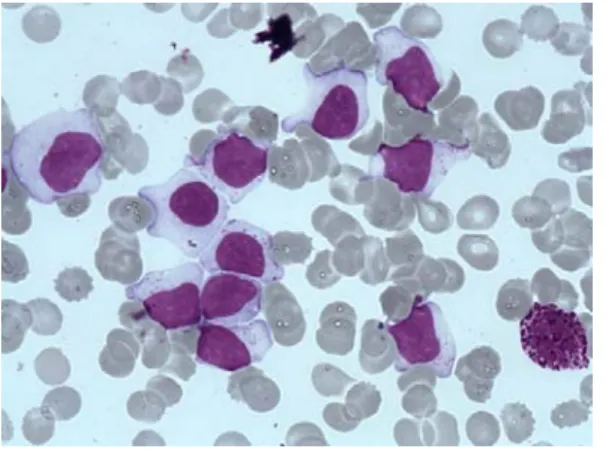
The first step of diagnosis is based on identification of increased numbers of circulating LGL. Initially, a LGL count > 2 x 10.9/l (normal value: < 0.3 x 10.9/l ) was mandatory13,14 but a lower number may be compatible with the diagnosis if these cells are clonal and the patient displays other clinical or hematological features such as rheumatoid arthritis or cytopenias. Indeed, it has been found that some patients with relatively low LGL counts (even less than 1x10.9/l) have a clonal disorder.9,14,15,16 Leukemic LGL are easily identified on blood smears by their specific morphology; however they are not cytologically distinguishable from normal reactive cytotoxic lymphocytes. They display a large size (15-18μ), an abundant cytoplasm containing typical azurophilic granules and a reniform or round nucleus with mature chromatin (Fig 1 A). Blood smears must be examined carefully in cases of normal lymphocyte counts and in rare cases in which the clonal lymphocytes do not present with typical LGL morphology. LGL excess in bone marrow (> 10% LGL) is detected in the majority of cases.13(Figure 1B)
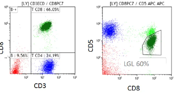
Immunophenotyping
T-LGL leukemias show a constitutive mature post-thymic phenotype. In the vast majority of cases, T-LGL leukemia shows a CD3+, TCR αβ+, CD4-, CD5dim, CD8+, CD16+, CD27-,CD28-, CD45R0-, CD45RA+, CD57+ phenotype which represents a constitutively activated T cell phenotype (Fig 2A). 17,18,19 CD3+/CD56+ T-LGL leukemias may have a more aggressive behavior associated with Stat5b
mutations.20,21 A rare subset of LGL leukemia is CD4+ with or without coexpression of CD8. Patents with this LGL leukemia subtype almost never have cytopenias, splenomegaly or autoimmune phenomena22,23, and clonal LGL seem to be driven by CMV.24 Recently, it was found that Stat5b mutations are frequent in this LGL
subtype.25
T-LGL leukemic cells are characterized by a terminal-effector memory phenotype defined by the expression of CD45RA and lack of CD62L expression.26 Leukemic LGL constitutively express IL2 Rβ (p75, CD122) but not IL2 Rα (p55, CD25).27,28 Few cases are TCR γδ+/CD4-/CD8-.29,30
NK-LGL leukemia and NK-LGL lymphocytosis are characterized by the following phenotype: CD2+/sCD3-/CD3ε+/TCRαβ-/CD4-/CD8+/CD16+/CD56+ (Fig 2B)13. Fas (CD95) and Fas-Ligand (CD178) are strongly expressed in LGL leukemia.31,32
Restricted KIR expression is often seen in both in T and NK LGL leukemia.19,33
Clonality
Evidence of T-LGL clonality is routinely assessed using TCR γ-PCR analyses. Deep sequencing of TCR has demonstrated a restricted diversity of TCR repertoire.34 Vβ TCR gene repertoire analysis can also be ascertained using flow cytometry (FCM) and serves as presumptive evidence for clonality.35,36(Fig 2C) The current Vβ MoAbs panel covers 75% of the Vβ spectrum with a high correlation between Vβ FCM and TCRγ-PCR results. Fluctuations in clonal dominance, as determined by CDR3 sequencing, are seen in up to one third of patients.37
It is difficult to assess clonality of NK LGL as these cells do not express TCR. Restricted expression of activating isoforms of killer immunoglobin-like receptors (KIR) has been utilized as a surrogate marker for a monoclonal expansion (Fig. 2B).33,38,39,40
Clonal chromosomal abnormalities have been reported in a few cases of LGL leukemia.1
Molecular findings
Constitutive activation of Stat3 in all LGL leukemia patients was first discovered in 2001.41 In 2012, common somatic gain-of-function Stat3 mutations were
demonstrated in 28% to 75 % of T-LGL leukemia and 30% to 48% of NK LGL lymphocytosis. 6,42,43,44(Fig 2D) These differences may be due to the sequencing technique and the selection of patients. Detection of identical Stat3 mutations in both T and NK subtypes suggests a unifying pathogenesis for these similar disorders.45 Mutations are primarily located in exons 20 and 21 encoding the Src homology 2 (SH2) domain, which drives the dimerization and activation of the Stat protein. D661 and Y640 account for two third of mutations.43 Activating mutations outside the SH2 domain are rarely detected.46 Such mutations are located in the DNA-binding and coiled-coil domain of Stat3. Utilization of deep sequencing has demonstrated presence of multiple subclones containing different Stat3 mutations in distinct LGL
populations in individual patients.47 These findings suggest a potential need for sequencing the entire Stat3 gene. Whether or not Stat3 mutations are correlated with specific clinico-biological features remains uncertain and subject of ongoing research. The ECOG prospective clinical trial suggested that a particular Stat3 mutation,
Y640F, predicted response to initial therapy with methotrexate.48 The first evidence of Stat5b mutations in human disease was discovered in LGL leukemia, but this mutation is not frequent (2%).20 The N642H mutation in particular was associated with a more aggressive disease and a unusual CD3+CD56+ phenotype.49
Marrow Features
The diagnosis of LGL leukemia as discussed above is readily established using blood studies, so that marrow aspirate/biopsy is not routinely recommended as part of initial evaluation. However, marrow evaluation can be helpful when the diagnosis is not straight-forward (eg LGL count not increased). Such studies are also of value when considering other diseases that are part of the differential, including MDS or aplastic anemia or when considering the diagnosis of pure red cell aplasia (PRCA) as potential etiology of profound anemia in patients with LGL leukemia.
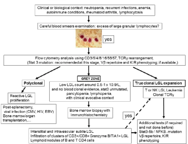
Individual or small clusters of LGLs may be sometimes identified. They are difficult to identify as they mimick granulocytic or monocytic precursors. As LGL infiltration of marrow is often a subtle finding, trephine biopsy with immunohistochemistry is recommended. 7,50,51,52 A “grey zone” where the diagnosis of LGL leukemia is not certain, should be considered in cases of low LGL count, or even lymphopenia associated with unexplained cytopenia in which the diagnosis can be easily missed (Figure 3).
Classical histological features are depicted in figure 4. Clusters of eight CD8+/TiA1+ cells or six Granzyme B+ lymphocytes are considered as characteristic
histopathologic findings of LGL leukemia, and support this diagnosis in uncertain cases.51,53 Very close topographic distribution between dendritic cells and LGL in patient marrow samples have been shown, suggesting constant antigen
presentation54. Trilineage hematopoiesis in bone marrow biopsies of LGL patients is normal or increased in the majority of cases. Apoptotic figures, increased
macrophages, and eosinophilia have been described, suggesting an underlying dysmyelopoiesis. In neutropenic patients, a typical finding is decrease in granulocyte precursors and left-shift maturation. However, degree of marrow infiltration by LGL
does not correlate with degree of cytopenia.50 Increase of erythroid precursors is described in 30-40% of the cases. Reticulin fibrosis is usually present, ranging from grade II or III in about 50-60% of the cases.55
Clinico-biological features
LGL leukemia principally affects elderly people with a median age of 60 years13,7,8,56,57,58,59. Very rare pediatric cases have been reported and less than 25% of adult patients are younger than 50 years old. About one third of patients are asymptomatic at the time of diagnosis. Initial presentation is mainly related to neutropenia and includes recurrent oral aphthous ulcerations, fever secondary to bacterial infections. These infections typically involve skin, oropharynx and perirectal areas, but severe sepsis may occur. However, some patients may have profound and persistent neutropenia without any infections over a very long period of time. The frequency of recurrent infections varies in different series from 15 to 39%. Fatigue and B symptoms are observed in 20 to 30 % of cases. Splenomegaly is reported with a frequency varying from 20 to 50 % and lymphadenopathy is rare.15 Half of the patients present with lymphocyte counts between 4 x 10.9/L and 10 x 10.9/L, and the LGL count usually ranges from 1 to 6 x 10.9/L. A lower LGL count (0.5 to 1 x 10.9/L) may be observed in 7% to 36% of cases. Severe neutropenia (<0.5 x 10.9/L) and moderate neutropenia (<1.5 x 10.9/L) are observed in 16% to 48%, and 48% to 80% of cases, respectively. Anemia is frequent; transfusion dependent patients are observed in 10 to 30% of cases.PRCA occurs in 8 to 19% of the cases and moderate thrombocytopenia is observed in less than 25% of patients.60
While not routinely performed, increased soluble Fas-Ligand (sFas-L) is a good surrogate marker of LGL leukemia.61 We and others showed that LGL leukemia patients had increased serum levels of interferon α2, MCP-1 (an attractive factor of monocytes, T and NK cells to sites of inflammation), EGF, IL6, IL8, and IL18.48,62,63 High β2 microglobulin level is observed in 70% of cases.Rheumatoid factor and antinuclear antibody are detected in 60 % and 40% of patients, respectively.15 Serum protein electrophoresis usually shows polyclonal hypergammaglobulinemia due to increased IgG and/or IgA subclasses. Defects in downregulation of Ig secretion in
LGL leukemia could partly explain the development of autoantibodies and clonal B-cell malignancies observed in this disease.64
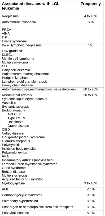
The associated comorbid conditions are reported in table 2. Rheumatoid arthritis (RA) is the most common associated disease, occurring in 10 to 18 % of
patients.7,18,65,66 Systemic lupus erythematosus (SLE), Sjogren’s syndrome, autoimmune thyroid disorders, coagulopathy, vasculitis with cryoglobulinemia and inclusion body myositis have occasionally been reported.65,67,68,69 Pulmonary artery hypertension (PAH) has been observed.70 Association with myeloid malignancies, and bone marrow failure syndromes, i.e Aplastic anemia (AA), paroxysmal nocturnal hemoglobinuria (PNH) and myelodysplasia (MDS), is also reported.71,72,73 Recurrent Stat3 mutations in de novo AA and MDS suggest similar pathogenesis.42 Efficacy of immunosuppressive treatments directed against T lymphocyte- mediated immune response is a strong argument for a common role of auto reactive T cells in all of these diseases.
Pathogenesis of LGL Leukemia
A model of LGL leukemia pathogenesis is shown in Figure 5. LGL leukemia is at the intersection of a clonal lymphoproliferative disease, chronic inflammation, and
autoimmunity. We hypothesize that an unknown antigen is the initial activating event, resulting in oligoclonal LGL expansion. Such chronic and persistent antigen
exposure leads to Stat3 activation and emergence of a dominant clone. A shift from oligoclonal to clonal dominance is supported by serial studies of T cell repertoire utilization.37 Somewhat surprisingly, clonal drift was observed with emergence over time of a different Vbeta clone in 37% of patients.37 We speculate that clonal drift results from emergence of LGL clones recognizing different epitopes/peptides of the same chronic antigen. This theory could also explain the observation of clonal T cell LGL populations seen in patients with chronic NK lymphocytosis as well as
emergence of a new NK clone in patient with established T cell LGL leukemia.74 LGL leukemia is characterized by profound dysregulation of the normal process of AICD. The fundamental pathogenic feature of LGL leukemia is activation of a
survival network which keeps the leukemic LGL alive and functioning as killer cells .75 The central hub of this survival network is Stat3. We hypothesize that activating mutations of Stat3 support clonal dominance by causing a higher level of
transcription of survival components. This theory is supported by the observation that STAT3 mutations are correlated with a larger clone size in LGL leukemia patients.48 Disease manifestations such as cytopenias and autoimmune diseases then result from production of proinflammatory cytokines mediated by STAT 3
regulation as well as attack on marrow and joints by the STAT 3- activated LGL. The key molecular signaling components in LGL leukemia are depicted in Figure 6. Key features of the model are further summarized below.
An initial viral antigenic stimulation
LGL leukemic cells represent an expanded population of a effector memory cytotoxic T cells, suggesting chronic antigen stimulation.26 A role for HTLV retroviral infection has been suggested primarily by serologic studies demonstrating cross-reactivity to HTLV epitopes. Such seroreactivity is directed towards the envelope protein BA21 and is seen in 30 to 50% of the patients. However, there is no definite evidence for HTLV-I infection in LGL leukemia patients76,77,78,79,80 while HTLV-2 has been described only occasionally. 81,82
Two cases of LGL leukemia associated with B indolent lymphoma were reported in patient infected with HCV infection. Both were successfully treated by antiviral therapy. This underlies a potential link between HCV chronic infection and LGL leukemia.83 Recently Sandberg et al analyzed CDR3 β and α chain in 26 T CD8+
αβTCR LGL patients and found heterogeneity in CDR3 region, suggesting that a unique antigen driving LGL leukemia may be unlikely.84
Initiating role of IL-15 and PDGF
IL15 pro-inflammatory cytokine and PDGF play a crucial role in LGL leukemia expansion by promoting NK cell or leukemic LGL survival.75,85 IL15 receptor-α -/- mice display a NK cell and CD8+ memory T cell defect. LGL cell growth is dependent of IL15 and IL15α chain mRNA was detectable in LGL purified cells.86,87 In a network model of LGL leukemia survival signaling, IL15 and PDGF were predicted to be the master activation switches necessary for leukemic LGL survival.75 IL15 has also been implicated in LGL leukemogenesis using a unique IL15 transgenic mouse model demonstrating an increase of global DNA methylation level in LGL cells.88 Activation of survival pathways by PDGF occurs through an autocrine loop in leukemic LGL.89
Constitutive activation of Jak-Stat3 signaling pathway
Constitutive activation of Stat3 (pStat3), in our opinion, plays the fundamental pathogenic role in LGL leukemia, as the transcriptome of LGL leukemia patients is that of Stat3-regulated genes.6,41 In vitro inhibition of Stat3 by STA-21 restores
apoptosis of LGL cell independent of Stat3 mutational status. This finding implies that Stat3 mutation is not itself mandatory to explain LGL clonal expansion. It raises the question of mechanism of Stat3 activation in unmutated patients. The
pro-inflammatory cytokine IL6 is able to activate Jak-Stat3 pathway and high amount of IL6 is observed in LGL patients.63 Inhibition of this cytokine restores LGL apoptosis. Dendritic cells could trigger the clonal proliferation and maintain LGL expansion by IL6 production. Because SOCS3 transcription is induced by IL-6 through P-Stat3, SOSC3 was postulated to be high in LGL leukemia. Surprisingly, Teramo et al, showed that SOCS3 mRNA and protein level was significantly decreased in LGL leukemia. Demethylation agent restored SOSC3 expression in leukemic LGL and consequently decreased Stat3 and Mcl-1 expression. No mutation or methylation of SOSC3 promoter has been found to explain this down regulation.63 Although Stat3 activation plays the most important pathogenenic role, multiple other cell survival pathways are dysregulated and these are briefly described below.
Resistance to Fas/Fas ligand mediated apoptosis
Leukemic LGL are resistant to Fas mediated apoptosis.31 However, apoptosis is restored using IL2 stimulation suggesting an inhibition of this pathway and not a complete defect. Moreover, soluble Fas (sFas), detected in LGL patient sera, acts as a decoy receptor able to inhibit Fas-dependent apoptosis.61 Decreased level of an inhibitory protein named c-FLIP is found in leukemic LGL contributing to DICS formation defect. This protein blocks the initial event of Fas-mediated apoptosis through FADD (Fas-associated protein with death domain) and caspase 8 recruitment.26
Ras-Raf-1-MEK1-ERK pathway
Overactive Ras plays a role in the survival signaling in LGL leukemia. Constitutive activation of Ras and extracellular-regulated kinase (ERK) was found in NK-LGL leukemia and G12 KRAS mutation was detected in LGL leukemia cell line.90
Blockade of either ERK or Ras activity may restore Fas sensitivity in leukemic LGLs.91
Dysregulation of PI3K/Akt pathway
Activated Akt is observed in leukemic LGL. Pro-inflammatory proteins RANTES, IL-18 and MIP-1b which activate PI3K pathway, are upregulated in LGL patients.62 Increased activity of the PI3K-AKT signaling axis has been found in T-LGL cells and participated in apoptosis inhibition.92
Activation of NFkB pathway
Leukemic LGL exhibit higher level of c-Rel (a member of NFκB member family) and high NFκB activity. NFκB inhibition induces leukemic LGL apoptosis. It has been shown that NFκB acts downstream of the PI3K-AKT pathway to prevent apoptosis through Mcl-1 independently of Stat3.75 Recently, a recurrent non-synonymous mutation in the gene encoding a NFκB signaling inhibitor, TNFAIP3, was found in three patient out of 39 patients (8%), underlying the important role of NFκB activity in LGL leukemia.93
Dysregulation of sphingolipid rheostat in LGL leukemia
Molecular expression profile analysis in LGL leukemia shows a predominant expression of pro-survival sphingolipids like S1P.94 Sphingosine kinase 1 (SphK1) which converts sphingosine into SP1 is increased in LGL leukemia and SphK1 inhibition leads to leukemic LGLs apoptosis. SP1 binding to SP1R activates pro survival signal through ERK1/2 signaling.95 Moreover, expression of S1P receptor, mainly S1PR5, is increased in LGL leukemia.96,97
PROGNOSIS
T and chronic NK-LGL leukemia are considered indolent diseases. The vast majority of patients will eventually need treatment at some point during disease evolution. Disease related deaths are mainly due to severe infections which occur in less than 10% of the patient population.10,15,98 Overall survival at 10 years is close to 70%.
Conversely, the prognosis of aggressive NK LGL leukemia is very poor since patients are refractory to treatment. 11,12
THERAPY
Treatment options are similar for T-LGL leukemia and chronic NK-cell lymphocytosis. Indications for treatment include severe neutropenia (ANC < 0.5 x 10.9/L), moderate neutropenia (ANC >0.5 x 10.9/L) associated with recurrent infections, symptomatic or transfusion dependent anemia and associated autoimmune conditions requiring therapy9. Standard treatment of LGL leukemia is immunosuppressive therapy but such treatment recommendations are primarily based on small retrospective series. The most clinical experience has been reported using low-dose methotrexate,
cyclophosphamide, and cyclosporine as single agents (see below).
Supportive therapy
Supportive care could be considered for patients suffering from anemia or
neutropenia using EPO or G-CSF, respectively. However, such treatment does not address the underlying illness. G-CSF delivered as a single agent may be effective in rapidly increasing ANC. This could be of some value in the setting of patients with episodes of severe febrile neutropenia in which a rapid neutrophil response would be desirable. However, CSF is not effective in all LGL leukemia patients. Of note, G-CSF may induce exacerbation of splenomegaly and articular symptoms.99,100
Therapy with EPO in LGL leukemia patients has been reported only infrequently and with disappointing results.15
First line therapy
Immunosuppressive therapy is the foundation of treatment for LGL leukemia based on the rationale that leukemic LGL represent constitutively activated cytotoxic lymphocytes.
First line therapy relies on use of single immunosuppressive oral agents such as methotrexate (MTX, 10mg/m²/week), cyclophosphamide (100 mg per day), or
cyclosporine (CyA, 3 mg/kg per day). A minimum of 4 months of therapy is required before assessing response. Since the initial publication on the efficacy of MTX, this drug has been considered until now as the best first line option, mainly for
neutropenic patients.101 Oral cyclophosphamide has been preferentially used in patients with predominant anemia and particularly PRCA.60,102
Based on retrospective studies, the overall response rate (ORR) appears quite broad ranging from 21% to 85% (median around 50%), with similar responses to each of the 3 drugs, making comparison difficult. The complete response rate is relatively low, around 21% for MTX, 33% for cyclophosphamide, and less than 5% for CyA.9 Duration of response is around 21 months for MTX. However relapse rate may be high, with the French series reporting rates of 67%, compared to 13% for patients receiving cyclophosphamide.9,15 Cyclophosphamide as first line therapy may induce high ORR with almost 50% of CR rate for either neutropenic or anemic patients.103 CyA corrects cytopenia without elimination of the LGL clone.15,16,104 It has been suggested that HLADR4 (observed in 32% of LGL leukemia) could be highly predictive of CyA responsiveness.105
The results of the first large prospective study of immunosuppressive agents in LGL leukemia was recently reported.48 Fifty-five patients received MTX at first line and non-responders were switched to cyclophosphamide. The ORR was 38% in step 1 (which is lower compared to the results of the large retrospective studies) and 64% in step 2. These data indicate that a high proportion of patients may respond to
cyclophosphamide even after having failed to respond to MTX.15,100 As discussed earlier, Stat3 Y640F mutation may be predictive biomarker for response to
methotrexate but this observation needs validation in a larger cohort of patients. An important randomized trial (NCT01976182) investigating first line MTX versus cyclophosphamide is ongoing in France will hopefully determine the best choice of initial therapy in this disease.
In case of failure of primary therapy, a switch between MTX and cyclophosphamide should be considered. In our practice, CyA is reserved for patients failing both drugs. Both MTX and CyA are maintained indefinitely as long as these medications are reasonably tolerated and disease response is maintained. Long term use and monitoring of low dose MTX follows guideline recommendations of its use in RA. Hepatic (hepatitis) and lung dysfunction (hypersensitivity pneumonitis) may occur requiring regular evaluation of liver tests and chest X-ray. Our recommendation is to
stop cyclophosphamide after 8-12 months because of its mutagenic potential. Renal function and blood pressure have to be carefully monitored during CyA treatment.9 The effects of prednisone as single agent in the treatment of LGL leukemia are not convincing but may decrease some inflammatory symptoms related to RA and in rare cases it may transiently improve neutropenia.
Second line therapy
It is difficult to make any treatment recommendations for patients refractory to the first line agents, because of the rarity of the disease and general absence of prospective data. To the authors’ knowledge there is only one ongoing clinical trial in the United States, which investigates alentuzumab. 110 The US author’s practice is to refer refractory patients for consideration of participation in this study being conducted at the NIH.
Clinical experience using a number of different options in a smaller number of
patients has been reported for LGL leukemia patients and these could be considered for refractory patients. These results are summarized below:
i) Purine analogs.
Less than 50 patients have been reported to be treated with either fludarabine, 2 CDA, deoxycoformycine, or bendamustine.15,104,106,107,108 The overall response rate looks promising at around 79%. Remissions are usually obtained rapidly and the patients may be treated with a maximum of 1 to 3 courses in order to limit toxicity. Fludarabine has been used in combination with Mitoxantrone with long lasting
complete remission. Whether or not purine analogs should be integrated into first line therapy is an open question.
ii) Combined chemotherapy
Polychemotherapy based on CHOP like or Ara-C containing regimen has not demonstrated efficacy in chronic refractory form of disease and should be reserved for the rare cases of aggressive LGL leukemia. In some cases of multi-refractory patients, stem cell transplantation (SCT) could be considered. We recently reported the EBMT series of 15 patients receiving auto or allogeneic SCT.109 Six patients remained disease-free post transplantation.
iii) Immunotherapy
Alentuzumab (campath) which is a humanized monoclonal antibody (anti CD52) has been proposed for refractory patients in very limited series. The overall response rate
is around 60%.9,15,16,110 However, general availability of this drug is now limited and infectious risks limit use of this agent. Efficacy of extracorporeal photopheresis has been reported recently with documented CR.111
Rituximab, the specific antiCD20 monoclonal antibody, has been paradoxically used in patients having both RA and LGL leukemia. It was suggested that eradication of B cell expansion and autoimmune antigens could have led to eradication of reactive LGL clones. In our opinion, this is not a reasonable option for LGL leukemia.112,113
iv) Splenectomy
Splenectomy may be considered in patients with symptomatic splenomegaly
associated or not with cyopenias. An overall review of the literature shows an ORR of 56% but sustained responses are infrequent.15,16,114,115
v) Targeted therapy
Considering the pathogenesis of LGL leukemia, different specific pathways
conceivably could be targeted using either cytokine antagonistic agents, monoclonal antibodies (MoAbs) directed against membrane receptors, Jak/Stat inhibitors, NFκB or farnesyl transferase/Ras inhibitors.
Regarding IL15 targeting, a clinical trial (NCT 00076180)has tested effects of Hu-Mikβ1 which blocks IL-15 trans presentation to T cells that express IL-2/IL-15Rβ (CD122). Administration of Hu-Mikβ1 to patients was safe but clinical responses were not observed .116A phase I study with an anti-CD2 monoclonal antibody (siplizumab)
was conducted in 2005 in patients with relapsed/ refractory CD2+ T-cell
lymphoma/leukemia including T-LGL leukemia (NCT00123942). To our knowledge, the results of this trial have not been published.
Based on constitutive ras activation in leukemic LGL, a phase II study investigating use of farnesyltransferase inhibitor (tipifarnib) enrolled a total of eight patients. No clinical responses were observed despite a significant improvement in marrow
hematopoiesis and increased hematopoietic colony growth in vitro.117
A Stat inhibitor, OPB-31121, had a significant antitumor effect on leukemia cells with Stat-addictive oncokinases.118 Stat3 inhibition could be a good therapeutic option in LGL leukemia. Of interest, low dose MTX was recently identified as a very active inhibitor of Jak/Stat pathway, perhaps now explaining its efficacy in LGL leukemia.119 However, in complete responders, STAT3 mutated sub clones are still detected.47 There are recent exciting data concerning Jak3 pathway inhibition. Jak3 is now considered as a target for immunosuppression.Tofacitinib citrate (CP690550), a Jak3
specific inhibitor hasdemonstrated impressive activity in refractory RA.120,121 Recently, 9 patients with refractory LGL leukemia and RA were treated with Tofacitinib citrate. Hematologic response was observed in 6/9 cases, with
improvement in neutropenia observed in 5/7 patients.122 Since NFκB is constitutively active in T-LGL, Bortezomib could be considered a promising agent for investigation in LGL leukemia as supported by demonstrating efficacy in the LGL leukemia model of IL15 transgenic mice.88 In LGL leukemia, proapoptotic signals (ceramide) are downregulated and prosurvival signals (S1PR5) are up-regulated95,96. We showed that FTY720, a S1PR agonist, induced apoptosis of LGL cells and that C6-ceramide encapsulated in nanoliposomes led to apoptosis in an NK-LGL rat leukemia model by targeting survivin. A trial of Ceramide Nanoliposomes is ongoing in patients with advanced solid tumors (NCT02834611) and this could be a potential therapeutic candidate for LGL leukemia.
Conclusions
It appears that immunosuppressive agents such as MTX, cyclophosphamide and CyA are limited in their capacity to eradicate the LGL clone and induce long lasting remission. Due to lack of prospective comparative clinical trials, there is no way to yet decide which one of these three agents is best proposed as first line therapy. There is an urgent unmet need to develop better therapeutics for LGL leukemia, as this disease remains incurable. Very important progress has been made in our understanding of LGL clonal expansion primarly based on the importance of Stat3 activation/mutation in pathogenesis of illness. This knowledge suggests the
possibility of using targeted therapy such as Jak/Stat inhibitors, which are currently under development, have entered clinical trials, or already FDA-approved for other medical conditions.
Acknowledgments
Authorship Contributions
Drs. Lamy, Moignet and Loughran participated in the writing of this manuscript.
Conflict of Interest Disclosures
Bibliography
1 Loughran TP, Kadin ME, Starkebaum G, Abkowitz JL, Clark EA, Disteche C et
al. Leukemia of Large Granular Lymphocytes: Association with Clonal
Chromosomal Abnormalities and Autoimmune Neutropenia,
Thrombocytopenia, and Hemolytic Anemia. Ann Intern Med 1985; 102(2): 169-175.
2 Loughran TP. Clonal diseases of large granular lymphocytes. Blood 1993;
82(1): 1–14.
3 Hossfeld DK. E.S. Jaffe, N.L. Harris, H. Stein, J.W. Vardiman (eds). World Health Organization Classification of Tumours: Pathology and Genetics of Tumours of Haematopoietic and Lymphoid Tissues. Ann Oncol 2002; 13: 490-NaN-491.
4 Swerdlow SH, Campo E, Harris NL, Jaffe ES, Pileri SA, Stein H et al. WHO
Classification of Tumours of Haematopoietic and Lymphoid Tissues, Fourth Edition. 2008.
5 Swerdlow SH, Campo E, Pileri SA, Harris NL, Stein H, Siebert R et al. The 2016 revision of the World Health Organization classification of lymphoid neoplasms. Blood 2016; 127(20): 2375–90.
6 Koskela HLM, Eldfors S, Ellonen P, van Adrichem AJ, Kuusanmäki H,
Andersson EI et al. Somatic STAT3 mutations in large granular lymphocytic leukemia. N Engl J Med 2012; 366(20): 1905–13.
7 Lamy T, Loughran TP. Clinical features of large granular lymphocyte leukemia.
Semin Hematol 2003; 40(3): 185–195.
8 Sokol L, Loughran TP. Large Granular Lymphocyte Leukemia. Oncologist
2006; 11(3): 263–273.
9 Lamy T, Loughran TP. How I treat LGL leukemia. Blood 2011; 117(10): 2764–
74.
10 Dinmohamed AG, Brink M, Visser O, Jongen-Lavrencic M. Population-based
analyses among 184 patients diagnosed with large granular lymphocyte leukemia in the Netherlands between 2001 and 2013. Leukemia 2016; 30(6): 1449–1451.
11 Imamura N, Kuramoto A. Effect of splenectomy in aggressive large granular lymphocyte leukaemia. Br J Haematol 1988; 69(4): 577–8.
Aggressive natural killer-cell leukemia revisited: large granular lymphocyte leukemia of cytotoxic NK cells. Leukemia 2004; 18(4): 763–70.
13 Loughran TPJ. Clonal diseases of large granular lymphocytes. Blood 1993;
82(1): 1–14.
14 Semenzato G, Zambello R, Starkebaum G, Oshimi K, Loughran TP. The
lymphoproliferative disease of granular lymphocytes: updated criteria for diagnosis. Blood 1997; 89(1): 256–60.
15 Bareau B, Rey J, Hamidou M, Donadieu J, Morcet J, Reman O et al. Analysis of a French cohort of patients with large granular lymphocyte leukemia: a report on 229 cases. Haematologica 2010; 95(9): 1534–41.
16 Mohan SR, Maciejewski JP. Diagnosis and therapy of neutropenia in large granular lymphocyte leukemia. Curr Opin Hematol 2009; 16(1): 27–34. 17 Lundell R, Hartung L, Hill S, Perkins SL, Bahler DW. T-cell large granular
lymphocyte leukemias have multiple phenotypic abnormalities involving pan-T-cell antigens and receptors for MHC molecules. Am J Clin Pathol 2005; 124(6): 937–946.
18 O’Malley DP. T-cell large granular leukemia and related proliferations. Am J
Clin Pathol 2007; 127(6): 850–859.
19 Bigouret V, Hoffmann T, Arlettaz L, Villard J, Colonna M, Ticheli A et al. Monoclonal T-cell expansions in asymptomatic individuals and in patients with large granular leukemia consist of cytotoxic effector T cells expressing the activating CD94:NKG2C/E and NKD2D killer cell receptors. Blood 2003;
101(8): 3198–3204.
20 Rajala HLM, Eldfors S, Kuusanmaki H, van Adrichem AJ, Olson T, Lagstrom S
et al. Discovery of somatic STAT5b mutations in large granular lymphocytic
leukemia. Blood 2013; 121(22): 4541–4550.
21 Gentile TC, Uner AH, Hutchison RE, Wright J, Ben-Ezra J, Russell EC et al. CD3+, CD56+ aggressive variant of large granular lymphocyte leukemia. Blood 1994; 84(7): 2315–21.
22 Garrido P, Ruiz-Cabello F, Bárcena P, Sandberg Y, Cantón J, Lima M et al. Monoclonal TCR-Vbeta13.1+/CD4+/NKa+/CD8-/+dim T-LGL lymphocytosis: evidence for an antigen-driven chronic T-cell stimulation origin. Blood 2007;
109(11): 4890–8.
Balanzategui A et al. TCRalphabeta+/CD4+ large granular lymphocytosis: a new clonal T-cell lymphoproliferative disorder. Am J Pathol 2003; 163(2): 763– 71.
24 Rodríguez-Caballero A, García-Montero AC, Bárcena P, Almeida J,
Ruiz-Cabello F, Tabernero MD et al. Expanded cells in monoclonal
TCR-alphabeta+/CD4+/NKa+/CD8-/+dim T-LGL lymphocytosis recognize hCMV antigens. Blood 2008; 112(12): 4609–16.
25 Andersson E, Tanahashi T, Sekiguchi N, Gasparini VR, Bortoluzzi S, Kawakami T et al. High incidence of activating STAT5B mutations in CD4-positive T-cell large granular lymphocyte leukemia. Blood 2016; 128(20): 2465-8.
26 Yang J, Epling-Burnette PK, Painter JS, Zou J, Bai F, Wei S et al. Antigen activation and impaired Fas-induced death-inducing signaling complex
formation in T-large-granular lymphocyte leukemia. Blood 2008; 111(3): 1610– 1616.
27 Zambello R, Trentin L, Facco M, Siviero M, Galvan S, Piazza F et al. Analysis of TNF-receptor and ligand superfamily molecules in patients with
lymphoproliferative disease of granular lymphocytes. Blood 2000; 96(2): 647– 54.
28 Oshimi K, Shinkai Y, Okumura K, Oshimi Y, Mizoguchi H. Perforin gene expression in granular lymphocyte proliferative disorders. Blood 1990; 75(3): 704–8.
29 Bourgault-Rouxel AS, Loughran TP, Zambello R, Epling-Burnette PK,
Semenzato G, Donadieu J et al. Clinical spectrum of gammadelta+ T cell LGL leukemia: analysis of 20 cases. Leuk Res 2008; 32(1): 45–8.
30 Sandberg Y, Almeida J, Gonzalez M, Lima M, Bárcena P, Szczepañski T et al. TCRgammadelta+ large granular lymphocyte leukemias reflect the spectrum of normal antigen-selected TCRgammadelta+ T-cells. Leukemia 2006; 20(3): 505–13.
31 Lamy T, Liu JH, Landowski TH, Dalton WS, Loughran TP. Dysregulation of CD95/CD95 ligand-apoptotic pathway in CD3(+) large granular lymphocyte leukemia. Blood 1998; 92(12): 4771–4777.
32 Perzova R, Loughran TP. Constitutive expression of Fas ligand in large granular lymphocyte leukaemia. Br J Haematol 1997; 97(1): 123–6.
33 Zambello R, Falco M, Della Chiesa M, Trentin L, Carollo D, Castriconi R et al. Expression and function of KIR and natural cytotoxicity receptors in NK-type lymphoproliferative diseases of granular lymphocytes. Blood 2003; 102(5): 1797–1805.
34 Clemente MJ, Przychodzen B, Jerez A, Dienes BE, Afable MG, Husseinzadeh
H et al. Deep sequencing of the T-cell receptor repertoire in CD8+ T-large granular lymphocyte leukemia identifies signature landscapes. Blood 2013;
122(25): 4077–85.
35 Langerak AW, Van Den Beemd R, Wolvers-Tettero ILM, Boor PPC, Van
Lochem EG, Hooijkaas H et al. Molecular and flow cytometric analysis of the Vbeta repertoire for clonality assessment in mature TCRalphabeta T-cell proliferations. Blood 2001; 98(1): 165–173.
36 Lima M, Almeida J, Santos AH, dos Anjos Teixeira M, Alguero MC, Queirós ML
et al. Immunophenotypic analysis of the TCR-Vbeta repertoire in 98 persistent
expansions of CD3(+)/TCR-alphabeta(+) large granular lymphocytes: utility in assessing clonality and insights into the pathogenesis of the disease. Am J
Pathol 2001; 159(5): 1861–8.
37 Clemente MJ, Wlodarski MW, Makishima H, Viny AD, Bretschneider I, Shaik M
et al. Clonal drift demonstrates unexpected dynamics of the T-cell repertoire in
T-large granular lymphocyte leukemia. Blood 2011; 118(16): 4384–4393. 38 Fischer L, Hummel M, Burmeister T, Schwartz S, Thiel E. Skewed expression
of natural-killer (NK)-associated antigens on lymphoproliferations of large granular lymphocytes (LGL). Hematol Oncol 2006; 24(2): 78–85.
39 Epling-burnette PK, Painter JS, Chaurasia P, Bai F, Wei S, Julie Y et al. Dysregulated NK receptor expression in patients with lymphoproliferative disease of granular lymphocytes Dysregulated NK receptor expression in patients with lymphoproliferative disease of granular lymphocytes. Blood 2011;
103(9): 3431–3439.
40 Scquizzato E, Teramo A, Miorin M, Facco M, Piazza F, Noventa F et al. Genotypic evaluation of killer immunoglobulin-like receptors in NK-type lymphoproliferative disease of granular lymphocytes. Leukemia 2007; 21(5): 1060–1069.
41 Epling-Burnette PK, Liu JH, Catlett-Falcone R, Turkson J, Oshiro M, Kothapalli R et al. Inhibition of STAT3 signaling leads to apoptosis of leukemic large
granular lymphocytes and decreased Mcl-1 expression. J Clin Invest 2001;
107(3): 351–362.
42 Jerez A, Clemente MJ, Makishima H, Rajala H, Gomez-Segui I, Olson T et al. STAT3 mutations indicate the presence of subclinical T-cell clones in a subset of aplastic anemia and myelodysplastic syndrome patients. Blood 2013;
122(14): 2453–2459.
43 Fasan a, Kern W, Grossmann V, Haferlach C, Haferlach T, Schnittger S. STAT3 mutations are highly specific for large granular lymphocytic leukemia.
Leukemia 2013; 27(7): 1598–600.
44 Ohgami RS, Ma L, Merker JD, Martinez B, Zehnder JL, Arber DA. STAT3
mutations are frequent in CD30+ T-cell lymphomas and T-cell large granular lymphocytic leukemia. Leukemia 2013; 27(11): 2244–2247.
45 Jerez A, Clemente MJ, Makishima H, Koskela H, LeBlanc F, Ng KP et al. STAT3 mutations unify the pathogenesis of chronic lymphoproliferative disorders of NK cells and T-cell large granular lymphocyte leukemia. Blood 2012; 120(15): 3048–3057.
46 Andersson E, Kuusanmäki H, Bortoluzzi S, Lagström S, Parsons A, Rajala H et
al. Activating somatic mutations outside the SH2-domain of STAT3 in LGL
leukemia. Leukemia 2016; 30(5): 1204–8.
47 Rajala HLM, Olson T, Clemente MJ, Lagström S, Ellonen P, Lundan T et al. The analysis of clonal diversity and therapy responses using STAT3 mutations as a molecular marker in large granular lymphocytic leukemia. Haematologica 2015; 100(1): 91–9.
48 Loughran TP, Zickl L, Olson TL, Wang V, Zhang D, Rajala HLM et al.
Immunosuppressive therapy of LGL leukemia: prospective multicenter phase II study by the Eastern Cooperative Oncology Group (E5998). Leukemia 2015;
29(4): 886–94.
49 Rajala HLM, Porkka K, Maciejewski JP, Loughran TP, Mustjoki S. Uncovering the pathogenesis of large granular lymphocytic leukemia-novel STAT3 and STAT5b mutations. Ann Med 2014; 46(3): 114–22.
50 Burks EJ, Loughran TP. Pathogenesis of neutropenia in large granular lymphocyte leukemia and Felty syndrome. Blood Rev 2006; 20(5): 245–266. 51 Morice WG, Kurtin PJ, Tefferi A, Hanson CA. Distinct bone marrow findings in
immunoperoxidase stains for CD8, TIA-1, and granzyme B. Blood 2002; 99(1): 268–274.
52 Osuji N, Beiske K, Randen U, Matutes E, Tjonnfjord G, Catovsky D et al. Characteristic appearances of the bone marrow in T-cell large granular lymphocyte leukaemia. Histopathology 2007; 50(5): 547–554.
53 Evans HL, Burks E, Viswanatha D, Larson RS. Utility of immunohistochemistry in bone marrow evaluation of T-lineage large granular lymphocyte leukemia.
Hum Pathol 2000; 31(10): 1266–73.
54 Zambello R, Berno T, Cannas G, Baesso I, Binotto G, Bonoldi E et al. Phenotypic and functional analyses of dendritic cells in patients with
lymphoproliferative disease of granular lymphocytes ( LDGL ). Mol Med 2005;
106(12): 3926–3931.
55 Mailloux AW, Zhang L, Moscinski L, Bennett JM, Yang L, Yoder SJ et al. Fibrosis and Subsequent Cytopenias Are Associated with Basic Fibroblast Growth Factor-Deficient Pluripotent Mesenchymal Stromal Cells in Large Granular Lymphocyte Leukemia. J Immunol 2013; 191(7): 3578–3593.
56 Lamy T, Loughran TP. Current concepts: large granular lymphocyte leukemia.
Blood Rev 1999; 13(4): 230–40.
57 Poullot E, Zambello R, Leblanc F, Bareau B, De March E, Roussel M et al. Chronic natural killer lymphoproliferative disorders: characteristics of an international cohort of 70 patients. Ann Oncol 2014; 25(10): 2030–5. 58 Watters RJ, Liu X, Loughran TP. T-cell and natural killer-cell large granular
lymphocyte leukemia neoplasias. Leuk Lymphoma 2011; 52(12): 2217–25.
59 Steinway SN, LeBlanc F, Loughran TP. The pathogenesis and treatment of
large granular lymphocyte leukemia. Blood Rev 2014; 28(3): 87–94.
60 Go RS, Li CY, Tefferi A, Phyliky RL. Acquired pure red cell aplasia associated with lymphoproliferative disease of granular T lymphocytes. Blood 2001; 98(2): 483–5.
61 Liu JH, Wei S, Lamy T, Li Y, Epling-Burnette PK, Djeu JY et al. Blockade of Fas-dependent apoptosis by soluble Fas in LGL leukemia. Blood 2002; 100(4): 1449–1453.
62 Kothapalli R, Nyland SB, Kusmartseva I, Bailey RD, McKeown TM, Loughran
TP. Constitutive production of proinflammatory cytokines RANTES, MIP-1beta and IL-18 characterizes LGL leukemia. Int J Oncol 2005; 26(2): 529–535.
63 Teramo A, Gattazzo C, Passeri F, Lico A, Tasca G, Cabrelle A et al. Intrinsic and extrinsic mechanisms contribute to maintain the JAK/STAT pathway aberrantly activated in T-type large granular lymphocyte leukemia. Blood 2013;
121(19): 3843–3854.
64 Viny AD, Lichtin A, Pohlman B, Loughran T, Maciejewski J. Chronic B-cell dyscrasias are an important clinical feature of T-LGL leukemia. Leuk
Lymphoma 2008; 49(5): 932–8.
65 Zhang R, Shah MV, Loughran TP. The root of many evils: indolent large granular lymphocyte leukaemia and associated disorders. Hematol Oncol 2010; 28(3): 105–17.
66 Sokol L, Loughran Jr TP. Large Granular Lymphocyte Leukemia. Oncologist 2006; 11(3): 263-73.
67 Audemard A, Lamy T, Bareau B, Sicre F, Suarez F, Truquet F et al. Vasculitis associated with large granular lymphocyte (LGL) leukemia: presentation and treatment outcomes of 11 cases. Semin Arthritis Rheum 2013; 43(3): 362–6. 68 Murphy PW, Brett LK, Verla-Tebit E, Macik BG, Loughran TP. Acquired
inhibitors to factor VIII and fibrinogen in the setting of T-cell large granular lymphocyte leukemia. Blood Coagul Fibrinolysis 2015; 26(2): 211–213.
69 Greenberg SA, Pinkus JL, Amato AA, Kristensen T, Dorfman DM. Association
of inclusion body myositis with T cell large granular lymphocytic leukaemia.
Brain 2016; 139(Pt 5): 1348–60.
70 Rossoff LJ, Genovese J, Coleman M, Dantzker DR. Primary Pulmonary
Hypertension in a Patient With CD8/T-cell Large Granulocyte Leukemia*: Amelioration by Cladribine Therapy. Chest 1997; 112(2): 551-3.
71 Go RS, Tefferi A, Li C, Lust JA, Phyliky RL, Dc W et al. Lymphoproliferative disease of granular T lymphocytes presenting as aplastic anemia Brief report Lymphoproliferative disease of granular T lymphocytes presenting as aplastic anemia. Blood 2000; 96(10): 3644–3646.
72 Risitano a M, Maciejewski JP, Muranski P, Wlodarski M, O’Keefe C, Sloand EM et al. Large granular lymphocyte (LGL)-like clonal expansions in
paroxysmal nocturnal hemoglobinuria (PNH) patients. Leukemia 2005; 19(2): 217–22.
73 Maciejewski JP, O’Keefe C, Gondek L, Tiu R. Immune-mediated bone marrow
T cell clones. Folia Histochem Cytobiol 2007; 45(1): 5–14.
74 Gattazzo C, Teramo A, Passeri F, De March E, Carraro S, Trimarco V et al. Detection of monoclonal T populations in patients with KIR-restricted chronic lymphoproliferative disorder of NK cells. Haematologica 2014; 99(12): 1826– 33.
75 Zhang R, Shah MV, Yang J, Nyland SB, Liu X, Yun JK et al. Network model of survival signaling in large granular lymphocyte leukemia. Proc Natl Acad Sci U
S A 2008; 105: 16308–16313.
76 Starkebaum G, Loughran TP, Kalyanaraman VS, Kadin ME, Kidd PG, Singer
JW et al. Serum reactivity to human T-cell leukaemia/lymphoma virus type I proteins in patients with large granular lymphocytic leukaemia. Lancet (London,
England) 1987; 1(8533): 596–9.
77 Loughran TP, Hadlock KG, Perzova R, Gentile TC, Yang Q, Foung SK et al. Epitope mapping of HTLV envelope seroreactivity in LGL leukaemia. Br J
Haematol 1998; 101(2): 318–24.
78 Sokol L, Agrawal D, Loughran TP. Characterization of HTLV envelope seroreactivity in large granular lymphocyte leukemia. Leuk Res 2005; 29(4): 381–7.
79 Loughran TP, Sherman MP, Ruscetti FW, Frey S, Coyle T, Montagna RA et al.
Prototypical HTLV-I/II infection is rare in LGL leukemia. Leuk Res 1994; 18(6): 423–9.
80 Pawson R, Schulz TF, Matutes E, Catovsky D. The human T-cell lymphotropic
viruses types I/II are not involved in T prolymphocytic leukemia and large granular lymphocytic leukemia. Leukemia 1997; 11(8): 1305–11.
81 Loughran TP, Coyle T, Sherman MP, Starkebaum G, Ehrlich GD, Ruscetti FW
et al. Detection of human T-cell leukemia/lymphoma virus, type II, in a patient
with large granular lymphocyte leukemia. Blood 1992; 80(5): 1116–9.
82 Heneine W, Chan WC, Lust JA, Sinha SD, Zaki SR, Khabbaz RF et al. HTLV-II
infection is rare in patients with large granular lymphocyte leukemia. J Acquir
Immune Defic Syndr 1994; 7(7): 736–7.
83 Poullot E, Bouscary D, Guyader D, Ghandour C, Roussel M, Fest T et al. Large granular lymphocyte leukemia associated with hepatitis C virus infection and B cell lymphoma: improvement after antiviral therapy. Leuk Lymphoma 2013;
84 Sandberg Y, Kallemeijn MJ, Dik WA, Tielemans D, Wolvers-Tettero ILM, van Gastel-Mol EJ et al. Lack of common TCRA and TCRB clonotypes in
CD8(+)/TCRαβ(+) T-cell large granular lymphocyte leukemia: a review on the role of antigenic selection in the immunopathogenesis of CD8(+) T-LGL. Blood
Cancer J 2014; 4: e172.
85 Hodge DL, Yang J, Buschman MD, Schaughency PM, Dang H BW.
Interleukin-15 Enhances Proteasomal Degradation of Bid in normal lymphocytes: implications for large granular lymphocyte leukemiass. Cancer Res 2009;
69(9): 3986–94.
86 Zambello R, Facco M, Trentin L, Sancetta R, Tassinari C, Perin a et al.
Interleukin-15 triggers the proliferation and cytotoxicity of granular lymphocytes in patients with lymphoproliferative disease of granular lymphocytes. Blood 1997; 89(1): 201–211.
87 Chen J, Petrus M, Bamford R, Shih JH, Morris JC, Janik JE et al. Increased serum soluble IL-15Ralpha levels in T-cell large granular lymphocyte leukemia.
Blood 2012; 119(1): 137–143.
88 Mishra A, Liu S, Sams GH, Curphey DP, Santhanam R, Rush LJ et al. Aberrant Overexpression of IL-15 Initiates Large Granular Lymphocyte Leukemia
through Chromosomal Instability and DNA Hypermethylation. Cancer Cell 2012; 22(5): 645–655.
89 Yang J, Liu X, Nyland S. Platelet-derived growth factor mediates survival of leukemic large granular lymphocytes via an autocrine regulatory pathway.
Blood 2010; 115(1): 51–60.
90 Mizutani N, Ito H, Hagiwara K, Kobayashi M, Hoshikawa A, Nishida Y et al. Involvement of KRAS G12A mutation in the IL-2-independent growth of a human T-LGL leukemia cell line, PLT-2. Nagoya J Med Sci 2012; 74(3-4): 261– 271.
91 Epling-Burnette PK, Bai F, Wei S, Chaurasia P, Painter JS, Olashaw N et al. ERK couples chronic survival of NK cells to constitutively activated Ras in lymphoproliferative disease of granular lymphocytes (LDGL). Oncogene 2004;
23(57): 9220–9229.
92 Schade AE, Wlodarski MW, Maciejewski JP. Pathophysiology defined by
altered signal transduction pathways: The role of JAK-STAT and PI3K signaling in leukemic large granular lymphocytes. Cell Cycle 2006; 5(22): 2571–2574.
93 Johansson P, Bergmann A, Rahmann S, Wohlers I, Scholtysik R, Przekopowitz M et al. Recurrent alterations of TNFAIP3 (A20) in T-cell large granular
lymphocytic leukemia. Int J Cancer 2016; 138(1): 121–124.
94 Shah MV, Zhang R, Irby R, Kothapalli R, Liu X, Arrington T et al. Molecular profiling of LGL leukemia reveals role of sphingolipid signaling in survival of cytotoxic lymphocytes. Blood 2008; 112(3): 770–781.
95 LeBlanc FR, Liu X, Hengst J, Fox T, Calvert V, Petricoin EF et al. Sphingosine kinase inhibitors decrease viability and induce cell death in natural killer-large granular lymphocyte leukemia. Cancer Biol Ther 2015; 16(12): 1830–1840. 96 Liu X, Ryland L, Yang J, Liao A, Aliaga C, Watts R et al. Targeting of survivin
by nanoliposomal ceramide induces complete remission in a rat model of NK-LGL leukemia. Blood 2010; 116(20): 4192–4201.
97 Kothapalli R, Kusmartseva I, Loughran TP. Characterization of a human sphingosine-1-phosphate receptor gene (S1P5) and its differential expression in LGL leukemia. Biochim Biophys Acta - Gene Struct Expr 2002; 1579(2-3): 117–123.
98 Pandolfi F, Loughran TP, Starkebaum G, Chisesi T, Barbui T, Chan WC et al. Clinical course and prognosis of the lymphoproliferative disease of granular lymphocytes. A multicenter study. Cancer 1990; 65(2): 341–8.
99 Litam PP, Friedman HD, Loughran TP. Splenic extramedullary hematopoiesis in a patient receiving intermittently administered granulocyte colony-stimulating factor. Ann Intern Med 1993; 118(12): 954–5.
100 Lamy T, LePrise PY, Amiot L, Drenou B, Fauchet R, Genetet N et al. Response to granulocyte-macrophage colony-stimulating factor (GM-CSF) but not to G-CSF in a case of agranulocytosis associated with large granular lymphocyte (LGL) leukemia. Blood 1995; 85(11): 3352–3.
101 Loughran TPJ, Kidd PG, Starkebaum G. Treatment of large granular lymphocyte leukemia with oral low-dose methotrexate. Blood 1994; 84(7): 2164–2170.
102 Fujishima N, Sawada K, Hirokawa M, Oshimi K, Sugimoto K, Matsuda A et al. Long-term responses and outcomes following immunosuppressive therapy in large granular lymphocyte leukemia-associated pure red cell aplasia: a Nationwide Cohort Study in Japan for the PRCA Collaborative Study Group.
103 Moignet A, Hasanali Z, Zambello R, Pavan L, Bareau B, Tournilhac O et al. Cyclophosphamide as a first-line therapy in LGL leukemia. Leukemia 2014;
28(5): 1134–6.
104 Osuji N, Matutes E, Tjonnfjord G, Grech H, Del Giudice I, Wotherspoon A et al. T-cell large granular lymphocyte leukemia: A report on the treatment of 29 patients and a review of the literature. Cancer 2006; 107(3): 570–578.
105 Battiwalla M, Melenhorst J, Saunthararajah Y, Nakamura R, Molldrem J, Young NS et al. HLA-DR4 predicts haematological response to cyclosporine in T-large granular lymphocyte lymphoproliferative disorders. Br J Haematol 2003;
123(3): 449–453.
106 Zaja F, Baldini L, Ferreri AJM, Luminari S, Grossi A, Salvi F et al.
Bendamustine salvage therapy for T cell neoplasms. Ann Hematol 2013; 92(9): 1249–1254.
107 Ma SY, Au WY, Chim CS, Lie AKW, Lam CCK, Tse E et al. Fludarabine, mitoxantrone and dexamethasone in the treatment of indolent B- and T-cell lymphoid malignancies in Chinese patients. Br J Haematol 2004; 124(6): 754– 761.
108 Fortune AF, Kelly K, Sargent J, O’Brien D, Quinn F, Chadwick N et al. Large granular lymphocyte leukemia: natural history and response to treatment. Leuk
Lymphoma 2010; 51(5): 839–45.
109 Marchand T, Lamy T, Finel H, Arcese W, Choquet S, Finke J et al.
Hematopoietic stem cell transplantation for T-cell large granular lymphocyte leukemia: a retrospective study of the European Society for Blood and Marrow Transplantation. Leukemia 2016; 30(5): 1201–1204.
110 Dumitriu B, Ito S, Feng X, Stephens N, Yunce M, Kajigaya S et al.
Alemtuzumab in T-cell large granular lymphocytic leukaemia: interim results from a single-arm, open-label, phase 2 study. Lancet Haematol 2016; 3(1): e22–e29.
111 Garban F, Carras S, Drillat P, Jacob MC, Fabre B, Callanan M et al. Extracorporeal photopheresis as a curative treatment strategy in non
epidermotropic T-cell lymphoma and large granular lymphocyte leukemia. Ann
Oncol 2012; 23(9): 2386–90.
112 Raposo A, Cerqueira M, Costa J, Sousa Neves J, Teixeira F, Afonso C. Rheumatoid arthritis and associated large granular lymphocytic leukemia-
successful treatment with rituximab. Acta Reumatol Port 2015.
http://www.ncbi.nlm.nih.gov/pubmed/26337633 (accessed 29 Jul 2016). 113 Cornec D, Devauchelle-Pensec V, Jousse-Joulin S, Marhadour T, Ugo V,
Berthou C et al. Long-term remission of T-cell large granular lymphocyte leukemia associated with rheumatoid arthritis after rituximab therapy. Blood 2013; 122(9): 1583–6.
114 Subbiah V, Viny AD, Rosenblatt S, Pohlman B, Lichtin A, Maciejewski JP. Outcomes of splenectomy in T-cell large granular lymphocyte leukemia with splenomegaly and cytopenia. Exp Hematol 2008; 36(9): 1078–1083.
115 Dhodapkar M V, Li CY, Lust JA, Tefferi A, Phyliky RL. Clinical spectrum of clonal proliferations of T-large granular lymphocytes: a T-cell clonopathy of undetermined significance? Blood 1994; 84(5): 1620–1627.
116 Waldmann TA, Conlon KC, Stewart DM, Worthy TYA, Janik JE, Fleisher TA et
al. Phase 1 trial of IL-15 trans presentation blockade using humanized
Mik-Beta-1 mAb in patients with T-cell large granular lymphocytic leukemia. Blood 2013; 121(3): 476–484.
117 Epling-Burnette PK, Sokol L, Chen X, Bai F, Zhou J, Blaskovich MA et al. Clinical improvement by farnesyltransferase inhibition in NK large granular lymphocyte leukemia associated with imbalanced NK receptor signaling. Blood 2008; 112(12): 4694–4698.
118 Hayakawa F, Sugimoto K, Harada Y, Hashimoto N, Ohi N, Kurahashi S et al. A novel STAT inhibitor, OPB-31121, has a significant antitumor effect on
leukemia with STAT-addictive oncokinases. Blood Cancer J 2013; 3: e166. 119 Thomas S, Fisher KH, Snowden JA, Danson SJ, Brown S, Zeidler MP.
Methotrexate Is a JAK/STAT Pathway Inhibitor. PLoS One 2015; 10(7): e0130078.
120 Fleischmann R, Kremer J, Cush J, Schulze-Koops H, Connell CA, Bradley JD
et al. Placebo-controlled trial of tofacitinib monotherapy in rheumatoid arthritis. N Engl J Med 2012; 367(6): 495–507.
121 Traynor K. FDA approves tofacitinib for rheumatoid arthritis. Am J Health Syst
Pharm 2012; 69(24): 2120.
122 Bilori B, Thota S, Clemente MJ, Patel B, Jerez A, Afable M et al. Tofacitinib as a novel salvage therapy for refractory T cell large granular lymphocytic
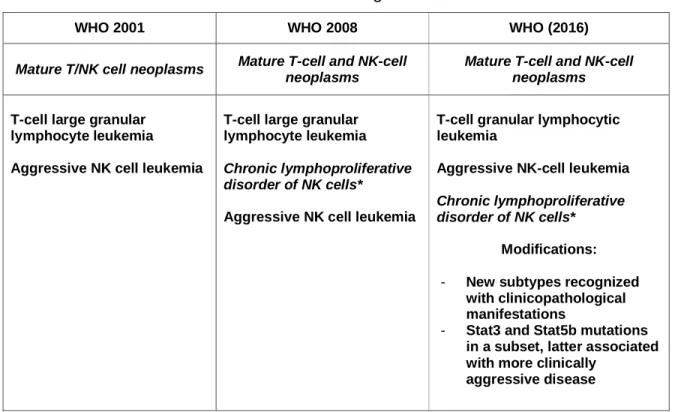
Table 1: Classification of LGL leukemia according to the WHO classification
WHO 2001 WHO 2008 WHO (2016)
Mature T/NK cell neoplasms Mature T-cell and NK-cell neoplasms
Mature T-cell and NK-cell neoplasms T-cell large granular
lymphocyte leukemia Aggressive NK cell leukemia
T-cell large granular lymphocyte leukemia Chronic lymphoproliferative disorder of NK cells*
Aggressive NK cell leukemia
T-cell granular lymphocytic leukemia
Aggressive NK-cell leukemia Chronic lymphoproliferative disorder of NK cells*
Modifications:
- New subtypes recognized with clinicopathological manifestations
- Stat3 and Stat5b mutations in a subset, latter associated with more clinically
aggressive disease
Table 2: Principal associated diseases with LGL leukemia
Associated diseases with LGL leukemia Frequency Neoplasms 4 to 10% Autoimmune cytopenia PRCA AIHA ITP Evans syndrome 5 %
B-cell lymphoid neoplasms Low-grade NHL
DLBCL
Mantle cell lymphoma Multiple myeloma CLL
Hairy cell leukemia
Waldenstrom macroglobulinemia Hodgkin lymphoma
Lymphomatoid granulomatosis Heavy chain disease
5%
Autoimmune diseases/connective tissue disorders Rheumatoid arthritis
Systemic lupus erythematosus Vasculitis Systemic sclerosis Endocrinopathy APECED Type I MEN Hashimoto Grave disease CIBD Celiac disease Gougerot-Sjogren syndrome Glomurolenephritis Polymyositis
Inclusion body myositis Poly/multinevritis RPA
Inflammatory arthritis (unclassified) Lambert-Eaton myasthenic syndrome Good syndrome
Behcet disease Multiple sclerosis
Acquired factor VIII inhibitor
10 to 20% 10 to 18% Myelodysplasia 3 to 10% AML < 1% Hemophagocytic syndrome < 1% Pulmonary hypertension < 1%
Post organ or hematopoietic stem cell transplant < 1%
PRCA: pure red cell aplasia; AIHA: autoimmune hemolytic anemia;
ITP: idiopathic thrombocytopenic purpura; APECED: polyendocrinopathy-candidosis-ectodermal dystrophy; MEN: multiple endocrine neoplasia; CIBD: chronic
inflammatory bowel disease; RPA: rhizomelic pseudopolyarthritis; AML acute myeloid leukemia;
NHL: non-Hodgkin’s lymphoma; DLBCL: diffuse large B-cell lymphoma
Figure 1: Large granular lymphocytes on blood (A) and marrow smears (B)
Fig 1A: A typical large granular lymphocyte on blood smear. (Wright-Giemsa stain: original magnification x 1000 Caméra RETIGA 2000 ). (with the courtesy of Dr
Ly-Sunnaram. Rennes University Hospital)
Fig 1 Bone marrow smear showing numerous LGL with a basophilic neutrophil. (Wright-Giemsa stain: original magnification x 1000 Caméra RETIGA 2000). (with
Fig 2: Flow cytometry and TCR γ gene rearrangement analysis in LGL leukemia
A: Flow cytometry analysis of a typical case of T CD3+ LGL leukemia showing CD3+/CD5dim/CD8+ (dark green).
B: Flow cytometry analysis of a case of NK LGL leukemia showing a CD3-/CD8-/CD16+/CD56+ phenotype (pink). KIR monotypic expression using CD161 and NkB1.
C: Clonality assessment of the same case of fig 3A: (Left: specific Vβ MoAbs showing restricted expression of Vβ13.1 > 90%, Right: detection of clonal TCR γ gene recombination by geneScaning analysis, single peak in green).
Vβ13.1 92%
D: Stat 3 mutation detection in a case of T LGL leukemia using Sanger sequencing.
STAT3
wt
G
A A
C C
A
T
A
C
A
C
A A A G
C
A
2051 A>T
STAT3
mutated
Y640F
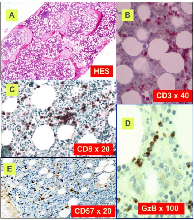
Figure 4: Bone marrow features of LGL leukemia
H&E staining of marrow biopsy reveals a slightly hypercellular marrow with subtle increase in interstitial lymphocytes (A). CD3 staining reveals the LGL interstitial infiltration. (B). Immunoperoxydase staining for CD8 demonstrates clusters of at least 8 cytotoxic lymphocytes CD8+. (C) Linear array of intravascular LGL demonstrated by Granzyme B staining (D). CD57 staining reveals the LGL interstitial infiltration (E). (with the courtesy of Pr
Figure 5: model of LGL Leukemia Pathogenesis unknown antigen Oligoclonal cytotoxic lymphocyte population resulting from an unknown antigen Monoclonal cytotoxic lymphocyte population with STAT3 activation/mutation leading to three outcomes. Chronic antigen stimulation STAT3 activation/ mutation
Production of inflammatory cytokines
Functional killer cells release cytotoxic granules
STAT3-mediated survival network
Outcome: persisting clone due to profound dysregulation of apoptosis (resistance to Fas/FasL mediated death, Mcl-1 upregulation...)
)
Outcomes: symptoms (neutropenia, anemia, fatigue), autoimmune disease (RA)
STAT3
apoptosis
Outcomes: symptoms (neutropenia, anemia, fatigue), autoimmune disease (RA)
1
2
3
Figure Legends: Activation and expansion occur, resulting in an oligoclonal cytotoxic lymphocyte population (colored outline represents a distinct clone, blue triangle represents an unknown antigen). STAT3 is activated and may acquire a mutation. Chronic antigen stimulation leads to expansion of one dominant (monoclonal) cytotoxic lymphocyte population (all are outlined in black signifying the monoclonal population). Three outcomes occur. In panel 1, production of inflammatory cytokines (starburst shapes representing: IFN-γ, IL-8, IL-10, IL1-β, IL-12p35, IL-18, IL1Ra, RANTES, MIP1-α,
MIP1-β) causes symptoms such as neutropenia, anemia, and fatigue, and can also cause
autoimmune disease such as rheumatoid arthritis (RA). In panel 2, the functional killer cells release cytotoxic granules containing perforin and granzyme B (purple dots); this leads to the same outcomes as in 1. In panel 3, the STAT3-mediated survival network results in a persisting clone due to profound dysregulation of apoptosis, including resistance to Fas/FasL mediated death and upregulation of Mcl-1.
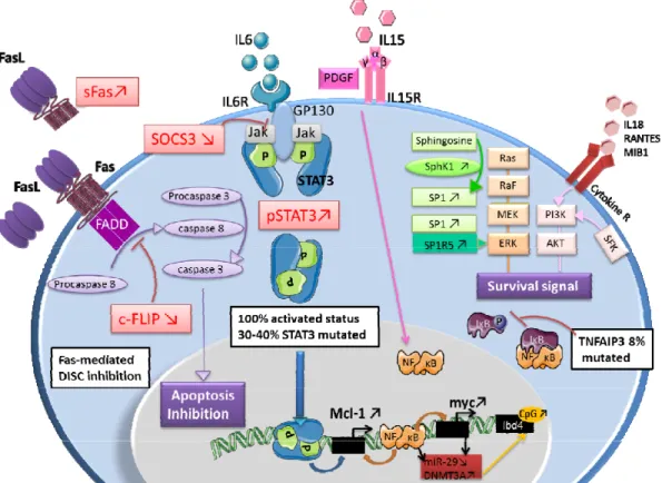
Figure 6: Key molecular abnormalities in LGL leukemia
Figure Legends:
Fas-mediated DISC inhibition: LGL leukemia cells are resistant to Fas mediated apoptosis. Soluble
Fas (sFas) acts as a decoy receptor able to inhibit Fas-dependent apoptosis. Increased level of an inhibitory protein named c-FLIP contributes to the DICS formation defect. Jak/Stat3 pathway: Stat3 is constitutively activated in LGL leukemia and is responsible for the transcription of Bcl2 and Mcl1 protein expression. Inhibition of Stat3 restores apoptosis of LGL cells whatever Stat3 mutation status witch implies that Stat3 mutation is not itself mandatory to explain LGL clonal expansion. SOCS3 witch inhibits Jak/Stat3 pathway is significantly decreased in LGL leukemia. Survival signal: LGL leukemia shows a predominant expression of pro-survival sphingolipids (S1P). SphK1 which converts
sphingosine into SP1 is increased in LGL leukemia and SphK1 inhibition leads to leukemic LGLs apoptosis. SP1 binding to SP1R activate pro survival signal through ERK1/2 signaling. Moreover, expression of S1P receptor, mainly S1PR5, is increased in LGL leukemia. Ras-Raf-1-MEK1-ERK, PI3K/AKT pathway are upregulated in LGL leukemia and they inhibition lead to LGL apoptosis. Increased activity of the PI3K-AKT signaling axis is found in T-LGL cells and participate to apoptotic inhibition. NFκB activity is upregulated in LGL leukemia. NFκB acts downstream of the PI3K-AKT pathway to prevent apoptosis through Mcl-1 independently of Stat3. A recurrent non-synonymous
mutation in the gene encoding an NFκB signaling inhibitor, TNFAIP3, was found in 8% of LGL
leukemia patients. IL15 and PDGF: IL15 promotes myc expression through NFKB pathway (model of IL15 transgenic mouse). IL15 is associated to an increase of global DNA methylation level in LGL leukemia through DNMT3A upregulation. The down regulation of miR-29 is responsible for the up regulation of DNMT3A witch induce methylation of the tumor suppressor gene Ibd4.
Abbreviations: Fas: First Apoptosis Signal, FasL : FasLigand, sFAS : soluble Fas, FADD : Fas-associated protein with death domain, c-FLIP : cellular FADD-like IL1 converting enzyme inhibitory protein, DISC : death inducing signaling complex, IL6: interleukin-6, IL6R: IL6 receptor, GP130: Glycoprotein 130, Jak: Janus Kinase, STAT3 : Signal transducer and activator of transcription 3, pSTAT3: phosphoSTAT3, IL15 Interleukin15, IL15R: IL15 receptor, PDGF : platelet-derived growth factor, SphK1: Sphingosine kinase 1, SP1: specificity protein 1, SP1R5 : specificity protein 1 receptor 5, Ras : Raf MEK : mitogen-activated protein kinase, ERK : extracellular-signal-regulated kinase, PI3K :phosphatidyl Inositol 3-Kinase, SFK : Src Family Kinase, NFκB : nuclear factor kappa B, Mcl1 : Myeloid cell leukemia1,