Robustness and efficacy of an inhibitory consortium against E. coli O26:H11 in
raw milk cheeses
Marie Frétina, Christophe Chassarda, Céline Delbèsa, René Lavignea, Etienne Rifaa, Sébastien Theila, Benoit Fernandezb, Patrice Laforceb, CécileCallona*
aUniversité Clermont Auvergne, INRAE, VetAgro Sup, UMR 545 Fromage, 20 Côte de Reyne, F-15000 Aurillac, France
bLallemand Specialty Cultures SAS, 19 rue des briquetiers 31 700 Blagnac, France
* Corresponding author. Tel: 33 (0)4 71 45 64 12; fax: 33 (0)4 71 45 64 13. E-mail address: cecile.callon@inra.fr (C. Callon).
Abstract (198 words)
Safety in raw milk cheeses being a major public health issue, the aim of this study was to validate a new bio preservation strategy by evaluating the efficacy of an inhibitory bacterial consortium (Hafnia alvei, Lactobacillus plantarum and Lactococcus lactis) on the growth of E. coli O26:H11 in uncooked pressed cheeses manufactured with different raw milk batches (6 farms, 3 periods). The pathogen was inoculated at very low concentrations (0.5 and 0.05 cfu mL-1), close to reality. The inhibitory power of the consortium was determined by culture analyses, and 16S rDNA sequencing of milk batches and cheeses was performed to evaluate the impact of milk microbial composition on the consortium's inhibition capacities. Raw milk batches differed in their fat and protein contents, microbial counts and diversity indices. The consortium's strong inhibitory power and adaptability were confirmed by a reduction of STEC levels (average of 2.8 log cfu g-1) in all cheeses, whatever the level of STEC inoculated into the milk. Differences in the growth and inhibition of E. coli in the cheeses depended on the microbial composition of the raw milk batches. Further research using a transcriptomic approach will help to improve understanding of the interactions between the strains.
Keywords:
Bio preservation; inhibitory consortium; Shiga toxin producing E. coli; raw milk batches; cheeses; 16S rRNA gene metabarcoding.
1. Introduction
Shiga-toxin-producing Escherichia coli (STEC) are widely recognized as emerging pathogens causing foodborne disease, and the control and prevention of milk-borne food pathogens are of prime importance for public health. The STEC serotype E. coli O157:H7 is associated with the majority of outbreaks. Non-O157 E. coli serogroups such as O26, O45, O103, O111, O121 and O145 have also been responsible for infections (Mathusa, Chen, Enache, & Hontz, 2010). It is difficult to have a general overview of STEC contamination in cheese because measurement methods have varied from one country to another. Worldwide, serotype O26:H11 is second only to E. coli O157:H7 for causing HUS (Hemolytic Uremic Syndrome), and is the one most often found in dairy products such as raw-milk cheeses (Bonanno, Delubac, Michel, & Auvray, 2017). Miszczycha et al. (2014) showed that E. coli O26:H11 grew better and was more persistent than E. coli O157:H7 in various experimentally contaminated raw-milk cheeses. E. coli O26:H11 is better adapted to uncooked pressed cheeses with short ripening times than to other cheese technologies such as cooked cheeses (cow’s milk), blue sheep's milk cheeses or lactic goat’s milk cheeses (Miszczycha et al. (2013).
Raw milk is a potential source of food-borne pathogens. The presence of STEC in milk is likely to arise from the farm environment, and especially direct contamination by faecal matter during milking (Baylis, 2009; Farrokh et al., 2013). Several studies have shown that faecal shedding of STEC can vary depending on the season, with an increase during the summer months (Berry & Wells, 2010; Fernández, Rodríguez, Arroyo, Padola, & Parma, 2009; Hancock, Besser, Rice, Herriott, & Tarr, 1997; Van Donkersgoed et al., 2001). As ruminants are healthy carriers of STEC, it is difficult to eradicate milk contamination at farm level. It is therefore important to control STEC from milk production to cheese-making.
The microbial and biochemical characteristics of raw milk batches play an important role in the safety and sensory properties of cheeses. Michel, Hauwuy, & Chamba (2001)
showed that the microbial and/or biochemical composition of raw milk could influence the growth of a STEC strain in raw-milk cheeses.
Environmental factors, the cows' feeding system and the season have a considerable influence on milk composition. Microbial composition can also be influenced by a combination of milk production practices (e.g. cow cleanliness, feeding, housing conditions) and by geographical origin (Kim et al., 2017; Mallet et al., 2012; Verdier-Metz, Michel, Delbès, & Montel, 2009). Seasonal variations result in varied milk composition, mainly due to animal feeding practices. Pasture feeding not only has a direct effect on the nutritional value
of the milk but may also result in a lower microbial load in the raw milk (Nateghi, Yousefi, Zamani, Gholamian, & Mohammadzadeh, 2014). The preservation of microbial diversity is necessary to benefit from all its potentialities: microbial ecosystems can have a protective effect or inhibit some pathogenic and spoilage microorganisms (Millet, Saubusse, Didienne, Tessier, & Montel, 2006; Retureau, Callon, Didienne, & Montel, 2010).
Biopreservation has attracted increasing interest as a means of naturally managing the microbiological safety of raw-milk cheeses. Some microorganisms isolated from such cheeses have proved to be antagonistic towards foodborne pathogens. Studies have reported the considerable contribution of lactic acid bacteria (LAB), in particular the genera Enterococcus, Lactobacillus, Lactococcus, Leuconostoc and Streptococcus (Buriti, Haíssa, Cardarelli, & Saad, 2007; Favaro, Barretto Penna, & Todorov, 2015; González et al., 2007; Hajikhani, Beyatli, & Aslim, 2007; Milioni et al., 2015; Ogawa et al., 2001) as biocontrol agents. This antibacterial activity may often be due to the production of organic acids, with a consequent reduction in the pH of the cheese, or the production of antimicrobial substances such as H2O2, diacetyl and bacteriocins (Dal Bello et al., 2010). Only a few bacterial species are able to inhibit STEC in cheeses. Among the species of Hafnia alvei, a member of the Enterobacteriaceae family and often isolated from raw milk and various types of cheese, one strain has been shown to be able to reduce the growth of E. coli O26:H11 in uncooked pressed cheeses (Callon, Arliguie, & Montel, 2016; Delbès-Paus et al., 2013). The inhibitory potential of several strains antagonistic to E. coli O26:H11 in association with H. alvei has been screened to compare their effectiveness (Callon et al., 2016). The consortium H. alvei, Lactobacillus plantarum and Lactococcus lactis was the most interesting one, decreasing E. coli O26:H11 and O157:H7 populations in cheeses by 3 log cfu g-1 when inoculated into milk at 10² cfu mL-1. However, very few protective cultures are currently marketed, underlining the difficulty of developing such effective cultures for the cheese industry.
The infective dose of pathogenic STEC is very low: only about ten viable cells are required (Schmid-Hempel & Frank, 2007). In cheeses, this low number is difficult to quantify and experiments have often been undertaken with higher inoculation levels. The aim of the present study was to validate a bio preservation strategy against E. coli in raw milk cheese. To do this, we evaluated the efficiency of an inhibitory consortium (H. alvei, Lactobacillus plantarum and Lactococcus lactis) on the growth of a Shigatoxin-producing E. coli O26:H11 strain in uncooked pressed cheeses made from different raw milk batches inoculated at very low concentrations, close to reality. Given the microbial variability of raw milk (see above), the robustness and efficiency of the inhibitory consortium was tested with raw milk batches
taken from six different farms and at three different times of year. Microbial analyses performed to determine the inhibitory power of the consortium, and 16S rDNA high throughput sequencing of the milk batches and cheeses, enabled us to evaluate the impact of the microbial composition of the raw milk batches on their inhibition properties.
2. Materials and methods
2.1 Strains and culture conditions
2.1.1 Anti-microbial consortium
Two strains from the collection held by UMRF 545 (Unité Mixte de Recherche sur le Fromage, INRA, France) (Lactobacillus plantarum (FH3) and Lactococcus lactis (D5.3)) and one strain from the UMR 782 AgroParisTech collection (Hafnia alvei (B16)) were used in the present study. All strains were isolated from milk products. They were revived and counted on Man Rogosa Sharpe (MRS), Terzaghi and Sandine M17 and Brain Heart Infusion (BHI), respectively. They were kept in cryobeads (AEB 400100; AES Laboratories, Combourg, France) and maintained at -80°C until the cheese-making day.
2.1.2. STEC strain
Escherichia coli O26:H11 strain 21765, isolated from Camembert cheese implicated in a human HUS case in 2005, was provided by Laboratoire d’Etudes des Microorganismes Alimentaires Pathogènes (VetAgro Sup, Marcy l’Etoile, France). The strain was incubated in 9 mL of BPW (Buffered Peptone Water) broth at 37°C overnight, and then stored at 4°C for 24 h until inoculation. The E. coli O26:H11 culture was counted on BHI agar medium incubated at 37°C for 24 h.
2.2 Experimental design and cheese-making
Cheeses were made at three different periods of the year (P1: winter, P2: spring and P3: summer). Raw milk batches (10 L) were collected after milking on six farms located within a 50 km diameter area in central France and selected according to their milk production practices, giving a range from the most intensive to the most extensive systems (F1, F2, F3, F4, F5, F6). Milk was transferred to the experimental cheese plant under refrigerated conditions and stored at 4°C overnight. Uncooked pressed cheeses (450 g, Saint-Nectaire-type cheeses) were manufactured with 5 L of each raw milk batch. On each cheese-making day, two cheeses were made from each milk batch. For the controls, the milk was inoculated only with the STEC strain at 0.5 or 0.05 cfu mL-1 and a commercial starter culture (MY800,
Streptococcus thermophilus, Lactobacillus delbrueckii spp. bulgaricus, Danisco, Paris La Défense, France) at 6.106 cfu mL-1. For the assayed cheeses, a consortium composed of H. alvei (B16), Lb. plantarum (FH3) and Lc. lactis (D5.3) was also added at 107 cfu mL-1. Then, 2 mL of rennet (Beaugel 500, Villefranche sur Soane, France) containing 520 mg of active chymosin per liter was added to each vat. The inoculated milk was processed according to the uncooked pressed cheese technology described by Callon, Saubusse, Didienne, Buchin, & Montel (2011). Cheeses were washed at 5 and 8 days of ripening with a commercial culture of Penicillium fuscoglaucum and Debaryomyces hansenii (Laboratoire Interprofessionnel de Production, Aurillac, France) and ripened in INRA’s cellars for 28 days (9°C, 96% relative humidity and 5% ventilation).
2.3 Milk and cheese sampling and culture analysis
Raw milk samples from each vat were analysed the day before the cheese-making day. Mesophilic bacteria were enumerated on Plate Count Agar (PCA) with milk at 30°C for 48 h, and coliforms were counted on Violet Red Bile Glucose (VRBG) agar incubated at 30°C (total coliforms) and 42°C (faecal coliforms). An enrichment step was performed to check whether STEC strains could be detected in these raw milk batches. Therefore, ten millilitres of raw milk was added to 90 mL of BPW supplemented with cefixim-tellurite (Savoye, Rozand, Bouvier, Gleizal, & Thevenot, 2011), incubated overnight at 42°C and enumerated after 24 h at 42°C on Chrom ID coli medium (Biomerieux, France) with cefixim-tellurite, following the manufacturer’s recommendations.
In inoculated milk batches, H. alvei (B16) was enumerated on Plate Count Agar with cristal violet as Gram-positive inhibitor (PCAI) at 30°C for 24 h, Lb. plantarum (FH3) on Facultative Heterofermentative (FH) (Isolini, Grand & Glättli, 1990) incubated at 30°C for 3 days under anaerobic conditions (Anaerocult A, VWR International, Fontenay-sous-Bois, France), and Lc. lactis (D5.3) and St. thermophilus on M17 for 48 h at 30°C or 42°C respectively, in order to confirm their inoculation levels in the raw milk batches.
Core cheese samples (25g) were taken at 6 h, 24 h, 8 d, 18 d and 28 d and STEC strain was enumerated by serial dilutions plated on Chrom ID coli medium with cefixim-tellurite incubated at 42°C for 24 h. When the STEC strain could not be enumerated in a cheese sample at 28 d (counts of STEC <1 log cfu mL-1, supplementary data Table S2), an enrichment step was also performed. Ten g of cheese were blended with 90 mL of BPW supplemented with cefixim-tellurite incubated overnight at 42°C and enumerated after 24 h at 42°C on Chrom ID coli medium.
The strains' inhibitory power (IP) was expressed as delta log cfu g-1 (log cfu g-1 of STEC in control cheeses, - log cfu g-1 of STEC in assay cheeses).
2.4 Physical-chemical analysis
Samples were taken from the vat milk used for cheese-making before the strains were added. Fat, protein and lactose contents were assessed by an automated infrared test method using a MilkoScan apparatus (Milkoscan FT 6000 milk analyzer, Foss Electric, Hillerød, Denmark) according to the FIL-IDF 141B method (IDF, 1996). Milk and cheese pH was determined at each sampling using a 926 VTV pH-meter with Ingold electrode 406 MX (Mettler-Toledo S.A., Viroflay, France).
2.5 16S rRNA gene sequencing of microbial communities in raw milk batches and cheeses 2.5.1 DNA extraction
To extract DNA from the raw milk batches, 7 mL of SDS (sodium dodecyl sulphate 20% solution; Sigma-Aldrich, Japan) was added to 70 mL milk samples and heated at 30°C for 30 min. The fat layer and the supernatant were extracted after 30 min of centrifugation (5,300 g, 4°C). Next, 1 mL of PBS (phosphate buffered saline; Sigma-Aldrich, USA) was added to each cell pellet. The suspension was transferred to a 2-ml tube and centrifuged for 5 min at 13,000 g, at room temperature. The supernatant was removed and the pellet was stored at – 20°C.
The subsequent DNA extraction steps were identical for milk pellets and cheeses ripened for 28 days: total DNA was extracted from milk pellets and 28-day-old cheeses according to the phenol-chloroform extraction method adapted from Monnet, Correia, Sarthou, & Irlinger (2006). The milk pellets and cheese samples (10g were randomly picked up from cheese, homogenized and the DNA extraction was performed on 250 mg of the mixture) were suspended in a mixture composed of 250 µL of guanidine thiocyanate (4 M) in Tris-HCl (0.1 M, pH 7.8), 40 µL of N-lauryl sarcosine (100 g L-1) and 200-mg of zirconium beads (50:50, 0.1 mm and 0.5 mm diameters). The suspension was homogenized in a bead mill homogenizer (Precellys Evolution; Bertin Technologies SAS, Ozyme, France) (one 20-s run at a speed of 6,500 m/s). Next, 75 µL of a mixture of lysozyme (40 mg mL-1) / lyticase (5,000 U mL-1) / TES (Tris 50 mM, EDTA 1 mM, sucrose 6.7%, pH 8) was added to the suspension. After incubation at 37°C for 30 min, 40 µL of proteinase K (14 mg mL-1) and 200 µL of SDS 20% were added. The tubes were incubated for 30 min in a water bath at 55°C, and 200 µL of sodium phosphate buffer (0.2 M), 200 µL of a 50 mM acetate in 10 mM EDTA buffer, and
500 µL of phenol-chloroform-isoamyl alcohol (25:24:1, pH 8) were then added. The tubes were vigorously shaken (one 45-s run at a speed of 10,000 m/s). Suspensions were heated at 55°C for 2 min, chilled on ice for 2 min, shaken again for 45 s at 10,000 m/s, heated at 70°C for 2 min and chilled again on ice for 2 min. After centrifuging for 30 min at 18,000 g, each supernatant was transferred to a 2-mL Phase Lock Gel Heavy. Two washing steps were performed with phenol-chloroform-isoamyl alcohol and a third with chloroform-isoamyl alcohol (24:1). The aqueous phase was recovered, mixed with 5 µL of RNase A (20 mg/mL), and incubated for 30 min at 37°C. At the end, the nucleic acids were precipitated using Genomic DNA Clean & Concentrator-10 (ZymoResearch, Ozyme, USA) according to the manufacturer’s recommendations. All the DNA solutions were stored at -20°C.
2.5.2 Sequencing and data analysis
The 16S rRNA genes (1,450 bp) from milk samples were amplified using the universal bacterial primers W02 (5’-GNTACCTTGTTACGACTT-3’) and W18 (5’-AGAGTTTGATCMTGGCTCAG-3’) as described by Verdier-Metz et al. (2012). The variable region V3-V4 of the 16S rRNA gene (~510 bp) was amplified from 2 μL of extracted and pre-amplified DNA (milk) or extracted DNA (cheese) with primers MSQ-16SV3F (5′-TACGGRAGGCWGCAG-3′) (Poirier et al., 2018) and PCR1R-460 (5′-TTACCAGGGTATCTAATCCT-3′), as described by Frétin et al. (2018). The amplified products were sequenced using Illumina Miseq technology (INRA, GeT-PLaGE platform, Toulouse, France). Raw sequence data were deposited in the Sequence Read Archive of the National Center for Biotechnology Information (SRA accession: PRJNA578621).
The sequence data were processed and analysed by procedures previously described by Frétin et al. (2018), using the FROGS pipeline on the Galaxy interface (Escudié et al., 2018). Briefly, sequences were screened for quality using the following parameters: minimum sequence length of 400 bp, maximum sequence length of 500 bp, no ambiguous bases in the entire sequence and no mismatches in the primer sequence. Chimeras were removed and operational taxonomic units (OTUs) were clustered using Swarm, with an aggregation distance of 3. Only OTUs that made up 0.005% or more of the total sequences were considered (Bokulich et al., 2013). Taxonomy was assigned to the OTUs against a curated version of the SILVA132-16S database and then compared manually with that obtained against NCBI database.
Statistical analyses and boxplots were performed with R for Windows (version 3.5.0). The inhibitory activity of the consortium on STEC strains in raw-milk cheeses was analysed using paired t-test. The inhibitory power values were compared by the Benjamini-Hochberg statistical test. The Bray Curtis dissimilarity matrix was used to perform the ordination analysis by metric multidimensional scaling (MDS). The permutational MANOVA (PERMANOVA) test was performed to detect the effect of milk origin and cheese-making period on the milk microbiota.
To investigate the effect of the milk microbiota on the growth of E. coli O26:H11, the 36 raw milk samples were split into two groups, STEC+ and STEC-, based on the average E. coli O26:H11 count in the control cheeses (without the inhibitory consortium), taking into account the level of STEC inoculated: E. coli O26:H11 counts (The average of counts at 6h, 24h, 8d, 18d and 28d) above the median were classed as STEC+ and E. coli O26:H11 counts below the median were classed as STEC-. The averages of the STEC levels in the STEC- group were significantly different to those in the STEC+ group (Wilcox test, for 0.5 cfu STEC mL-1 inoculated, N=9, P<0.001, for 0.05 cfu STEC mL-1 inoculated, N=9, P<0.001) (Data not shown). Genus differences between the groups were assessed by pairwise comparison of sequence counts using Negative Binomial Wald Tests from the DESeq2 package (Love, Huber, & Anders, 2014).
Differences in the relative abundance of the consortium's 3 species (in terms of the STEC+ and STEC- groups) in different cheeses according to the milk batch used – milk origin (6 farms) and cheese-making period (3 periods) – were assessed by pairwise comparison of sequence counts using Negative Binomial Wald Tests from the DESeq2 package, Metacoder package and MetagenomeSeq package.
3. Results and Discussion
In our study, E. coli O26:H11 was artificially inoculated into raw milk at a very low level (less than 1 cfu mL-1), unlike most cheese manufacturing experiments where this level has usually been 10² cfu mL-1 (Callon et al., 2016; Miszczycha et al., 2013; Montet et al., 2009).
3.1 Characteristics of 36 raw milk batches
Enrichment of all raw milk batches confirmed that no STEC strain was present prior to inoculation.
The 36 raw milk batches were characterized by biochemical composition, total mesophilic flora and enterobacteria counts and alpha-diversity of bacterial community (Fig.
1). Biochemical analysis showed that the 36 raw milk batches differed widely in their fat and protein contents. Fat content was between 33.6 and 44.7 g L-1, with a median of 38.7 g L-1. Protein content was between 29.7 and 37.1 g L-1, with a median of 32.6 g L-1. These median values are in agreement with the average content of fat (35-40 g L-1) and protein (30-35 g L-1) in cow's milk (Vilain, 2010). The level of total mesophilic bacteria also varied widely between the 36 raw milk batches, ranging between 2.78 and 5.64 log cfu mL-1. The same trend was observed for total enterobacteria on VRBG at 30°C (from 0 to 4.95 log cfu mL-1) and at 42°C (from 0 to 4.58 log cfu mL-1). Species richness (Chao1) and diversity (Simpson and Shannon indices) were calculated for each raw milk batch. The Chao1 value was close to the number of OTUs detected, in all the milk batches. The diversity indices and the number of OTUs detected revealed less diversity in a few raw milk batches. However, the median of the Simpson index was 0.73. These data confirmed that the 36 milk batches used to make the cheeses were different from each other.
3.2 Inhibitory activity of the consortium against growth of an E. coli O26:H11 strain
The growth of E. coli O26:H11 in assay cheeses (with inhibitory consortium) and control cheese (without inhibitory consortium) was evaluated at different stages of ripening (6h, 24h, 8d, 18d and 28d), for both levels of STEC inoculation and for all milk batches and periods (Table S2; Fig. 2). Throughout ripening, the level of E. coli O26:H11 was significantly (P < 0.001) lower in assay cheeses (with consortium) than in control cheeses, for all cheeses (6 raw milk batches and 3 periods, N=18) and for both inoculation levels. The consortium showed a significant impact on the growth of STEC whichever level of STEC was inoculated into the milk.
In the control cheeses, the level of E. coli O26:H11 increased sharply during the first 6h of manufacturing and reached 2.2 and 3 log cfu g-1 in cheeses inoculated at 0.05 and 0.5 cfu mL-1 respectively. This effect has been attributed to the entrapment of bacteria in the curd during coagulation followed by the draining of the whey (Miszczycha et al., 2013; Schlesser et al., 2006; Vernozy-Rozand et al., 2005). The STEC strain reached its maximum level at 24h with 3.4 and 4.1 log cfu g-1,respectively. These results are in agreement with those of
Miszczycha et al. (2013) and Callon et al. (2016) who reported that the level of STEC in uncooked pressed cheese increased sharply during the first 6h of cheese making, reached the maximum at 24h and remained stable during cheese ripening. In the absence of the inhibitory consortium, the decrease in pH caused by the starter culture may have slowed the growth of E. coli O26:H11 before the STEC activated its acid resistance mechanisms (Montet et al.,
2009). The starter culture (which included S. thermophilus) helped to reduce the pH sharply during the first hours of cheese-making (Fig. S1). These results confirm that E. coli O26:H11 is well adapted to uncooked pressed cheeses (Miszczycha et al. (2013).
In the assay cheeses, the level of E. coli O26:H11 reached a plateau at 6h of maturation. For the 0.5 cfu mL-1 dose, its mean level was 1.8 log cfu g-1 at that stage and remained constant until the end of ripening. The inhibitory power (IP defined as log cfu g-1 of STEC in control cheeses, - log cfu g-1 of STEC in assay cheeses) of the consortium against the STEC strain varied from 1.2 to 3 delta log cfu g-1 during ripening. For the 0.05 cfu mL-1 dose, the mean level of STEC was below the detection threshold (0.7 log cfu g-1). The consortium's IP against the STEC strain was, on average, 2.7 delta log cfu g-1 from 24h. The presence of LAB in the inhibitory consortium (Lc. lactis D5.3 and Lb. plantarum FH3) favoured the decrease in pH (Fig. S1) by producing organic acids (e.g. lactic acid). The faster acidification due to molecular mechanisms of antagonistic strains may be involved in the inhibition of E. coli O26:H11 in the assay cheeses. The results showed a significant reduction of E. coli O26:H11 levels (on average 2.8 log cfu g-1) in all assay cheeses manufactured with different milk batches artificially contaminated by a small number of STEC cells. These results confirm previous research (Callon et al. 2016) in which these strains were first tested both on a high level of STEC (102 cfu mL-1) and on a low level of STEC (0.05 cfu mL-1), but with only one milk batch.
3.3 Influence of raw milk bacterial community on the growth of E. coli O26:H11
The relative abundance of OTUs in milks are presented in table S1. The rDNA-based metabarcoding analysis revealed 41 OTUs present in the raw milk batches. Metric multidimensional scaling (MDS) of the relative abundance of bacterial OTUs showed that bacterial community structure had a strong influence on the growth of E. coli O26:H11 in raw-milk cheeses (Fig. 3A). This result was supported by the PERMANOVA test (P = 0.007). Differential analysis of raw milk bacterial profiles after counting E. coli O26:H11 in control cheeses revealed numerous significant differences in the abundance of bacterial genera (Fig. 4) and pointed to a link between the milk microbiota and the growth of STEC in cheese. In the raw milk batches, 11 of 17 genera were found to differ significantly in their abundance depending on STEC group. The Lactococcus genus was among those that differed most significantly (Padj = 7.61e-04; log2FoldChange = 2.70) between the STEC+ and STEC- groups. The raw milk batches associated with the lowest growth of E. coli O26:H11 in the control cheeses (STEC- cheeses) were characterized by a greater abundance of the LAB
genera Lactococcus, Lactobacillus and Leuconostoc and also Acinetobacter, Serratia and Hafnia of the Enterobacteriaceae family and Macrococcus (ripening bacteria). Conversely, the abundance of Romboutsia and Paeniclostridium, which are both genera of the Peptostreptococcaceae family, and Turicibacter, was significantly higher in the raw milk batches associated with the highest levels of E. coli O26:H11 in control cheeses. Many of these microorganisms are part of the gut microbiota and the increase in their presence in milk may be due to greater faecal contamination in intensive dairy systems. The complex microbiota of raw milk is both a potential reservoir of pathogenic or spoilage microorganisms and a source of bacteria with bactericidal or bacteriostatic properties. Retureau et al. (2010)
showed the ability of the cheese microbiota to control and/or prevent the growth of Listeria monocytogenes in cheese. Bluma & Ciprovica (2015) observed that the diversity of LAB in raw milk was strictly related to herd management practices such as equipment, environmental hygiene and animal welfare.
The MDS also showed that milk origin had a strong impact (P < 0.001) on bacterial community structure (Fig. 3B). No difference in raw milk bacterial community structure was observed between farms F1 and F4, farms F2 and F4, or farms F1 and F2, but the other pairs tested were all significantly different. The origin of the milk also influenced the growth of E. coli O26:H11. The six milk batches from farm F1 (4, 10, 16, 22, 28, 34) and five milk batches from farm F2 (5, 11, 17, 29, 35) appeared to be more favourable to the growth of E. coli O26:H11 than the six milk batches from farm F4 (1, 7, 13, 19, 25, 31) and five milk batches from farm F5 (12, 18, 24, 30, 36) (Fig. 3A and 3B). Besides the raw milk bacterial community, several antimicrobial factors could slow the growth of E. coli O26:H11 in milk batches from farmsF4 and F5. The dairying system on farms F4 and F5 was considered more extensive than the one on farms F1 and F2. The extensive system was characterized by cows of local breeds fed a pasture-based diet, in contrast to intensive system with specialized dairy breeds fed mainly hay and concentrate. The cows' diet (e.g. pasture, hay, grass silage, or maize silage) affects milk fat composition, and free fatty acids released during lipolysis may have an antibacterial effect (Frétin et al., 2019). Other compounds with inhibitory activity, such as terpenes, have been found in the milk of pasture-fed cows (Bugaud, Buchin, Coulon, Hauwuy, & Dupont (2001)).
Milk samples were also clustered according to cheese-making period (P = 0.005) (Fig. 3C). Bacterial community structure differed significantly between periods P1 and P2 (Padj < 0.01) as well as between P1 and P3 (Padj < 0.05), but no difference was observed between periods P2 and P3. The growth of E. coli O26:H11 was higher in the raw-milk cheeses made
with the milk batches of period P1 period (winter) than in those made from milk batches of period P3 (summer) (Fig. 3A and 3C). Several authors have observed a change in raw milk microbiota over several months as weather conditions changed (Gao et al., 2017; Li et al., 2018). Overall, our results suggest that seasonal factors such as weather conditions on the farm can affect the propensity of milk to allow STEC to grow in cheese.
3.4 Influence of inhibitory consortium in uncooked pressed cheeses
The relative abundance of 16S OTUs in all cheeses are presented in table S3.
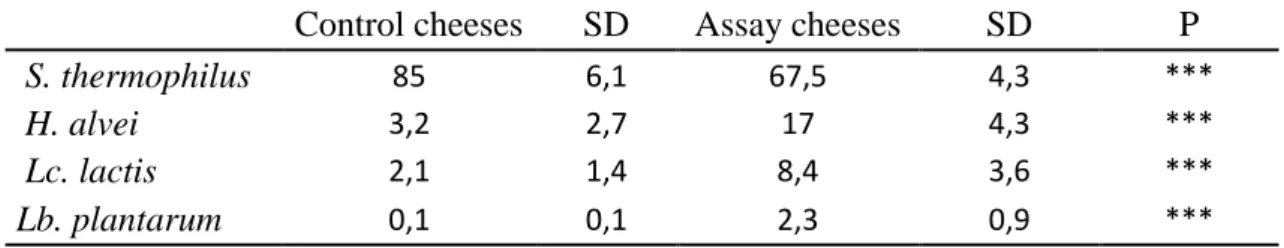
At 28 days of ripening, DNA sequences assigned to the species composing the anti-microbial consortium, namely H. alvei, Lc. lactis and Lb. plantarum were detected in greater relative abundance in the 36 assay cheeses than in the control cheeses (Table 1). The predominant strain was S. thermophilus which accounted for 67.5% of the bacterial sequences, followed by H. alvei 17%, Lc. lactis 8.5% and Lb. plantarum 2.3%. These four strains, inoculated into the milk, were the most abundant in the raw-milk cheeses at the end of ripening.
Differential analysis was also performed to compare the relative abundance of the four consortium species in 28-day-old cheeses depending on the two groups of raw milk batches (STEC+ and STEC-), the origins of the milk batches (6 farms) and the cheese making periods (3 periods). The relative abundance of the strains was not significantly different between these conditions (data not shown). These results suggest that the influence of the indigenous milk microbiota on the establishment and growth of the consortium strains is limited.
It is obvious that the starter culture and the consortium species constitute the dominant population in cheese, given their high level of inoculation in milk (6.106 and 107 cfu/mL). Inoculation of strains into milk at a high concentration, combined with inoculation with a starter culture, may change the microbial balance in cheese (Settanni & Moschetti, 2014). Indeed, the inoculation of the consortium is reflected in the diversity indices of the community. The richness was significantly lower in assay cheeses than in control cheeses (fig. 5) by 16S metabarcoding analyses. But the shannon indices indicated that a species was over-represented (starter St. thermophilus) in control cheeses whereas the dominant species (consortium strains and S. thermophilus) were more evenly balanced in assay cheeses. However, the decrease of the diversity could be a bias due to the method and a deeper sequencing would maybe reveal the presence of a larger diversity masked by several predominant species.
Altogether, these results suggest the anti-STEC consortium is very robust since the maximal level of E. coli O26:H11 in assay cheeses remained below 2.92 log cfu g-1 whatever the tested milk and the ripening stage. Differences observed in the inhibitory power among the assay cheeses (supplementary data Table S2) were probably not due to a differential growth of the inhibitory consortium in the cheeses. They may have been due to differential expression of the inhibitory properties of the consortium, depending on cheese indigenous microbiota composition. Differential analyses of the fat, protein and lactose contents of the STEC+ and STEC- groups of raw milk batches were also non-significant (results not shown), indicating that the growth of STEC in the cheeses was not correlated with the biochemical characteristics of the milk batches.
4. Conclusion
In this experiment, we aimed to evaluate the robustness of a bacterial consortium (composed of H. alvei B16, Lc. lactis D5.3 and Lb. plantarum FH3) against STEC in cheeses made from different raw milk batches. The pathogenic E. coli O26:H11 strain was inoculated at very low levels, close to those of natural contamination. This is the first time this inhibitory consortium has been tested on a large number of raw milk batches (n=36). We have shown its efficiency in all the raw-milk cheeses during the ripening, with an average inhibitory power of 2.8 log ufc g-1. The growth of E. coli O26:H11 differed from one cheese to another depending on the microbial composition of the raw milk and the period of milk production. Our results disclose an interaction between the milk microbiota and the growth of STEC. Several genera of LAB (Lactococcus, Lactobacillus, Leuconostoc) and of Gram negative bacteria (Acinetobacter, Serratia, Hafnia) were detected in greater abundance in raw milk batches used to make cheeses in which the growth of STEC was the weakest, in contrast to other genera (Romboutsia, Paeniclostridium and Turicibacter). We have highlighted that raw milk from two extensive farming system (farms F4 and F5) seemed to be less favourable to the growth of STEC.
Further research using a transcriptomic approach (e.g. RNA-Seq analysis) will be needed to better understand the positive or negative interactions between these strains.
Declaration of competing interest
Patrice Laforce and Benoit Fernandez are employed by Lallemand Specialty Cultures (Blagnac, France).
The remaining authors declare that the research was conducted in the absence of any commercial or financial relationships that could be construed as a potential conflict of interest.
Acknowledgements
This study was financially supported by Auvergne regional authority (France), by the European Community via FEDER and by Lallemand Specialty Cultures SAS (France) fundingunder a Bourse Innovation Transfert Program (contrat 23000923 projet 460).
References
Baylis, C. L. (2009). Raw milk and raw milk cheeses as vehicles for infection by Verocytotoxin-producing Escherichia coli. International Journal of Dairy Technology, 62(3), 293-307. https://doi.org/10.1111/j.1471-0307.2009.00504.x
Berry, E. D., & Wells, J. E. (2010). Chapter 4-Escherichia coli O157:H7 : Recent Advances in Research on Occurrence, Transmission, and Control in Cattle and the Production Environment. In S. L. Taylor (Éd.), Advances in Food and Nutrition Research (Vol. 60, p. 67-117). https://doi.org/10.1016/S1043-4526(10)60004-6
Bluma, A., & Ciprovica, I. (2015). Diversity of lactic acid bacteria in raw milk. Research for Rural Development. International Scientific Conference Proceedings (Latvia).
Consulté à l’adresse
http://agris.fao.org/agris-search/search.do?recordID=LV2016000350
Bokulich, N. A., Subramanian, S., Faith, J. J., Gevers, D., Gordon, J. I., Knight, R., … Caporaso, J. G. (2013). Quality-filtering vastly improves diversity estimates from Illumina amplicon sequencing. Nature Methods, 10(1), 57-59. https://doi.org/10.1038/nmeth.2276
Bonanno, L., Delubac, B., Michel, V., & Auvray, F. (2017). Influence of Stress Factors Related to Cheese-Making Process and to STEC Detection Procedure on the Induction of Stx Phages from STEC O26:H11. Frontiers in Microbiology, 8. https://doi.org/10.3389/fmicb.2017.00296
Bugaud, C., Buchin, S., Coulon, J.-B., Hauwuy, A., & Dupont, D. (2001). Influence of the nature of alpine pastures on plasmin activity, fatty acid and volatile compound composition of milk. Le Lait, 81(3), 14. https://doi.org/10.1051/lait:2001140
Buriti, F. C. A., Haíssa, Cardarelli, R., & Saad, S. M. I. (2007). Biopreservation by Lactobacillus paracasei in Coculture with Streptococcus thermophilus in Potentially Probiotic and Synbiotic Fresh Cream Cheeses. Journal of Food Protection, 70(1), 228-235. https://doi.org/10.4315/0362-028X-70.1.228
Callon, C., Arliguie, C., & Montel, M. C. (2016). Control of Shigatoxin-producing Escherichia coli in cheese by dairy bacterial strains. Food Microbiology, 53, 63-70. https://doi.org/10.1016/j.fm.2015.08.009
Callon, C., Saubusse, M., Didienne, R., Buchin, S., & Montel, M. C. (2011). Simplification of a complex microbial antilisterial consortium to evaluate the contribution of its flora in uncooked pressed cheese. International Journal of Food Microbiology, 145(2–3), 379-389. https://doi.org/10.1016/j.ijfoodmicro.2010.12.019
Dal Bello, B., Rantsiou, K., Bellio, A., Zeppa, G., Ambrosoli, R., Civera, T., & Cocolin, L. (2010). Microbial ecology of artisanal products from North West of Italy and antimicrobial activity of the autochthonous populations. LWT - Food Science and Technology, 43(7), 1151-1159. https://doi.org/10.1016/j.lwt.2010.03.008
Delbès-Paus, C., Miszczycha, S., Ganet, S., Helinck, S., Veisseire, P., Pochet, S., … Montel, M.-C. (2013). Behavior of Escherichia coli O26:H11 in the presence of Hafnia alvei in a model cheese ecosystem. International Journal of Food Microbiology, 160(3), 212-218. https://doi.org/10.1016/j.ijfoodmicro.2012.10.019
Escudié, F., Auer, L., Bernard, M., Mariadassou, M., Cauquil, L., Vidal, K., … Pascal, G. (2018). FROGS : Find, Rapidly, OTUs with Galaxy Solution. Bioinformatics, 34(8), 1287-1294. https://doi.org/10.1093/bioinformatics/btx791
Farrokh, C., Jordan, K., Auvray, F., Glass, K., Oppegaard, H., Raynaud, S., … Cerf, O. (2013). Review of Shiga-toxin-producing Escherichia coli (STEC) and their significance in dairy production. International Journal of Food Microbiology, 162(2), 190-212. https://doi.org/10.1016/j.ijfoodmicro.2012.08.008
Favaro, L., Barretto Penna, A. L., & Todorov, S. D. (2015). Bacteriocinogenic LAB from cheeses – Application in biopreservation? Trends in Food Science & Technology, 41(1), 37-48. https://doi.org/10.1016/j.tifs.2014.09.001
Fernández, D., Rodríguez, E. M., Arroyo, G. H., Padola, N. L., & Parma, A. E. (2009). Seasonal variation of Shiga toxin-encoding genes (stx) and detection of E. coli O157 in dairy cattle from Argentina. Journal of Applied Microbiology, 106(4), 1260-1267. https://doi.org/10.1111/j.1365-2672.2008.04088.x
Frétin, M., Martin, B., Buchin, S., Desserre, B., Lavigne, R., Tixier, E., … Ferlay, A. (2019). Milk fat composition modifies the texture and appearance of Cantal-type cheeses but not their flavor. Journal of Dairy Science, 102(2), 1131-1143. https://doi.org/10.3168/jds.2018-15534
Frétin, M., Martin, B., Rifa, E., Isabelle, V.-M., Pomiès, D., Ferlay, A., … Delbès, C. (2018). Bacterial community assembly from cow teat skin to ripened cheeses is influenced by grazing systems. Scientific Reports, 8(1), 200. https://doi.org/10.1038/s41598-017-18447-y
Gao, M. L., Hou, H. M., Teng, X. X., Zhu, Y. L., Hao, H. S., & Zhang, G. L. (2017). Microbial diversity in raw milk and traditional fermented dairy products (Hurood cheese and Jueke) from Inner Mongolia, China. Genetics and Molecular Research : GMR, 16(1). https://doi.org/10.4238/gmr16019451
González, L., Sandoval, H., Sacristán, N., Castro, J. M., Fresno, J. M., & Tornadijo, M. E. (2007). Identification of lactic acid bacteria isolated from Genestoso cheese throughout ripening and study of their antimicrobial activity. Food Control, 18(6), 716-722. https://doi.org/10.1016/j.foodcont.2006.03.008
Hajikhani, R., Beyatli, Y., & Aslim, B. (2007). Antimicrobial activity of enterococci strains isolated from white cheese. International Journal of Dairy Technology, 60(2), 105-108. https://doi.org/10.1111/j.1471-0307.2007.00304.x
Hancock, D. D., Besser, T. E., Rice, D. H., Herriott, D. E., & Tarr, P. I. (1997). A longitudinal study of Escherichia coli O157 in fourteen cattle herds. Epidemiology & Infection, 118(2), 193-195. https://doi.org/10.1017/S0950268896007212
IDF. 1996. Whole milk: Determination of milk fat, protein and lactose content-guide for the operation of mid-infra-red instruments. IDF Standard 141B. International Dairy Federation, Brussels, Belgium.
Isolini, D., Grand, M., Glättli, H. (1990). Selektivmedien zum Nachweis von obligat und fakultativ heterofermentativen Laktobazillen. Schweizerische milchwirtschaftliche Forschung 19, 57-59.
Kim, I. S., Hur, Y. K., Kim, E. J., Ahn, Y.-T., Kim, J. G., Choi, Y.-J., & Huh, C. S. (2017). Comparative analysis of the microbial communities in raw milk produced in different regions of Korea. Asian-Australasian Journal of Animal Sciences, 30(11), 1643-1650. https://doi.org/10.5713/ajas.17.0689
Li, N., Wang, Y., You, C., Ren, J., Chen, W., Zheng, H., & Liu, Z. (2018). Variation in Raw Milk Microbiota Throughout 12 Months and the Impact of Weather Conditions. Scientific Reports, 8(1), 2371. https://doi.org/10.1038/s41598-018-20862-8
Love, M. I., Huber, W., & Anders, S. (2014). Moderated estimation of fold change and dispersion for RNA-seq data with DESeq2. Genome Biology, 15(12), 550. https://doi.org/10.1186/s13059-014-0550-8
Mallet, A., Guéguen, M., Kauffmann, F., Chesneau, C., Sesboué, A., & Desmasures, N. (2012). Quantitative and qualitative microbial analysis of raw milk reveals substantial diversity influenced by herd management practices. International Dairy Journal, 27(1), 13-21. https://doi.org/10.1016/j.idairyj.2012.07.009
Mathusa, E. C., Chen, Y., Enache, E., & Hontz, L. (2010). Non-O157 Shiga Toxin–Producing Escherichia coli in Foods. Journal of Food Protection, 73(9), 1721-1736. https://doi.org/10.4315/0362-028X-73.9.1721
Michel, V., Hauwuy, A., & Chamba, J.-F. (2001). La flore microbienne de laits crus de vache : Diversité et influence des conditions de production. Le Lait, 81(5), 18. https://doi.org/10.1051/lait:2001151
Milioni, C., Martínez, B., Degl’Innocenti, S., Turchi, B., Fratini, F., Cerri, D., & Fischetti, R. (2015). A novel bacteriocin produced by Lactobacillus plantarum LpU4 as a valuable candidate for biopreservation in artisanal raw milk cheese. Dairy Science & Technology, 95(4), 479-494. https://doi.org/10.1007/s13594-015-0230-9
Millet, L., Saubusse, M., Didienne, R., Tessier, L., & Montel, M. C. (2006). Control of Listeria monocytogenes in raw-milk cheeses. International Journal of Food Microbiology, 108(1), 105-114. https://doi.org/10.1016/j.ijfoodmicro.2005.11.004 Miszczycha, S. D., Perrin, F., Ganet, S., Jamet, E., Tenenhaus-Aziza, F., Montel, M.-C., &
Thevenot-Sergentet, D. (2013). Behavior of Different Shiga Toxin-Producing Escherichia coli Serotypes in Various Experimentally Contaminated Raw-Milk Cheeses. Appl. Environ. Microbiol., 79(1), 150-158. https://doi.org/10.1128/AEM.02192-12
Miszczycha, S. D., Thévenot, J., Denis, S., Callon, C., Livrelli, V., Alric, M., … Thevenot-Sergentet, D. (2014). Survival of Escherichia coli O26:H11 exceeds that of Escherichia coli O157:H7 as assessed by simulated human digestion of contaminated raw milk cheeses. International Journal of Food Microbiology, 172, 40-48. https://doi.org/10.1016/j.ijfoodmicro.2013.11.029
Monnet, C., Correia, K., Sarthou, A.-S., & Irlinger, F. (2006). Quantitative Detection of Corynebacterium casei in Cheese by Real-Time PCR. Appl. Environ. Microbiol., 72(11), 6972-6979. https://doi.org/10.1128/AEM.01303-06
Montet, M. P., Jamet, E., Ganet, S., Dizin, M., Miszczycha, S., Dunière, L., … Vernozy-Rozand, C. (2009). Growth and Survival of Acid-Resistant and Non-Acid-Resistant Shiga-Toxin-Producing Escherichia coli Strains during the Manufacture and Ripening of Camembert Cheese [Research article]. https://doi.org/10.1155/2009/653481
Nateghi, L., Yousefi, M., Zamani, E., Gholamian, M., & Mohammadzadeh, M. (2014). The effect of different seasons on the milk quality. 4(1), 550-552.
Ogawa, M., Shimizu, K., Nomoto, K., Tanaka, R., Hamabata, T., Yamasaki, S., … Takeda, Y. (2001). Inhibition of in vitro growth of Shiga toxin-producing Escherichia coli O157:H7 by probiotic Lactobacillus strains due to production of lactic acid. International Journal of Food Microbiology, 68(1), 135-140. https://doi.org/10.1016/S0168-1605(01)00465-2
Poirier, S., Rué, O., Coeuret, G., Champomier-Vergès, M.C., Louxand, V., Chaillou, S. (2018). Detection of an amplification bias associated to Leuconostocaceae family with a universal primer routinely used for monitoring microbial community structures within food products. BMC Research Notes, 11:802. https://doi.org/10.1186/s13104-018-3908
Retureau, E., Callon, C., Didienne, R., & Montel, M.-C. (2010). Is microbial diversity an asset for inhibiting Listeria monocytogenes in raw milk cheeses? Dairy Science & Technology, 90(4), 375-398. https://doi.org/10.1051/dst/2010010
Savoye, F., Rozand, C., Bouvier, M., Gleizal, A., & Thevenot, D. (2011). Optimized enrichment for the detection of Escherichia coli O26 in French raw milk cheeses : Enrichment optimization of E. coli O26. Letters in Applied Microbiology, 52(6), 603-609. https://doi.org/10.1111/j.1472-765X.2011.03044.x
Schlesser, J. E., Gerdes, R., Ravishankar, S., Madsen, K., Mowbray, J., & Teo, A. Y.-L. (2006). Survival of a Five-Strain Cocktail of Escherichia coli O157:H7 during the 60-Day Aging Period of Cheddar Cheese Made from Unpasteurized Milk. Journal of Food Protection, 69(5), 990-998. https://doi.org/10.4315/0362-028X-69.5.990
Schmid-Hempel, P., & Frank, S. A. (2007). Pathogenesis, Virulence, and Infective Dose. PLOS Pathogens, 3(10), e147. https://doi.org/10.1371/journal.ppat.0030147
Settanni, L., & Moschetti, G. (2014). New trends in technology and identity of traditional dairy and fermented meat production processes : Preservation of typicality and
hygiene. Trends in Food Science & Technology, 37(1), 51-58. https://doi.org/10.1016/j.tifs.2014.02.006
Van Donkersgoed, J., Berg, J., Potter, A., Hancock, D., Besser, T., Rice, D., … Klashinsky, S. (2001). Environmental sources and transmission of Escherichia coli O157 in feedlot cattle. The Canadian Veterinary Journal, 42(9), 714-720.
Verdier-Metz, I., Gagne, G., Bornes, S., Monsallier, F., Veisseire, P., Delbès-Paus, C., & Montel, M.-C. (2012). Cow Teat Skin, a Potential Source of Diverse Microbial Populations for Cheese Production. Applied and Environmental Microbiology, 78(2), 326-333. https://doi.org/10.1128/AEM.06229-11
Verdier-Metz, I., Michel, V., Delbès, C., & Montel, M.-C. (2009). Do milking practices influence the bacterial diversity of raw milk? Food Microbiology, 26(3), 305-310. https://doi.org/10.1016/j.fm.2008.12.005
Vernozy-Rozand, C., Mazuy-Cruchaudet, C., Bavai, C., Montet, M. P., Bonin, V., Dernburg, A., & Richard, Y. (2005). Growth and survival of Escherichia coli O157:H7 during the manufacture and ripening of raw goat milk lactic cheeses. International Journal of Food Microbiology, 105(1), 83-88. https://doi.org/10.1016/j.ijfoodmicro.2005.05.005 Vilain, A.-C. (2010). Qu’est-ce que le lait ? Revue Française d’Allergologie, 50(3), 124‑127.
Table 1 : Differential analyses on relative abundances (supplementary data Table S3) of the 3 species of the inhibitory consortium and of St. thermophilus in 28 days old cheeses according to the treatment (control/assay).
Control cheeses SD Assay cheeses SD P
S. thermophilus 85 6,1 67,5 4,3 ***
H. alvei 3,2 2,7 17 4,3 ***
Lc. lactis 2,1 1,4 8,4 3,6 ***
Lb. plantarum 0,1 0,1 2,3 0,9 ***
Fig 1
Characteristics of 36 raw milk batches before inoculation of bacterial strains: fat, protein and lactose content, microbial analysis by counts on selective media (total mesophilic bacteria on PCA, total Enterobacteria on VRBG at 30°C, faecal coliforms on VRBG at 42°C), diversity of microbial composition by 16S rDNA high throughput sequencing.
10 20 30 40 50 Observed Chao1 variable N um ber of bac terial OT U s div alpha Enterobacteria at 42°C Enterobacteria at 30°C Total Mesophilic bacteria
Fig 2
A) B)
Levels of the E. coli O26:H11 strain at each stage of ripening (6h, 24h, d8, d18, d28) in assay cheeses and control cheeses at two levels of E. coli O26:H11 inoculation A) E. coli O26:H11 inoculated at 0.5 cfu mL-1, and B) E. coli O26:H11 inoculated at 0.05 cfu mL-1. Values reported are the mean from 6 raw-milk cheeses made in each of 3 periods (n=18). *P < 0.05; **P < 0.01; ***P < 0.001
IP: inhibitory power of the consortium against the E. coli O26:H11 strain, expressed in delta log cfu g-1. Values with different superscript letters differ at P < 0.05 by statistical Benjamini-Hochberg test.
Fig 3
STEC+ STEC-
B) C)
Beta-diversity of 36 raw milk batches. Metric multidimensional scaling (MDS) based on the Bray Curtis algorithm of bacterial communities. A: comparison of milk samples for which the growth of E. coli O26:H11 in raw-milk cheeses during 28 days of ripening was high ( STEC+) vs. low ( STEC-). The numbers indicate the 36 milk samples, the details are given in Supplementary Table S1. B: comparison based on milk sample origin (6 milk producers, F1 to F6). C: comparison based on the 3 sampling periods. P-values obtained after permutational MANOVA analysis (Adonis statistical test) indicate significance between sample groups.
A)
24 Fig 4 1 2 3 4 5 6
Differential analysis performed on the sequence counts of 17 genera detected in raw milk 7
batches. The 36 raw milk samples were divided into two groups according to the count of E. 8
coli O26:H11 in the cheeses: E. coli O26:H11 count higher than the median (STEC+) and 9
E. coli O26:H11 count lower than the median (STEC-). The 11 genera identified here were 10
significantly different between the STEC+ and STEC- groups. 11 12 13 14 15 16 17 18 19 20 21 22 23 24 25 26 27 28 29
25 Fig 5 30 31 32 33 34 35 36 37 38 39 40 41 42 43 44 45 46 47 48 49 50 51 52 53 54 55 56 57
Alpha-diversity inferred from 16S rRNA sequence data of cheeses: number of observed 58
OTUs, Shannon diversity indices in assay and control cheeses. 59
*** The treatment factor was significant on observed diversity and Shannon indices 60 (ANOVA, P<0.001). 61 62 63 64 65 66 67 68 69 70 71 72 73 ** * ** * Alpha diversity indices