HAL Id: hal-01547108
https://hal.archives-ouvertes.fr/hal-01547108
Submitted on 18 Jan 2021
HAL is a multi-disciplinary open access
archive for the deposit and dissemination of
sci-entific research documents, whether they are
pub-lished or not. The documents may come from
teaching and research institutions in France or
abroad, or from public or private research centers.
L’archive ouverte pluridisciplinaire HAL, est
destinée au dépôt et à la diffusion de documents
scientifiques de niveau recherche, publiés ou non,
émanant des établissements d’enseignement et de
recherche français ou étrangers, des laboratoires
publics ou privés.
Multivalent Binding to Pathogenic Bacterial Lectins
K. Buffet, I. Nierengarten, N. Galanos, E. Gillon, M. Holler, A. Imberty, S.E.
Matthews, S. Vidal, S.P. Vincent, J.F. Nierengarten
To cite this version:
K. Buffet, I. Nierengarten, N. Galanos, E. Gillon, M. Holler, et al.. Pillar[5]arene-Based Glycoclusters:
Synthesis and Multivalent Binding to Pathogenic Bacterial Lectins. Chemistry - A European Journal,
Wiley-VCH Verlag, 2016, 22 (9), pp.2955-2963. �10.1002/chem.201504921�. �hal-01547108�
Pillar[5]arene-based glycoclusters: synthesis and multivalent
binding to pathogenic bacterial lectins
Kevin Buffet,
[a]Iwona Nierengarten,
[b]Nicolas Galanos,
[c,d]Emilie Gillon,
[d]Michel Holler,
[b]Anne
Imberty,*
[d]Susan E. Matthews,
[e]Sébastien Vidal,*
[c]Stéphane P. Vincent,*
[a]and Jean-François
Nierengarten*
[b]Abstract: The synthesis of pillar[5]arene-based glycoclusters has been readily achieved by CuAAC conjugations of azido- and alkyne-functionalized precursors. The lectin binding properties of the resulting glycosylated multivalent ligands have been studied by at least two complementary techniques in order to provide a good understanding. Three lectins were selected from bacterial pathogens based on their potential therapeutic applications as anti-adhesives, namely LecA and LecB from Pseudomonas aeruginosa and BambL from Burkholderia ambifaria. As a general trend, multivalency improved the binding to lectins and a higher affinity can be obtained by increasing to a certain limit the length of the spacer arm between the carbohydrate subunits and the central macrocyclic core.
Introduction
Several opportunistic pathogens involved in lung infections in immune-compromised patients have been shown to produce soluble lectins that bind to human oligosaccharide epitopes in lung tissues.[1] Among these, Pseudomonas aeruginosa and the
members of the Burkholderia cepacia complex (BCC) are of special interest because of their emerging resistance to
antibiotics, and the burden they represent for both cystic fibrosis patients and also patients under ventilation.[2] LecA from P.
aeruginosa is a galactose-specific lectin with a tetrameric,
calcium-dependent structure.[3] LecA is involved in destruction of
the alveolar barrier in animal models of infection[4] and has
recently been demonstrated to be necessary for the intracellular uptake of the bacterium.[5] Two soluble fucose-specific lectins
have been also characterized from these bacteria. LecB from P.
aeruginosa is also a calcium-dependent tetramer with strong
affinity for fucose and fucose-containing oligosaccharides such as the Lewis a antigen.[6, 7] LecB is a virulence factor that is
involved in adhesion on airway epithelial cells.[4] BambL from
Burkholderia ambifaria, a member of the BCC, is a trimer that
assembles as a -propeller with six-fucose binding sites.[8]
BambL binds to blood group oligosaccharides and is a close relative of AFL from, Aspergillus fumigatus, another airborne pathogen that triggers inflammation on cultured epithelial cells.[9]
LecB and BambL have very different topologies (Figure 1) and therefore can be expected to bind differently to multivalent ligands.
Figure 1. Crystal structures of three soluble bacterial lectins with peptide chains represented by ribbons with different colours, carbohydrate ligands represented as sticks, and calcium ions represented by green spheres. The following complexes are displayed from left to right: LecA/galactose (PDB code 1OKO), LecB/fucose (1GZT) and BambL/fucose (3ZZV).
The design of glycoclusters based on aromatic scaffolds has attracted much attention recently taking advantage of the intrinsic physico-chemical properties of the different cores.[10] We
have recently designed a series of multivalent scaffolds based on calixarenes,[11-15] resorcinarenes,[16] porphyrins,[12, 14, 17, 18] and
fullerenes.[19-27] In all cases, the grafting of carbohydrate
residues onto these central cores yielded glycoclusters displaying improved lectin binding properties in comparison to their monovalent natural ligands.[28, 29]
Since 2008, when the synthesis of pillar[5]arenes[30] was first
described, they and subsequently pillar[6]arenes[31] have rapidly
[a] Dr. K. Buffet, Prof. S. P. Vincent
University of Namur (UNamur), Académie Louvain
Département de Chimie, Laboratoire de Chimie Bio-Organique rue de Bruxelles 61, B-5000 Namur (Belgium)
E-mail: [email protected]
[b] Dr. I. Nierengarten, Dr. M. Holler, Dr. J.-F. Nierengarten Laboratoire de Chimie des Matériaux Moléculaires, Université de Strasbourg et CNRS (UMR 7509), Ecole Européenne de Chimie, Polymères et Matériaux (ECPM)
25 rue Becquerel, 67087 Strasbourg Cedex 2 (France) E-mail: [email protected]
[c] N. Galanos, Dr. S. Vidal
Institut de Chimie et Biochimie Moléculaires et Supramoléculaires, CO-Glyco, UMR , CNRS
Université Claude Bernard Lyon ,
Boulevard du Novembre , F- Villeurbanne (France) E-mail: [email protected]
[d] N. Galanos, E. Gillon, Dr. A. Imberty
CERMAV - CNRS and Université Grenoble Alpes BP 53, 38041, Grenoble (France)
E-mail: [email protected]
[e] Dr. S. E. Matthews School of Pharmacy University of East Anglia
Norwich, NR4 7TJ, United Kingdom.
Supporting information for this article is given via a link at the end of the document.
become major macrocyclic host molecules. Exploitation of the π-electron rich core has enabled applications in the preparation of sensors, rotaxanes, supramolecular polymers and vesicles.[32-40]
Unlike their sister molecules the calixarenes and resorcinarenes, pillararenes offer a uniquely rigid and symmetrical structure with inherent planar chirality and higher levels of functionality. Thus the per-functionalised materials offer 10 or 12 derivatisation points whilst, by using non-symmetrically functionalised alkoxybenzene derivatives, platforms featuring 5 or 6 functionalised positions can also be readily accessed. Additionally a number of different methods including co-cyclisation and post-co-cyclisation modification enable highly selective partial functionalisation.
The use of pillararenes in biological applications has to-date been minimal. Hou and co-workers have developed both pillar[5]arene dimers[41] and hydrazide functionalised
pillar[5]arenes[42] as transmembrane channel mimics for protons
and water molecules respectively. The potential of carboxyl functionalised pillararenes as controlled release drug delivery systems has been demonstrated through the formation of parquet-pillar[6]arene complexes which afforded a reduction in toxicity of the paraquat on complexation but also pH dependent release of the guest.[43] Additionally, the use of cationic
functionalised pillararenes for DNA complexation and gene delivery has recently been reported.[44] As part of this research,
we have reported the synthesis of a mannose glycocluster based on a per-functionalised pillar[5]arene scaffold which was able to inhibit the adhesion of Escherichia coli to red blood cells through the inhibition of FimH-mediated bacterial adhesion.[45]
Subsequently, Huang described the synthesis of pillar[5]arene derivatives featuring 5 galactose moieties and investigated their self-assembly properties into vesicles and on prolonged standing to nanotubes. These assemblies were shown to be of low toxicity to human cells and to facilitate agglutination of
Escherichia coli.[46]
In this study, we describe the development of a family of pillar[5]arene based glycoclusters incorporating galactose or fucose and their evaluation for binding to the lectins LecA and LecB from Pseudomonas aeruginosa and BambL from
Burkholderia ambifaria.
Results and Discussion
Synthesis
The most convenient method for the conjugation of two molecular entities is probably the Cu(I)-assisted azide-alkyne cycloaddition (CuAAC).[47-52] The high yields obtained in such
1,3-dipolar cycloadditions are particularly relevant when a high number of reactions must be performed at once on a single molecular scaffold. The synthesis of the pillar[5]arene-based glycoclusters was based on already developed azido- or alkyne-functionalized pillar[5]arene scaffolds.[44, 53-56] Conjugation of
deca-azide 1[56] with a series of propargylated derivatives (2a-f)
provided the acetylated glycoclusters 3a-f (Scheme 1). It is worth noting that a 1:1 mixture of diastereoisomers was obtained
upon conjugation of 1 with carbohydrates owing to the chirality of the pillar[5]arene ring system.[45] For all the compounds, their
separation was unsuccessful even after several careful column chromatographies. Although these isomeric mixtures could affect the interpretation of the binding studies towards lectins, the spatial arrangements of the carbohydrate epitopes for decavalent scaffolds will not differ greatly and the general presentation of the binding partners to the lectins will be statistically homogeneous. Finally, deprotection of the acetate ester moieties of 3a-f afforded the desired water-soluble glycoclusters 4a-f. While a single galactoside was used in this series, a number of different fucosides were incorporated to investigate the role of both the linker and valency in achieving selectivity and the interpretation of parameters governing the binding properties towards multimeric lectins. Thus fucosides were incorporated without linker arm (4b), with linear and flexible oligoethyleneglycol-based linker arms (4c-d) or with bifurcated oligoethyleneglycol-based linker arms (4e-f) providing up to twenty carbohydrate epitopes.
Scheme 1. Synthesis of pillar[5]arene-based glycoclusters using a deca-azido
core scaffold. Reagents and conditions: (a) 2a, CuI, iPr2NEt, DMF, 110°C, 15
min µwaves, 56% (3a) or 2b-f, CuSO4•5H2O, sodium ascorbate, CH2Cl2/H2O,
84% (3b), 80% (3c), 72% (3d), 77% (3e), 71% (3f); (b) MeOH, MeONa, r.t., 99% (4a), 79% (4b), 89% (4c), 85% (4d), 84% (4e), 72% (4f).
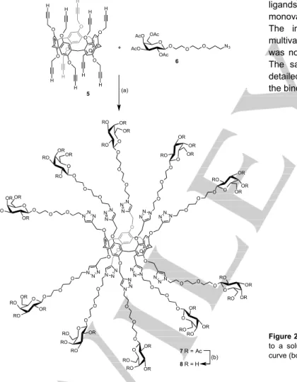
Additionally, deca-alkyne pillar[5]arene 5[44] was conjugated with
azido-functionalized galactoside 6 to afford the acetylated
O O O O O O O O O O N N N N N N N N N NNN N N N R R R R R N N N N N N N N N NNN N N N R R R R R R H 3a-f (a) O O O O O O O O O N3 N3 N3 N3 N3 N3 N3 N3 N3 N3 O 1 2a-f O O R'O R'O OR' OR' O O OR' OR' R'O H3C O O OR' OR' R'O H3C O O O O OR' OR' R'O H3C O O O O O OR' R'O R'O H3C N NN O O O OR' OR' R'O H3C N N N O O O O OR' OR' R'O H3C O O O N N N O NNN O O O O OR' OR' OR' H3C O O O 3a R' = Ac 4a R' = H (b) 3b R' = Ac 4b R' = H (b) 3c R' = Ac 4c R' = H (b) 3d R' = Ac 4d R' = H (b) 3e R' = Ac 4e R' = H (b) 3f R' = Ac 4f R' = H (b)
macromolecule 7 and further deacetylation led to the desired hydroxylated glycocluster 8. The post-functionalization of deca-alkyne 5 is sensitive to steric effects; all attempts to prepare a
per-galactosylated derivative from 5 and 1-azido-1-deoxy--D
-galactopyranoside tetraacetate failed and only partially substituted products were obtained. The introduction of a linker unit between the reactive azide group and the acetylated galactoside moiety overcame the negative steric effects and complete functionalization could be achieved.
A series of two galactosylated and five fucosylated pillar[5]arene-based glycoclusters was thus readily synthesized from the corresponding azido- or alkynyl-functionalized macrocyclic cores. All the new compounds were fully characterized by 1H and 13C NMR, and IR spectroscopy. Their
structures were further confirmed by mass spectrometry. The monodispersity of the acetylated glycoclusters was also evidenced by their chromatograms recorded on a HPLC equipped with a PLgel size exclusion column (see ESI).
Scheme 2. Synthesis of a galactosylated decavalent pillar[5]arene-based
glycocluster using a deca-propargylated core scaffold. Reagents and
conditions: (a) 1-azido-3,6-dioxa-octyl 2,3,4,6-tetra-O-acetyl--D -galactopyranoside, CuSO4•5H2O, sodium ascorbate, CH2Cl2/H2O, 43%; (b)
MeOH, MeONa, r.t., 16 h, >95%.
Binding studies
In-depth determination of the binding properties of multivalent ligands towards multivalent receptors is always a complex issue due to the varied binding modes adopted between partners and the bioanalytical techniques used. In order to obtain a broad view of the multivalent interactions involved in such systems, the use of a single assay is not recommended. Thus, the designed glycoclusters were evaluated using up to four complementary techniques including a hemagglutination inhibition assay (HIA), an enzyme-linked lectin assay (ELLA), isothermal titration microcalorimetry (ITC) and surface plasmon resonance (SPR). In this latter case, the affinity was evaluated by the kinetic approach, based on determination of kinetic constants (kon and
koff), and was verified by plotting response at equilibrium against
analyte concentration.
LecA Binding studies.
The two galactosylated glycoclusters were evaluated as LecA ligands (Table 1). These compounds were compared to the monovalent reference -GalOMe (methyl -D-galactopyranoside). The initial HIA study highlighted good binding of these multivalent ligands to the lectin although the increase in affinity was not large compared to other known multivalent ligands.[29]
The same observation holds for the ELLA assays. A more detailed description of the thermodynamic parameter governing the binding mode was obtained by ITC (Figure 2).
Figure 2. Raw ITC data (top) obtained by injections of glycocluster 8 (190 µM)
to a solution of LecA (79.5 µM) and the corresponding integrated titration curve (bottom). O O O O O O O O O O H H H H H H H H H H O O O O O O O O O O 5 (a) O O AcO AcO OAc OAc O O N3 O O OR RO OR OR O O O O OR RO RO OR O O O O OR RO RO OR O O O O OR OR RO OR O O O O OR OR RO OR O O O O OR OR RO RO O O O O RO OR OR RO O O O O RO OR OR RO O O O O RO RO OR RO O O O O RO RO OR OR O O N N N N N N N N N NN N NNN N NN N N N NN N N NN N NN + 6 7 R = Ac 8 R = H (b)
Table 1. HIA, ELLA ITC and SPR measurements for binding of pillar[5]arene-based multivalent glycoclusters 4a and 8 to LecA.
Ligand HIA ELLA ITC SPR
MIC[a] (µM) IC50 (M) [b] N[c] -H (kJ/mol) -TS (kJ/mol) KD (nM) [b] K D th (nM) KD cin (nM) kon (M-1.s-1) koff (M-1.s -1) -GalOMe 6250 658 1 0.8[d] 39[d] 15[d] 70000[d] 1 n.m.[e,f] 4a 2 26 25 0.16±0.01 125.9±71.0 89.3 413±87 169 68 8 76.4×10³ 0.62×10⁻³ 8 5 218 3 0.20±0.01 115.2±2.2 78.3 366±129 191 47 2 109×103 0.25×10⁻³
[a] Minimum inhibitory concentration (MIC) required to inhibit the agglutination of erythrocytes. [b] Calculated as the ratio of the value obtained for the monovalent reference (methyl -D-galactopyranoside = -GalOMe) to the ligand’s value. [c] Stoichiometry of binding defined as the number of
glycoclusters per monomer of LecA. [d] Data from a previous report.[12] [e] n.m. = Not measured. [f] RU shift was too weak due to the low molecular
weight of -GalOMe.
Table 2. ELLA and ITC measurements for binding of pillar[5]arene-based multivalent glycoclusters 4b-f
to LecB.
Ligand ELLA ITC
IC50 (nM) [a] N[b] -H (kJ/mol) -TS (kJ/mol) KD (nM) [a] -FucOMe 440 1 0.77±0.03[c] 41.3±1[c] 4.9 [c] 430±10[c] 1 4b 90 5 0.16±0.01 180.3±6.2 146.0 990±90 0.4 4c 30 15 0.13±0.002 202.8±4.2 164.8 220±10 1.9 4d 30 15 0.13±0.01 250.2±1.9 212.8 280±0.33 1.5 4e 6 74 0.09±0.01 300.9±4.1 261.5 150±20 2.9 4f 20 22 0.09±0.01 329.2±4.3 290.2 180±10 2.4
[a] Calculated as the ratio of the value obtained for the monovalent reference (methyl -L-fucopyranoside = -FucOMe) to the ligand’s value. [b] Stoichiometry of binding defined as the number of glycoclusters per monomer of LecB. [c] Data from a previous report.[57]
Table 3. ELLA, ITC and SPR measurements for binding of pillar[5]arene-based multivalent glycoclusters 4b-f to BambL.
Ligand ELLA ITC SPR
IC50 (M) [a] N[b] -H (kJ/mol) -TS (kJ/mol) KD (nM) [a] K D th (nM) KD cin (nM) kon (M-1.s-1) koff (M-1.s-1) -FucOMe 2544 1 2.02[c] 47.8±1.8[c] 13.5[c] 960±30[c] 1 466 475 579000 275x10-3 4b 14.8 172 0.30±0.10 235.5+85.4 194.5 60.0±13.0 16 48.9 24.3 659000 16x10-2 4c 0.05 50880 0.29±0.06 365.5±0.2 321.4 19.0±0.5 50 14.2 0.49 629000 3.08x10⁻⁴ 4d 0.095 26780 0.35±0.02 201.4±5.0 160.4 57.0±17.6 17 14.0 0.64 275000 1.76x10⁻⁴ 4e 0.0085 299294 0.14±0.01 500.1±11.1 455.6 17.0±1.0 56 18.7 n.m.[d,e] 4f 0.00007 36342860 0.061±0.008 665 ± 30 621 27±10 36 6.1 n.m.[d,e]
[a] Calculated as the ratio of the value obtained for the monovalent reference (methyl -L-fucopyranoside = -FucOMe) to the ligand’s value. [b] Stoichiometry of binding defined as the number of glycoclusters per monomer of BambL. [c] Data from a previous report.[58] [d] n.m. = Not measured. [e] Compounds 4e and
Both glycoclusters displayed KD values in the sub-micromolar
range. The longer linker arms gave slightly better binding when comparing the decavalent glycoclusters 4a and 8. The stoichiometry (N) for the complex generated between the lectin and the glycoclusters (N = 0.16 and 0.20 for 4a and 8 respectively) indicates that only five LecA tetramers are effectively interacting with glycoclusters 4a and 8. SPR experiments using LecA bound to the chip resulted in typical curves when circulating the multivalent ligand, with a fast association phase and slow dissociation, especially for high valency ligands (see ESI). Nevertheless, the dissociation constant obtained from the kinetic data (KD cin) is in the same
range as the one obtained at equilibrium (KD th). The obtained
affinities are more accurate than those obtained with ITC. SPR appears to be a more suitable method in this particular case, which may be due to the immobilization of the lectin on the chips avoiding the formation of aggregates, a common problem for solution experiments.
LecB Binding studies.
The fucosylated glycoclusters 4b-f were evaluated as potential LecB ligands (Table 2). Unfortunately, SPR experiments could not be performed due to alteration of the lectin’s binding properties when covalently immobilized on the sensor chip surface or to high aggregation when the lectin was used in solution. Therefore, the binding properties of 4b-f were investigated by ELLA and ITC which could be performed with concentration low enough to avoid too strong aggregation. ELLA experiments revealed that the decavalent ligands displayed nanomolar IC50 values with moderate improvement ( values) in
comparison to -FucOMe (methyl -L-fucopyranoside) used as a
reference monovalent ligand. However, increasing the length of the linker arm improved the binding to LecB in the decavalent glycocluster series going from 4b to 4c and then 4d. Increasing both the linker arm and valency in the icosavalent series gave the best ligand 4e however increasing the length of the spacer further 4f proved detrimental. This tendency was not observed in the ITC measurements which gave micromolar KD values to
LecB that were not a significant improvement compared to the monosaccharide ligand methyl -L-fucopyranoside. Compound
4b appears even as a weaker ligand, presumably the short
spacer preventing the fucose to be located in the most efficient way in the binding site. As a typical example, the ITC data obtained from ligand 4e are shown in Figure 3. The stoichiometry (N) of the lectin-glycocluster complexes varied from seven or eight LecB monomers bound to the decavalent ligand 4c-d (N = 0.13) to a maximum of ten for the icosavalent glycoclusters 4e-f (N = 0.09). The poor performance of the clusters towards LecB in solution may be explained by the topology of the lectin which presents its four binding sites far from each other and with different orientations at the tips of a tetrahedron. The multivalent ligands could therefore only induce aggregation, with no effective clustering of neighbouring binding sites on the lectin surface.
Figure 3. Raw ITC data (top) obtained by injections of glycocluster in a
solution of lectin and the corresponding integrated titration curve (bottom).. Left : 4f (150 µM) in LecB (110 µM); right : 4f (30 µM) in BambL (30 µM)
BambL Binding studies.
The binding properties towards BambL were then investigated for the six fucosylated glycoclusters (Table 3). ELLA experiments provided spectacular results and a value of nearly 36 million was observed for the icosavalent glycocluster 4f with the longest linker arm composed of two triethyleneglycols. The IC50 value was therefore in the picomolar range (IC50 = 70 pM),
which is the best result for a ligand reported to date for this lectin. The affinity was also highly improved for the shorter icosavalent ligand 4e ( ~ 300 000) reaching a nanomolar IC50 value.
Further investigations by ITC moderated the ELLA observations since the values were below 60 for these fucosylated glycoclusters. The best ELLA ligands 4e and 4f were shown to have a KD value around 20 nM and an interaction with high
enthalpic and entropic contributions. The very strong ELLA activity of 4f was not comparable with its affinity in solution, and was most likely due to very strong aggregative properties. Indeed, the ITC data in Table 3 and Figure 2 were obtained at very low concentration to obtain a reasonable stoichiometry value (N = 0.06, compatible with 20 fucose). When higher concentrations were used, very low values of N were obtained (N = 0.013) with much higher affinity (7 nM) that are more representative of a large aggregative network being formed in solution.
Similarly the affinities obtained by SPR were very high, ranging from 50 to 6 nM when measured at equilibrium (KD th). The
values obtained by the kinetic approach (KD cin) were very
different, but the dissociation of the compounds was very slow which introduces large errors in the estimation of koff. The two
highest affinity ligands, 4e and 4f could not be evaluated with the kinetic approach because of their flat dissociation curves (see ESI).
Conclusions
The potential therapeutic applications of multivalent glycoclusters targeting bacterial adhesion lectins call for the design of core scaffolds presenting various valencies and topologies in order to understand the specificities of binding. Pillar[5]arenes are ideal candidates in this endeavor since they provide decavalent scaffolds on a minimal molecular architecture leading to dense presentation of carbohydrate epitopes on their periphery. The synthesis of pillar[5]arene-based glycoclusters was readily achieved by CuAAC conjugations from the azido- and alkyne-functionalized precursors. The binding properties of the glycosylated multivalent ligands were studied for three lectins and by at least two complementary techniques in order to provide a good overview. The most important data were obtained from ITC and SPR experiments. Three lectins were selected from bacterial pathogens based on their potential therapeutic applications as anti-adhesive targets. Generally, multivalency improved the binding to lectins and the length of the spacer arm usually improved the affinity. The two galactosylated glycoclusters studied herein are strong ligands of LecA and an influence of both valency and linker arm for binding was observed. However, these ligands displayed binding properties similar or slightly poorer than previously reported glycoclusters.[29] In contrast, the
fucosylated glycoclusters are amongst the most potent ligands for both LecB and Bambl. These results show clearly the potential of pillar[5]arene scaffolds for the design of bioactive multivalent nanomolecules. One important problem to solve is the stereochemistry of the pillar[5]arene core and the development of optically pure pillar[5]arene derivatives is an important challenge for future biological applications. On the other hand, the possibility to prepare rotaxanes with glycopillar[5]arene as the macrocyclic component offers the possibility of producing heteroglycoclusters targeting several lectins[59] or fluorescent derivatives for confocal microscopy
studies. Therefore, the present results pave the way towards the development of a new generation of glycoclusters for various biological applications.
Experimental Section
Synthesis.General. All reagents were used as purchased from commercial sources without further purification. Compounds 1,[56] 2a,[24] 2b-f,[60] 5[44] and 6[61]
were prepared according to previously reported procedures. Solvents were distilled over CaH2 (CH2Cl2), Mg/I2 (MeOH), Na/benzophenone
(THF) or purchased dry. Reactions under microwave activation were performed on a Biotage Initiator system. Thin Layer Chromatography (TLC) was carried out on aluminium sheets coated with silica gel 60 F254
(Merck). TLC plates were inspected by UV light (λ = 254 nm, 365 nm) and developed by treatment with a mixture of 10% H2SO4 in EtOH/H2O
(95:5 v/v) followed by heating. Silica gel 60 (230-400 mesh, 0.040-0.063
mm) for column chromatography was purchased from E. Merck. Optical rotation was measured using a Perkin Elmer polarimeter. NMR spectra were recorded on a Bruker AC 300 or AC 400 spectrometer with solvent peaks as reference. The following abbreviations were used to define the multiplicities: s = singlet, d = doublet, t = triplet, q = quadruplet, m = multiplet, and their combinations. Complete signal assignments were based on 1D and 2D NMR correlations COSY and HSQC. High resolution (HR-ESI-QToF) mass spectra were recorded using a Bruker MicroTOF-Q II XL spectrometer and MALDI-TOF-LD+ were recorded using a Waters QTOF1 spectrometer. Infrared spectra were recorded on a Perkin–Elmer Spectrum One spectrometer. Elemental analyses were performed by the analytical service of the Chemistry Department of the University of Strasbourg (France).
Compound 3a. A mixture of 1 (29 mg, 0.022 mmol), 2a (129 mg, 334 mmol), CuI (2 mg, 0.011 mmol) and DIPEA (55 L, 334 mmol) in DMF (3 mL) was heated at 110°C for 15 min under microwave irradiation. The crude mixture was concentrated and co-evaporated with toluene 6 times. Column chromatography (CH2Cl2/MeOH 99/1 to 95/5) gave 3a as a 1:1
mixture of two diastereoisomers (65 mg, 56%). Colourless glassy product. IR (neat): 1745 (C=O) cm-1. 1H NMR (400 MHz, CDCl3): δ =7.80 and
7.77 (2 s, 10H), 6.56 (s, 10H), 5.34 (m, 10H), 5.14 (m, 10H), 5.02-4.85 (m, 20H), 4.81-4.56 (m, 40H), 4.30-4.00 (m, 40H), 3.98-3.83 (m, 10H), 3.25 and 3.23 (2 s, 10H), 2.11-1.85 (m, 120H) ppm. 13C NMR (100 MHz,
CDCl3): δ = 170.4, 170.2, 170.0, 169.6, 169.5, 149.5, 144.4, 144.3, 128.7,
123.4, 116.0, 100.3, 70.7, 68.7, 67.3, 67.0, 62.7, 62.6, 61.1, 61.0, 50.2, 29.6, 20.7 (2 peaks), 20.6, 20.5 ppm. HR-ESI-QTOF (positive mode): m/z 1721.5847 ([M+3H]3+, calcd. for C225H283N30O110: 1721.5824).
Compound 3b. A mixture of 1 (95 mg, 0.073 mmol), 2b (263 mg, 0.80 mmol), CuSO4•5H2O (1.8 mg, 0.007 mmol) and sodium ascorbate (4.0
mg, 0.022 mmol) in CH2Cl2/H2O (1:1, 8 mL) was vigorously stirred at
room temperature under Ar. After 72 h, H2O was added (15 mL) and the
aqueous layer was extracted with CH2Cl2 (3 x). The combined organic
layers were dried (MgSO4), filtered and concentrated. Column
chromatography (SiO2, CH2Cl2 containing 1.9% of methanol) followed by
gel permeation chromatography (Biobeads SX-1, CH2Cl2) gave 3b as a
1:1 mixture of two diastereoisomers (280 mg, 84%). Colourless glassy product. IR (neat): 1741 (C=O) cm-1. 1H NMR (300 MHz, CDCl3): δ = 8.07
and 7.88 (2 s, 10H), 6.60 and 6.45 (2 s, 10H), 5.30 (m, 10H), 5.20 (m, 20H), 5.12 (m, 10H), 4.74 (m, 40H), 4.31 (m, 5H), 4.15 (m, 20H), 3.98 (m, 5H), 3.36 and 3.26 (2 s, 10H), 2.16 (s, 30H), 2.01 and 1.99 (2 s, 30H), 1.97 and 1.96 (2 s, 30H), 1.08 and 0.96 (2 d, J = 6 Hz, 30H) ppm. 13C NMR (100 MHz, CDCl3): δ = 170.8, 170.7, 170.5, 170.5, 170.1, 170 .1, 149.6, 149.5, 144.6, 144.3, 128.8, 128.6, 123.8, 123.7, 116.0, 115.7, 95.9, 95.9, 71.2, 71.2, 68.2, 68.1, 68.1, 68.0, 67.5, 67.3, 64.9, 64.8, 61.6, 61.4, 50.4, 29.4, 29.1, 20.9, 20.8, 20.8, 16.0, 15.9 ppm. ESI-TOF-MS (positive mode): m/z 1528.966 (100%, [M+3H]3+, calcd. for
C205H263N30O90: 1528.23), 2291.937 (5%, [M+2H]2+, calcd. for
C205H262N30O90: 2291.84).
Compound 3c. As described for 3b, this compound was prepared from 1 (117 mg, 0.09 mmol), 2c (412 mg, 0.99 mmol), CuSO4•5H2O (2.2 mg,
0.009 mmol) and sodium ascorbate (5.0 mg, 0.027 mmol) in CH2Cl2/H2O
(1:1, 10 mL). Column chromatography (SiO2, CH2Cl2 containing 2.8% of
methanol) followed by gel permeation chromatography (Biobeads SX-1, CH2Cl2) gave 3c as a 1:1 mixture of two diastereoisomers (389 mg, 80%).
Colourless glassy product. IR (neat): 1740 (C=O) cm-1. 1H NMR (400
MHz, CDCl3): δ = 7.85 (s, 10H), 6.61 (s, 10H), 5.34 (m, 10H), 5.29-5.25 (m, 10H), 5.10 (m, 20H), 4.78-4.65 (m, 40H), 4.30-4.14 (m, 30H), 3.77 (m, 10H), 3.64 (m, 60H), 3.52 (broad s, 10H), 3.25 (broad s, 10H), 2.15 (s, 30H), 2.05 (s, 30H), 1.97 (s, 30H), 1.05 (d, J = 4 Hz, 30H) ppm. 13C NMR (100 MHz, CDCl3): δ = 170.8, 170.6, 170.2, 149.6, 145.3, 128.8, 123.3, 116.1, 96.4, 71.3, 70.7, 70.2, 69.9 (2x), 68.3, 68.2 (2x), 67.6, 64.7, 64.4,
51.0, 50.2, 29.5, 21.0, 20.9, 20.8, 15.9 ppm. MALDI-TOF-MS: m/z 5465.765 ([M]+, calcd. for C
245H340N30O110: 5465.20).
Compound 3d. As described for 3b, this compound was prepared from 1 (97 mg, 0.075 mmol), 2d (382 mg, 0.83 mmol), CuSO4•5H2O (2.0 mg,
0.008 mmol) and sodium ascorbate (4.5 mg, 0.023 mmol) in CH2Cl2/H2O
(1:1, 8 mL). Column chromatography (SiO2, CH2Cl2 containing 3.0% of
methanol) followed by gel permeation chromatography (Biobeads SX-1, CH2Cl2) gave 3d as a 1:1 mixture of two diastereoisomers (320 mg, 72%).
Colourless glassy product. IR (neat): 1740 (C=O) cm-1. 1H NMR (400
MHz, CDCl3): δ = 7.84 (m, 10H), 6.59 (m, 10H), 5.35 (m, 10H), 5.29 (m, 10H), 5.12 (m, 5 H), 5.09 (m, 15H), 4.77 – 4.65 (m, 40H), 4.30 – 4.11 (m, 30H), 3.78 (m, 10H), 3.63 (m, 110H), 3.25 (broad s, 10H), 2.16 (s, 30H), 2.06 (s, 30H), 1.97 (s, 30H), 1.12 (d, J = 8 Hz, 30H) ppm. 13C NMR (100 MHz, CDCl3): δ = 170.8, 170.6, 170.2, 149.6, 145.4, 128.8, 123.3, 116.1, 96.4, 71.3, 70.8, 70.7, 70.6, 70.2, 69.8, 68.3, 68.1, 67.6, 67.5, 64.6, 64.4, 50.3, 29.5, 20.9, 20.8, 20.8, 16.0 ppm. MALDI-TOF-MS: m/z 5905.73 ([M]+, calcd. for C265H380N30O120: 5905.4667). Anal. Calcd. for
C265H380N30O120.2C6H12 (6074.31): C, 54.77; H, 6.71; N, 6.92. Found: C,
54.75; H, 6.41; N, 6.65.
Compound 3e. As described for 3b, this compound was prepared from 1 (40 mg, 0.031 mmol), 2e (315 mg, 0.34 mmol), CuSO4•5H2O (0.8 mg,
0.003 mmol) and sodium ascorbate (2.0 mg, 0.009 mmol) in CH2Cl2/H2O
(1:1, 8 mL). Column chromatography (SiO2, CH2Cl2 containing 5.0% of
methanol) followed by gel permeation chromatography (Biobeads SX-1, CH2Cl2) gave 3e as a 1:1 mixture of two diastereoisomers (252 mg, 77%).
Colourless glassy product. IR (neat): 1740 (C=O) cm-1. 1H NMR (400
MHz, CDCl3): δ = 7.97 (m, 30H), 6.61 (s, 10H), 5.30 (m, 20H), 5.25 (m, 30H), 5.15 (broad s, 20H), 5.04 (m, 20H), 4.78 (m, 30H), 4.62 (m, 40H), 4.49 (m, 20H), 4.33-4.12 (m, 70H), 3.65 (s, 20H), 3.58 (s, 40H), 3.51 (s, 20H), 3.21 (broad s, 10H), 2.13 (s 60H), 1.98 (s, 30H), 1.97 (s, 30H), 1.93 (s, 60H), 1.12 (s, 30H), 1.10 (s, 30H) ppm. 13C NMR (100 MHz, CDCl3): δ = 170.7, 170.6, 170.6, 170.5, 170.1, 149.5, 145.0, 143.7, 143.6, 128.7, 125.4, 125.3, 123.4, 115.9, 115.9, 95.3, 95.0, 71.2, 70.7, 70.4, 70.1, 69.7, 68.2, 68.2, 67.9, 67.9, 67.5, 64.8, 64.8, 64.4, 64.4, 60.9, 60.8, 50.6, 50.6, 50.3, 50.3, 29.8, 20.9, 20.9, 20.8 (2x), 20.7 (2x), 16.0, 16.0 ppm. MALDI-TOF-MS: m/z 10547.46 ([M]+, calcd. for C455H620N90O200:
10550.13). Anal. Calcd. for C455H620N90O200.4CH2Cl2 (10890.02): C,
50.62; H, 5.81; N, 11.58. Found: C, 50.52; H, 5.60; N, 11.40.
Compound 3f. As described for 3b, this compound was prepared from 1 (40 mg, 0.031 mmol), 2f (400 mg, 0.34 mmol), CuSO4•5H2O (0.8 mg,
0.003 mmol) and sodium ascorbate (2.0 mg, 0.009 mmol) in CH2Cl2/H2O
(1:1, 8 mL). Column chromatography (SiO2, CH2Cl2 containing 5.5% of
methanol) followed by gel permeation chromatography (Biobeads SX-1, CH2Cl2) gave 3f as a 1:1 mixture of two diastereoisomers (289 mg, 71%).
Colourless solid. IR (neat): 1744 (C=O) cm-1. 1H NMR (400 MHz, CDCl3):
δ = 7.76 (broad s, 30H), 6.62 (s, 10H), 5.34 (m, 10H), 5.32 (m, 10H), 5.26
(s, 30H), 5.10 and 5.09 (2 s, 10H), 5.07 and 5.04 (2 broad s, 30H), 4.75 - 4.58 (m, 70H), 4.47 (m, 20H), 4.29-4.08 (m, 70H), 3.77 (m, 20H), 3.64 - 3.48 (m, 300H), 3.22 (broad s, 10H), 2.14 (s 60H), 2.04 (s, 60H), 1.96 (s, 60H), 1.11 (s, 30H), 1.10 (s, 30H) ppm. 13C NMR (100 MHz, CDCl3): δ = 170.7, 170.5, 170.1, 149.6, 145.0, 128.7, 128.7, 124.9, 123.5, 123.4, 116.0, 105.6, 96.4, 82.8, 81.8, 71.3, 70.8, 70.7, 70.6, 70.4, 70.2, 70.1, 69.8, 69.7, 68.8, 68.3, 68.1, 67.6, 66.8, 64.6, 64.4, 50.7, 50.3, 27.0, 20.9, 20.9, 20.9, 20.8, 20.8, 16.3, 16.0 ppm. MALDI-TOF-MS: m/z 13193.35 ([M+H]+, calcd. for C575H861N90O260: 13193.71).
Compound 7. As described for 3b, this compound was prepared from 5 (20 mg, 0.020 mmol), 6 (133 mg, 0.263 mmol), CuSO4•5H2O (25 mg,
0.100 mmol) and sodium ascorbate (40 mg, 0.200 mmol,) in CH2Cl2/H2O
(1:1, 4 mL). Column chromatography (SiO2, CH2Cl2 containing 9% of
methanol) gave 7 as a 1:1 mixture of two diastereoisomers (52 mg, 43%).
Colourless glassy product. IR (neat): 1746 (C=O) cm-1. 1H NMR (400
MHz, CDCl3): δ = 7.90 (s, 10H), 6.90 (s, 10H), 5.35 (d, J = 2.3 Hz, 10H), 5.15 (dd, J = 9.9, 8.7 Hz, 10H), 4.99 (dd, J = 9.9, 2.3 Hz, 10H), 4.91-4.78 (m, 20H),4.56-4.44 (m, 30H), 4.11 (m, 20H), 3.89 (m, 40H), 3.77-3.49 (m, 80H),2.12, 2.01, 1.98 (3s, 120H) ppm. 13C NMR (100 MHz, CDCl3): δ = 170.5, 170.4, 170.2, 169.6, 149.8, 144.3, 128.6, 124.2, 115.5, 101.4, 71.0, 70.7, 70.6, 70.5, 70.2, 69.5, 69.3, 69.2, 68.9, 67.2, 62.3, 61.3, 50.2, 29.6, 20.9, 20.8, 20.7 ppm. HR-ESI-QTOF (positive mode): m/z 3023.6128 (8%, [M+2H]2+, calcd. for C265H362N30O130: 3023.6365),
2015.0849 (89%, [M+3H]3+, calcd. for C265H363N30O130: 2015.0900),
1512.3273 (100%, [M+4H]4+, calcd. for C
265H364N30O130: 1512.3222).
General procedure for the Zemplén deacetylations. MeONa (10 eq.) was added to a solution of acetylated glycocluster (1 eq.) in distilled MeOH. The mixture was stirred at r.t. for 16 h, then filtered over a short column of DowexTM 50WX8-200 or Amberlite IR 120 (H+ resin form). The
resin was washed with water. The obtained filtrate was concentrated to afford the corresponding deprotected glycocluster.
Compound 4a. Obtained from 3a (150 mg, 0.029 mmol) as a pale yellow foam (100 mg, quantitatively). 1H NMR (500 MHz, DMSO-d6 + D2O): δ =
8.31 (s, 10H), 6.73 and 6.68 (2s, 10H), 5.05-4.74 (m, 30H), 4.72-4.56 (m, 10H), 4.52-4.37 (m, 10H), 4.30-4.19 (m, 10H), 4.12 (bs, 10H), 3.66-3.60 (m, 10H), 3.57-3.50 (m, 20H), 3.45-3.22 (m, 30H), 3.21-3.08 (m, 10H) ppm. 13C NMR (125 MHz, DMSO-d6 + D2O): δ = 148.8, 144.2, 144.0,
128.5, 124.3, 114.9, 102.8, 102.6, 75.4, 73.4, 70.6, 68.3, 67.6, 61.33, 60.6, 49.9, 28.3 ppm. HR-ESI-QTof (positive mode): m/z 1183.4278 ([M+3Na]3+, calcd. for C
145H200N30Na3O70: 1183.4230).
Compound 4b. Obtained from 3b (50 mg, 0.01 mmol) as a colorless foam (26 mg, 79%). 1H NMR (400 MHz, D2O): δ = 8.13 and 8.05 (2 s,
10H), 6.30 and 6.26 (2 s, 10H), 4.94 (d, J = 3.4 Hz, 10H), 4.74-4.50 (m, 40H),3.99 (s, 10H), 3.74-3.70 (m, 20H), 3.62 - 3.43 (m, 30H), 3.25-3.18 (m, 10H), 0.94 and 0.90 (2 d, J = 6.2 Hz, 30H) ppm. 13C NMR (100 MHz,
D2O): δ = 149.7, 144.4, 144.2, 129.2, 129.0, 125.2, 115.7, 98.7, 98.5,
71.7 (2 peaks), 69.5, 68.1, 67.3, 67.2, 66.7, 60.8, 60.6, 50.0, 15.28 ppm. MALDI-TOF-MS-LD+: m/z 3344.3 ([M+Na]+, calcd. for C145H200N30O60Na:
3344.34), 3199.4 ([M-C6H11O4+Na]+, calcd. for C139H189N30O56Na:
3199.2). HR-ESI-QTof (positive mode): m/z 3344.3445 ([M+Na]+, calcd.
for C145H200N30O60Na: 3344.34).
Compound 4c. Obtained from 3c (50 mg, 9.14 µmol) as a colorless foam (34 mg, 89%). 1H NMR (400 MHz, D2O): δ = 8.04 and 7.95 (2 s,
10H), 6.41 and 6.32 (2 s, 10H), 4.55-4.46 (m, 30H), 4.11-3.24 (m, 140H), 1.09 (s, 30H) ppm. 13C NMR (100 MHz, D2O): δ = 149.9, 144.1, 129.0,
125.1, 116.0, 98.6, 71.8, 69.7, 69.6, 69.5, 68.8, 68.1, 67.5, 66.7, 66.5, 63.0, 50.2, 50.0, 15.4 ppm. MALDI-TOF-MS-LD+: m/z 4227.1 ([M+Na]+,
calcd. for C185H280N30O80Na: 4227.4), 4081.0 [M-C6H11O4+Na]+, 4057.9
[M-C6H11O4]+, 3935.9 [M-C12H22O8+Na]+, 3910.9 [M-C12H22O8]+, 3789.8
[M-C18H33O12+Na]+, 3763.8 [M-C12H22O8]+.
Compound 4d. Obtained from 3d (48 mg, 8.13 µmol) as a colorless foam (32 mg, 85%). 1H NMR (400 MHz, D2O): δ = 8.05 (broad s, 10H),
6.37 (s, 10H), 4.54 (s, 30H), 4.04-3.48 (m, 180H), 1.10 (d, J = 6.6 Hz, 30H) ppm. 13C NMR (100 MHz, D2O): δ = 149.8, 129.2, 125.2, 116.0,
98.6, 71.8, 69.7, 69.5, 68.9, 68.1, 67.66, 67.74, 66.6, 63.0, 50.2, 15.4 ppm. MALDI-TOF-MS-LD+: m/z 4667.3 ([M+Na]+, calcd. for
C205H320N30O90Na: 4667.9), 4521.3 [M-C6H11O4+Na]+, 4373.2
[M-C12H22O8+Na]+, 4229.1 [M-C18H33O12+Na]+, 4206.1 [M-C18H33O12]+,
4084.1 [M-C24H44O16+Na]+, 4061.1 [M-C24H44O16]+, 3914.0
[M-C30H55O20]+, 3769.0 [M-C36H66O24]+, 3621.9 [M-C42H77O28]+, 3477.8
Compound 4e. Obtained from 3e (56 mg, 5.31 µmol) as a colorless foam (36 mg, 84%). 1H NMR (400 MHz, D2O): δ = 8.02 (s, 20H), 7.96 (s, 10H) 6.40 and 6.39 (2s, 10H), 4.93 (d, J = 3.0 Hz, 20H), 4.72-4.27 (m, 30H), 4.02-3.69 (m, 72H), 3.55-3.28 (m, 110H), 1.06 (d, J = 6.4 Hz, 60H) ppm. 13C NMR (100 MHz, D2O): δ = 149.8, 144.2, 144.0, 128.9, 126.0, 124.9, 115.7, 98.4, 76.7, 71.7, 69.7, 69.5, 69.4, 69.0, 67.9, 66.7, 63.1, 60.4, 50.9, 49.9, 15.3 ppm. MALDI-TOF-MS-LD+: m/z 8051.1 ([M+Na]+,
calcd. for C335H500N90O140Na: 8051.1), 7904.8 ([M-C6H11O4+Na]+, 7758.7
([M-C12H22O8+Na]+.
Compound 4f. Obtained from 3f (50 mg, 3.79 µmol) as a colorless foam (31 mg, 72%). 1H NMR (400 MHz, D2O): δ = 7.99 (s, 20H), 7.96 (s, 10H),
6.39 (s, 10H), 4.63-4.24 (m, 130H), 3.98 (q, J = 6.6 Hz, 20H), 3.81-3.51 (m, 100H), 3.36-3.30 (m, 90H), 1.12 (d, J = 6.6 Hz, 60H) ppm. 13C NMR
(100 MHz, D2O): δ = 149.9, 144.3, 144.0, 129.0, 126.1, 124.9, 115.8, 98.
7, 76.7, 71.8, 69.8, 69.6, 69.5, 69.1, 68.1, 67.4, 66.8, 66.6, 63.0, 51.0, 49.9, 15.4 ppm. MALDI-TOF-MS-LD+: m/z 10693.8 ([M+Na]+, calcd. for
C455H740N90O200Na: 10694.1), 10546.6 ([M-C6H11O4+Na]+, calcd. for
C449H729N90O196Na: 10546.0).
Compound 8. Obtained from 7 (35 mg, 0.006 mmol) as a pale yellow foam (25 mg, quantitatively). 1H NMR (400 MHz, DMSO-d6 + D2O): δ =
8.24 (s, 10H), 6.97 (s, 10H), 5.06 (d, J = 10.0 Hz, 10H), 4.77 (d, J = 10.0 Hz, 10H), 4.57-4.38 (m, 20H), 4.08 (d, J = 6.7 Hz, 10H), 3.86-3.74 (m, 30H), 3.63 (bs, 10H), 3.58-3.42 (m, 100H), 3.42-3.21 (m, 30H) ppm. 13C
NMR (100 MHz, DMSO-d6 + D2O): δ = 148.8, 143.2, 128.1, 124.5, 114.4,
103.6, 75.2, 73.5, 70.5, 69.72, 69.67, 69.5, 68.8, 68.1, 67.7, 61.5, 60.4, 49.4, 28.8 ppm. HR-ESI-QTof (positive mode): m/z 2204.9186 (8%, [M+2Na]2+, calcd. for C185H280N30Na2O90: 2204.9055), 1476.9385 (100%,
[M+3Na]3+, calcd. for C185H280N30Na3O90: 1476.9311).
Hemagglutination Inhibition Assays (HIA). Determination of lectin
concentration: Hemagglutination inhibition assays (HIA) were performed
in U-shaped 96-well microtitre plates. Rabbit erythrocytes were purchased from Biomérieux and used without further washing. Erythrocytes were diluted to a 4% solution in NaCl (150 mM). Lectin solutions of 2 mg/mL were prepared in TRIS-HCl 20 mM, NaCl 100 mM and CaCl2 100 μM. The hemagglutination unit (HU) was first obtained by
the addition of 25 μL of the 4% erythrocyte solution to 25 μL aliquots of sequential (two-fold) lectin dilutions. The mixture was incubated at 25°C for 60 minutes. The HU was measured as the minimum lectin concentration required for hemagglutination. Lectin concentrations of 4 HU were used for the lectin inhibition assays. For LecA, this concentration was found to be 8 μg/mL. Determination of minimum
inhibitory concentration (MIC): Subsequent inhibition assays were then
carried out by the addition of 12.5 μL lectin solution (at the required concentration) to 25 μL of sequential dilutions of glycoclusters, monomer molecules and controls. These solutions were then incubated at 25°C for 2 h then 12.5 μL of 4% erythrocyte solution was added followed by an additional incubation at 25°C for 30 minutes. The minimum inhibitory concentration (MIC) for each molecule was determined for each duplicate.
Enzyme-Linked Lectin Assays (ELLA). Determination of lectin
concentration: 96-Well microtitre plates (Nunc Maxisorb) were coated
with -PAA-Gal for LecA and -PAA-Fuc for BambL: 100 μL of 5 μg/mL in carbonate buffer, pH 9.6 for 1h at 37°C then blocking at 37°C for 1h with 100 μL per well of 3% (w/v) BSA in PBS. Lectin solution (100 μL) were diluted (1/2) starting from 30 μg/mL. After 1h incubation at 37°C and 3 washings with T-PBS (PBS containing 0,05% Tween 20), 100 μL of horseradish streptavidin–peroxidase (HRP) conjugate (dilution 2:5000; Boehringer-Mannheim) was added and left for 1h at 37°C. Colouration was developed using 100 μL per well of 0.05 M phosphate/citrate buffer containing o-phenylenediamine dihydrochloride (0.4 mg/mL) and urea
hydrogen peroxide (0.4 mg/mL) (OPD kit, Sigma-Aldrich) for 15 minutes and stopped with 50 μL of 30% sulfuric acid. Absorbance at 490 nm was then read using a microtiter plate reader (BioRad 680). Biotinylated lectins concentration were determined by plotting the relative absorbance versus lectin concentration. The concentration which leads to the highest response in the linear area was selected as the standard lectin concentration for the subsequent inhibition experiments. Determination of
inhibition potency (IC50): ELLAs were conducted in the same conditions
as above. Inhibitor solutions (50 μL) were submitted to serial dilutions (1/3) with PBS-BSA 0.3% (w/v). Then, 50 μL of biotinylated lectin solution (0.12 μg/mL for LecA and 3 ng/mL for BambL) were added in each well and the plates were incubated for 1h at 37°C. Plots of inhibition percentage versus inhibitor concentration and sigmoidal fitting provided IC50 determination.
Surface plasmon resonance (BambL). SPR inhibition experiments were performed on a Biacore X100 instrument (GE Healthcare) at 25°C in HBS buffer (10 mM Hepes/NaOH, pH 7.5, 150 mM NaCl, 0.05% Tween 20) at a flow rate of 10 mL.min-1. Streptavidin was immobilized on
a research grade CM5 chip using standard procedures, and 200 resonance units of biotinylated polyacrylamide--L-fucopyranoside probe
(Lectinity, 200 mg.mL-1) were captured on channel 2. Inhibition
experiments were performed with the fucosylated channel 2, and plots represent the subtracted data (channel 2-channel 1). Inhibition studies consisted of the injection (association 180 s, dissociation 180 s) of incubated (30 min at room temperature) mixtures of BambL (0.2 mM) and various concentrations of inhibitor (3-fold cascade dilutions). For each inhibition assay, BambL was injected to observe the full adhesion of the lectin onto the sugar-coated surface (0% inhibition). The chip was fully regenerated by two successive injections of L-fucose (120 s, 1 M in
running buffer). Binding was measured as resonance units over time after blank subtraction, and data were then evaluated by using the Biacore X100 evaluation software, version 2.0. For IC50 determination,
the response was considered as the amount of lectin bound to the sugar surface at the end of injection. Inhibition curves were obtained by plotting the percentage of inhibition against the inhibitor concentration (logarithmic scale).
Surface plasmon resonance. The studies were conducted using a Biacore X100 instrument (GE Healthcare) at 25°C with a CM5 sensor chip. A continuous flow of HEPES buffer (10 mM HEPES, 150 mM NaCl, 100 µM CaCl2, twin 20 0.05 %, pH 7.5) was maintained over the sensor
surface at a flow rate of 10 μL.min-1. The CM5 sensor chip was activated
with an injection of a solution containing N-ethyl-N’-(3-diethyl-aminopropyl)carbodiimide (EDC) (0.2 M) and N-hydroxysuccinimide (NHS) (0.05 M) for 7 minutes. LecA (100 μg.mL-1) in NaOAc buffer (pH
4.2) was injected over the activated flow cell at flow rate of 10 μL.min-1
for 10 minutes to achieve a ~1000 RU. The same procedure was applied for BamblL achieving 200 RU capture The immobilization procedure was completed by an injection of ethanolamine hydrochloride (1 M) (70 μL), followed by a flow of the buffer (100 μL.min-1) in order to eliminate
physically adsorbed compounds. Ethanol amine alone was used in channel 1 as a reference. For the LecA chip, Compounds 4a and 8 were injected using the single-cycle mode with 5 concentrations varying from 0.5 nm to 5 µM concentration. By the end of measures the sensor chip was regenerated with 100 seconds pulse of methyl β-D -galactopyranoside (100 mM) followed by an injection of running buffer for 300 seconds.
Microcalorimetry. Recombinant lyophilized lectin was dissolved in buffer (LecA: 20 mM TRIS-HCl, 100 µM CaCl2, pH 7.5, NaCl 100 mM; LecB:
20 mM TRIS-HCl, 100 µM CaCl2, pH 7.5, NaCl 100 mM; BambL: 20 mM
TRIS-HCl, pH 7.5, NaCl 150 mM) and degassed. The protein concentration was checked by measuring A280 by using a theoretical
molar extinction coefficient (LecA: 28000, LecB: 6990, BambL: 40450). Carbohydrate ligands were dissolved in the same buffer, degassed, and loaded in the injection syringe. ITC was performed with a VP-ITC microcalorimeter (Microcal, GE Healthcare). The lectin solution was placed in a 1.4478-mL sample cell at 25°C. Titration was performed with 10 µL injections of carbohydrate ligands every 400 s. Data were fitted with MicroCal Origin 7 software according to standard procedures. The fitted data yielded the stoichiometry (N), association constant (Ka), and
enthalpy of binding (H). Other thermodynamic parameters (i.e. changes in free energy (G) and entropy (S)) were calculated from the equation G = H – TS = – RTlnKa, in which T is the absolute temperature and R
= 8.314 J.mol-1.K-1. Two independent titrations were performed for each
ligand tested.
Acknowledgements
The authors thank the Université Claude Bernard Lyon 1, the Université de Strasbourg, the International Center for Frontier Research in Chemistry (icFRC) and the CNRS for financial support. This project was also supported by COST Action CM-1102 MultiGlycoNano and the Agence Nationale de la
Recherche (ANR, Programme Blanc 2011, Sweet60s). A.I.
acknowledges support from GDR Pseudomonas and Labex ARCANE (ANR-11-LABX-003). Dr F. Albrieux, C. Duchamp, N. Henriques and J.-M. Strub are gratefully acknowledged for mass spectrometry analyses. N.G. thanks the Région Rhône-Alpes (Cluster de Recherche Chimie) for additional funding and the Royal Society for funding his internship undertaken in S.E.M.’s laboratory (JP090449).
Keywords: Pillar[5]arene • Glycocluster • Click chemistry •
Lectin • Glycobiology
[1] A. Imberty, A. Varrot, Curr. Opin. Struct. Biol. 2008, 18, 567-576. [2] E. C. Adam, B. S. Mitchell, D. U. Schumacher, G. Grant, U. Schumacher,
Am. J. Respir. Crit. Care Med. 1997, 155, 2102-2104.
[3] G. Cioci, E. P. Mitchell, C. Gautier, M. Wimmerova, D. Sudakevitz, S. Pérez, N. Gilboa-Garber, A. Imberty, FEBS Lett. 2003, 555, 297-301. [4] C. Chemani, A. Imberty, S. de Bentzman, P. Pierre, M. Wimmerová, B. P.
Guery, K. Faure, Infect. Immun. 2009, 77, 2065-2075.
[5] T. Eierhoff, B. Bastian, R. Thuenauer, J. Madl, A. Audfray, S. Aigal, S. Juillot, G. E. Rydell, S. Müller, S. de Bentzmann, A. Imberty, C. Fleck, W. Römer, Proc. Natl. Acad. Sci. U.S.A. 2014, 111, 12895-12900.
[6] E. Mitchell, C. Houles, D. Sudakevitz, M. Wimmerova, C. Gautier, S. Pérez, A. M. Wu, N. Gilboa-Garber, A. Imberty, Nature Struct. Biol. 2002,
9, 918-921.
[7] S. Perret, C. Sabin, C. Dumon, M. Pokorná, C. Gautier, O. Galanina, S. Ilia, N. Bovin, M. Nicaise, M. Desmadril, N. Gilboa-Garber, M. Wimmerova, E. P. Mitchell, A. Imberty, Biochem. J. 2005, 389, 325-332.
[8] A. Audfray, J. Claudinon, S. Abounit, N. Ruvoën-Clouet, G. Larson, D. F. Smith, M. Wimmerová, J. Le Pendu, W. Römer, A. Varrot, A. Imberty, J.
Biol. Chem. 2012, 287, 4335-4347.
[9] J. Houser, J. Komarek, N. Kostlanova, G. Cioci, A. Varrot, S. C. Kerr, M. Lahmann, V. Balloy, J. V. Fahy, M. Chignard, A. Imberty, M. Wimmerova,
PloS ONE 2013, 8, e83077.
[10] Y. M. Chabre, R. Roy, Chem. Soc. Rev. 2013, 42, 4657-4708.
[11] S. Cecioni, R. Lalor, B. Blanchard, J.-P. Praly, A. Imberty, S. E. Matthews, S. Vidal, Chem. Eur. J. 2009, 15, 13232-13240.
[12] S. Cecioni, S. Faure, U. Darbost, I. Bonnamour, H. Parrot-Lopez, O. Roy, C. Taillefumier, M. Wimmerová, J.-P. Praly, A. Imberty, S. Vidal, Chem.
Eur. J. 2011, 17, 2146-2159.
[13] D. Sicard, S. Cecioni, M. Iazykov, Y. Chevolot, S. E. Matthews, J.-P. Praly, E. Souteyrand, A. Imberty, S. Vidal , M. Phaner-Goutorbe, Chem.
Commun. 2011, 47, 9483-9485.
[14] S. Cecioni, S. E. Matthews, H. Blanchard, J.-P. Praly, A. Imberty, S. Vidal,
Carbohydr. Res. 2012, 356, 132-141.
[15] A. M. Boukerb, A. Rousset, N. Galanos, J.-B. Méar, M. Thépaut, T. Grandjean, E. Gillon, S. Cecioni, C. Abderrahmen, K. Faure, D. Redelberger, E. Kipnis, R. Dessein, S. Havet, B. Darblade, S. E. Matthews, S. de Bentzmann, B. Guéry, B. Cournoyer, A. Imberty, S. Vidal,
J. Med. Chem. 2014, 57, 10275-10289.
[16] Z. H. Soomro, S. Cecioni, H. Blanchard, J.-P. Praly, A. Imberty, S. Vidal, S. E. Matthews, Org. Biomol. Chem. 2011, 9, 6587-6597.
[17] H. Vedala, Y. Chen, S. Cecioni, A. Imberty, S. Vidal, A. Star, Nano Lett.
2011, 11, 170-175.
[18] Y. Chen, H. Vedala, G. P. Kotchey, A. Audfray, S. Cecioni, A. Imberty, S. Vidal, A. Star, ACS Nano 2012, 6, 760-770.
[19] J. Luczkowiak, A. Munoz, M. Sanchez-Navarro, R. Ribeiro-Viana, A. Ginieis, B. M. Illescas, N. Martin, R. Delgado, J. Rojo, Biomacromolecules
2013, 14, 431-437.
[20] M. Durka, K. Buffet, J. Iehl, M. Holler, J.-F. Nierengarten, S. P. Vincent,
Chem. Eur. J. 2012, 18, 641-651.
[21] M. Sánchez-Navarro, A. Muñoz, B. M. Illescas, J. Rojo, N. Martín, Chem.
Eur. J. 2011, 17, 766-769.
[22] M. Durka, K. Buffet, J. Iehl, M. Holler, J.-F. Nierengarten, J. Taganna, J. Bouckaert, S. P. Vincent, Chem. Commun. 2011, 47, 1321-1323. [23] S. Cecioni, V. Oerthel, J. Iehl, M. Holler, D. Goyard, J.-P. Praly, A. Imberty,
J.-F. Nierengarten, S. Vidal, Chem. Eur. J. 2011, 17, 3252-3261. [24] J.-F. Nierengarten, J. Iehl, V. Oerthel, M. Holler, B. M. Illescas, A. Munoz,
N. Martin, J. Rojo, M. Sanchez-Navarro, S. Cecioni, S. Vidal, K. Buffet, M. Durka, S. P. Vincent, Chem. Commun. 2010, 46, 3860-3862.
[25] P. Compain, C. Decroocq, J. Iehl, M. Holler, D. Hazelard, T. Mena Barragán, C. Ortiz Mellet, J.-F. Nierengarten, Angew. Chem. Int. Ed. 2010,
49, 5753-5756.
[26] R. Rísquez-Cuadro, J. M. García Fernández, J.-F. Nierengarten, C. Ortiz Mellet, Chem. Eur. J. 2013, 19, 16791-16803.
[27] A. Muñoz, D. Sigwalt, B. M. Illescas, J. Luczkowiak, L. Rodríguez-Pérez, I. Nierengarten, M. Holler, J.-S. Remy, K. Buffet, S. P. Vincent, J. Rojo, R. Delgado, J.-F. Nierengarten, N. Martín, Nature Chem. 2015, DOI:10.1038/nchem.2387.
[28] A. Bernardi, J. Jimenez-Barbero, A. Casnati, C. De Castro, T. Darbre, F. Fieschi, J. Finne, H. Funken, K.-E. Jaeger, M. Lahmann, T. K. Lindhorst, M. Marradi, P. Messner, A. Molinaro, P. V. Murphy, C. Nativi, S. Oscarson, S. Penades, F. Peri, R. J. Pieters, O. Renaudet, J.-L. Reymond, B. Richichi, J. Rojo, F. Sansone, C. Schaffer, W. B. Turnbull, T. Velasco-Torrijos, S. Vidal, S. Vincent, T. Wennekes, H. Zuilhof, A. Imberty, Chem.
Soc. Rev. 2013, 42, 4709-4727.
[29] S. Cecioni, A. Imberty, S. Vidal, Chem. Rev. 2015, 115, 525-561. [30] T. Ogoshi, S. Kanai, S. Fujinami, T.-A. Yamagishi, Y. Nakamoto, J. Am.
Chem. Soc. 2008, 130, 5022-5023.
[31] D. Cao, Y. Kou, J. Liang, Z. Chen, L. Wang, H. Meier, Angew. Chem. Int.
Ed. 2009, 48, 9721-9723.
[32] D. Cao, H. Meier, Asian J. Org. Chem. 2014, 3, 244-262. [33] T. Ogoshi, J. Incl. Phenom. Macrocycl. Chem. 2012, 72, 247-262. [34] P. J. Cragg, K. Sharma, Chem. Soc. Rev. 2012, 41, 597-607.
[35] M. Xue, Y. Yang, X. Chi, Z. Zhang, F. Huang, Acc. Chem. Res. 2012, 45, 1294-1308.
[36] M. Holler, N. Allenbach, J. Sonet, J.-F. Nierengarten, Chem. Commun.
2012, 48, 2576-2578.
[37] N. L. Strutt, H. Zhang, S. T. Schneebeli, J. F. Stoddart, Acc. Chem. Res.
2014, 47, 2631-2642.
[38] T. Ogoshi, T.-A. Yamagishi, Eur. J. Org. Chem. 2013, 2961-2975. [39] T. Ogoshi, T. Yamagishi, Chem. Commun. 2014, 50, 4776-4787. [40] H. Zhang, Y. Zhao, Chem. Eur. J. 2013, 19, 16862-16879.
[41] W. Si, L. Chen, X.-B. Hu, G. Tang, Z. Chen, J.-L. Hou, Z.-T. Li, Angew.
Chem. Int. Ed. 2011, 50, 12564-12568.
[42] X.-B. Hu, Z. Chen, G. Tang, J.-L. Hou, Z.-T. Li, J. Am. Chem. Soc. 2012,
134, 8384-8387.
[43] G. Yu, X. Zhou, Z. Zhang, C. Han, Z. Mao, C. Gao, F. Huang, J. Am.
Chem. Soc. 2012, 134, 19489-19497.
[44] I. Nierengarten, M. Nothisen, D. Sigwalt, T. Biellmann, M. Holler, J.-S. Remy, J.-F. Nierengarten, Chem. Eur. J. 2013, 19, 17552-17558. [45] I. Nierengarten, K. Buffet, M. Holler, S. P. Vincent, J.-F. Nierengarten,
Tetrahedron Lett. 2013, 54, 2398-2402.
[46] G. Yu, Y. Ma, C. Han, Y. Yao, G. Tang, Z. Mao, C. Gao, F. Huang, J. Am.
Chem. Soc. 2013, 135, 10310-10313.
[47] I. Nierengarten, J.-F. Nierengarten, Chem. Rec. 2015, 15, 31-51. [48] C. E. Hoyle, A. B. Lowe, C. N. Bowman, Chem. Soc. Rev. 2010, 39,
1355-1387.
[49] I. Nierengarten, J.-F. Nierengarten, Chem. Asian J. 2014, 9, 1436-1444. [50] G. Franc, A. K. Kakkar, Chem. Soc. Rev. 2010, 39, 1536-1544. [51] C. E. Hoyle, C. N. Bowman, Angew. Chem. Int. Ed. 2010, 49, 1540-1573. [52] C. Remzi Becer, R. Hoogenboom, U. S. Schubert, Angew. Chem. Int. Ed.
2009, 48, 4900-4908.
[53] I. Nierengarten, S. Guerra, M. Holler, L. Karmazin-Brelot, J. Barberá, R. Deschenaux, J.-F. Nierengarten, Eur. J. Org. Chem. 2013, 3675-3684.
[54] H. Deng, X. Shu, X. Hu, J. Li, X. Jia, C. Li, Tetrahedron Lett. 2012, 53, 4609-4612.
[55] G. Yu, Z. Zhang, J. He, Z. Abliz, F. Huang, Eur. J. Org. Chem. 2012, 5902-5907.
[56] I. Nierengarten, S. Guerra, M. Holler, J.-F. Nierengarten, R. Deschenaux,
Chem. Commun. 2012, 48, 8072-8074.
[57] C. Sabin, E. P. Mitchell, M. Pokorná, C. Gautier, J.-P. Utille, M. Wimmerová, A. Imberty, FEBS Lett. 2006, 580, 982-987.
[58] A. Audfray, J. Claudinon, S. Abounit, N. Ruvoën-Clouet, G. Larson, D. F. Smith, M. Wimmerová, J. Le Pendu, W. Römer, A. Varrot, A. Imberty, J.
Biol. Chem. 2012, 287, 4335-4347.
[59] S. P. Vincent, K. Buffet, I. Nierengarten, A. Imberty,J.-F. Nierengarten,
Chem. Eur. J. 2015, DOI:10.1002/chem.201504110.
[60] K. Buffet, E. Gillon, M. Holler, J.-F. Nierengarten, A. Imberty, S. P. Vincent,
Org. Biomol. Chem. 2015, 13, 6482-6492.
[61] S. Cecioni, M. Almant, J.-P. Praly, S. Vidal in Synthesis of
Azido-Functionalized Carbohydrates for the Design of Glycoconjugates, Vol. 1
Entry for the Table of Contents
FULL PAPER
Glycoclusters
Pillar[5]arene-based glycoclusters with up to 20 peripheral sugars have been prepared by azide-alkyne “click” conjugations. Their binding properties have been studied for three bacterial lectins, namely LecA and LecB from
Pseudomonas aeruginosa and BambL
from Burkholderia ambifaria.
K. Buffet, I. Nierengarten,N. Galanos, E.
Gillon, M. Holler, A. Imberty,* S. E. Matthews, S. Vidal,* S. P. Vincent,* and J.-F. Nierengarten*
Page No. – Page No.
Pillar[5]arene-based glycoclusters: synthesis and multivalent binding to pathogenic bacterial lectins
![Table 3. ELLA, ITC and SPR measurements for binding of pillar[5]arene-based multivalent glycoclusters 4b-f to BambL](https://thumb-eu.123doks.com/thumbv2/123doknet/14426296.514275/5.892.63.822.793.1028/table-ella-measurements-binding-pillar-multivalent-glycoclusters-bambl.webp)

