HAL Id: hal-03019000
https://hal.archives-ouvertes.fr/hal-03019000
Submitted on 16 Dec 2020
HAL is a multi-disciplinary open access
archive for the deposit and dissemination of
sci-entific research documents, whether they are
pub-lished or not. The documents may come from
teaching and research institutions in France or
abroad, or from public or private research centers.
L’archive ouverte pluridisciplinaire HAL, est
destinée au dépôt et à la diffusion de documents
scientifiques de niveau recherche, publiés ou non,
émanant des établissements d’enseignement et de
recherche français ou étrangers, des laboratoires
publics ou privés.
thickness skin after exposure to heavy metals
Laurent Chavatte, Milène Juan, Sandra Mounicou, Emmanuelle Leblanc
Noblesse, Karl Pays, Carine Nizard, Anne-Laure Bulteau
To cite this version:
Laurent Chavatte, Milène Juan, Sandra Mounicou, Emmanuelle Leblanc Noblesse, Karl Pays, et
al.. Elemental and molecular imaging of human full thickness skin after exposure to heavy metals.
Metallomics, Royal Society of Chemistry, 2020, 12 (10), pp.1555-1562. �10.1039/d0mt00121j�.
�hal-03019000�
ARTICLE
a.
Université de Pau et des Pays de l’Adour, E2S UPPA, CNRS, Institut des Sciences Analytiques et de Physico-Chimie Pour l'Environnement et les Materiaux (IPREM), UMR5254, Hélioparc, 64053 Pau, France.
b.CIRI, Centre International de Recherche en Infectiologie, CIRI, 69007 Lyon, France. c.Institut National de la Santé et de la Recherche Médicale (INSERM) Unité U1111,
69007 Lyon, France. d.
Ecole Normale Supérieure de Lyon, 69007 Lyon, France. e.
Université Claude Bernard Lyon 1 (UCBL1), 69622 Lyon, France. f.
Unité Mixte de Recherche 5308 (UMR5308), Centre national de la recherche scientifique (CNRS), 69007 Lyon, France.
g.LVMH Recherche. Life Science Department, 185 Avenue de Verdun, 45800. Saint Jean de Braye, France.
h.
* To whom correspondence should be addressed: Anne-Laure Bulteau. LVMH Recherche. Life Science Department, 185 Avenue de Verdun, 45800. Saint Jean de Braye. France.Email: [email protected]. Phone: 33-02-38-25-84-19. orcid.org/0000-0002-3315-5100
Elemental and molecular imaging of human full thickness skin
after exposure to heavy metals
Laurent Chavatte,a-f Milène Juan, g Sandra Mounicou, a Emmanuelle Leblanc Noblesse, g Karl Pays,g Carine Nizard g andAnne-Laure Bulteau, *g
Compelling evidences suggest that heavy metals have potentially harmful effects to the skin. However, knowledge about cellular signaling events and toxicity subsequent to metals exposure to human skin cells is still poorly documented. The aim of this study was to focus on the interaction between 4 different heavy metals (lead, nickel, cadmium, and mercury) at doses mimicking chronic low-level of environmental exposure and the effect on skin to get better insight into metals-cell interactions. We provide evidence that 2 metals (lead and nickel) can permeate skin and accumulate at high concentrations in the dermis. Skin barrier was disrupted after metals exposure and it was accompanied by apoptosis, DNA damage and lipid oxidation. Skin antioxidant enzymes such as glutathione peroxidase and methionine sulfoxide reductase are also heavy metals targets. Taken together, our findings provide insight into potential mechanisms of metals-induced oxidative stress production and the cellular consequences of these events.
Introduction
Heavy metals contamination is a major environmental concern particularly in China1. They are found in various media including soil, water air and food2, 3. Heavy metals are known to be persistent in the human body, with excretion half-lives that last for decades4. At the interface between the body and the environment, the skin is directly exposed to chemical oxidants, pollutants, all of which are potent inducers of reactive oxygen species (ROS). Although, epidemiological and clinical studies highlight the adverse effects of heavy metals on human health, very little research is available to date regarding cutaneous effects 3. A major mechanism by which ambient heavy metals such as cadmium, nickel exert their detrimental effects is through the generation of oxidative stress which is an important contributor to skin aging5, 6. They can cause several genotoxic effects on cells, including oxidative stress, DNA breakage and protein modification7-9. Moreover, they can,
especially nickel, induce the overexpression of collagenases enzymes leading to the weakening of the extracellular matrix resulting in a loss of fitness and elasticity of the skin10. Besides their high level of toxicity, they can also induce contact allergies and other serious skin diseases11. They also may affect cell growth and proliferation of keratinocytes 12. Studies of heavy metals have analyzed the effects of one single metal. However, we must consider that people are exposed to a cocktail of metals and a synergic effect of each pollutant cannot be excluded. Therefore, in our study, we considered the joint exposure to pollutants. The exact mechanisms by which heavy metals can cause skin damage has yet to be elucidated. Based on current evidence, there may be two potential mechanisms (i) generation of free radicals, (ii) induction of inflammatory response and disruption of the skin barrier11. Ex vivo experiments based on excised epidermis model could help to provide information on heavy metals induced biological processes, skin penetration and their rates. Permeability of metals through the skin is highly variable since they can penetrate the epidermis through different routes. Furthermore, metals are retained in the skin which could lead to a reservoir and extended exposure even after their removal from the outer surface of the skin13. Dermal exposure can lead to systemic inflammation11. Therefore, it is important to localize and measure the amount of each type of metals in the skin. Imaging mass spectrometry is based on the measurement of the abundance of elemental or molecular ions generated locally by means of a laser and offers a typical resolution of 20-50m. Elemental imaging by laser ablation (LA ICP-MS) allows a sensitive imaging of metals and thus an ultimate way of tracking the penetration of heavy metals into the skin 14.
ARTICLE Metallomics
2 | Metallomics, 2020, 00, 1-3 This journal is © The Royal Society of Chemistry 20xx
The current study was undertaken to characterize the effects of heavy metals (lead, mercury, nickel and cadmium) exposure on skin and to assess how alterations in skin integrity may contribute to oxidative stress.
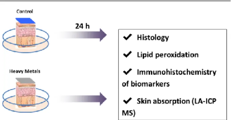
Figure 1. Effect of heavy metals exposure on skin explants. Metals exposure protocol. Skin full thickness models were cultured for 4 days in a survival medium and at day 4 a mixture of 4 heavy metals was deposited on a filter paper and applied on explants. Control samples received only filter paper. 24 hours after the explants were taken and prepared for histology, LA-ICP MS or immunostaining.
Results
Heavy metals induced changes in skin morphology and an increase of skin oxidative damages.
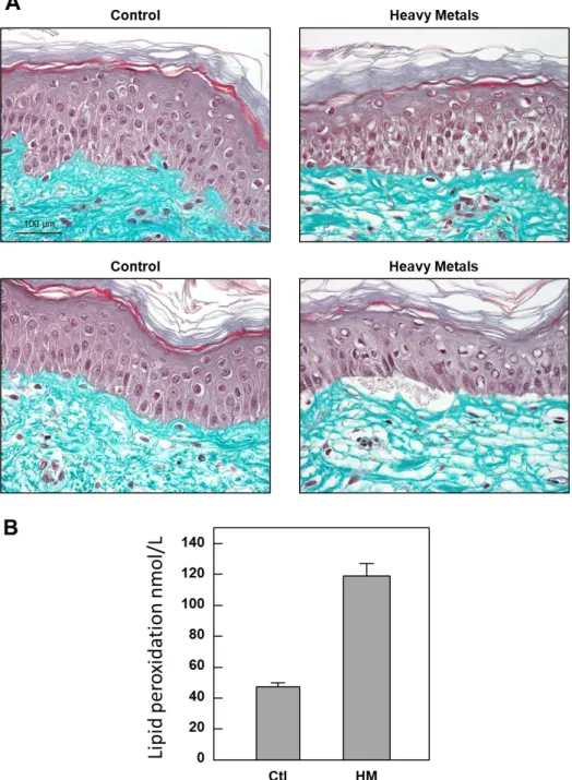
Skin explants were exposed to the metals cocktail (nickel, lead, mercury and cadmium) for 4 days (Figure 1). 24 hours after the last exposure, we first analyzed the skin morphology in the samples (Figure 2A). Light transmission microscopy visualization of Masson’s Trichrome 5-µm thick sections showed that each skin piece used as control maintained good morphology throughout the experiments as assessed by the constant presence of the stratum corneum layer (SC), a compact viable epidermis and a collagen-filled dermis. No macroscopic modification of the skin architecture was observed in PBS-exposed specimens, indicating that the protocols used from explants to fixation did not damage the
skin. However, we found a significant alteration in skin
morphology and structure in the explants exposed to metals compared to untreated controls (Figure 2A). We found slight defects underlying cell death in the specimens exposed to pollution. The anomalies observed included spongiosis, pyknotic nuclei, and even epidermal detachment. Dermis was also altered with a decrease in collagen density. To better understand the exact nature of the cell death observed in the explants exposed to pollution, we investigated quantification of the Malonaldehyde (MDA) which is a measure of skin oxidative damages (Figure 2B). As reported by other authors, the devastating effect of heavy metals on skin viability is mostly owning to an increase in reactive oxygen species (ROS)
in the cells8, 15. Interestingly, exposure to pollutants induced a marked increase of MDA in the medium of explants indicating that heavy metals have induced an oxidative stress in the skin.
Heavy metals compromised skin barrier.
To further investigate the effects of the pollutants on epidermal function, we examined the expression of differentiation markers and desmosomal proteins by immunostaining (Figure 3). As expected, we observed a dramatic reduction in the expression levels of all the differentiation markers between control and treated explants. Significant differences in the amount of early, (desmoglein 1) and late (loricrin) differentiation markers were observed in skin from heavy metals-exposed samples suggesting that skin barrier was disrupted.
Heavy metals compromised cellular antioxidant systems in cells and induced DNA damage.
In response to stressing agents including metals and oxidative stress, cells are able to induce the metal binding-proteins metallothionein16. As expected metallothionein expression was induced by exposure to heavy metals (Figure 3). These proteins are small protein composed of 80 amino acids and contain up to 15 cysteine residues that can chelate metals and to avoid any toxic effect of the free metal in the cytosol. Glutathione peroxidase (Gpx) are involved in detoxification of hydrogen peroxidde and lipid peroxidation products and methionine sulfoxide reductase (MSR) are involved in the repair of oxidatively modified proteins in the skin17. To determine how these enzymes are involved in skin protection after exposure to pollutants, these proteins were detected by immunostaining in skin biopsies. Surprisingly, GPX2 and MSR labelling were less pronounced in the heavy metals- treated epidermis compared to the control ones (Figure 3). To determine if metals exposure could induce DNA damage, we looked at phosphorylation of H2AX which is used to quantify accumulation of DNA damage. Immunofluorescence against histone H2AX phosphorylated at Ser-139 revealed that metals exposure induced an increase in DNA damage in keratinocytes (Figure 3). This result indicates that heavy metals exposure can be associated with antioxidant enzymes downregulation, that may explain the accumulation of oxidative damages in the skin as shown in Figure 2B.
Figure 2. Effect of heavy metals exposure on skin structure and oxidative. A. Masson’s trichrome staining of the explants at Day 5 . Two representative pictures are shown for
each conditions, control and heavy metals treated (HM). B. Quantification of Lipid peroxidation. MDA was determined in the survival media at 24 hours after heavy metals exposure using a kit as described in the methods.
Metals localization inside the skin
One goal of this study was the localization of metals in order to check their penetration in the skin. The nickel containing signature of the detected compounds is asserted by the cluster due to the two major isotopes of the nickel, 58Ni and, 60Ni, 13C is used as a control. Nickel accumulates into the dermis and was absorbed by the skin (Figure 4). Mercury was detected by the cluster due to 200Hg and 202Hg. Mercury accumulates in the epidermis and it is not able to penetrate the dermis. Cadmium (112Cd, 112Cd, 114Cd) is detected only in the epidermis whereas
lead (206Pb and 208Pb) is found both in the dermis and the epidermis.
In conclusion, we demonstrated in this study that exposure to pollution decreased cell viability and antioxidant enzymes activities in the ex vivo human skin model. These anomalies are confirmed at the histological level, by the presence of cell necrosis features including spongiosis, pyknotic nuclei and dermo epidermal detachment. This may be explained by the fact that heavy metals could penetrate the epidermis up to the dermis and are retained.
ARTICLE Metallomics
4 | Metallomics, 2020, 00, 1-3 This journal is © The Royal Society of Chemistry 20xx
Figure 3. Heavy metals induce changes in skin proteins expression at day 5. Immunofluorescence staining of 5 proteins in epidermis using specific antibodies. Metallothionein
(response to metals), Gamma H2AX which is a measure of DNA damage, Desmoglein and Loricrin (proteins from the skin barrier) and antioxidant proteins (Gpx2 and PMSR)
proteins in ex vivo human skin. Representative pictures of skin slides of non-stressed (Control) and heavy metals stressed (HM) are shown. Following confocal laser microscopy acquisition, pictures were analyzed, and relative expression of the different proteins was quantified and standardized to the epidermis area (LeicaQWin software).
Discussion
This study evaluated the effects of heavy metals (nickel, cadmium, lead and mercury) exposure on skin. This is the first study directly exposing human skin explants to such a cocktail.
In vitro models are a promising approach to characterize the
impact of pollution on skin.
Interaction between metals and cell membranes play a key role in metals-induced toxicity by inducing ROS production 15.
This ROS production can damage proteins, DNA of the cells but also can trigger inflammation by activation of cytokines and might be involved in the initiation or pathogenesis of allergic or non-allergic cutaneous inflammation 18, 19. We have shown that exposure to metals lead to DNA damage that may be explained mainly by ROS production (Figure 3). We found that exposure to metals resulted in an increase of lipid peroxidation due to ROS production (Figure 2B).
ARTICLE
Figure 4. Figure 4. Penetration of heavy metals in human full thickness skin at day 5. A. Pictures of skin slices (E, epidermis and D, dermis). B. LA ICP -MS qualitative images. C. Heat map of metals (nickel, mercury, cadmium, lead). The signal intensity heatmaps were obtained using a lab -written application for Matlab (Mathworks, Natick, MA) as described in 31.
Malondialdehyde is an indirect mediator of oxidative stress, formed by lipid peroxidation of polyunsaturated fatty acids in, for example, cholesterol, phospholipids and triglycerides20. It is highly reactive and able to readily modify the sulfhydryl group of cysteine residues and the imidazole group of histidine residues leading to protein oxidation. Various types of protein oxidative modifications are directly or indirectly induced by ROS by reactions with secondary products of oxidative stress
21
. Cysteine and methionine residues are particularly prone to oxidative modifications, but they might not be directly linked to protein damage, since they also participate in cellular signaling events22. We have shown that glutathione peroxidase and methionine sulfoxide reductase were inhibited upon pollutant exposure suggesting that they may be targets of oxidative stress (Figure 3). Our results suggest that ROS and lipid peroxidation products may react directly with these enzymes which active site contains a cysteine. Decrease in the antioxidant enzymes capacities of the skin will result in an increase in oxidative stress and to oxidative damage leading to intrinsic aging of the skin23.
We also wanted to investigate if the heavy metals can permeate the skin and thus lead to a storage compartment of these pollutants in the skin. Because metals can penetrate through diverse routed and that surface modification with diverse ligand could modify hydrophobicity of the skin and thus metals speciation, penetration of each metals is specific13. Moreover, thinner facial skin is more permeable than abdominal skin24. Skin permeation is influenced by age, vascular and collagen composition25. We found that all the four metals through intact human skin under physiological conditions (Figure 4). Based on our LA-ICP MS analysis, the order of permeability is Pb>Ni>Hg>Cd. It was known that lead was easily absorbed into the skin through sweat glands and hair follicles26. We found that the dermal absorption of lead is important that may contribute to the total body burden. We found that nickel is capable of permeating through the skin confirming a study done human volunteers exposed to metallic nickel for 30 minutes27. As filaggrin is a very rich in histidine which has huge affinity for nickel, it is most likely that nickel is bound to filaggrin in the stratum corneum and that the damage to skin barrier due to the cocktail of metals lead to a better penetration of nickel into dermis28. Another dangerous effect caused by nickel is the induction of matrix-metalloproteinases (MMP) especially the one that can degrade
collagen leading to a loss of elasticity and fitness of the skin 10. Because nickel is retained in the dermis it can lead to a permanent increased expression of these enzymes. Regarding mercury and cadmium, they penetrate only in the epidermis as it was shown before that their solubility into the SC is a rate limiting process13. The concentration applied to the skin can therefore influence their permeability. The high concentration of cadmium in the epidermis may explain the induction of metallothionein, as this metal is by far the best activator of metallothionein expression. These in vitro studies indicate large variations in the results of metals permeability through the skin. It is evident that permeation of metals may be influenced by pH and speciation of the metals 13.
Our results demonstrate that two metals, nickel and lead can permeate skin whereas mercury and cadmium are localized in the epidermis leading to disruption of skin barrier. Our results also demonstrated that exposure of keratinocytes to pollutants induced a significant decline in antioxidants enzymes. This was accompanied by DNA damage. We also observed an increase in lipid peroxidation products malondialdehyde (MDA) which is involved in the formation of oxidatively modified proteins. Taken together, these findings suggest that antioxidant systems may be particularly vulnerable to inactivation in conditions associated with metals exposure resulting in accumulation of oxidatively modified proteins, mitochondrial dysfunction and cell death.
Experimental
Chemicals and antibodies
All chemicals were purchased from Sigma-Aldrich (Saint Quentin Falavier, France). The antibodies used in the study were rabbit antibodies against, Desmoglein (Life technology, 326000), Gamma H2AX (Upstate, 07627), Loricrin (Covance,
PRB-145-P100), GPX2 (Abcam, ab64322) and MSR, a gift from Dr Bertand Friguet, (Sorbonne University, Paris, 30) and mouse antibodies metallothionein (Dako M063901-2)
Skin preparation
12 mm diameter explants were prepared from a 36 years old woman abdominoplasty and placed in a survival medium at 37°C in a humid atmosphere enriched with 5 percent CO2.
With respect to the ethical permissions, for studies using skin biopsies, the principle requirements of the Declaration of Helsinki were considered to protect the rights, safety and well-being of subjects participating in the study. Before initiating the studies, the investigator had obtained written consent from the participants and full approval from the Freiburg Ethics Commission International for the protocol, protocol amendment(s), if applicable. All participants who provided their skin biopsies for this research provided their written informed consent to participate in this study and for their data to be used for research purposes.
Exposure to heavy metals.
On day four, the survival medium was renewed and heavy metals were deposited on a patch applied on explants at these concentrations; Hg 2.5 g/mL, Ni 2.5 g/mL, Pb 2.5 g/mL, Cd 2.5 g/mL (ICP multi-element standard V, Merck, Germany). On day 5 (24 hours after exposure), explants were taken and prepared for histology, biochemistry or immunostaining or LA-ICP-MS imaging. Three explants per conditions were prepared.
Histology
After 24 hours in buffered formalin, samples were dried and impregnated in paraffin, 5 µm sections were made and mounted on slides for histological studies. 3 explants per conditions were prepared and stained using Masson’s trichrome staining. The general morphology was evaluated by microscopy analysis (Leica optical microscope type DMLB) at the magnification of 40. 5 pictures per explants were taken.
LA-ICP MS imaging
Biopsies of human skin were embedded in a Tissue-Tek OCT (Sakura Finetek USA, Inc.), frozen in liquid nitrogen and stored at -80°C. Sections of 10 µm were cut on a cryostat, mounted on a glass slide and desiccated.
A 213 nm nanosecond laser (NewWave, UP-213) was directly coupled with ICP MS (Agilent 7700cs) using a 60 cm Tygon tube (5.0 mm i.d.) as described in 31.
The scanning was performed at the repetition rate of 20 Hz, fluence of 14 J/cm2, spot size of 55 μm, and scan speed of 40 μm/s. Each LA ICP MS image is composed of between 20 and 40 laser scans transects (125 µm between each line) depending on the skin thin section dimensions.
Ablated material was carried to ICPMS by 500 mL/min flow of He mixed just before the torch entrance with 760 mL/min of Ar gas. ICP MS was used in the collision cell mode using He (10 mL/min) as collision/reaction gas. A 1.5 mm i.d. injector torch and Pt cones were used. Working conditions were optimized in terms of detection limits and speed by the ablation of a raw tissue partially impregnated with heavy metals. 13C, 58Ni, 60Ni,
111
Cd, 112Cd, 114Cd, 200Hg, 206Pb, 208Pb were monitored with 0.1 s as integration time except 13C (0.01s). Acquisition time per skin thin section was approximately 40 min.
The signal intensity heatmaps were obtained using a lab-written application for Matlab R2009b (Mathworks, Natick, MA).
Immunofluorescence assays
Biopsies of human skin were embedded in a Tissue-Tek OCT (Sakura Finetek USA, Inc.), frozen in liquid nitrogen and stored at -80°C. Sections of 10 µm were cut on a cryostat, placed on Superfrost+, mounted on slides (Dako, Trappes, France) and fixed in acetone at -20°C for 10 min. Then, they were rinsed with phosphate-buffered saline (PBS) and incubated for 30 min with PBS containing BSA 1% at room temperature. After three times washing with PBS, slides were incubated with primary antibodies, overnight at 4°C in a dark humid chamber. After another cycle of washing, they were incubated with the appropriate secondary Alexa Fluor 568 or 488 labeled antibody at 1/200 dilution (Molecular Probes) for 1 h at room temperature. Nuclei were stained with DAPI (300 M, Sigma-Aldrich, St.Louis, MO, USA). Negative controls were realized without primary antibodies (data not shown). After a final rinsing with PBS, the slides were mounted with the Dako fluorescent mounting system and stored at 4°C, protected from light, until analyzing with a SP5 Leica confocal microscope (magnification: 6630). Quantification analysis was performed using the LeicaQWin Software. Quantifications of all the skin markers were standardized to the epidermis surface.
MDA assay
MDA was determined in the survival medium on day 5 using the lipid peroxidation kit determination from Abcam (Cambridge, MA, USA).
Statistical analysis
Results were expressed as mean ± SEM and analyzed using GraphPad Prism 5 Software. The Mann–Whitney and one-way ANOVA tests were used to compare data sets. Statistical significance was set at P < 0.05.
Conclusions
Finally, and although more experiments are needed to completely understand the penetration mechanisms of metals, this study represents a proof of principle and provides a breakthrough in the field of skin absorption which allows us to envisage potential toxicological risks of metals. Further investigations using Nano-SIMS technology will help us to localize the metals at the level of the organites to decipher better their effects on cell physiology29.
Conflicts of interest
ARTICLE Metallomics
8 | Metallomics, 2020, 00, 1-3 This journal is © The Royal Society of Chemistry 20xx
Notes and references
1. S. Cheng, Heavy metal pollution in China: origin, pattern and control, Environ Sci Pollut Res Int, 2003, 10, 192-198. 2. J. O. Nriagu and J. M. Pacyna, Quantitative assessment of
worldwide contamination of air, water and soils by trace metals, Nature, 1988, 333, 134-139.
3. Q. R. Wang, Y. S. Cui, X. M. Liu, Y. T. Dong and P. Christie, Soil contamination and plant uptake of heavy metals at polluted sites in China, J Environ Sci Health A Tox Hazard Subst Environ Eng, 2003, 38, 823-838.
4. L. D. Thomas, S. Hodgson, M. Nieuwenhuijsen and L. Jarup, Early kidney damage in a population exposed to cadmium and other heavy metals, Environ Health Perspect, 2009, 117, 181-184.
5. J. Krutmann, W. Liu, L. Li, X. Pan, M. Crawford, G. Sore and S. Seite, Pollution and skin: from epidemiological and mechanistic studies to clinical implications, J Dermatol Sci, 2014, 76, 163-168.
6. A. Vierkotter, T. Schikowski, U. Ranft, D. Sugiri, M. Matsui, U. Kramer and J. Krutmann, Airborne particle exposure and extrinsic skin aging, J Invest Dermatol, 2010, 130, 2719-2726. 7. M. Caicedo, J. J. Jacobs, A. Reddy and N. J. Hallab, Analysis of metal ion-induced DNA damage, apoptosis, and necrosis in human (Jurkat) T-cells demonstrates Ni2+ and V3+ are more toxic than other metals: Al3+, Be2+, Co2+, Cr3+, Cu2+, Fe3+, Mo5+, Nb5+, Zr2+, J Biomed Mater Res A, 2008, 86, 905-913. 8. H. Orhan, H. Gurer-Orhan, E. Vriese, N. P. Vermeulen and J. H. Meerman, Application of lipid peroxidation and protein oxidation biomarkers for oxidative damage in mammalian cells. A comparison with two fluorescent probes, Toxicol In Vitro, 2006, 20, 1005-1013.
9. S. V. Rana, Metals and apoptosis: recent developments, J Trace Elem Med Biol, 2008, 22, 262-284.
10. M. Cammarota, M. Lamberti, L. Masella, P. Galletti, M. De Rosa, N. Sannolo and M. Giuliano, Matrix metalloproteinases and their inhibitors as biomarkers for metal toxicity in vitro, Toxicol In Vitro, 2006, 20, 1125-1132.
11. J. P. Thyssen and T. Menne, Metal allergy--a review on exposures, penetration, genetics, prevalence, and clinical implications, Chem Res Toxicol, 2010, 23, 309-318.
12. B. Perfetto, M. Lamberti, M. T. Giuliano, N. Canozo, M. Cammarota and A. Baroni, Analysis of the signal transduction pathway of nickel-induced matrix metalloproteinase-2 expression in the human keratinocytes in vitro: preliminary findings, J Cutan Pathol, 2007, 34, 441-447.
13. A. Franken, F. C. Eloff, J. Du Plessis and J. L. Du Plessis, In Vitro Permeation of Metals through Human Skin: A Review and Recommendations, Chem Res Toxicol, 2015, 28, 2237-2249.
14. A. Sussulini and J. S. Becker, Application of laser microdissection ICP-MS for high resolution elemental mapping in mouse brain tissue: a comparative study with laser ablation ICP-MS, Talanta, 2015, 132, 579-582.
15. M. Valko, H. Morris and M. T. Cronin, Metals, toxicity and oxidative stress, Curr Med Chem, 2005, 12, 1161-1208. 16. N. Thirumoorthy, A. Shyam Sunder, K. Manisenthil Kumar, M.
Senthil Kumar, G. Ganesh and M. Chatterjee, A review of metallothionein isoforms and their role in pathophysiology, World J Surg Oncol, 2011, 9, 54.
17. V. M. Labunskyy, D. L. Hatfield and V. N. Gladyshev, Selenoproteins: molecular pathways and physiological roles, Physiol Rev, 2014, 94, 739-777.
18. D. He, L. Wu, H. K. Kim, H. Li, C. A. Elmets and H. Xu, IL-17 and IFN-gamma mediate the elicitation of contact hypersensitivity responses by different mechanisms and both are required for optimal responses, J Immunol, 2009, 183, 1463-1470.
19. S. P. Saunders, T. Moran, A. Floudas, F. Wurlod, A. Kaszlikowska, M. Salimi, E. M. Quinn, C. J. Oliphant, G. Nunez, R. McManus, E. Hams, A. D. Irvine, A. N. McKenzie, G. S. Ogg and P. G. Fallon, Spontaneous atopic dermatitis is mediated by innate immunity, with the secondary lung inflammation of the atopic march requiring adaptive immunity, J Allergy Clin Immunol, 2016, 137, 482-491. 20. M. M. Gaschler and B. R. Stockwell, Lipid peroxidation in cell
death, Biochem Biophys Res Commun, 2017, 482, 419-425. 21. E. R. Stadtman, Protein oxidation and aging, Free Radic Res,
2006, 40, 1250-1258.
22. J. M. Lim, G. Kim and R. L. Levine, Methionine in Proteins: It's Not Just for Protein Initiation Anymore, Neurochem Res, 2019, 44, 247-257.
23. M. Rinnerthaler, J. Bischof, M. K. Streubel, A. Trost and K. Richter, Oxidative stress in aging human skin, Biomolecules, 2015, 5, 545-589.
24. R. J. Feldmann and H. I. Maibach, Regional variation in percutaneous penetration of 14C cortisol in man, J Invest Dermatol, 1967, 48, 181-183.
25. S. Gattu and H. I. Maibach, Modest but increased penetration through damaged skin: an overview of the in vivo human model, Skin Pharmacol Physiol, 2011, 24, 2-9. 26. J. L. Stauber, T. M. Florence, B. L. Gulson and L. S. Dale,
Percutaneous absorption of inorganic lead compounds, Sci Total Environ, 1994, 145, 55-70.
27. M. G. Ahlstrom, K. Midlander, T. Menne, C. Liden, J. D. Johansen, A. Julander and J.P. Thyssen. Nickel depostion and penetration into the stratum corneum after short metallic nickel contact: An experimental study, Contact Dermatisis, 2019, 80, 86-93.
28. A. D. Irvine, W. H. McLean and D. Y. Leung, Filaggrin mutations associated with skin and allergic diseases, N Engl J Med, 2011, 365, 1315-1327.
29. J. Nunez, R. Renslow, J. B. Cliff, 3rd and C. R. Anderton, NanoSIMS for biological applications: Current practices and analyses, Biointerphases, 2017, 13, 03B301.
30. F. Cabreiro, C. R. Picot, M. Perichon, B. Friguet and I. Petropoulos, Overexpression of methionine sulfoxide reductases A and B2 protects MOLT-4 cells against zinc-induced oxidative stress, Antioxid Redox Signal, 2009, 11, 215-225.
31. J. Bianga, A. Bouslimani, N. Bec, F. Quenet, S. Mounicou, J. Szpunar, B. Bouyssiere, R. Lobinski and C. Larroque, Complementarity of MALDI and LA ICP mass spectrometry for platinum anticancer imaging in human tumor, Metallomics, 2014, 6, 1382-1386.