HAL Id: hal-02880966
https://hal.univ-reims.fr/hal-02880966
Submitted on 1 Oct 2020
HAL is a multi-disciplinary open access
archive for the deposit and dissemination of
sci-entific research documents, whether they are
pub-lished or not. The documents may come from
teaching and research institutions in France or
L’archive ouverte pluridisciplinaire HAL, est
destinée au dépôt et à la diffusion de documents
scientifiques de niveau recherche, publiés ou non,
émanant des établissements d’enseignement et de
recherche français ou étrangers, des laboratoires
Glucosinolates in Reseda lutea L.: Distribution in plant
tissues during flowering time
Eleonora Pagnotta, Sabine Montaut, Roberto Matteo, Patrick Rollin,
Jean-Marc Nuzillard, Luca Lazzeri, Manuela Bagatta
To cite this version:
Eleonora Pagnotta, Sabine Montaut, Roberto Matteo, Patrick Rollin, Jean-Marc Nuzillard, et al..
Glucosinolates in Reseda lutea L.: Distribution in plant tissues during flowering time. Biochemical
Systematics and Ecology, Elsevier, 2020, 90, pp.104043. �10.1016/j.bse.2020.104043�. �hal-02880966�
Glucosinolates in Reseda lutea L.: distribution in plant tissues during flowering time
Authors:
Eleonora Pagnottaa*, Sabine Montautb, Roberto Matteoa, Patrick Rollinc, Jean-Marc
Nuzillardd, Luca Lazzeria, Manuela Bagattaa
a CREA-Council for Agricultural Research and Economics, Research Centre for Cereal and
Industrial Crops, via di Corticella 133, 40128 Bologna, Italy
b Department of Chemistry and Biochemistry, Biomolecular Sciences Programme,
Laurentian University, 935 Ramsey Lake Road, Sudbury, ON P3E 2C6 Canada
c Université d’Orléans et CNRS, ICOA, UMR 7311, BP 6759, F-45067 Orléans, France
d Université de Reims Champagne Ardenne, CNRS, ICMR UMR 7312, 51097 Reims,
France
* Corresponding author: Email address: eleonora.pagnotta@crea.gov.it, CREA-Council for Agricultural Research and Economics, Research Centre for Cereal and Industrial Crops, via di Corticella 133, 40128 Bologna, Italy
2
Abstract
Reseda lutea L. belongs to the Resedaceae family included in the order of Brassicales. R. lutea is a plant worthy of investigation on an ecological level for its ability to adapt to extreme
environmental conditions and for its capacity to attract honeybees and wild pollinators. In the ancient pharmacotherapy it was also known for its healing properties. R. lutea glucosinolates (GSLs) were investigated by HPLC-UV considering their accumulation pattern and their quality profiles during flowering time. 3-hydroxybenzyl GSL and 2-(α-L -rhamnopyranosyloxy)benzyl GSL were identified by NMR and HPLC-MS of the desulfo derivatives, while benzyl GSL, indol-3-ylmethyl GSL and traces of 2-phenylethyl GSL were identified by HPLC-UV comparison with authentic standards. Our data showed that the uncommon 2-(α-L-rhamnopyranosyloxy)benzyl GSL, until now identified as the main GSL in
R. lutea, reached its highest content in the racemes during the full flowering stage, the most
pollinator attractive phenological phase of the plant. The 2-(α-L-rhamnopyranosyloxy)benzyl GSL then decreased during late flowering, when the presence of 3-hydroxybenzyl GSL increased. This is the first report of 3-hydroxybenzyl GSL in R. lutea as well as of the full characterization by means of NMR and HPLC-APCI-MS of the desulfated derivative of 2-(α-L-rhamnopyranosyloxy)benzyl GSL. Finally, the identified R. lutea GSL profiles are discussed with reference to the actual knowledge on the Reseda genus GSLs. The results added new evidence to complete the characterization of the GSL profile of this species.
Keywords: Brassicales, Resedaceae, wild mignonette, 2-(α-L-rhamnopyranosyloxy)benzyl glucosinolate, 3-hydroxybenzyl glucosinolate
1. INTRODUCTION
Reseda lutea L., also known as yellow mignonette or wild mignonette, belongs to the
Resedaceae family encompassing ca. 12 genera and 107 species, mostly originating from the Mediterranean basin (Abdallah and de Wit, 1978; Christenhusz and Byng, 2016), with endemic species well adapted to different habitats, from arid and semi-arid areas to mountainous regions and archipelagos (Martίn-Bravo et al., 2007). R. lutea is the most ubiquitous species of the genus Reseda and, together with R. alba, R. luteola, and
R. phyteuma, is spread and distributed across many temperate areas of the world.R. lutea
is mostly a biennial or short-lived perennial herbaceous plant, well adapted to rocky slopes, fallow fields, and roadsides (Doǧan et al., 2008). It grows wild throughout Italy from sea level up to an altitude of 1,500 m (Pignatti, 1982; Conti et al., 2005), mainly in sunny scarcely colonized habitats, with a flowering time usually from May until September. R. lutea was introduced to lists of interesting species at an ethnopharmacological level for its healing properties (Cakilcioglu et al., 2011), anti-inflammatory and antibacterial activities (Kumarasamy et al., 2002; Bremner et al., 2009) together with a relevant toxicity and antiproliferative effect in cell lines (Bedoya et al., 2001; Radulović et al., 2014).
Interesting ecological aspects of R. lutea have been highlighted such as its adaptability to extreme temperatures (up to 50°C and below – 25°C), to altitudes from 0 to 2300 m in Anatolia (Doǧan, 2001), to annual 100 mm to 625 mm rainfall (Heap et al., 1987). In arid and semi-arid regions of Iran, at 1,900 m a.s.l. it was considered a precious native revegetating species, with a very early growth cycle in spring, useful for green forage or hay, and regarded also as a primary succession plant preventing erosion with its deep root system (Moghaddam, 1977; Jochimsen and Janzen, 1991). Another important R. lutea ecological feature that has been reported is its ability to attract pollinators, and is, in the UK, included among the five plant species with the highest number of flower-visiting insects
4 (Balfour et al., 2015) and among the ten most important plants feeding the honey bee Apis
mellifera (Wood et al., 2015).
As all the other families in the order Brassicales, Resedaceae contain glucosinolates (GSLs) in their tissues. GSLs and the enzyme myrosinase are the constitutive defense system of plants which confer resistance to insect herbivores upon the hydrolysis of the GSLs by myrosinase and the release of toxic bioactive molecules such as isothiocyanates (ITCs) (Müller et al., 2015).
GSLs are -D-glucopyranosyl NO-sulfated thiohydroximates, bearing an R- amino acid-derived side chain (Agerbirk and Olsen, 2012). To date, more than 140 GSL structures have been classified according to the various R-chains issued from GSL biosynthesis (Montaut and Rollin, 2016; Agerbirk et al., 2018). GSLs are classified on the basis of their biosynthetic precursor amino acids, among which the most common leading to the GSL structures so far identified are: methionine (Met), alanine (Ala), phenylalanine (Phe), tyrosine (Tyr), tryptophan (Trp), valine (Val), leucine (Leu), isoleucine (Ile), and glutamic acid (Glu) (Agerbirk and Olsen, 2012).
The GSLs previously identified or suggested in R. lutea are benzyl GSL (1), 2-(α-L -rhamnopyranosyloxy)benzyl GSL (2), and indol-3-ylmethyl GSL (3); they are listed in Table 1, including source tissues and sample handling procedures.
1: benzyl GSL, 2: 2-(α-L-rhamnopyranosyloxy)benzyl GSL, 3: indol-3-ylmethyl GSL.
Considering that i) R. lutea is reported to be very attractive for wild pollinators and honey-bees; ii) no studies to date have analyzed the GSL accumulation pattern during the flowering phenological phase of this species; iii) intact GSLs were detected in unripe and mature honeys (Truchado et al., 2010), in pollen of plants of Brassica juncea and Brassica napus (Dungey et al., 1988), and in bee pollen (Ares et al., 2015); iv) natural habitats and their wild native flowering plants are decreasing as well as the feeding resources for many insect species including pollinators whose survival is worldwide threatened (Burkle et al., 2013; Hallmann et al., 2017); v) pollinators may benefit from eating nectar rich in secondary metabolites, such as ITC derived from GSL hydrolysis, with potential antimicrobial activity (Padla et al., 2012; Romeo et al., 2018), we found it interesting and enlightening to investigate the quantitative and qualitative profile of GSLs in different tissues of R. lutea
Glucosinolates Tissue Extraction of GSLs or autolytic hydrolysis products
References
1 8 or 12 week-old
plants, whole fresh plants
Tissues macerated in H2O
(pH 7-8), and extracted in ether
Cole, 1976
1 Seed Defatted seed meal
extracted
in boiling methanol
Daxenbichler et al., 1991
2 and 3 Root Milled lyophilized roots extracted in boiling methanol
Vierheilig et al., 2000
2 fresh leaf and seed Air-dried leaves and oven dried seeds extracted in methanol
Mithen et al., 2010
2 1
fresh flower and fruit root
After endogenous myrosinase hydrolysis in distilled water, extraction of isothiocyanates in diethyl ether
Radulović et al., 2014
6 during the flowering period in plants sown and grown in an experimental open-field trial. Preliminary observations of pollinator visits were made. Furthermore, seeds were identified as the best source for the isolation and purification of the 2-(α-L-rhamnopyranosyloxy)benzyl GSL which was fully characterized by means of NMR and HPLC-APCI-MS for the first time in this study as its desulfated derivative.
2. MATERIALS AND METHODS
2.1 Plant material
Original seeds of R. lutea were purchased from the Trieste Botanical Gardens “Civico Orto Botanico”. Seeds were sown in March 2016 in a 90 m2 experimental field of the CREA-CI
farm located in Budrio (Bologna) in the Po Valley area (Emilia Romagna region, 44°32’00”N; 11°29’33” E, altitude 28 m a.s.l.). Since R. lutea exhibits a flowering gradient within its
racemes, we considered the beginning of the flowering stage to be when the first flowers bloomed in most inflorescences.
The end of the flowering period was definitively chosen when almost only the capsules were present on the inflorescences, with few flowers at their extremities.
From the beginning of the flowering stage, four moments of sampling were chosen, from the first days of June to the end of July, corresponding to the following phenological phases: 1) small inflorescences characterized only by floral buds at 81 days after planting (DAP); 2) completely developed inflorescences with flowers and few buds, at 96 DAP; 3) inflorescences with all flowers opened and some fruits at their base, at about 111 DAP; 4) inflorescences with almost all fruits and rarely some flowers at their tops, at 124 DAP.
For each sampling time three replicate plants, randomly chosen, were collected and for each plant, the whole inflorescences, the stem together with leaf tissues and the root tissue were frozen at – 20°C in separate plastic bags, freeze-dried, and then separately analyzed.
2.2 Chemicals.
Methanol (MeOH) and acetonitrile (ACN) (HPLC grade, Lichrosolv) were purchased from Merck Millipore (Burlington, Massachusetts, USA). Ethanol (99.8% purity), formic acid (HCO2H) 98% purity, and trimethylamine (Et3N) > 99% purity, were from Sigma Aldrich (St.
Louis, Missouri, USA). Water of HPLC grade (Millipore) was produced using a Milli-Q Synthesis A 10 (Molsheim, France) system. All of the solvents were of analytical grade. The standard of allyl GSL was isolated as previously reported (Wathelet et al., 2004) at HPLC purity >99%, and stored at -20°C until required. Sulfatase from Helix pomatia (Sigma Aldrich, USA) was purified according to ISO 9167-1:1992/Amd 1:2013 and stored at -20°C until required.
2.3 Glucosinolate analysis by HPLC-UV
GSLs were extracted from seeds (250 mg) and from finely powdered freeze-dried herbs, i.e. stems and leaves (350 mg), racemes (200 mg) and roots (500 mg), using boiling 70% ethanol, and they were analyzed by HPLC-UV after enzymatic desulfation following the ISO 9167-1:1992/Amd 1:2013 method with some minor modifications as previously reported (Pagnotta et al., 2017). The desulfated GSLs were detected by monitoring their absorbance at 229 nm and initially identified with respect to their retention times and UV spectra according to our library (Nastruzzi et al., 2000; Galletti et al., 2015; Pagnotta et al., 2017, Müller et al., 2018). The amounts were estimated using allyl GSL as internal standard; the response factor 0.5 measured for 4-hydroxybenzylGSL (relative to allyl GSL) was arbitrarily
8 adopted for the phenolic GSL and O-glycosylated benzyl GSL detected in our experiments when unavailable in literature (Wathelet et al., 2004).
2.4 Desulfated glucosinolate analysis of Reseda lutea seeds by HPLC-APCI-MS
Seeds (10 g) were extracted in 100 mL boiling 70% ethanol. The filtered extract was loaded onto seven DEAE Sephadex A-25 mini-columns, conditioned with 25 mM sodium acetate buffer (pH 5.6). After washing with the same buffer (3 mL), purified sulfatase (0.1 U) was loaded onto the mini-column which was then left overnight at 20 °C. The crude extract containing partially concentrated desulfated GSLs was eluted with 3 mL of water for each mini-column, pooled and finally freeze-dried to afford 445 mg of powdered material. A 1.8 mg sample was dissolved in 1 mL of MeOH-H2O (7:3 v/v) and filtered through a firmly packed
plug of cotton in a Pasteur pipet prior to HPLC analysis. The analysis was performed by injecting a 10 L aliquot of the solution of crude extract into an Agilent Technologies HP 1100 (New Castle, US-DE) high-performance liquid chromatograph equipped with a quaternary pump G1311A, automatic injector G1313A, photodiode-array detector (wavelength range 190-600 nm) G1315B, vacuum degasser G1322A, and a Hypersil ODS column (5 m, 4.6 200 mm). The two mobile-phase solvents, MeOH and H2O, were
prepared with 0.15% triethylamine and 0.18% formic acid added as ion-pairing reagent. Both solutions were filtered using 0.45 m nylon membranes. The initial mobile phase was 100% HPLC-grade H2O with ion-pairing reagent. At 10 min, the mobile phase was switched to a
linear gradient of 100% H2O with ion-pairing reagent to 100% MeOH with ion-pairing reagent
over 60 min. After each run, the initial mobile phase conditions were set and the system was allowed to equilibrate. The flow rate was kept constant at 1 mL min-1. The column
temperature was held at room temperature. The HPLC is interfaced to an Agilent model 6120 single quadrupole mass spectrometer (Toronto, ON) with a Chemstation data system
LCMSD B.03.01. The standard APCI source was operated with a capillary voltage of -4.5 kV and 3 kV and temperature of 325°C. The corona discharge current was held at 4 A (positive) and 7 A (negative), respectively. The system was operated in the negative and positive ion modes. Nitrogen was used as nebulizing and drying gas at a flow rate of 5 L min- 1 (60 psig). The mass spectrometer was programmed to perform full scans between
m/z 100 and 1,000.
2.5 Isolation of the major desulfated glucosinolates from Reseda lutea seeds and fruits.
Desulfated 2-(α-L-rhamnopyranosyloxy)benzyl GSL was isolated after desulfation of seeds
GSLs, while desulfated 3-hydroxybenzyl GSL was isolated after desulfation of GSLs in unripe fruits of R. lutea. Concerning 2-(α-L-rhamnopyranosyloxy)benzyl GSL 400 mg of the
powdered material prepared in section 2.4 from R. lutea seeds were dissolved in 1 mL of water and filtered. For preparative chromatography a HPLC (Hewlett-Packard chromatograph 1100) equipped with a diode array detector, and a Zorbax SB-C18 column
(150 mm 4.6 mm, 3.5 µm) was used. The column was eluted at a flow rate of 0.8 mL min- 1
with aqueous ACN (solvent A: water; solvent B: ACN) at 30°C following the program: 0-1 min, isocratic 4% B; 0-1-9 min linear gradient 4-0-16% B; 9-0-16 min isocratic 0-16% B, 0-16-20 min linear gradient 16%-4% B, 20-25 min isocratic 4 % B. The desulfated GSLs were detected by monitoring their absorbance at 229 nm and the fractions corresponding to the major GSL in R. lutea seeds were collected from 7 runs (injection volume = 100 µL).
Concerning 3-hydroxybenzyl GSL, 20 g of finely powdered freeze-dried R. lutea unripe fruits were extracted two times in boiling 70% aqueous ethanol. The extract (160 mL) was divided among eight DEAE Sephadex A-25 mini-columns, and the pool of partially concentrated desulfated GSLs was produced and freeze-dried as previously described. The obtained powder was dissolved in 2 mL of water and filtered. For preparative HPLC, we used the
10 same instrument above described, equipped with a Pursuits XRs C18 column (250 mm × 10.0 mm, 5 µm) and a Pursuits XRs C18 guard column (50 mm × 10.0 mm, 5 µm) (Agilent Technologies). The column was eluted at a flow rate of 3.5 mL min-1 with aqueous ACN
(solvent A: water; solvent B: ACN) at 30°C following the program: 0-1 min, isocratic 1% B; 1-24 min linear gradient 1-22% B; 24-32.50 linear gradient 22%-1% B, 32.50-35 min isocratic 1 % B. The desulfated GSLs were detected by monitoring their absorbance at 229 nm and the fractions corresponding to the major GSL in unripe R. lutea fruits were collected from 9 runs (injection volume = 100 µL).
After removal of ACN with a gentle air stream, the combined fractions were freeze-dried and subjected to HPLC-UV analysis and to NMR spectroscopy for structural identification.
2.6 Structure elucidation of the major desulfated glucosinolate in Reseda lutea
NMR spectra were recorded at 600.16 MHz for 1H and 150.91 MHz for 13C on a Bruker
Avance III spectrometer (Wissembourg, France) equipped with a TCI cryoprobe. The sample was dissolved in D2O (Eurisotop, Saint-Aubin, France) and its temperature regulated
at 25°C. In the absence of an internal reference substance, the 1D spectra calibration parameters (the so-called “spectrum reference” SR) were imposed according to previous studies of GSLs in D2O and with TSP as internal reference (Ibrahim et al., 2018). All spectra
were recorded with standard pulse sequences from the Bruker TopSpin 3.2 pulse sequence library.
3. RESULTS
3.1 Glucosinolate profiles in Reseda lutea by HPLC-UV
Stems and leaves, racemes and roots of R. lutea showed several differences in their GSL profiles when analyzed by HPLC-UV (Fig. 1). The prevalent peak in aerial tissues from the early to the full flowering stages, which was present from 80 to 100 days after planting (DAP)
in our field conditions, eluted at 16.5 min. This peak was identified as desulfated 2 by means of HPLC-APCI-MS, and NMR techniques as described next in the paragraph 3.2. The assignment of 3-hydroxybenzyl GSL (4), until now never reported in R. lutea, was initially hypothesized by the retention time and UV spectrum of desulfated 4 which overlapped with those characterized in Lepidium densiflorum (Pagnotta et al., 2017), then by HPLC-APCI-MS (APCI--MS m/z measured: 390.1 (100%) [M+HCOO]-, and 194.9 (10%) [Glc-S]-), and
finally confirmed on purified desulfated 4 from R. lutea fruits by 1H-NMR and by 13C NMR
(see https://www.dropbox.com/s/o0gyhy036t37eme/Reseda_lutea_flowers_SI.zip?dl=0 for a temporarily access and https://doi.org/10.5281/zenodo.3262193 for a permanent access). The 1H NMR spectrum of desulfated 4 shows a pattern of signals from aromatic protons that
is characteristic of the presence of two ring substituents in meta position. A broad singlet can be assigned to H-2', only submitted to long-distance couplings with other aromatic protons. Two doublets and a triplet from which two 7.5 Hz coupling constants were measured can be assigned to H-4', H-6' and H-5', respectively. The peak of 2 was the major one in aerial tissues from the beginning to the full flowering stage and it reached its maximum content in the inflorescences during the full bloom. After this phase, when many inflorescences showed an increasing number of flowers transformed into capsules, that is starting from 100 DAP, the amount of 2 decreased in racemes, and in stems and leaves (Fig. 1). In racemes, this drop seemed to be partially counteracted by the rise of 4, which is also the only GSL found in seeds together with the most abundant 2. Mature seeds are indeed very poor in GSLs in comparison to other tissues, as they contain 13 ± 2 mol g-1
12
Fig. 1. Content of the major glucosinolates in Reseda lutea racemes, stems and leaves, and
roots. Glucosinolates (GSLs) include benzyl- (1), 2-(α-L- rhamnopyranosyloxy)benzyl- (2), indol-3-ylmethyl- (3), and 3-hydroxybenzyl- (4), GSLs. The results represent the mean of three independent analyses, and they are reported on a dry weight basis.
0
20
40
60
81
96
111
124
GS
L
(
m
ol/g
DW
)
Days after planting (DAP)
Flower
1
2
3
4
0
10
20
30
81
96
111
124
GS
L
(
m
ol/g
DW
)
Days after planting (DAP)
Stems and leaves
1
2
3
4
0
50
100
81
96
111
124
GS
L
(
m
ol/g
DW
)
Days after planting (DAP)
Roots
1
2
3
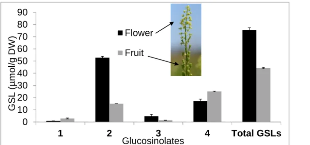
For a better understanding of GSL distribution among the inflorescence tissues, both flowers and unripe fruits were analyzed immediately after the full flowering stage, when capsules were already well-formed. Considering that R. lutea has a scalar flowering period, the same inflorescence provided open flowers at its top and fruits at its base. Flowers were particularly rich in total GSLs with 76 ± 2 mol g-1, composed of 70% and 23% of 2 and 4, respectively;
fruits had 44.3 ± 0.6 mol g-1 total GSLs, with proportions of 57% and 34% of 4 and 2,
respectively (Fig. 2).
Fig. 2. Glucosinolate contents in Reseda lutea flower and fruit. 1: benzyl GSL; 2: 2-(α-L -rhamnopyranosyloxy)benzyl GSL; 3: indol-3-ylmethyl GSL; 4: 3-hydroxybenzyl GSL. The results represent the mean of three independent analyses and are reported on a dry weight basis.
GSL profiles, finally, remained broadly stable in roots during the entire flowering time and were characterized by a high concentration of 1, a minor content of 2 and 3 (Fig. 1), and traces of 4 and 2-phenylethyl GSL (5), which was detected by HPLC with a previously purified standard (Müller et al., 2018).
0
10
20
30
40
50
60
70
80
90
1
2
3
4
Total GSLs
GS
L
(
µ
m
ol/g
DW
)
Glucosinolates
Flower
Fruit
14 We found a high content of 1 in roots, ranging from 20 to 75 µmol/g, and only traces of 5, a combination known in other species of the Brassicales order, such as in roots of Tropaelum
majus and Carica papaya, known to be arbuscular mycorrhizal fungi hosts (van Dam et al.,
2009).
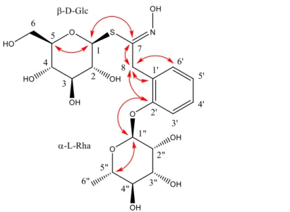
3.2 Structure elucidation of 2-(α-L-rhamnopyranosyloxy)benzyl glucosinolate
Compound 2 was isolated starting from mature seeds, which show the simplest GSL profile. The 1H, 13C, 1H-1H COSY, 1H-1H TOCSY, 1H-1H ROESY, 1H-13C edited-HSQC, and 1H-13C
HMBC of desulfated compound 2 were recorded for the first time. The TOCSY spectrum showed four spin systems, an aromatic one, two monosaccharide units and an isolated AB spin system (Fig. 3).
Fig. 3. Structure of desulfated 2-(α-L-rhamnopyranosyloxy)benzyl glucosinolate (2) with evidenced key HMBC correlations.
The resonance pattern of the four aromatic ring protons (H-3’ to H-6’) corresponded to an
ortho-disubstituted benzene ring. The HSQC, COSY, and HMBC spectra provided the
13C NMR chemical shifts of the protonated and non-protonated carbons. One of the latter
was deshielded (δ 156.1, C-2’) and correlated in HMBC with the anomeric proton (δ 5.6, H-1’’) of a sugar unit. This sugar unit is an aldohexose that bears a methyl group at position 6 (3 H-6’’). The value of the 1H-1H coupling constants were compatible with the presence of
α-L-rhamnose. The ROESY spectrum confirmed the spatial proximity of H-1” with H-3’ and therefore confirms that the rhamnosyl group is bound to the aromatic ring.
The protons of the isolated AB spin system (H-8a and H-8b) correlated in the HMBC spectrum with three aromatic carbons (1’, 2’, and 6’), thus allowing to directly bind C-8 and C-1’, as confirmed by a ROESY correlation between the two H-C-8 and H-6’. A fourth HMBC correlation of the two H-8 signals with a carbon at δ 157.7 suggested the binding of C-8 with the imino-type carbon C-7, supported by the very strong coupling constant between the two H-8 (17 Hz) that reflected the presence of a neighbouring sp2 carbon atom. The
HMBC spectrum correlated the chemical shifts of C-7 with the one of the other anomeric proton (δ 4.85, H-1). The 1H-1H coupling pattern of this proton is typical of the thioglucosyl
unit of a benzylic GSL (desulfated or not), in which H-2 and H-3 are strongly coupled (Ibrahim et al., 2018). The coupling constants within the H-3/H-4/H-5/H-6a/H-6b spin sub-system were compatible with a -D-gluco unit. The C-1/S and S/C-7 bonds were confirmed by the 7/H-1 HMBC correlation and by the low, characteristic, chemical shift of the anomeric C-1 (δ 84.C-1). The HSQC, COSY, and HMBC spectra provided a full assignment of the 13C NMR
chemical shifts within the thioglucosyl unit. The reported H-1/H-2 and H-2/H-3 coupling constants were those obtained by a thorough analysis of the 1H NMR spectrum of the
isomeric desulfated 4-(α-L-rhamnopyranosyloxy)benzyl GSL for which the multiplet patterns of the 1H NMR resonances in the thioglucose unit are identical but very hard to decipher due
16 of C-7 (δ 157.7) which was very close to the one in desulfated 4-(α-L -rhamnopyranosyloxy)benzyl GSL (δ 157.5) and about 8 ppm lesser than the one in the corresponding intact GSL (δ 165.6) (Ibrahim et al., 2018). The proposed structure was consistent with the HPLC-APCI-MS analysis: APCI--MS m/z measured: 536.0
(5%)[M+HCOO]-, 490.1 (40%) [M-H]-, 340.1 (15%) [M-Glc-H+HCOO]-, 328.0 (7%) [M-Glc]
-, 324.1 (100%) [RhaOPhCH2CN+HCOO]-, 195.1 (20%) [Glc-S]-. NMR raw data and spectra
are available at
https://www.dropbox.com/s/o0gyhy036t37eme/Reseda_lutea_flowers_SI.zip?dl=0 for a
temporarily access and at https://doi.org/10.5281/zenodo.3262193 for a permanent access
4. DISCUSSION
4.1 Ecological role of Reseda lutea with reference to its glucosinolate profiles.
No ecological studies have been performed on R. lutea, except for a statistical analysis of plant elements such as nitrogen, phosphorous, potassium and calcium in relation with characteristics of soil in Western Anatolia, where it is considered a plant with a great economic value as a source of natural dye in the carpet industry (Doǧan, 2001). Besides its high content in luteolin pigment, other interesting aspects of this plant have been considered since the ‘90s. It was recommended as a species suited for the improvement of apiculture in North Europe, as a fresh feed source in spring and summer, and as a dry feed source in Australia and Iran; finally as a plant used to prevent erosion thanks to its fast growing roots (Doǧan, 2001). Recently its content in GSL-derived ITCs (Radulović et al., 2014) and flavonol glycosides (Kızıltaş et al., 2019) were considered in relation to their possible beneficial effects on human health.
In this study the plants were subjected to a preliminary survey to identify the insects visiting them during flowering. Many insects were attracted by R. lutea inflorescences during the full flowering phase. They were identified and classified into different orders. Hymenoptera were
the most abundant, in particular the honey-bee Apis mellifera and other endangered wild pollinators belonging to this order. It was also evident that the number of honey-bees in particular, started to decrease during the late blooming phase and it dropped to 0 visits at the end of the flowering period (data not shown). This observation, made in open field, highlights that the greatest number of pollinators visiting the plants is coincident with the maximum peak of accumulation of 2 in the open flowers of the inflorescence, during full flowering time.
Several studies highlighted that the consumption of plant secondary metabolites favours a better resistance of bees to parasite and pathogen infection (Manson et al., 2010; Simone-Finstrom and Spivak, 2012; Richardson et al., 2015, 2016).
Like herbivores, pollinators eating nectar or pollen expose the adults and the larvae to the effect of plant secondary metabolites, whose preferential ingestion has been hypothesized as a self-medication mechanism used by pollinators (Simone-Finstrom and Spivak, 2012; Baracchi et al., 2015). In Muller et al. 2015, the taste-mediated deterrence of the non-volatile ITC moringin (4-(α-L-rhamnopyranosyloxy)benzyl ITC), to GSL adapted insect larvae of P.
napi and P. brassicae was reported. Moringin is the degradation product of 4- (α-L-rhamnopyranosyloxy)benzyl GSL, the characteristic GSL of the Moringaceae family which, together with the Resedaceae family are characterized by a R-side chain carrying a rhamnose moiety.
The similarity of the two GSL structures allows us to formulate a hypothesis on the possible ecological role of 2 as a deterrent and defensive molecule against pathogens.
In roots, one potential ecological role of 1 has been demonstrated on engineered plants of
A. thaliana overexpressing 1 in which herbivory by the generalist Spodoptera littoralis was
inhibited. In particular, benzyl ITC had a detrimental effect on the growth of generalist larvae (Bejai et al., 2012).
18
4.2 Chemosystematic
Phylogenetic studies about evolutionary relationships of Brassicales highlighted the presence of a Brassicales “core” including two clades: one that comprises Brassicaceae, Cleomaceae and Capparaceae families and the second, the “GRFT” clade, containing Resedaceae together with Gyrostemonaceae, Borthwickiaceae which was proposed as a new family (Su et al., 2012) and three genera Forchhammeria, Tirania, and Stixis (Cardinal-McTeague et al., 2016), with the validation of Stixaceae for the last one as a suprageneric name (Doweld and Reveal, 2008). Following the last Angiosperm Phylogeny Group (APG IV, 2016), Borthwickiaceae, and Stixaceae are thus far included in the Resedaceae family. In contrast to the GSLs derived from the chain-elongated forms of Met, which predominate in Brassicaceae, Cleomaceae and Capparaceae, main GSLs in Resedaceae derive from chain elongated Phe, whereas in Gyrostemonaceae and Tovariaceae, they are synthesized from branched-chain amino acids; however, GSLs derived by Leu, Ile and Val were reported in some species of Reseda genus (Mithen et al., 2010).
The identified structures of the GSLs to date attributed to Reseda genus are listed in Table 2. In this study we confirmed the presence of 1, 2, 3 in the GSL profiles of R. lutea and identified for the first time the presence of 4 in all aerial tissues, and traces of 5 in roots. The O-glycosylated benzyl GSL 2, appeared as the major GSL in all tissues of R. lutea except for fresh fruits where we observed a predominance of 4. 2 was firstly identified in the inflorescences of R. odorata where it is abundant, and then found in other plant tissues of this species (Olsen and Sorensen, 1979). It is considered the most representative GSL of the genus Reseda (Mithen et al., 2010) even if its presence was confirmed to date only in
R. lutea and R. odorata (Table 2). R. lutea GSLs profile depicted in this study appear similar
to that found in R. media, except for the presence of 2 which was not revealed in R. media. Differently from Vierheilig et al., (2000) who reported the presence of the indole GSL 3 only in the root of R. lutea, in our study 3 was identified in all tissues except for seeds. The special
metabolism of GSLs in R. lutea appears potentially connected to the metabolism of its flavonoids, in view of the presence of O-α-L-rhamnopyranosyl residues recently found in kaempferol and in isorhamnetin glycosylated forms in the aerial parts of R. lutea (Kızıltaş et al., 2019).
Table 2. Glucosinolate overview in Reseda genus
Species Glucosinolates Reference
R. odorata o-(α-L-rhamnopyranosyloxy)
benzyl GSL (2)
Olsen and Sorensen, 1979
R. media m-hydroxybenzyl GSL (4),
benzyl GSL (1), 2-phenethyl GSL (5), indol-3-ylmethyl GSL (3) in traces
Olsen and Sorensen, 1980
R. luteola (2S)-2-hydroxy-2-phenylethyl GSL, 2-phenylethyl GSL (5), indol-3-ylmethyl GSL (3) and (2R)-2-hydroxy-2-phenylethyl GSL (minor) Agerbirk et al., 2018 (2S)-2-hydroxy-2-phenylethyl GSL, indol-3-ylmethyl GSL (3), and an hydroxyalkyl GSL -HPLC-MS Griffiths et al., 2001
R. alba 2-hydroxy-2-methylpropyl GSL Gmelin and Kjær, 1970
(2S)-2-hydroxy-2-phenylethyl GSL identified by HPLC-MS; (2S)-2-hydroxy-3-butenyl GSL, indol-3-ylmethyl GSL (3), 1-methoxyindol-3-ylmethyl GSL identified by HPLC-DAD and comparison with standard compounds Ludwig‐Müller, et al., 1999 Sesamoides canescens or R. canescens, synomym of S. 2-(α-L -arabinopyranosyloxy)-2-phenylethyl GSL, 2-hydroxy-Olsen et al., 1981
20
interrupta and S. pigmea, synonym of R. phyteuma
phenylethyl GSL, 2-phenylethyl GSL (5)
Declaration of interest
The authors declare that there is no conflict of interest with regard to this research.
Acknowledgements
We thank anonymous reviewers for helpful and constructive comments. This research was financially supported by the Italian Ministry of agricultural, food, forestry and tourism policies (Mipaaft, grant RGV-FAO 2017-2019, CREA-CI M.B.), by the Natural Sciences and Engineering Research Council of Canada (NSERC Research Tool and Instruments, S.M. grant 315095-05) and by the Canadian Foundation for Innovation (Leaders
Opportunity Fund) – Ontario Research Fund (S.M. grant 11666). CNRS, Conseil Régional Champagne Ardenne, Conseil Général de la Marne, Ministry of Higher Education and Research (MESR), and EU-programme FEDER to the PlAneT CPER project is acknowledged by JMN.
References
Abdallah, M.S., de Wit, H.C.D., 1978. The Resedaceae. A taxonomical revision of the family (final installment). Meded. Landbouwhogeschool Wageningen 78–14: 99–416.
Agerbirk, N., Olsen, C.E., 2012. Glucosinolate structures in evolution. Phytochemistry 77, 16–45. https://doi.org/10.1016/j.phytochem.2012.02.005.
Agerbirk, N., Matthes, A., Erthmann, P.Ǿ., Ugolini, L., Cinti, S., Lazaridi, E., Nuzillard, J.M., Müller, C., Bak, S., Rollin, P., Lazzeri, L., 2018. Glucosinolate turnover in Brassicales species to an oxazolidin-2-one, formed via the 2-thione and without formation of thioamide. Phytochemistry 153, 79-93. https://doi.org/10.1016/j.phytochem.2018.05.006.
Ares, A. M., Nozal, M. J., & Bernal, J. 2015. Development and validation of a liquid chromatography-tandem mass spectrometry method to determine intact glucosinolates in bee pollen. J. Chromatog., B 1000, 49-56 https://doi.org/10.1016/j.jchromb.2015.07.017.
Balfour, N.J., Fensome, K.A., Samuelson, E.E.W., Ratnieks, F.L.W., 2015. Following the dance: Ground survey of flowers and flower-visiting insects in a summer foraging hotspot identified via honey bee waggle dance decoding. Agric. Ecosyst. Environ. 213, 265-271.
https://doi.org/10.1016/j.agee.2015.08.007.
Baracchi, D., Brown, M.J.F., Chittka, L., 2015. Behavioural evidence for self-medication in bumblebees? F1000Res. 4: 73. https://doi.org/10.12688/f1000research.6262.3.
Mis en forme : Anglais (États-Unis) Mis en forme : Anglais (États-Unis) Mis en forme : Anglais (États-Unis)
Mis en forme : Français (France) Mis en forme : Français (France)
22 Bedoya, L.M., Sanchez-Palomino, S., Abad, M.J., Bermejo, P., Alcami, J., 2001. Anti-HIV activity of medicinal plant extracts. J. Ethnopharmacol. 77, 113–116.
https://doi.org/10.1016/S0378-8741(01)00265-3.
Bejai, S., Fridborg, I., Ekbom, B., 2012. Varied response of Spodoptera littoralis against
Arabidopsis thaliana with metabolically engineered glucosinolate profiles. Plant Physiol.
Bioch. 50, 72-78. https://doi.org/10.1016/j.plaphy.2011.07.014.
Bremner, P., Rivera, D., Calzado, M.A., Obón, C., Inocencio, C., Beckwith, C., Fiebich, B.L., Muñoz, E., Heinrich, M., 2009. Assessing medicinal plants from South-Eastern Spain for potential anti-inflammatory effects targeting nuclear factor-Kappa B and other pro-inflammatory mediators. J. Ethnopharmacol. 124 (2), 295–305. https://doi.org/10.1016/j.jep.2009.04.035.
Burkle, L.A., Marlin, J.C., Knight, T.M., 2013. Plant-pollinator interactions over 120 years: loss of species, co-occurrence, and function. Science 339, 1611-1615.
https://doi.org/10.1126/science.1232728.
Cakilcioglu, U., Khatun, S., Turkoglu, I., Hayta, S., 2011. Ethnopharmacological survey of medicinal plants in Maden (Elazig–Turkey). J. Ethnopharmacol. 137, 469–486. https://doi.org/10.1016/j.jep.2011.05.046.
Cardinal-McTeague, W. M., Sytsma, K. J., Hall, J. C., 2016. Biogeography and diversification of brassicales: A 103 million year tale. Mol. Phylogenet. Evol. 99, 204-224.
Christenhusz, M.J.M., Byng, J.W., 2016. The number of known plants species in the world
and its annual increase. Phytotaxa, 261, 201–217.
http://dx.doi.org/10.11646/phytotaxa.261.3.1.
Cole, R.A., 1976. Isothiocyanates, nitriles, and thiocyanates as products of autolysis of glucosinolates in Cruciferae. Phytochemistry 15, 759–762.
https://doi.org/10.1016/S0031-9422(00)94437-6.
Conti, F., Bonacquisti, S., Abbate G., 2005. An annotated checklist of the Italian vascular flora. Palombi Editori, Roma: 152.
Daxenbichler, M. E., Spencer, G. F., Carlson, D. G., Rose, G. B., Brinker, A. M., Powell, R. G., 1991. Glucosinolate composition of seeds from 297 species of wild plants. Phytochemistry 30, 2623-2638. https://doi.org/10.1016/0031-9422(91)85112-D.
Doǧan, Y., 2001. A Study on the autecology of Reseda lutea L. (Resedaceae) distributed in Western Anatolia. Turk. J. Bot. 25, 137-148.
Doǧan, Y., Mert, H.H., Akcan, K., 2008. Anatomical studies of Reseda lutea (Resedaceae). Phytol. Balcan. 14, 91-95.
Doweld, A., Reveal, J.L. 2008. New suprageneric names for vascular plants. Phytologia 90, 416–417.
24 Dungey, S.G., Sang, J.P., Rothnie, N.E., Palmer, M.V., Burke, D.G., Knox, R.B., Williams, E.G., Hilliard, E.P., Salisbury, P.A., 1988. Glucosinolates in the pollen of rapeseed and indian mustard. Phytochemistry 27, 815-817.
https://doi.org/10.1016/0031-9422(88)84098-6.
Galletti, S., Bagatta, M., Branca, F., Argento, S., De Nicola, G. R., Cianchetta, S., Ninfali, P., 2015. Isatis canescens is a rich source of glucobrassicin and other health-promoting compounds. J. Sci. Food Agric. 95, 158-164. https://doi.org/10.1002/jsfa.6697.
Gmelin, R., Kjær, A., 1970. 2-hydroxy-2-methylpropyl glucosinolate in Reseda alba, Phytochemistry 9, 599-600. https://doi.org/10.1016/S0031-9422(00)85698-8.
Griffiths, D.W., Deighton, N., Birch, A.N.E., Patrian, B., Baur, R., Städler, E, 2001. Identification of glucosinolates on the leaf surface of plants from the Cruciferae and other closely related species. Phytochemistry, 57, 693-700.
https://doi.org/10.1016/S0031-9422(01)00138-8.
Hallmann, C.A., Sorg, M., Jongejans, E., Siepel, H., Hofland, N., Schwan, H., Stenmans, W., Müller, A., Sumser, H., Hörren, T., Goulson, D., De Kroon, H., 2017, More than 75 percent decline over 27 years in total flying insect biomass in protected areas. PLoS ONE 12: e0185809. https://doi.org/10.1371/journal.pone.0185809.
Heap, J.W., Willcocks, M.C., Kloot, P.M., 1987. The biology of Australian weeds. 17. Reseda
Ibrahim, N., Allart-Simon, I., De Nicola, G. R., Iori, R., Renault, J. H., Rollin, P., Nuzillard, J. M. 2018. Advanced NMR-based structural investigation of glucosinolates and desulfated glucosinolates. J. Nat. Prod. 81, 323-334. https://doi.org/10.1021/acs.jnatprod.7b00776.
ISO 9167-1:1992/Amd 1:2013. Graines de colza – Dosage des glucosinolates – Partie 1:Méthode par chromatographie liquide à haute performance (1992).
Jochimsen, M., Janzen, D. 1991. Structure and phytomass production of a pioneer community. Modern Ecology: Basic and Applied Aspects, 39-60.
https://doi.org/10.1016/B978-0-444-89183-9.50008-3.
Kızıltaş, H., Küçüksolak, M., Duman, S., Bedіr, E. 2019. Flavonol glycosides from Reseda
lutea L. Phytochem. Lett. 30, 150-153. https://doi.org/10.1016/j.phytol.2019.01.027.
Kumarasamy, Y., Cox, P.J., Jaspars, M., Nahar, L., Sarker, S.D., 2002. Screening seeds of Scottish plants for antibacterial activity. J. Ethnopharmacol. 83, 73–77.
https://doi.org/10.1016/S0378-8741(02)00214-3.
Ludwig‐Müller, J., Bennett, R.N., Kiddle, G., Ihmig, S., Ruppel, M. And Hilgenberg, W., 1999. The host range of Plasmodiophora brassicae and its relationship to endogenous glucosinolate content. New Phytologist, 141, 443-458.
https://doi.org/10.1046/j.1469-8137.1999.00368.x.
Manson, J.S., Otterstatter, M,C., Thomson, J.D., 2010. Consumption of a nectar alkaloid reduces pathogen load in bumble bees. Oecologia 162, 81–89.
Mis en forme : Anglais (États-Unis) Mis en forme : Anglais (États-Unis) Mis en forme : Anglais (États-Unis)
Mis en forme : Anglais (États-Unis) Mis en forme : Anglais (États-Unis)
26 Martίn-Bravo, S., Meimberg, H., Luceño, M., Märkl, W., Valcárcel, V., Bräuchler, C., Vargas, P., Heubl, G., 2007. Molecular systematics and biogeography of Resedaceae based on ITS and trnL-F sequences. Mol. Phylogenet. Evol., 44, 1105-1120.
https://doi.org/10.1016/j.ympev.2006.12.016.
Mithen, R., Bennett, R., Marquez, J., 2010. Glucosinolate biochemical diversity and innovation in the Brassicales. Phytochemistry 71, 2074–2086.
https://doi.org/10.1016/j.phytochem.2010.09.017.
Moghaddam, M.R., 1977. Reseda lutea: a multipurpose plant for arid and semiarid lands. J. Range Manag. 30 (1), 71-72. https://doi.org/10.2307/3897343.
Montaut, S., Rollin, P., 2016. “Glucosinolates and their distribution” in “Broccoli: Cultivation, Nutritional Properties and Effects on Health, Chapter: 2, Publisher: Nova Publishers Inc., Editors: B. Juurlink: New York, USA, 2016; pp 9-31.
Müller, C., Van Loon, J., Ruschioni, S., De Nicola, G. R., Olsen, C. E., Iori, R., Agerbirk, N., 2015. Taste detection of the non-volatile isothiocyanate moringin results in deterrence to glucosinolate-adapted insect larvae. Phytochemistry 118, 139-148.
https://doi.org/10.1016/j.phytochem.2015.08.007.
Müller, C., Schulz, M., Pagnotta, E., Ugolini, L., Yang, T., Matthes, A., Lazzeri, L., Agerbirk, N., 2018. The role of the glucosinolate-myrosinase system in mediating greater resistance of Barbarea verna than B. vulgaris to Mamestra brassicae larvae. J. Chem. Ecol. 44, 1190-1205. https://doi.org/10.1007/s10886-018-1016-3.
Nastruzzi, C., Cortesi, R., Esposito, E., Menegatti, E., Leoni, O., Iori, R., Palmieri, S, 2000. In vitro antiproliferative activity of isothiocyanates and nitriles generated by myrosinase-mediated hydrolysis of glucosinolates from seeds of cruciferous vegetables. J. Agric. Food Chem. 48, 3572-3575. https://doi.org/10.1021/jf000191p.
Olsen, O., Sorensen, H., 1979. Isolation of glucosinolates and the identification of o-(α-L-rhamnopyranosyloxy) benzylglucosinolate from Reseda odorata. Phytochemistry 18, 1547-1552. https://doi.org/10.1016/S0031-9422(00)98494-2.
Olsen, O., Sorensen, H., 1980. Glucosinolates and amines in Reseda media. Phytochemistry 19, 1783-1787. https://doi.org/10.1016/S0031-9422(00)83813-3.
Olsen, O., Rasmussen, K.W., Sørensen, H., 1981. Glucosinolates in Sesamoides
canescens and S. pygmaea: Identification of 2-(α-L -arabinopyranosyloxy)-2-phenylethylglucosinolate. Phytochemistry 20, 1857-1861.
https://doi.org/10.1016/0031-9422(81)84021-6.
Padla, E. P., Solis, L. T., Levida, R. M., Ragasa, C. Y., Shen, C., 2012. Antimicrobial isothiocyanates from the seeds of Moringa oleifera Lam. Z. Naturforsch. C 67, 557-564.
https://doi.org/10.1515/znc-2012-11-1205.
Pagnotta, E., Agerbirk, N., Olsen, C.E., Ugolini, L., Cinti, S., Lazzeri, L., 2017. Hydroxyl and methoxyl derivatives of benzylglucosinolate in Lepidium densiflorum with hydrolysis to isothiocyanates and non-isothiocyanate products: substitution governs product type and
28 mass spectral fragmentation. J. Agric. Food Chem. 65, 3167-3178. https://doi.org/10.1021/acs.jafc.7b00529.
Pignatti, S., 1982. Flora d'Italia. Bologna, Edagricole, volume I: 483-484.
Radulović, N.S., Zlatković, D.B., Ilić-Tomić, T., Senerović, L., Nikodinovic-Runic, J., 2014. Cytotoxic effect of Reseda lutea L.: A case of forgotten remedy. J. Ethnopharmacol. 153,125-132. https://doi.org/10.1016/j.jep.2014.01.034.
Richardson, L.L., Adler, L.S., Leonard, A.S., Andicoechea, J., Regan, K.H., Anthony, W.E., Manson, J.S., Irwin, R.E., 2015. Secondary metabolites in floral nectar reduce parasite infections in bumblebees. Proc. R. Soc. London, Ser. B 282: 20142471.
http://dx.doi.org/10.1098/rspb.2014.2471.
Richardson, L.L., Bowers, M.D., Irwin, R.E., 2016. Nectar chemistry mediates the behavior of parasitized bees: consequences for plant fitness. Ecology 97, 325-337.
https://doi.org/10.1890/15-0263.1.
Romeo, L., Cariccio, V. L., Iori, R., Rollin, P., Bramanti, P., Mazzon, E., 2018. The α-cyclodextrin/moringin complex: A new promising antimicrobial agent against
Staphylococcus aureus. Molecules 23, 2097. https://doi.org/10.3390/molecules23092097.
Simone-Finstrom, M.D., Spivak, M., 2012. Increased resin collection after parasite
challenge: a case of self-medication in honey bees? PLoS One 7:e34601.
https://doi.org/10.1371/journal.pone.0034601.
Mis en forme : Anglais (Royaume-Uni) Mis en forme : Anglais (Royaume-Uni) Mis en forme : Anglais (Royaume-Uni)
Mis en forme : Français (France) Mis en forme : Français (France) Mis en forme : Français (France)
Su, J.X., Wang, W., Zhang, L.B., Chen, Z.D., 2012. Phylogenetic placement of two enigmatic genera, Borthwickia and Stixis, based on molecular and pollen data, and the description of a new family of Brassicales, Borthwickiaceae. Taxon 61, 601–611. https://doi.org/10.1002/tax.613009.
Truchado, P., Tourn, E., Gallez, L.M., Moreno, D.A., Ferreres, F., Tomás-Barberán, F.A., 2010. Identification of botanical biomarkers in argentinean Diplotaxis honeys: flavonoids and glucosinolates. J. Agric. Food Chem. 58, 12678-12685.
https://doi.org/10.1021/jf103589c.
van Dam, N.M., Tytgat, T.O.G., Kirkegaard, J.A., 2009. Root and shoot glucosinolates: a comparison of their diversity, function and interactions in natural and managed ecosystems. Phytochem. Rev. 8, 171-186. https://doi.org/10.1007/s11101-008-9101-9.
Vierheilig, H., Bennett, R., Kiddle, G., Kaldorf, M., Ludwig-Müller, J., 2000. Differences in glucosinolate patterns and arbuscular mycorrhizal status of glucosinolate-containing plant species. New Phytol. 146, 343-352. https://doi.org/10.1046/j.1469-8137.2000.00642.x.
Wathelet, J.P., Iori, R., Leoni, O., Rollin, P., Quinsac, A., Palmieri, S., 2004. Guidelines for glucosinolate analysis in green tissues used for biofumigation. Agroindustria, 257-266. http://hdl.handle.net/2268/16127.
Wood, T.J., Holland, J.M., Goulson, D., 2015. Pollinator-friendly management does not increase the diversity of farmland bees and wasps. Biol. Conserv. 187, 120-126. https://doi.org/10.1016/j.biocon.2015.04.022.