HAL Id: tel-02065313
https://tel.archives-ouvertes.fr/tel-02065313
Submitted on 12 Mar 2019HAL is a multi-disciplinary open access archive for the deposit and dissemination of sci-entific research documents, whether they are pub-lished or not. The documents may come from teaching and research institutions in France or abroad, or from public or private research centers.
L’archive ouverte pluridisciplinaire HAL, est destinée au dépôt et à la diffusion de documents scientifiques de niveau recherche, publiés ou non, émanant des établissements d’enseignement et de recherche français ou étrangers, des laboratoires publics ou privés.
New inhibitors of quinolinate synthase as potential
antibacterial drugs
Jaione Saez Cabodevilla
To cite this version:
Jaione Saez Cabodevilla. New inhibitors of quinolinate synthase as potential antibacterial drugs. Bacteriology. Université Grenoble Alpes, 2018. English. �NNT : 2018GREAV047�. �tel-02065313�
THÈSE
Pour obtenir le grade de
DOCTEUR DE LA COMMUNAUTÉ UNIVERSITÉ
GRENOBLE ALPES
Spécialité : Chimie Biologie Arrêté ministériel : 25 mai 2016
Présentée par
« Jaione SAEZ CABODEVILLA »
Thèse dirigée par Sandrine OLLAGNIER, DR, CNRS, et codirigée par Yung-Sing WONG, CR, CNRS
préparée au sein du Laboratoire de Chimie et Biologie des métaux (CEA, Grenoble) et du Département de
Pharmacochimie Moléculaire (UGA, Grenoble)
dans l'École Doctorale Chimie et Sciences du Vivant
Nouveaux inhibiteurs de la
quinolinate synthase comme
potentiels antibactériens
Thèse soutenue publiquement le « 6 décembre 2018», devant le jury composé de :
Monsieur David PIGNOL
Directeur de Recherches, CEA Cadarache, Président Madame Myriam SEEMANN
Directrice de Recherches au CNRS, Strasbourg, Rapportrice Monsieur Erwan GALARDON
Chargé de Reserches au CNRS, Paris, Rapporteur Madame Isabelle SCHALK
Directrice de Recherches au CNRS, Strasbourg,examinatrice Monsieur Nicolas SPINELLI
Maître de conférences à l’UGA, Grenoble, examinateur Madame Sandrine OLLAGNIER
Directrice de Recherches au CNRS, Grenoble, directrice de thèse Monsieur Yung-Sing WONG
Chargé de Recherches au CNRS,Grenoble, invité Monsieur Olivier HAMELIN
ABBREVIATION LIST
3HP 3-hydroxyphthalic acid 4AP 4-aminophthalic acid 4-DMAP 4-dimethylaminopyridine 4HP 4-hydroxyphthalic acid 4MP 4-mercaptophthalic acid 5MPDC 5-mercaptopyridine-2,3-dicarboxylic acid 5MPzDC 5-mercaptopyrazine-2,3-dicarboxylic acid 6MPDC 6-mercaptopyridine-2,3-dicarboxylic acid
ADP Adenosine diphosphate Aib 2-aminoisobutyric acid AIBN 2,2’-Azo-bis-isobutyronitrile
Ala Alanine
AMP Adenosine monophosphate
Arg Arginine
AS Ammonium sulfate
Asp Aparagine
ATP Adenosine triphosphate BLY Bio-Layer Interferometry
Boc tert-butyloxycarbonyl
BPB Bromophenol blue
BSA Bovine serum albumin
cADPR Cyclic ADP-ribose
CD Circular dichroism
CHES N-Cyclohexyl-2-aminoethanesulfonic acid
Cys Cysteine
DCC N,N'-Dicyclohexylcarbodiimide
DFT Density functional theory DHAP Dihydroxyacetone phospate
DMF N,N-dimethylformamide
DMP 1,2-dimercaptophthalic acid DMPD N,N-dimethyl-p-phenylenediamine
DMSO Dimethyl sulfoxide
DNA Deoxyribonucleic acid
DTHPA Dithiohydroxyphthalic acid
DTT Dithiothreitol
EDCI 1-ethyl-3-(3-dimethylaminopropyl)carbodiimide EDTA Ethylenediaminetetraacetic acid
EPR Electron paramagnetic resonance
Eq Equivalents
ESI Electrospray ionization
Et Ethyl
Et2O Diethyl ether
EtOAc Ethyl acetate
EtOH Ethanol
FAD Flavin adenine dinucleotide
G3P Glyceraldehyde-3-phosphate
Glu Glutamic acid
h Hour
HATU Hexafluorophosphate Azabenzotriazole Tetramethyl Uronium
HEPES 4-(2-hydroxyethyl)-1-piperazineethanesulfonic acid
His Histidine
HOBt Hydroxybenzotriazole
HPLC High performance liquid chromatography
HRMS High resolution mass spectrometry
Hunig base N,N-Diisopropylethylamine
IA Iminoaspartate
IARC International agency for research on cancer IPTG Isop opyl β-D-1-thiogalactopyranoside ITC Isothermal titration calorimetry LB Luria Miller-Broth
Lys Lysine
M9 Minimal medium
m-CPBA meta-chloro perbenzoic acid
MDT Multidrug therapy Me Methyl Me3N Trimethylamine MeOH Methanol min Minute MP Mercaptophthalic acid MS Mass spectrometry Na Nicotinic acid
NAD Nicotinamide adenine dinucleotide
NADP Nicotinamide adenine dinucleotide phosphate NaNM Nicotinic acid mononucleotide
Ni-NTA Nickel nitriloacetic acid
Nm Nicotinamide
NMN Nicotinamide mononucleotide
NMNAT Nicotinamide mononucleotide adenylyltransferase
NMR Nuclear magnetic resonance
NmR Nicotinamide riboside OAA Oxaloacetic acid
Oac Acetate
OD Optical density
PA Phthalic acid
PCA Perchloric acid
PEG Polyethylene glycol
PLP Pyridoxal-5- phosphate
PPI Proton pump inhibitor
PRPP Phosphoribosyl pyrophosphate
p-TsCl para-toluenesulfonyl chlorine
QA Quinolinic acid
RNA Ribonucleic acid
ROS Reactive oxygen species
rt Room Temperature
SAM S-adenosyl-L-methionine
SDS Sodium dodecyl sulfate
SDS-PAGE Sodium dodecyl sulfate polyacrylamide gel electrophoresis
Ser Serine
tBu tert-butyl
TFA Trifluoroacetic acid
THF Tetrahydrofuran
Thr Threonine
TLC Thin layer chromatography
TMSCl Trimethylsilyl chloride
TMSOH 2(trimethylsilyl ethanol
TPI Triose phosphate isomerase TRIS Tris(hidroxymethyl)aminomethane
Trp Tryptophan
Ts Tosyl
Tyr Tyrosine
UV Ultraviolet
Résumé
La résistance aux antibiotiques par les micro-organismes est une menace mondiale. C'est pourquoi la recherche de nouveaux médicaments revêt une importance capitale de nos jours.
Le nicotinamide adénine dinucléotide (NAD) est un cofacteur essentiel et un substrat dans de nombreuses réactions biologiques. Pour cette raison, les enzymes qui l'utilisent comme substrat et celles qui participent à sa biosynthèse sont largement étudiées en tant que cibles antibactériennes potentielles. Dans ce contexte, nous nous intéressons au développement de nouveaux agents antibactériens contre la quinolinate synthase (NadA). Cette enzyme participe à la voie de biosynthèse de novo du NAD chez les procaryotes et est essentielle chez deux pathogènes : Helicobacter pylori, responsable d'ulcères et de cancers gastriques, et Mycobacterium leprae, responsable de la lèpre. Le fait que NadA n'existe que chez les procaryotes et qu'elle est essentielle chez deux agents pathogènes font de NadA une cible intéressante pour la conception de nouveaux antibactériens. La quinolinate synthase est une enzyme [4Fe-4S] où l'un des atomes de Fe est coordonné par une molécule d'eau à l'état de repos et joue le rôle d'acide de Lewis durant la catalyse. La réaction catalysée par NadA consiste en la formation d'acide quinolinique (QA), précurseur du NAD, à partir d'iminoaspartate et de dihydroxyacétone phosphate. Sur la base de la découverte dans notre laboratoire du premier inhibiteur de NadA (le DTHPA), qui agit par coordination irréversible du fer catalytique du cluster, nous avons conçu et synthétisé une famille de molécules pouvant servir d'inhibiteurs plus spécifiques de NadA. Ces molécules contiennent un cycle benzène / pyridine ou pyrazine, deux carboxylates vicinaux et un thiol en tant que groupe coordinant du fer: l’acide 4-mercaptophtalique (4MP), l’a ide 6-mercaptopyridine-2, 3-dicarboxylique (6MPDC), l’a ide 5-mercaptopyrazine-2, 3-dicarboxylique (5MPzDC et l’a ide 5-mercaptopyridine-2, 3-dicarboxylique (5MPDC . Nous avo s dé o t é ue es olé ules i hi e t l’e zy e NadA in vitro avec un IC50 du
même ordre de grandeur que celui du DTHPA dizai e de µM . Pa l’utilisatio de diverses spectroscopies et de la cristallographie, nous avons démontré que l'inhibition s’effe tue par la coordination de ces molécules avec le fer catalytique du cluster via leur groupement thiol. Nous avons également étudié in vitro la spécificité de ces molécules. Nous avons montré que les molécules 4MP et 5MPDC étaient des inhibiteurs spécifiques de NadA lo s u’on les teste su l’a o itase B, u e enzyme [4Fe-4S] bactérienne, dont le cluster présente des propriétés structurales et fonctionnelles similaires à celles du cluster de NadA.
Enfin, nous avons étudié l'activité d'inhibitrice de l’e se le des olé ules in cellulo sur de la voie de biosynthèse du QA chez Escherichia coli avec le DTHPA comme contrôle. Alors que les molécules 6MPDC et 5MPzDC i hi e t la oissa e d’E. coli de manière indépendante de la voie de biosynthèse du QA, le 4MP et le 5MPDC (les deux inhibiteurs spécifiques in vitro) n'ont montré aucune activité inhibitrice in cellulo. Ce manque d'activité pouvant être dû à un manque de pénétration des molécules à l'intérieur des bactéries, nous avons envisagé de favoriser la pénétration à l'aide d'un vecteur transmembranaire, un analogue simplifié du tétra-cyclopeptide naturel FR235222. Nous avons synthétisé et couplé le cyclopeptide à l'inhibiteur 4MP. Malheureusement, aucune inhibition de la croissance d'E. coli n'a été observée. La thèse s’est te i ée e essaye t de comprendre la pénétration du tétra-cyclopeptide sur bactérie, en utilisant notamment des agents fluorophores.
Summary
Resistance to antibiotics is becoming a world-wide threat. Microorganisms are able to withstand drugs leading to persistence of diseases. This is why the research of new drugs is of great importance nowadays.
Nicotinamide adenine dinucleotide (NAD) is an essential cofactor and substrate in numerous biological reactions. For this reason, both the enzymes that use it as a substrate and those participating on its biosynthetic pathway are largely studied as potential antibacterial targets. In this context, we are interested in the development of new antibacterial drugs against quinolinate synthase enzyme (NadA). This enzyme participates in the prokaryotic NAD de novo biosynthetic pathway and is essential in two pathogens: Helicobacter pylori, cause of gastric ulcers and cancers, and Mycobacterium leprae, responsible of leprosy. The fact that NadA only exists in prokaryotes and the fact that it is essential for these two pathogens make it an interesting target for the design of new antibacterial drugs. Quinolinate synthase is a [4Fe-4S] cluster enzyme, where one of the Fe sites is coordinated by a water molecule in the resting state, and plays a Lewis acid role during catalysis. The reaction catalyzed by NadA consists in the formation of quinolinic acid (QA), precursor of NAD, from iminoaspartate and dihydroxyacetone phosphate. Based on the discovery in our laboratory of the first inhibitor of NadA (DTHPA) that coordinates irreversibly the catalytic iron site of the cluster, we designed and synthesized a family of molecules as potential more specific NadA inhibitors. These molecules contain a benzene/pyridine or pyrazine ring, two vicinal carboxylates and a thiol as an iron coordinating group: 4-mercaptophthalic acid (4MP), 6-mercaptopyridine-2, 3-dicarboxylic acid (6MPDC), 5-mercaptopyrazine-2, dicarboxylic acid (5MPzDC) and 5-mercaptopyridine-2, 3-dicarboxylic acid (5MPDC). We demonstrated that these molecules inhibit NadA enzyme in vitro in the same range as DTHPA. Using different spectroscopies and crystallography, we demonstrated that inhibition occurs by coordination of the molecules to the catalytic iron site through their thiol group. We investigated also in vitro the specificity of these molecules. We demonstrated that 4MP and 5MPDC molecules are specific NadA inhibitors when assayed on bacterial aconitase B, a [4Fe-4S] enzyme, whose cluster displays functional and structural properties similar to those of NadA.
Finally, we investigated the QA pathway inhibition activity of the four molecules in cellulo, in an Escherichia coli strain. Whereas 6MPDC and 5MPzDC molecules inhibit E. coli growth in a QA biosynthetic pathway independent manner, 4MP and 5MPDC (the two in vitro specific inhibitors) did not show any in cellulo inhibition activity. Since this lack of activity might be due to a lack of penetration of the molecules inside bacteria, we thought about assisting the penetration of the molecules using a transmembrane carrier, a simplified analogue of the tetra-cyclopeptide FR235222 natural product. We synthetized and coupled the cyclopeptide to the 4MP inhibitor. Unfortunately, no E. coli growth inhibition was observed. The Ph.D ended by investigating the penetration of the tetra-cyclopeptide inside bacteria, using some fluorophore agents.
1
Table of contents
INTRODUCTION ... 15
I. Nicotinamide Adenine Dinucleotide (NAD) ... 16
a. Structure ... 16
b. Physical-chemical properties ... 17
c. Functions of NAD/NADH and NADP/NADPH ... 18
(i) Role in redox metabolism ... 18
(ii) Role in non-redox metabolism ... 20
d. NAD physiological concentration and cell localization in eukaryotes ... 23
II. NAD biosynthesis ... 24
a. The de novo pathways ... 24
(i) Upstream pathway in prokaryotes ... 25
(ii) Upstream pathway, in eukaryotes ... 26
(iii) -Downstream pathway: common in prokaryotes and eukaryotes ... 29
b. Salvage and recycling pathways ... 31
(i) Prokaryotes and eukaryotes except mammals ... 32
(ii) Mammals ... 35
c. NAD biosynthesis regulation ... 36
(i) NAD metabolism regulation by NadR ... 36
(ii) NAD metabolism regulation by NiaR ... 37
(iii) NAD metabolism regulation by NrtR ... 37
III. NAD: a pharmacological target... 38
IV. Quinolinic acid production in prokaryotes: Quinolinate synthase system ... 39
a. Identification of enzymes ... 39
b. Identification of substrates ... 40
c. A NadA-NadB complex? ... 43
d. Characterization of L-aspartate oxidase (NadB) ... 44
e. NadA: A [4Fe-4S]2+ enzyme ... 46
(i) Purification and characterization of NadA apoprotein ... 46
(ii) Purification and characterization of the holoprotein ... 46
(iii) NadA [4Fe-4S]2+ cluster: sensitivity to oxygen ... 49
f. Structure of Quinolinate synthase NadA ... 52
2
(ii) First structure of holo-protein quinolinate synthase ... 54
g. Mechanism of Quinolinate synthase ... 57
h. DTHPA: The first NadA inhibitor ... 63
i. Structure-activity study based on DTHPA ... 66
V. Antibiotics in the XXI century ... 68
a. Antibiotic resistance-historical overview ... 68
(i) Gram-positive bacteria ... 71
(ii) Gram-negative bacteria ... 71
(iii) Mycobacteria ... 71
b. The search for new antibacterial agents ... 73
(i) Helicobacter pylori ... 73
(ii) Mycobacterium leprae ... 74
c. Across the cell membranes ... 75
d. Approaches to design and improve membrane permeability ... 76
e. The natural cyclic tetrapeptide FR235222 and its analogues ... 77
Ph.D OBJECTIVES ... 81
RESULTS CHAPTER I: ... 85
SYNTHESIS OF POTENTIAL QUINOLINATE SYNTHASE INHIBITORS ... 85
I. Synthesis of 6- mercaptopyridine- 2, 3-dicarboxylic acid (6MPDC) from QA ... 87
II. Synthesis of 5- mercaptopyrazine- 2, 3-dicarboxylic acid (5MPzDC) from 2- chloroquinoxaline 93 III. Synthesis of 5-mercaptopyridine- 2, 3-dicarboxylic acid (5MPDC) from 3-chloroquinoline .... 98
RESULTS CHAPTER II: ... 107
IN VITRO STUDIES OF 6MPDC, 5MPzDC, 6MPDC AND 4MP ON NadA ... 107
I. Production of 56Fe-S NadA, 57Fe-S NadA and NadB proteins ... 108
a. E. coli 56Fe-S NadA and 57Fe-S NadA ... 108
b. E.coli NadB ... 110
II. NadA inhibition with 6MPDC, 5MPzDC, 5MPDC and 4MP ... 111
III. Study of NadA inhibition mechanism ... 116
c. Mössbauer spectroscopy ... 116
d. UV-visible absorption spectroscopy ... 118
e. Circular dichroism ... 119
f. Crystal structures of NadA in complex with the molecules ... 122
3
V. Discussion ... 129
a. NadA inhibition mechanism ... 130
b. Specificity ... 132
VI. 5MPDC and TmNadA* ... 134
a. Discussion ... 136
RESULTS CHAPTER III: ... 139
IN CELLULO INHIBITION STUDY OF 6MPDC, 5MPzDC, 5MPDC AND 4MP ... 139
I. E. coli MG1655 as strain for in cellulo tests ... 140
II. In cellulo tests of the four NadA inhibitors: 6MPDC, 5MPzDC, 5MPDC and 4MP ... 141
a. Esterification of the carboxylic acids ... 142
b. Test under anaerobic conditions ... 144
c. Penetration and drug delivery strategy assisted by FR235222 simplified analogue . 146 (i) Synthesis of compound 22 ... 149
Synthesis of compound 23 ... 153
(ii) Synthesis of compound 24: coupling of 23 and 4MP ... 154
(iii) E. coli MG1655 growth tests with compounds 22 and 24 ... 155
III. Looking for visual penetration evidence ... 156
a. Synthesis of compound 40... 157
b. Synthesis of compound 41... 160
c. In cellulo assays of fluorophores and their cyclopeptide-analogues 38 and 39; 42 and 40; 50 and 41. ... 162
d. Fluorescence assays in cellulo ... 164
IV. DISCUSSION AND CONCLUSSION ... 166
GENERAL CONCLUSION AND PERSPECTIVES ... 169
MATERIALS AND METHODS ... 175
I. Biological materials... 176
a. Bacterial strains ... 176
b. Plasmids ... 176
c. Culture media ... 177
II. Molecular biology methods ... 177
a. Preparation of competent cells ... 177
b. Transformation of competent cells ... 177
4
a. Protein overexpression ... 178
b. Protein extraction ... 178
(i) Freeze-thaw cycles ... 178
(ii) Sonication ... 178
c. Protein purification ... 179
d. Reconstitution of NadB FAD cofactor ... 179
e. Enzymatic activity measurements ... 179
(i) in vitro NadA activity ... 179
(ii) in vitro NadB activity ... 181
(iii) in vitro AcnB activity ... 182
f. Biochemical analyses ... 183
(i) Protein quantification using Bradford method ... 183
(ii) Protein quantification using Nanodrop ... 183
(iii) SDS-PAGE: Electrophoresis ... 184
(iv) Iron content ... 185
(v) Sulfur content ... 186
IV. Biological methods ... 187
a. Functional complementation tests ... 187
b. E. coli MG1655 bacterial growth in the presence of potential inhibitors ... 188
V. Spectroscopic, biophysical and analytical methods ... 188
a. Reversed-phase high performance liquid chromatography (HPLC) ... 188
b. UV-visible absorption spectroscopy ... 190
c. Mössbauer spectroscopy ... 191
d. Circular dichroism ... 193
VI. Protein crystallization and X-Ray structures... 195
a. Crystallization ... 195
i. 4MP (4-mercaptopyridine-2,3-dicarboxylic acid) complex. ... 195
ii. 6MPDC (6-mercaptopyridine-2,3-dicarboxylic acid) complex. ... 195
iii. 5MPDC (5-mercaptopyridine-2,3-dicarboxylic acid) complex. ... 196
iv. TmNadA*-QA Complex (5MPDC-derived) ... 196
b. X-Ray Structure Determination. ... 196
VII. QA Production by 5MPDC and NadA in Solution... 199
VIII. Confocal microscopy ... 200
5
X. Chemistry experimental section ... 200
a. Products and solvents ... 200
b. Analytic devices ... 201
c. Synthesis of NadA inhibitors ... 201
i. 6MPDC: 6- mercaptopyridine- 2, 3-dicarboxylic acid ... 201
ii. 5MPzDC: 5- mercaptopyrazine- 2, 3- dicarboxylic acid ... 205
iii. 5MPDC: 5- mercaptopyridine- 2, 3- dicarboxylic acid ... 208
(i) Alternative synthesis of 5MPDC: 5- mercaptopyridine- 2, 3- dicarboxylic acid 211 d. Synthesis of molecules for in cellulo assays... 212
ANNEXES ... 233
I. ANNEX I: Aconitase B enzyme (AcnB) ... 234
II. ANNEX II: NadA, a highly structurally conserved enzyme in different organisms ... 236
a. Sequence alignment of NadA from different organisms ... 236
BIBLIOGRAPHY ... 241
7 Gizonak badu inguru latz bat menperatzeko premia,
burruka hortan bizi da eta hori du bere egia. Ekin ta ekin bilatzen ditu, saiatze hortan ezin gelditu, jakintza eta argia ; bide ilunak nekez aurkitu, lege berriak noizbait erditu, hortan jokatuz bizia.
9 A a sœu et es pa e ts…
11
REMERCIEMENTS
Je tie s à e e ie tout d a o d mes trois encadrants de thèse. Ma directrice Sandrine Ollagnier ; mon codirecteur Yung-Sing Wong ; et mon co-encadrant Olivier Hamelin.
- Me i “a d i e de a oi e ad pe da t i a s et de a oi fait o fia e a e Oli ie ua d j tais u e aie d uta te au la o. Vous a ez donné la confiance et la motivation pou ue je e la e da s ette th se. Me i de a oi app is toute la io hi ie ue je o ais, la igueu et le o p o is au t a ail. Me i de a oi o sa du te ps e ua d e tait pas ide t de le fai e. Ça a t u ai plaisi de pa tage auta t d a es avec toi, bien professionnellement comme personnellement. Je garde beaucoup de choses appris et beaucoup de moments importants partagés.
- Me i Yu g de ta patie e, d a oi su e assu e et e al e ua d j tais d o d e. Me i de toujou s a i e à t ou e le ôt positif de ha ue situatio , de a oi toujou s out et e ou ag . Me i aussi de tout e ue tu as pa tag a e oi et e ue tu as appris du côté scientifique mais aussi du côté personnelle.
- Olivier, merci pour tout. Sans toi rien aurait été pareil ; ni la thèse, mi ma période grenobloise. Merci de ton soutien, de ta confiance, de ta force, ton savoir être, ton dynamisme, et merci de ta calme et ta pêche à la fois. Merci de chaque moment partagé et de tout e ue tu as app is s ie tifi ue e t ais su tout da s le pe so el. Me i d a oi été là toujours, dans vraiment tous les moments.
Je e e ie gale e t toutes les pe so es ue j ai o ues pe da t es i a s da s es deu laboratoires :
- Leandro, merci pour le gros travail de synthèse, la rigueur et finesse de ton travail, le o p o is a e les dates li ites toujou s diffi iles à te i et ha gea ts… e i ai e t de to i estisse e t da s le p ojet et de tout e ue tu as appris.
- Je remercie aussi Stéphane Ménage et Ahcène Boumendjel, les directeurs des deux laboratoires où j ai t a aill LCBM et DPM . Me i pou u a ueil t s haleu eu , l i t t toujours montré vers moi et mon travail, et de la bonne humeur.
- Co e j ai t a aill da s t ois uipes diff e tes et ue j ai i te agit ai e t a e tout le monde pour des discussions scientifiques et/ou personnelles, je ne pourrais pas nommer toutes les personnes qui forment ou qui ont fait partie des équipes dans un moment donné pe de t es i a s BioCAT, BioCE et MedChe . Mais je tie s à ous e e ie tous. J ai
12 t t s ie e tou e et soute ue tout le te ps g â e à ous tous et j ai fait ette th se avec plein de conseils de la plupart de vous, merci. Je tiens à remercier en particulier les discussions scientifiques surtout au moment de la rédaction de thèse avec Patrice Cathy, Philippe Carpentier et Vincent Nivière. De la même façon, je tiens à remercier le soutien plutôt personnel de Nicolas Duraffourg, Philippe Carpentier et Christine Cavazza .
- Un petit mot pour remercier les thésards, post-do s et jeu es he heu s ue j ai ôto et do t j ai t ou des ais a is ui o t toujou s u e oi et o t toujou s soute u : Marila, Jordan, Émile, Mathieu, Benjamin, Sarah, Lucile, Steve, Leandro, Brayan, Mériem, Lie … e i.
Je e e ie tous os olla o ateu s sa s les uels ette th se au ait pas t possi le et ui o t appo t u t a ail u ial ui a pe is d a a e auta t a th se o e le p ojet de NadA. Me i pour votre travail, votre temps, votre investissement et votre soutien.
- Spectroscopie Mössbauer : Jean-Marc Latour et Martin Clémencey. - Spectroscopie UV-visible : Jean-Marc Latour
- Cristallographie : Juan Carlos Fontecilla Camps, Anne Volbeda, Claudine Darnault, Océane Gigarel et Oriane Renoux.
- Docking et calculs théoriques : Patricia Amara - Microscopie confocale : Claire Durmort
Fi ale e t, je eu e e ie les ge s ui o t pas fait pa tie de o e i o e e t p ofessio el mais qui ont été indispensables pou oi pou leu soutie , fo e, hu eu … grâce auxquels cette thèse a été possible.
- Aita, ama eta Ainhoa. Tengo que ser breve porque si no escribiría otra tesis agradeciéndoos todo lo ue sois pa a í ha is po í. “i esto a uí es po osot os… po que yo me he ocupado del estudio y trabajo, pero esta tesis la he hecho gracias a vuestro apoyo, fuerza, determinación y valores que me habéis transmitido y enseñado toda la vida. ESKERRIK ASKO beroena zuentzako da eta tesi hau ere, zuena da.
- Abuela Petra, gracias por todo el apoyo, por pensar tanto en mí y rezar siempre para que todo me salga bien. No solo durante la tesis, si no durante mis 27 años. Qué suerte tenerte. - Al resto de mi familia en general y en especial a mi abuela Pili y a todos los que me habéis
apoyado tanto e incluso acompañado en Grenoble el día de la lectura: Mariaje, Jose Antonio, Amaya, Cristina, Alex, Maria Eugenia, Aitor e Iñigo.
13 - Me i de tout o œu , Clai e. Pou a oi a o pag da s e he i , pou toujou s e soute i , e ou age et oi e e oi. Mais su tout pou tout e ue ous a ez app is et o t … telle e t p ieu . U e de es plus elles e o t es g e o loises. - Irakasteko jartzen duzun ilusio, gogo eta energiagatik kimikari gustoa hartu niola ez da
sekretu. Bide honi hasiera ematen lagundu zenidan eta nire aurrera-pauso eta lorpen guztiak pozez izi iza dituzu. Eske ile e o-bero bat, Felixe.
- Nire kuadrillari: Bea, Marina, Miren, Ainhoa eta Olaia. U o ea, a i oak… ez zaizkit i oiz falta izan. Eskerrik asko neskak, hau ere zuena da parte batean. Amaia, zurea ere bai!
- Maite, Maider eta Olaia. Zuei ere eskerrik asko, beti hor zaudetelako eta kimikako karrerak eman didan hoberena zaretelako.
- Mezkiritzeko lagunei. Arnas hartu behar izan dudanean leku ederrenean eta konpainia ederrenarekin egin ahal izan dudalako, eskerrik asko.
- I want to thank my American family, the Widemans, for the continued support and prayers and for being my main English teachers.
- A d last ut defi itel ot least… to f ie ds a I all my family) in Grenoble. Thank you for making these years the best of my life so far, for supporting me, for providing me love, laughter and lots of joy. You will always be part of me. And thank you for the music, for all that singing that put sound to these amazing adventure.
15
INTRODUCTION
16
I.
Nicotinamide Adenine Dinucleotide (NAD)
In 1906 Nicotinamide Adenine Dinucleotide (NAD) was discovered by Arthur Harden in yeast extracts due to its capacity to accelerate alcoholic fermentation1. However, its structure remained unknown for 30 years. It was between 1935 and 1936 that Hans Von Euler-Chelpin not only found its structure2 but Otto Heinrich Warburg also evidenced its redox role3. In 1940, nicotinamide adenyltransferase enzyme, an enzyme involved in NAD biosynthesis was identified by Arthur Kornberg4 and became the first identified enzyme in NAD biosynthetic pathway. Since then, the NAD biosynthesis has been and is still widely studied5,6. NAD has expanded beyond its role as a redox cofactor, since indeed, NAD+ and its metabolites also act as degradation substrates for a wide range of enzymes7.
a. Structure
Nicotinamide adenine dinucleotide is formed by two nucleotides bonded by a pyrophosphate bridge. The nitrogenous bases that form these nucleotides are adenine and nicotinamide. NAD refers to the two redox forms which exist in nature: NAD+ (oxidized form) and NADH (reduced form) (Scheme 1)8.
17
NAD also exists in a phosphorylated form, NADP. NADP has an additional phosphate group on the 2' position of the ribose ring that carries the adenine moiety. NADP stands for its two redox forms: NADP+ and NADPH. Both NAD and NADP have the same redox properties but there is a difference on the way they are used in the cell. Whereas NAD is involved mainly in catabolic reactions, NADP mainly takes part in anabolic reactions.
b. Physical-chemical properties
NAD(P) is an important redox cofactor in the cell; it can accept or donate electrons in different metabolic reactions. The electron transfer happens at the nicotinamide part. In order to reduce NAD(P)+, a hydride (H-, a proton and two electrons are involved) is transferred to the C4 of the nicotinamide base. The aromatization of the system is achieved
by reoxidation of the cofactor with the transfer of a hydride (2e-, H+) to a substrate (Scheme 2). The standard potential of the NAD(P)+/NAD(P)H couple is E°= -0.32 V 9.
Scheme 2: NAD(P)+/NAD(P)H redox couple. Only the nicotinamide part is shown in the figure 2, R represents the rest of the skeleton of NAD(P).
The NAD(P)+/NAD(P)H ratio is known as the redox state of the cell, which is a reflect of the cell metabolic activities as well as an indicator of cell health. In general, the ratio stays constant on a same cell compartment. Under aerobic conditions, the ratio of NAD+/NADH = 8-10, which means that the cofactor exists mainly on its oxidized form. However, the ratio of NADP+/NADPH = 0.005, meaning that in this case the cofactor exists mainly on its reduced form10.
Both NAD(P)+ and NAD(P)H present UV absorption band(s), allowing their identification and quantitation. NAD(P)+ has an absorbance peak at 260 nm with a molar extinction coefficient 260nm = 16900 M-1cm-1; NAD(P)H has an additional peak at 340 nm
18
allows to easily quantify NAD(P)+ and NAD(P)H, and to easily monitor their conversion during enzymatic reactions.
The two forms also differ with regard to fluorescence. NAD(P)+ does not present any fluorescence whereas NAD(P)H does at 460 nm. This makes it possible to control the binding of the cofactor to a molecule, for example, a protein.
c. Functions of NAD/NADH and NADP/NADPH
It has been well known for years that NAD(P) is a cofactor that plays a crucial role in the redox metabolism. However, it also plays a role in several non-redox processes such as substrate of bacterial DNA ligases, sirtuins or ADP-ribosyltransferases.
(i) Role in redox metabolism
NAD(P) is the redox cofactor of several oxidoreductases, one of the largest family of enzymes that catalyze electron transfer. Some of the most significant examples are alcohol dehydrogenases, lactate dehydrogenases or NAD-ubiquinone reductases.
NAD(P) cofactor is bound to proteins th ough the ‘oss a fold , a st u tu al otif found in proteins that bind to dinucleotide cofactors (FAD, NAD, NADP). It was first identified by Michael Rossmann in the lactate dehydrogenase enzyme11 (Figure 1). The Rossmann fold is composed of a series of alternating beta strand () and alpha helical () segments wherein the -strands are hydrogen bonded forming a -sheet. The initial beta-alpha-beta () fold is the most conserved segment of Rossmann folds12.
Figure 1: NAD-bound Rossmann fold motif. Structure of Crysptosporidium parvum lactate dehydrogenase. Red: NAD cofactor; purple: -helixes; Yellow: -sheets.
19
The redox reactions catalyzed by oxidoreductases are essential in cell metabolism with a particular role on the catabolism; in other words, on the break-down of nutrients and energy release. A big part of the energy produced in the catabolic reactions is stored as NADH and converted into ATP during cell respiration in mitochondria. Each molecule of NADH consumed, provides energy to form 3 ATP molecules at the last stage of oxidative phosphorylation (Figure 2)13.
Figure 2: Simplified scheme of NAD role in redox metabolism.
Even though NAD plays an essential role in catabolism, it is also involved in gluconeogenesis (as an electron transporter), which is an anabolic process. It also plays an important role in nitrogen assimilation in nitrifying bacteria14,15. In the case of NADP, it is widely used as a reducing agent mainly in anabolic reactions such as fatty acid synthesis or photosynthesis16.
A NAD-oxidoreductase that it is worth highlighting at this point is the NAD(P) transdehydrogenase enzyme. It couples the reduction of NADP+ to NADPH with the oxidation of NADH to NAD+. This way, it plays a primordial role on maintaining the equilibrium in the cell that exists between the catabolic reactions with NAD production and anabolic reactions with NADPH consumption17. The enzyme is present in both prokaryotes and in mitochondria of eukaryotes.
20
(ii) Role in non-redox metabolism
- NAD: substrate in ADP-ribosylation reactions
Adenosine diphosphate ribosylation is a reversible post-translational modification reaction that consists in the transfer of an ADP-ribose to a protein and/or the formation of ribose polymers (Scheme 3). These reactions are performed by ADP-ribosyltransferase (ARTs) or by poly-(ADP-ribose) polymerase (PARPs) enzymes, respectively18 and are involved in many cellular processes including DNA repair, gene regulation and apoptosis19.
Scheme 3: ribosyl transferase reaction. NAD is hydrolyzed by an ART yielding an ADP-ribosylated protein and nicotinamide.
- NAD: cyclic ADP-ribose precursor
Cyclic ADP-ribose (cADPR) is a potent intracellular calcium releasing agent. This is due to the binding of it to a ryanodine-type receptor. As a consequence, it plays an important role in the maintenance of calcium homeostasis in cells. After the formation of ADP-ribose from NAD+, it is cycled through the anomeric carbon of the terminal ribose and the N1 nitrogen of the adenine ring (Scheme 4)20.
21 Scheme 4: Formation of cyclic ADP-ribose from NAD.
- NAD: sirtuin substrate
Most of the sirtuins are NAD-dependent protein-deacetylases found in prokaryotes, eukaryotes and archaea. They catalyze the lysine deacetylation coupled to the hydrolysis of NAD and this hydrolysis yields -O-acetyl-ADP-ribose, the deacetylated substrate and nicotinamide21,22 (Scheme 5). Sirtuins play an important role in the regulation of many biological pathways; the deacetylation of histones is a key part in gene regulation, since it deactivates transcription whereas the acetylation activates it. For this reason, these enzymes are of big importance in processes like apoptosis or cellular aging23,24. Eukaryotic cells present seven types of sirtuins whereas prokaryotes and archaea only present one or two25.
22 Scheme 5: Reaction atal zed a si tui that ields -O-acetyl-ADP ribose, nicotinamide and a deacetylated protein.
- NAD: a substrate of NAD-dependent DNA ligases
DNA ligases participate in DNA replication and DNA repair. NAD is a substrate of some of the enzymes of this type in bacteria. In the case of eukaryotes, the substrate of DNA ligases is ATP26. They catalyze the phosphodiester bond formation. NAD provides the adenosine monophosphate (AMP) group, which binds to a lysine on the active site of the ligase27. The mechanism by which DNAis repaired is shown in Scheme 6.
Scheme 6: Reaction catalyzed by a DNA ligase. NAD is the AMP donor which allows the ligation of the two DNA extremities
As shown above, the consumption of NAD in different reactions leads to the formation of nicotinamide (Nm) in most of them. It is important to keep this in mind since this product, along with some others such as nicotinamide mononucleotide (NMN) might be recycled in the cells in order to re-synthetize NAD. This will be explained later on this introduction.
23
d. NAD physiological concentration and cell localization in eukaryotes
As previously explained, NAD plays a crucial role in the redox states and redox processes of the cells as well as in many other different biological processes. For this reason, NAD is found all over the cell in different compartments28. Its synthesis is mostly performed in the nucleus where it has an essential role in the regulation of gene expression. It can diffuse through the nuclear pores in order to reach the cytoplasm where it takes part in many different processes. The NADH produced during glycolysis is transported from the cytoplasm into the mitochondrial matrix to provide reducing equivalents for the energy metabolism (Krebs cycle and electron transport chain). The inner membrane of mitochondria is impermeable for NAD+, so it can only be imported inside these organelles through membrane carrier proteins called mitochondrial shuttles. The most common ones are malate-aspartate and glycerol-3-phosphate shuttles. Yet, no NAD+ transporter has been identified in mammals and whether there is a transport through the mitochondrial membrane remains unknown. However, because exogenous NAD+ was shown to increase mitochondrial NAD+ level more than cytoplasmic level29, it seems conceivable that mitochondrial transport mechanisms might exist for NAD. This seems to be the case also for its precursors or intermediates due to the presence of nicotinamide mononucleotide adenylyltransferase-3 enzyme (NMNAT-3) in the mitochondria, which is the enzyme that catalyzes the formation of NAD from NMN and ATP. This suggests an intramitochondrial NAD biosynthesis30.
The cellular compartments where the cofactor is found in the largest amounts are mitochondria. Depending on the cell type, up to 70 % of the total NAD of the cell can be found in these organelles31. Considering that intracellular NAD level has been estimated to be between 0.2 and 0.5 mM depending on the cell type (levels can change significantly in response to energy stresses), most reports indicate that mitochondrial NAD content is 250 µM, while nuclear and cytosolic levels seem to be much lower, around 70 µM. It is admitted that the concentration in the nucleus and cytosol is similar7.
24
II.
NAD biosynthesis
Due to its numerous and essential biochemical functions, NAD is an essential molecule in all living organisms. For this reason, organisms have developed different ways of producing it through both de novo and salvage pathways. Whereas some organisms are able to synthesize NAD through different ways, some others only possess a single pathway to synthesize it.
a. The de novo pathways
The de novo route of NAD is composed of two biosynthetic blocks: (i) The upstream pathway and (ii) the downstream pathway separated by a common molecule, nicotinic acid mononucleotide (NaMN). (i) The upstream pathway yields NaMN from L-tryptophan in eukaryotes and from L-Aspartate in prokaryotes. The last intermediate of this pathway (quinolinic acid, QA) as well as its last reaction (phosphoribosylation of QA) are common in both prokaryotes and eukaryotes. (ii) The downstream pathway converts NaMN into NAD(P) a d is la gel o se ed i euka otes a d p oka otes. Fo this easo , it is ofte alled the do st ea o o path a . The separation of the two pathways is illustrated by their different phylogenomic distribution (Scheme 7).
Scheme 7: NAD biosynthetic de novo pathway in eukaryotes and prokaryotes. They converge in a common intermediate from the upstream pathway, quinolinic acid (QA).
25
(i) Upstream pathway in prokaryotes
This three-step pathway is only required for the de novo synthesis of NAD. All the three genes involved on it are most of the time dispensable under favorable conditions when nicotinamide (Nm) or nicotinic acid (Na) are present in the growth medium. Many groups of bacteria, mainly Gram-positive pathogens, lack the genes of this biosynthetic pathway and are auxotroph for niacin to produce NaMN 32.
The conversion of L-Aspartate to NaMN is performed in three consecutive steps by three different enzymes: First, oxidation of Aspartate to iminoaspartate catalyzed by L-aspartate oxidase (NadB)33; second, condensation of iminoaspartate with dihydroxyacetone phosphate (DHAP, a central glycolytic intermediate) catalyzed by quinolinate synthase (NadA) to yield quinolinic acid (QA)34; and last, phosphoribosylation of QA by quinolinate ribosyltransferase (NadC) using phosphoribosyl pyrophosphate (PRPP) as second substrate. Each of these enzymes and corresponding reactions are briefly described below and a general scheme of this pathway is presented in Scheme 8.
a. L-aspartate oxidase (NadB)
This enzyme is encoded by nadB gene and catalyzes the oxidation of L-aspartate into iminoaspartate (Scheme 8). NadB is a flavoprotein containing a non-covalently bounded FAD (Flavin adenine dinucleotide). Importantly, NadB is able to use different electron acceptors such as oxygen or fumarate showing that NadB is able to work under aerobic and anaerobic conditions35,36.
In some organisms like Thermotoga maritima or Acheoglobus fulgidus, nadB gene is not present in their genome. In this case, the conversion from L-aspartate to iminoaspartate is performed by a non-homologous NAD(P)-dependent enzyme, L-aspartate dehydrogenase (NadX)37.
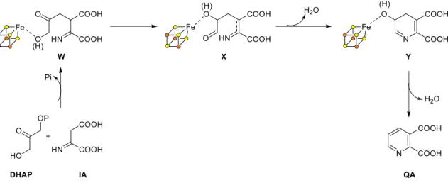
b. Quinolinate synthase (NadA)
Encoded by nadA gene, quinolinate synthase is a [4Fe-4S]2+ cluster metalloenzyme38,39. It catalyzes a complex reaction which consists in the condensation of two substrates (iminoaspartate and DHAP) to form an aromatic product, quinolinic acid (Scheme 8). The different structural and functional aspects will be detailed further on this
26
introduction. Very important insights have been understood during my PhD, especially concerning the mechanism.
c. Quinolinate phosphoribosyl transferase (NadC)
This step is common in both prokaryotic40 and eukaryotic upstream pathways41. NadC enzyme is conserved in the genomes of all species having a de novo NAD synthetic pathway. NadC catalyzes the formation of nicotinic acid mononucleotide (NaMN) from quinolinic acid and 5-phosphoribosyl-1-pyrophosphate (PRPP) (Scheme 8). More precisely, it specifically couples the phosphoribosyl group of a 5-phosphoribosyl-1-pyrophosphate (PRPP) with QA. The removal of the pyrophosphate group of PRPP is facilitated by the Mg2+ metal cofactor of the enzyme, which gives rise to the formation of an oxocarbenium species that is coupled to QA. This coupling yields an instable intermediate which is stabilized by a decarboxylation forming nicotinic acid mononucleotide.
Scheme 8: Prokaryotic upstream de novo pathway: From L-aspartate to Nicotinic acid mononucleotide (NaMN).
(ii) Upstream pathway, in eukaryotes
Eukaryotes and some bacteria (Pseudomonas aureginosa, Burkholderia fungorum,
Bacillus anthracis, Ralstonia metallidurans… 42 synthesize quinolinic acid by degradation of
L-tryptophan under aerobiosis. This pathway is known as the kynurenine pathway and involves the action of five different enzymes and a final spontaneous cyclisation reaction (Scheme 9). The last step of this upstream pathway is common to the prokaryotic one as previously stated, which consists in the formation of nicotinic acid mononucleotide. This pathway and the enzymes involved on it are detailed below.
27 a. Tryptophan-2,3-dioxygenase (TDO) or Indoleamine-2,3-dioxygenase (IDO)
This first step may be performed by any of the two enzymes TDO and IDO and consists on the oxidation of L-tryptophan to N-formylkynurenine. Tryptophan-2,3-dioxygenase or TDO is able to oxidize only L-tryptophan whereas indoleamine-2,3-dioxygenase or IDO oxidizes L-tryptophan and some analogues (D-tryptophan, tryptamine, se oto i … . Both enzymes contain a heme cofactor which allows the binding and activation of dioxygen and tryptophan43 (Scheme 9).
b. Kynurenine formamidase (KFA)
The second step of the upstream pathway consists in the deformylation of N-formylkynurenine to L-kynurenine and it is performed by kynurenine deformidase (KFA). Different structures of this enzyme have been solved from different organisms and it was demonstrated that the catalysis occurs via a catalytic triad (Ser-Asp-His) where the conserved serine catalyzes the deformylation by participating in a tetrahedral intermediate. Then, a hydrolysis step gives rise to the formation of the product and formic acid44 (Scheme 9).
c. Kynurenine-3- monooxygenase (KMO)
L-kynurenine is oxidized to 3-hydroxykynurenine by kynurenine-3-monooxygenase (KMO) (Scheme 9); a flavoprotein, containing a non-covalently bound FAD. This oxidoreductase enzyme uses NADP in order to regenerate its cofactor. The presence of this enzyme in an organism is not always related to quinolinic acid synthesis. For example, in some insects, the enzyme converts its substrate L-kynurenine into xanthurenic acid45 and in Pseudomonas, KMO participates in the formation of an intermediate in the synthesis of quinolobactine-type siderophores46.
d. Kynureninase (KYN)
Hydroxykynurenine is hydrolyzed by a Kynureninase (KYN) yielding 3-hydroxyanthranilic acid and free L-alanine (Scheme 9). This enzyme is the most studied one inside the superfamily of aspartate aminotransferases. This hydrolase is pyridoxal-5-phosphate (PLP) dependent47.
28 e. 3-hydroxyanthranilate-3,4-dioxygenase (HAD)
3-Hydroxyanthranilate-3,4-dioxygenase enzyme oxidizes 3-hydroxyanthranilic acid to form 2-amino-3-carboxymuconic acid semialdehyde (Scheme 9). HAD is a Fe-containing extradiol-dioxygenase. Each of the two enzyme subunits has two Fe binding sites. One of them is catalytic and is coordinated by three conserved amino acid residues, two histidines and a glutamate. The other Fe is coordinated by four cysteines forming a [Fe-4S] whose role remains unclear48.
f. Spontaneous cyclisation of 2-amino-3-carboxymuconic acid semialdehyde to form Quinolinic acid
This is the only non-enzymatic step in the eukaryotic de novo pathway. During this reaction, there is a spontaneous dehydration that gives rise to the cyclisation and aromatization of the product to form quinolinic acid49 (Scheme 9).
g. Quinolinate phosphoribosyltransferase (QPRT)
The last step of the upstream de novo pathway in eukaryotes is common with prokaryotic pathway (Scheme 9). QPRT enzyme is the eukaryotic homologue of the prokaryotic NadC. However, the eukaryotic and prokaryotic enzymes are structurally different. For this reason, NadC became an interesting target for the design of new antibacterial drugs.
29 Scheme 9: Eukaryotic upstream de novo pathway: From L-tryptophan to Nicotinic acid mononucleotide (NaMN).
The different steps of this de novo pathway in eukaryotes have all been characterized at a molecular level, mostly thanks to the crystal structures of all the enzymes50,51,52,53,54,48. Thispathway is performed under aerobic conditions facilitating the functional and structural studies with regard to the prokaryotic pathway where one of the enzymes, quinolinate synthase, has an oxygen-sensitive Fe-S cofactor.
(iii) -Downstream pathway: common in prokaryotes and eukaryotes
Nicotinic acid mononucleotide (NaMN) is converted during the downstream pathway into nicotinamide adenine dinucleotide (NAD) in two steps. The first step results in adenylation of NaMN catalyzed by NaMN adenyltransferase (NadD or NaMNAT) to form Nicotinic acid adenine dinucleotide (NaAD). The second step consists in an amidation of the NaAD by the ATP-dependent NAD synthase enzyme (NadE) to form NAD. NAD may be further phosphorylated by an ATP-dependent kinase (NadK) to form NADP.
The reactions in this pathway are common in prokaryotes and eukaryotes, but the enzymes that catalyze them are not structurally homologous, which make the prokaryotic enzymes interesting targets for the design of antibacterial drugs. This pathway also participates in the NAD salvage and recycling. Below is reported a short description of these enzymes and the corresponding reactions they catalyze.
a. Nicotinic acid mononucleotide adenyltransferase (NadD or NaMNAT)
Encoded by nadD gene, this enzyme catalyzes the adenylation of nicotinic acid mononucleotide (NaMN) to form nicotinic acid adenine dinucleotide (NaAD) using ATP (Scheme 10). The ea tio o sists of a u leophili atta k of the -phosphoribosyl group of NaMN on the -phosphate of ATP followed by the departure of the pyrophosphate group. The reaction is Mg2+ dependent and facilitated by two histidine residues of the ATP binding sequence55.
It has been shown that the mammal nicotinic acid mononucleotide adenyltransferase (NaMNAT) enzyme is able to use both NaMN and its amidated analogue, nicotinamide mononucleotide (NMN) as substrates56. However, the high resolution structure of NadD
30
from E. coli in complex with NaAD product provided a basis for the interpretation of its strict preference for NaMN as substrate (>1,000- fold based on comparison of Kcat/Km values for its
substrate). This involves some important conformational changes such as H-bonds that favor the deamidated form of the ligand55. This preference is conserved in most bacteria and other organisms except mammals.
b. NAD synthetase (NadE or NADS)
The last enzyme participating in NAD biosynthesis is NAD synthetase (NadE in prokaryotes and NADS in mammals) catalyzing the conversion of NaAD to NAD, by transforming the carboxylic acid group of nicotinic acid into an amide (Scheme 10). A Mg2+ cofactor is essential for this activity by activating ATP giving rise to the removal of a pyrophosphate group and formation of an adenylate species, as well as facilitating the nucleophilic attack of ammonia to the carbonyl group of the adenylate intermediate to form an amide57. Most of prokaryotes (E. coli58, Bacillus subtilis59, Helicobacter pylori60… use ammonia as nitrogen source whereas eukaryotes use glutamine61 (with Mycobacterium
tuberculosis as an exception in prokaryotes that can also use glutamine62). In this latter case,
the protein structure of NADS varies with regard to prokaryotes since these enzymes have to hydrolyze glutamine in order to obtain ammonia63,64. They contain an additional N-terminal nitrilase-like domain (Nit) that was shown to hydrolyze glutamine and channel ammonia directly to the amidotransferase domain.
c. From NAD to NADP: NAD kinase (NadF or NadK)
NAD Kinase enzyme is responsible for the phosphorylation of NAD to form NADP (Scheme 10), using ATP that acts as a phosphate donor65. The phosphorylation occurs at the hydroxyl group o the positio of the i ose ring carried by the adenine nucleotide moiety. The enzyme requires a divalent ion for its activity that varies depending on the organism66,67,68. In Salmonella typhimurium, an allosteric regulation of the enzyme has been demonstrated which enables the modulation of the equilibrium between NAD and NADP with regard to the cellular need69.
31 Scheme 10: Downstream pathway of NAD biosynthesis: from nicotinic acid mononucleotide to NAD/NADP. Common pathway for prokaryotes and eukaryotes.
b. Salvage and recycling pathways
Under conditions of NAD deficit, organisms still need to keep their NAD pool present (as essential) and this is why most of them, apart from the de novo synthetic pathway of NAD, possess salvage or recycling pathways to synthesize the cofactor. These pathways use pyridines and other byproducts of NAD consumption within the cell (recycling) or available on the medium (salvage) that come from the food. These salvage and recycling pathways exist from as many as five salvageable precursors which are nicotinamide (Nm), nicotinic acid (Na), nicotinamide riboside (NmR), nicotinic acid riboside (NaR) and nicotinamide mononucleotide (NMN) (Figure 3). Depending on the organism these pathways can be predominant with regard to the de novo pathway. In contrast to NAD biosynthetic pathway, salvage and recycling pathways appear to be subject to considerable variations even between closely related species. Without performing an exhaustive analysis, below, I present a general summary with the main salvage pathways.
Whatever the molecule that is recycled (Nm, Na, NmR, NaR or NMN), these precursors might be outside the cell and they have to pass the membrane through transporters or passive diffusion70,71,72. Transporters differ from one organism to another: in the case of pyridine salvage, it exists NiaX in Streptococcus73, NiaX, NiaY or NiaP in
32
Mycobateria70,71, or Tna1 in yeast74. PnuC exists in many bacteria and it is also capable to
transport nicotinamide riboside (NmR)75. For the transport of NmR, yeast have Nrt172,71.
Figure 3: Structures of nicotinamide (Nm), nicotinic acid (Na), nicotinamide riboside (NmR), nicotinic acid riboside (NaR) and nicotinamide mononucleotide (NMN).
(i) Prokaryotes and eukaryotes except mammals
Salvage pathway of pyridine ring (Nm and Na)
Many NAD consuming enzymes yield nicotinamide (Nm) as a byproduct and this latter can be recycled in order to synthesize NAD. For this purpose, many bacteria have the following enzymatic pathway (Scheme 11).
a. Nicotinamidase (PncA)
In contrast to mammals, bacteria are unable to transfer any phosphoribosyl group to nicotinamide (Nm)76; therefore Nm must be hydrolyzed to nicotinic acid (Na) in order to be used for NAD salvage. The nicotinamidase enzyme PncA catalyzes this hydrolysis reaction
33
that yields Na and ammonia77. The enzyme structure reveals a catalytic triad (Cys-Asp-Lys) and a divalent ion (Mn2+, Zn2+, Fe2+), that plays the role of a Lewis acid, as key elements for the mechanism. The interaction of the divalent ion with the catalytic triad activates the thiolate of the cysteine from the triad which performs a nucleophilic attack to the carbonyl of the amide group giving rise to the formation of Na and ammonia (Scheme 11)78.
b. Nicotinic acid phosphoribosyltransferase (PncB)
This enzyme is often encoded by the same operon as PncA32. It catalyzes the transfer of a phosphoribosyl group from PRPP to nicotinic acid (Na) forming nicotinic acid mononucleotide (NaMN) like NadC does, however without the last decarboxylation step. Even though the enzyme is able to perform this catalysis without ATP, the catalytic efficiency is increased in the presence of ATP by shifting the reaction equilibrium almost fully to the formation of the product (Scheme 11)79.
Scheme 11: Salvage of nicotinamide and nicotinic acid to form nicotinic acid mononucleotide NaMN that is going to be taken in charge by the enzymes of the de novo pathway NadD and NadE.
c. From NaMN to NAD
Once NaMN is synthesized, it is taken into the de novo pathway enzymes in order to transform it to NAD (NadD and NadE).
Salvage of pyridine nucleosides (NmR and NaR)
This salvage pathway is less conserved than the pyridine salvage pathway and differs a lot from one organism to another.
34
In the case of Escherichia coli, Salmonella typhimurium, or Haemophilus influenzae, nicotinamide riboside (NmR) is transformed to nicotinamide mononucleotide (NMN) by NadR enzyme. This last enzyme has also a nicotinamide mononucleotide adenylyltransferase activity and allows the production of NAD80,81,82. The enzyme is present in many bacteria and it is trifunctional; apart of the described catalytic activities, it also plays a crucial role on NAD metabolism regulation80. This will be discussed into more detail further in this introduction.
It is worth noting that this salvage pathway is the only way of NAD production in
Haemophilus influenzae, that does not possess a de novo NAD biosynthetic pathway83 and
which obtains NmR exogenously through the PnuC transporter84.
The bacteria Francisella tularensis is another particular case, which lacks the nadD gene on its genome. Therefore, after the formation of nicotinic acid mononucleotide NaMN, it cannot pursue with the de novo pathway. Instead, NaMN is amidated to NMN by NadE and subsequently adenylated to NAD by nicotinamide nucleotide adenyltransferase, NadM83,85.
The yeast Saccharomyces cerevisiae and Candida glabrata transform NmR into NMN using nicotinamide riboside kinase NRK1 enzyme86,87. Then, NMN is taken in charge by NadD as in the de novo pathway. However, NmR might also be transformed into nicotinamide (Nm) by uridine hydrolase (Urh1) or purine nucleotide phosphorylase (Pnp1) enzymes88. Once Nm is formed, it is transformed to nicotinic acid (Na) by nicotinamidase Pnc1 and then to nicotinic acid mononucleotide (NaMN) by nicotinate phosphoribosyl transferase NPT1. Na can also be obtained from the salvage of nicotinic acid riboside NaR with the action of Urh1 and Pnp1 enzymes88. At this point, the precursor is taken in charge by the enzymes of the de novo pathway to yield NAD.
Salvage of nucleotides (NMN)
Nicotinamide mononucleotide is a salvageable molecule in the NAD biosynthesis. Some organisms possess a nicotinamide mononucleotide deamidase, PncC, which catalyzes the conversion of NMN to NaMN. NaMN is then taken in charge by the enzymes of the de novo pathway. NMN is one of the exceptions of phosphorylated molecules that are able to cross the cell membrane, and it can be also obtained exogenously. However, this is not the case in all organisms. Salmonella enterica needs either to cleave nicotinamide (Nm) from NMN in the periplasm and it is Nm that enters the cell89; or to transform NMN into NmR
35
through a periplasmic AphA phosphatase and transport NmR through the membrane by PnuC. Once inside the cell, the NmR salvage route can proceed73.
(ii) Mammals
Mammals salvage pathways possess some features with regard to the rest of the organisms. Here are the main differences:
For the recycling of pyridines, mammals do not possess PncA nicotinamidase enzyme in order to hydrolyze Nm into Na. However, Nm might be directly recycled in mammals through its phosphoribosylation by nicotinamide phosphoribosyltransferase enzyme, NadV, with PRPP as second substrate to form NMN. This enzyme is also present in some bacteria like Haemophilus ducreyi or in bacteriophages90,91,92. The enzyme is able to couple the PRPP with Na to form NaMN. NadV is the equivalent to PncB in bacteria, except that it can couple PRPP to both Na and Nm, whereas PncB is specific for Na.
Concerning the recycling of NMN and NaMN, mammals possess a nicotinamide adenylyltransferase enzyme which exists in three isoforms in humans, depending on the compartment: NMNAT-1 in the nucleus; NMNAT-2 in Golgi apparatus; and NMNAT-3 in mitochondria. Compared to prokaryotes, it is the equivalent of NadD and NadR. However, once again, no substrate specificity is found in the mammal one, being able to transfer the adenylyl group to both NaMN and NMN. This yields directly NAD from NMN but NaAD from NaMN. The amidation of NaAD in order to obtain NAD is performed by NAD synthetase NadE93.
For the salvage of NmR and NaR, nicotinamide ribose kinase NRK catalyzes their transformation to NMR and NaMN respectively. The enzyme exists in two isoforms; NRK-1 is present in every tissue whereas NRK-2 is present in only some tissues. After phosphorylation, NMN and NaMN are taken in charge by the enzymes described above for the salvage of pyridine nucleotides.
To conclude this part of the introduction, I present a general view summarizing the NAD biosynthetic pathways (de novo and salvage/recycling) in Figure 12.
36 Scheme 12: de novo and salvage NAD production pathways. In black: the de novo pathway in prokaryotes and eukaryotes as well as their common pathway. In blue: different NAD salvage pathways in eukaryotes and prokaryotes.
c. NAD biosynthesis regulation
Bacteria possess different NAD biosynthesis regulation systems depending on the type of organism (NadR, NiaR or NrtR/Nudix). The existence of various regulatory mechanisms reflects the importance of maintaining NAD homeostasis in a variety of growth conditions.
(i) NAD metabolism regulation by NadR
This tri-functional protein regulator NadR is present in enterobacteria (E. coli, Salmonella, Yersinia, Shigella). The N-terminal domain (NadR_R) with a helix-turn-helix DNA binding motif provides the protein with a regulation activity; the nucleotide binding central domain (NadR_A) displays a nicotinamide mononucleotide (NMN) adenyltransferase activity; the C-terminal domain (NadR_K ) displays nicotinamide ribose kinase activity.
The basis of this transcriptional regulation is the following one: In the presence of NAD, the dinucleotide binds to the enzyme through its NadR_A domain. This interaction leads to the dimerization of the protein that can bind to DNA through the helix-turn-helix DNA-binding domain (NadR_R) in the specific site where the promoter of genes nadA, nadB,
37 pnuC, pncB and nadR is found. This way, the transcription of the genes involved in NAD biosynthesis is repressed as well as the transcription of the regulatory protein itself. However, in the absence of NAD, ATP binds to the protein on the same NAD binding domain (NadR_A) than NAD and the protein remains as a monomer unable to bind DNA. As a consequence, nadA, nadB, pnuC, pncB and nadR genes are transcripted and the NAD biosynthesis occurs94.
(ii) NAD metabolism regulation by NiaR
This second regulation system is present in some bacteria (Bacillus, Clostridium, Streptococcus, Thermotoga). The regulator NiaR (Niacin Repressor) was originally named YrxA95. NiaR is a transcriptional repressor that binds to DNA through is helix-turn-helix domain. In contrast to NadR, it is the nicotinic acid (Na) and not NAD that binds to the protein and induces its dimerization and therefore its interaction with DNA. In this case, the transcription of nadA, nadB, nadC, pncA, pncB, pnuC niaP (the last one identified as a nicotinic acid and nicotinamide transporter in bacteria) is repressed. In the absence of Na, the protein remains on its monomeric form unable to bind DNA. In this case, there is a derepression of the genes mentioned above96.
(iii) NAD metabolism regulation by NrtR
Some bacteria like Vibrio, Pseudomonas, Mycobacterium or Shewanella possess this last type or regulation system. The regulator is alled N t‘ fo Nudix-related transcriptional
Regulator , he e Nudi efe s to Nucleoside Diphosphate linked to a x part, a motif found
in a family of enzymes that catalyze the hydrolysis of several nucleoside diphosphate derivatives)97. The Nudix domain is found on the N-terminal part of the protein whereas the C-terminal contains a helix-turn-helix DNA-binding domain. In contrast to the two previous regulators (NadR and NiaR), the protein is in a dimeric form in the absence of ADP-ribose and bound to DNA. In the presence of ADP-ribose, the protein becomes monomeric and under this form it dissociates from DNA. In this case, the transcription of the genes nadA, nadB, nadC, nadD nadE, pncA, pncB, niaP, pnuC, nadR, nadM, nadV and prs (protein participating in the synthesis of PRPP) is possible. Since ADP-ribose is a NAD degradation product, it functions as a signal for the cell indicating that NAD must be resynthesized98.
38
III.
NAD: a pharmacological target
Nicotinamide adenine dinucleotide plays crucial roles in cellular processes as explained above. Maintenance of the NAD pool is of extreme importance for the good health of cells. Many diseases are associated to perturbations on the concentration levels of NAD coming either from a dysfunction in the NAD biosynthesis enzymes or in the NAD consuming enzymes. For this reason, any of these two kinds of enzymes are interesting targets for the design of pharmacological agents against a large variety of diseases.
Targeting the enzymes involved on NAD biosynthesis is particularly interesting because the NAD biosynthesis pathway of mammals differs from that of bacteria, and therefore, inactivation of the bacterial pathway has no impact on the eukaryotic one (human). However, due to the complexity and diversity of NAD biosynthesis and the presence of different salvage pathways which differ from one organism to another, this antibacterial strategy, despite being promising, is more complex than expected.
Currently, prokaryotic enzymes from both upstream and downstream pathways are explored as potential antibacterial targets. The enzymes driving the downstream conversion of nicotinic acid mononucleotide (NaMN) to NAD are present in almost all analyzed genomes and therefore represent promising broad-spectrum antibacterial targets32. Progress have been made in the development of bacterial NadD, NadE and NadK enzyme inhibitors99,100,101,102. The upstream NAD biosynthetic pathway is not conserved in all bacteria. Yet, it is a particularly interesting target for the design of new drugs since it allows targeting specific pathogens that lack all kind of NAD salvage pathway and for whom the de novo biosynthetic pathway of NAD is essential. From this point of view, two organisms have been proved to have only the de novo pathway: Helicobacter pylori (cause of gastroduodenal disease) and Mycobacterium leprae (cause of leprosy disease).
The main objective of my PhD was to find new antibacterial drugs against these pathogens by targeting quinolinate synthase or NadA, the enzyme responsible of quinolinic acid formation (NAD precursor). The essential nature of NadA was confirmed in H. pylori since a nadA mutation is lethal to this organism103(and unpublished data). Similarly, it has been validated that NAD biosynthesis is a drugable pathway in Mycobacteria such as M. leprae101. In the following two chapters of this introduction, I will perform a deep description of the


![Figure 11: A-left: Structure of TmNadA*. Three domains (green, purple and gold) define a tunnel that connects the [4Fe-4S] 2+ cluster to the molecular surface](https://thumb-eu.123doks.com/thumbv2/123doknet/12872035.369355/68.892.116.768.664.904/figure-structure-tmnada-domains-connects-cluster-molecular-surface.webp)