HAL Id: tel-03131503
https://tel.archives-ouvertes.fr/tel-03131503
Submitted on 4 Feb 2021HAL is a multi-disciplinary open access archive for the deposit and dissemination of sci-entific research documents, whether they are pub-lished or not. The documents may come from teaching and research institutions in France or abroad, or from public or private research centers.
L’archive ouverte pluridisciplinaire HAL, est destinée au dépôt et à la diffusion de documents scientifiques de niveau recherche, publiés ou non, émanant des établissements d’enseignement et de recherche français ou étrangers, des laboratoires publics ou privés.
Mateja Senicar
To cite this version:
Mateja Senicar. Mise au point de NéoLectines spécifiques des furanosides : bioingénierie, diagnostic et imagerie. Sciences agricoles. Université d’Orléans, 2019. Français. �NNT : 2019ORLE3037�. �tel-03131503�
UNIVERSITÉ D’ORLÉANS
ÉCOLE DOCTORALE
(SANTE, SCIENCES BIOLOGIQUES ET CHIMIE DU VIVANT) Institut de Chimie Organique et Analytique, UMR 7311, CNRS
Centre de Biophysique Moléculaire, UPR 4301, CNRS
THÈSE
présentée par:Mateja SENICAR
Soutenue le: 13 décembre 2019 Pour obtenir le grade de: Docteur de l’Université d’Orléans Discipline/Spécialité: Biologie – Biochimie
THÈSE dirigée par:
Richard DANIELLOU Professeur, Université d’Orléans – directeur de Thèse Svetlana V. ELISEEVA Chargée de recherche, CNRS – co-directrice de Thèse
RAPPORTEURS:
Laurent SALMON Professeur, Université Paris-Sud – Université Paris-Saclay Pascale DELANGLE Directrice de recherche, Université Grenoble-Alpes
JURY:
Laurent SALMON Professeur, Université Paris-Sud – Université Paris-Saclay (Président du jury) Pascale DELANGLE Directrice de recherche, Université Grenoble-Alpes
Tom DESMET Professeur, Université de Gent
Stéphane PETOUD Professeur, Directeur de recherche INSERM Ludovic LANDEMARRE Directeur d’entreprise GLYcoDiag
Richard DANIELLOU Professeur, Université d’Orléans – directeur de Thèse Svetlana V. ELISEEVA Chargée de recherche, CNRS – co-directrice de Thèse
Mise au point de NéoLectines spécifiques des furanosides:
bioingéniérie, imagerie et diagnostic
II
Multi enim sunt vocati, pauci vero electi. Matthaeus 22:14
Acknowledgments
III
Acknowledgements
This doctoral thesis was part of the NeoLect project and the work was supported by APR IR, Région Centre-Val de Loire, France.
I thank Prof. Laurent Salmon (Université Paris-Sud – Université Paris-Saclay) and Prof. Pascale Delangle (Directrice de recherche, Université Grenoble-Alpes) for accepting to review this thesis, as well as to Prof. Tom Desmet (Professeur, Université de Gent) and Prof. Stéphane Petoud (Professeur, Directeur de recherche INSERM) for accepting to be part of this thesis defence committee.
Thesis is life experience and job at the same like no other. I sincerely thank Prof. Richard Daniellou for giving me opportunity to be part of his Glycobio research team and to learn from him. He literally travelled on the other side of the world (Japan) to start this project. Thank You for trusting me with this thesis. I really enjoyed working on it. I hope that I was up to the expectations. Special thanks goes to Dr. Pierre Lafite and his support, advices and enthusiasm. Coming to lab every day felt like going to another family and I thank you all for that.
I thank Prof. Stéphane Petoud for welcoming me in his team and giving me opportunity to work with him. I specially thank Dr. Svetlana V. Eliseeva, my co-directrice, for all the help and support and for introducing me into the bright world of lanthanides. Many thanks to Dr Guillaume Collet for his help and advices during imaging experiments and for driving me to Patàpain! Thank to Anton, my CBM bureau coloc, and Keyron, bureau voisin, for the coffee breaks and fun.
Thought these three years many past and present members of GlycoBio team have contributed to unique work and high-spirit atmosphere. I specially thank Philippe for our daily small talks and his support to my French lessons. Many thanks to Jihen (Jedi) and her friendship during our long manips in the lab, her cheerful and energetic music choices, ensuring enthusiasm even during the most depressing days, Wednesday evenings in V&Bs and volley-ball tournaments (when my head injuries started 😉).
During these three years, I have shared my office, moods, ups-and downs and hopefully knowledge with many colocataires. So here it goes, season 2016/2017 (cast of the first year): Yasmina, Emilie (my best French teachers ever!) and Hyuna; season 2017/2018 (cast of the
IV
the final season, third year): Julien, Alexis, Martyna and Tomoki (by far, the craziest combination of team members. Thanks to you, I can be assured that none day will be like the other and your jokes, humor and fun will keep my spirits high!) and of course past and present members of GlycoDiag team, Blanka and Benoit and director Mr. Ludovic Landemarre.
To Arnaud, my colocataire de bureau, and Damien (Dam-Dam, Benjamin, Padawan or also known as “call me by my name” 😜), I will only say: “Le cerveau n'est pas une partie vitale, vise les bouboules plutôt" (This is exclusively internal joke, and should not be taken out of the context). See you guys!
I also thank my BTS stagiaires Antoine, Florian and Boris as well as my Edifice students (les petites) Martin, Alexis, Lucas, Chloé and Juliette for their participation and interest they showed during our teaching sessions.
Hélène and Eugénie, these three years definitely would not be the same without your company! You’re absolutely fabulous! Our petite pause clope, soirée picole, petits messages “coucou“ à ma voisine préfèrée, #Hélènechallenge and many other little inventions of ours were great fun and joy!
I thank Mélanie, for sharing her family time during Christmas holidays and introducing me to Saran where I enjoyed lovely Saturday mornings. I hope that mon poney préféré, JoJo, now officially retired, is happily grazing on pasture and is far from finishing as saucisson (bonne retraite à toi!).
Finally, gros bisous to mama & papa and their ever-present support! It seems that I’m about to become a doctor!
Cheers!
Mateja
Abbreviations
V
Abbreviations
6xHis-tag – hexahistidine fusion tag Araf – arabinofuranose Araf-ase – arabinofuranosidase BF – bright field bp – base pair
CAZy – Carbohydrate-Active Enzymes EC – Enzyme Commission
EDTA – ethylenediaminetetraacetic acid ELISA – enzyme-linked immunosorbent assay EPS –exopolysaccharides
FAD – flavine adenine dinucleotide Fruf – fructofuranose Fucf – fucofuranose Galf – galactofuranose Galf-ase – galactofuranosidase GalfT – galactofuranosyltransferase GFP – green fluorescent protein GHs – glycoside hydrolases GIPLs – glycoinosinositolphospholipids Glcf – glucofuranose GM – galactomannan
hIntL – human intelectin → intelectin-1 HPLC – high performance thin
layer chromatography
HRMS – high-resolution mass spectrometry IMAC – immobilized metal-affinity
chromatography
IPTG – isopropyl-β-D-1-thiogalactopyranoside
ITNL1 – intelectin-1 → human intelectin
IUBMB – Union of Biochemistry and Molecular Biology LPGs – lipophosphoglycans
LPS – lipopolysaccharide Manf – mannofuranose MC – metallacrown
MRI – magnetic resonance imaging NIR – near-infrared
NMR – nuclear magnetic resonance NPs – nanoparticles
NTA-Ni2+ - Ni2+ immobilized on
a nitrilotriacetic acid matrix
ORF – open reading frame
PAMPs – pathogen-associated molecular patterns PCR – polymerase chain reaction
PDB – Protein Data Bank
PET – positron emission tomography PET/MR – positron emission tomography/
magnetic resonance
pNP – para-nitrophenol
pNP-β-D-Galf – para-nitrophenyl β-D-galactofuranose
quinHA – quinaldichydroxamic acid
SDS-PAGE – sodium dodecyl sulfate-polyacrylamide
gel electrophoresis
SPECT – single-photon emission computed tomography tR – retention time
UDP-Galf – uracil diphosphate galactofuranose UDP-Galp – UDP-galactopyranose
UGM – UDP-galactopyranose mutase XCT – X-ray computed tomography
VI
Chapter I Bibliographical overview
I.General introduction ... 1
II. Carbohydrates – essential molecules of life ... 6
1. Natural glycofuranoses ... 6 2. Galactofuranose ... 7 2.1. Occurrence in nature ... 7 2.1. In bacteria ... 8 2.2. In fungi………..………10 2.3. In protozoa ... 11 2.4. In other organisms ... 13
3. Medical relevance & functional impact ... 14
3.1. Galactofuranose antigens – therapeutic & diagnostic target ... 14
3.2. Aspects of enzymatic biosynthesis & metabolism ... 16
UDP-Galactopyranose Mutase (UGM) ... 16
Galactofuranosyltransferase (GalfT) ... 17
II. Carbohydrate-degrading enzymes ... 19
2. Glycosidases – a general overview ... 19
2.1. Glycofuranoside hydrolases ... 22
2.2. Galactofuranosidases ... 22
2.2.1. Sources & activities – the early reports ... 22
2.2.2. Cloning & characterization of the first galactofuranosidase ... 26
III. Lanthanide (III) complexes & nanoparticles – near-infrared emitting probes for biological imaging ... 27
1. Optical imaging in the near-infrared domain ... 27
2. Lanthanide (III) coordination compounds – brief overview ... 28
IV. Objectives of the thesis ... 30
Table of Contents
VII
Chapter II Galactofuranosidase – an original and specific glycoside
hydrolase
I. Recombinant DNA technology in protein production ... 46
1. The basic principles of gene and plasmid manipulations ... 46
2. Expression systems for recombinant protein production ... 48
II. Obtaining a desired Galf-ase plasmid construct – the applications of gene (sub)cloning techniques ... 50
1. Plasmid construct pET-50b(+) – screening of Galf-ase gene insert bearing plasmid ... 50
2. Galf-ase gene subcloning – preparing pET-28a(+) plasmid construct ... 52
III. Production and characterization of recombinant Galf-ase ... 54
1. Overexpression and purification ... 54
2. Optimum conditions and kinetic parameters determination ... 56
2.1. Substrate specificity ... 56
2.2. Temperature and pH properties ... 57
2.3. Freeze-thaw stability... 58 2.4. Kinetic analysis ... 60 3. Inhibition studies... 62 4. Crystallization trials ... 64 Résumé ... 66 Bibliography... 67
Chapter III Tailoring galactofuranosidase activity – potential platform
for new applications
I. Glycosidase engineering ... 691. Thioglycosides – versatile tools in glycobiology ... 69
2. Thioglycoligases ... 72
II. Design and production of Galf-ase E464X mutants ... 74
1. Selection and creation of mutagenesis points ... 74
2. Overexpression and purification of Galf-ase E464X mutants ... 76
3. Galf-ase E464X mutants kinetic parameters determination – Identification of the acid/base catalyst ... Erreur ! Signet non défini. III. Application of Galf-ase E464X mutants in thioglycosides synthesis ... 79
1. Selection of appropriate Galf-ase E464X mutant ... 79
2. Galf-ase E464Q mutant catalysed synthesis of thiogalactofuranosides ... 80
VIII
Bibliography... 86
Chapter IV Galf-specific & lanthanide (III) complex loaded
nanoparticles for NIR imaging – concept validation
I. Engineering neolectins from glycosidases ... 911. Application in biotechnology, diagnostic and imaging ... 91
II. Construction of luminescent nanoparticle scaffold for neolectin bioconjugation & imaging applications ... 93
1. Construction and characterization of luminescent nanoparticle scaffold ... 93
1.1. Encapsulation procedure... 95
1.2. Surface functionalization ... 96
1.3. Photophysical properties of YbZn16(quinoHA)16 and corresponding NPs ... 98
2. Bioconjugation of luminescent nanoparticle scaffold ... 101
2.1. Galf-specific near-infrared imaging with Galf-ase functionalized luminescent nanoparticles103 III. Conclusion ... 104
Résumé ... 105
Bibliography... 106
Chapter V Conclusion & perspectives
V. Conclusion & perspectives ... 109Chapter VI Experimental part
6.1. Materials and methods ... 1176.2. Molecular biology ... 117
6.2.1. In silico sequence analysis ... 117
6.2.2. Polymerase chain reaction (PCR) ... 117
6.2.3. DNA analysis and purification ... 121
6.2.4. Bacterial strains and plasmids... 122
6.3. Biochemistry ... 123
6.3.1. Protein overexpression ... 123
6.3.2. Protein analysis... 123
6.3.3. Protein purification ... 125
6.3.4. Enzymatic activity assays ... 126
6.3.5. Furanothioglycosylation ... 130
Table of Contents
IX
6.4.1. Synthesis of YbIII/ZnII metallacrowns: LnZn16(quinoHA)16... 131 6.5. Nanotechnology ... 132 6.5.1. Encapsulation of YbIII/ZnII metallacrowns (LnZn16(quinoHA)16) in polystyrene nanoparticles: LnX@PS/COOH ... 132 6.5.2. Surface modification LnX@PS/COOH to PS/NTA and LnX@PS/NTA ... 132 6.5.3. Synthesis of PS/NTA-Ni2+ and LnX@PS/NTA-Ni2+ ... 133 6.5.4. Bioconjugation of 6xHis-tagged recombinant Galf-ase to PS/NTA-Ni2+ and LnX@PS/NTA-Ni2+...133 6.5.5. Characterization of PS/NTA-Ni2+ & LnX@PS/NTA-Ni2+ nanoparticles... 134
Chapter VII Appendix
7.1.Additional figures ... 136 7.1.1. Sequence alignment ... 136 7.1.2. Structural formulas of pNP-linked substrates ... 138 7.1.3. The HPLC chromatograms and ESI-HRMS spectras of thioglycosylation reactions by E464Q………..139 7.1.5. Short communication published in the journal Carbohydrate research ... 147
X
Chapter I Bibliographical overview
Figure I-1. Glycoside hydrolases as platform for versatile applications………1 Figure I-2. The carbohydrate layer on the cell surface is composed of the glycans (oligosaccharides) attached to membrane glycolipids and membrane glycoproteins.11……….6
Figure I-3. The equilibrium representation between cyclic forms of D-galactose in solution. The
rapid interconverted of cyclic, Galf and Galp, forms through acyclic form and favorable equilibrium for Galp are indicated by the arrows.19………...7
Figure I-4. A schematic representation of Mycobacterium tuberculosis cell wall structure. A) Depiction of the major cell wall features, including peptidoglycan, arabinogalactan and mycolic acids. B) Detailed structure of mycobacterial arabinogalactan composed of galactan and arabinan
domains.28,29………...9
Figure I-5. A schematic representation of Aspergillus fumigatus cell wall structure. A) Depiction of the major cell wall features, including chitin, glucan and galactomannan. B) Structural fragment of fungal galactomannan composed of mannan and galactan units.29,38………..11
Figure I-6. A schematic representation of main Galf-containing glycoconjugates present at the cell
surface of leishmanial promastigote.42………...……12
Figure I-7. Chemical structures of Galf-containing glycoconjugates. A) The structure of toxin prymnesin-1, produced by Prymnesiou parvum. B) The structure of agelagalastatin,
glycosphingolipid produced by Agelas sp..25………13
Figure I-8. Schematic representation of the Galf-glycan biosynthesis and the involvement of the three main enzymes, UDP-galactopyranose mutase (UGM), galactofuranosyltransferase (GalfT) and galactofuranosidase (Galf-ase)………..………16 Figure I-9. UGM-catalyzed izomerization of UDP-Galp and UDP-Galf………16 Figure I-10. Biosynthesis of Galf-glycan catalyzed by GalfT………18 Figure I-8. Schematic representation of A) inverting and B) retaining GH catalytic mechanis.21
Figure I-9. Ranges of wavelengths usually used for fluorescence optical imaging.113………27
Figure I-10. Schematic representation of the general architecture of the luminescent Ln3+
complexes and the antenna effect. A) Pendant antenna. B) Chelating antenna………..29 Figure I-11. A graphical abstract of the main thesis objectives………...32
Chapter II Galactofuranosidase – an original and specific glycoside
hydrolase
Figure II-1. Schematic representation of recombinant DNA cloning procedure………..47 Figure II-2. Schematic representation of pET-28a(+) vector backbone………....48
Table of Figures
XI
Figure II-3. Schematic representation of the pET vector and E. coli RosettaTM (DE3) host
expression system the control elements.6………...….50
Figure II-4. Schematic representation of pET-50b(+) plasmid construct cloning/expression region coding for N-terminal Nus protein and double 6xHis-tagged Galf-ase….…..………..51 Figure II-5. PCR and restriction enzyme analysis of pET-50b(+) plasmid construct. A) Agarose-gel electrophoresis of the PCR reaction indicating the amplification efficiency of Galf-ase insert gene between T7 and T7term. B) Agarose-gel electrophoresis of pET-50b(+) plasmid construct after EcoRI and HindIII double digestion reaction………...52 Figure II-6. Schematic representation of Galf-ase gene subcloning procedure. A) Preparation of pET-28a(+) plasmid construct, bearing Galf-ase gene insert. B) pET-28a(+) plasmid construct confirmation on agarose-gel electrophoresis after EcoRI and HindIII restriction enzyme analysis.53
Figure II-7. Confirmation of EcoRI restriction site mutagenesis on agarose-gel electrophoresis after pET-28a(+) plasmid construct EcoRI and HindIII restriction enzyme analysis………..54 Figure II-8. The design and production of N-terminal fusioned Galf-ase protein. A) Schematic representation of N-terminal single peptide 6xHis-tagged Galf-ase fusion protein. B) The SDS-PAGE analysis of Galf-ase after IPTG induction and NTA-Ni2+chromatography purification………55 Figure II-9. Schematic representation of pET-28a(+) plasmid construct cloning/expression region coding for N-terminal single peptide 6xHis-tagged Galf-ase………..56 Figure II-10. Schematic representation of hydrolytic reaction between Galf-ase and a panel of
pNP-linked pyranosyl and furanosyl monosaccharide……….56
Figure II-11. Substrate specificity screening of Galf-ase. A) Substrate specificity of Galf-ase tested towards 21 pNP-linked monosaccharide substrates. B) Structural formulas of pNP-β-D-Galf (1)
and the pNP-α-L-Araf (2)………57 Figure II-12. Determination of optimum activity conditions for Galf-ase. A) Influence of pH on Galf-ase hydrolytic activity towards pNP-β-D-Galf. B) Influence of temperature on Galf-ase
hydrolytic activity towards pNP-β-D-Galf……….58
Figure II-13. Effect of freeze and thaw cycles and storage conditions on Galf-ase residual
hydrolytic activity towards pNP-β-D-Galf………59
Figure II-14. Michaelis-Menten plot of pNP-β-D-Galf hydrolysis reaction catalysed by Galf-ase.
Mean values and SD error bars were calculated from three independent experiments and from a single protein preparation……….60
Figure II-15. Michaelis-Menten plot of pNP-α-L-Araf hydrolysis reaction catalysed by Galf-ase.
Mean values and SD error bars were calculated from three independent experiments and from a single protein preparation……….61 Figure II-16. Effects of concentration of inhibitors 3-6 on the hydrolytic activity of Galf-ase towards pNP-β-D-Galf. Mean values and SD error bars were calculated from three independent
XII
towards pNP-β-D-Galf by 4. Mean values and SD error bars were calculated from two independent
experiments and from a single protein preparation………...63 Figure II-14. Photographs of the crystals obtained during Galf-ase crystallization trials. A) Crystals submitted to X-ray crystallography proved to be of salt origin. B) One of the examples of the salt crystals obtained during Galf-ase crystallography trials………64
Chapter III Tailoring galactofuranosidase activity – potential platform
for new applications
Figure III-1. Structures of some naturally occurring thiosugars sharing sulfur-containing heterocycle structure………....69 Figure III-2. Structures of some naturally occurring thioglycosides………...70 Figure III-3. Proposed general mechanism for retaining thioglycoligase……….73 Figure III-4. Alignment of partial ORF Galf-ase (Streptomyces spp.) amino acid sequence and corresponding regions of its homologs. The conserved E464 and E573 amino acid residues are indicated with asterisks……….74 Figure III-5. Confirmation of pET-28a(+) plasmid construct integrity, after E464X mutant variants generation, on agarose-gel electrophoresis by EcoRI and HindIII restriction enzyme analysis………..76
Figure III-6. SDS-PAGE analysis of E464X Galf-ase mutant variants after NTA-Ni2+
chromatography purification………...76 Figure III-7. Structural formulas of amino acids selected for the generation of E464X Galf-ase mutant variants………...…….77
Figure III-8. Michaelis-Menten plot of pNP-β-D-Galf hydrolysis reaction catalysed by Galf-ase
E464S, E464C and E464Q mutant variants. Mean values and SD error bars were calculated from three independent experiments (*except E464C) and from a single protein preparation………...………..78
Table III-2. Comparison of Michaelis-Menten kinetics constants for hydrolysis of β-D-Galf by
E464X Galf-ases mutant variants………...79 Figure III-9. Influence of pH on E464Q Galf-ase mutant variant hydrolytic activity towards
pNP-β-D-Galf………..80
Figure III-10. Structural formulas of thiol acceptors used for E464Q Galf-ase catalyzed furanothioglycoligase reactions………..…80 Figure III-11. Proposed furanothioglycoligation mechanism catalyzed by E464Q Galf-ase mutant variant……….81 Figure III-12. HPLC chromatograms of representative thioglycosylation reactions by E464Q. A) Chromatogram of crude thioglycosylation reaction with thioaryl acceptor. B) Chromatogram of purified product fraction from thioglycosylation reaction……….82
Table of Figures
XIII
Figure III-13. Structural formulas of synthetized furanothioglycosides………83 Figure III-14. The measured HRMS-ESI spectrum of thioarylgalactofuranosyl ion in negative mode. The analysed sample is the fraction purified on C18 chromatography resin (eluent solvent mixture: A:B=6:4; A=0.1% formic acid (v/v) in water, B=0.1% formic acid (v/v) in acetonitrile) and corresponds to the peak (tR=9.19 min) detected on HPLC (Figure III-12. B)………83
Chapter IV Galf-specific & lanthanide (III) complex loaded
nanoparticles for NIR imaging – concept validation
Figure IV-1. A) X-ray crystal structure of DyZn16(quinHA)16 along the (left) a-axis and (right)
c-axis. B) Emission spectra of LnZn16(quinHA)16 (Ln = Yb, Nd, Er, quinHA = quinaldichydroxamic
acid).12……….93
Figure IV-2. Absorption spectra of YbZn16(HA)16 MCs (HA = pyzHA, quinHA, quinoHA) in
methanol or water solutions at room temperature……….94 Figure IV-3. Schematic representation of the three main steps in which internal and external functionalization of NPs can be achieved………..94 Figure IV-4. Schematic representation of the encapsulation step . A) Top-down view of the crystal structure of YbZn16(quinoHA)16 MC obtained by X-ray diffractometry on a single crystal, done at the Department of Chemistry of the University of Michigan by Tu N. Nguyen and Jeff W. Kampf (colour code: green, Yb; yellow, Zn; red, O; blue, N; grey, C; hydrogen atoms and solvents have been omitted for clarity). B) Encapsulation of YbZn16(quinoHA)16 complex inside PS/COOH NPs followed by subsequent colour change to orange……….95
Figure IV-5. Schematic representation of synthetic route for preparation of PS/NTA NPs:
activation of NPs carboxylic groups with EDC/sulfo-NHS to create the amide bond with AIDA……….…96
Figure IV-6. Photographs of the pellets of PS/NTA-Ni2+ NPs (A) before and (B) after incubation
with EDTA. The change of colour indicates the release of Ni2+ from the surface………97
Figure IV-7. Corrected and normalized (left) excitation (λem = 980 nm) and (right) emission
spectra of YbZn16(quinoHA)16 in DMF solution (50 µM, λex = 420 nm) or for aqueous suspensions of YbZn16(quinoHA)16@PS NPs (6 mg/mL, λex = 510 nm), room temperature………98
Figure IV-8. Changes in emission intensity at 980 nm for a solution of YbZn16(quinoHA)16 in DMF
(50 µM) or an aqueous suspension of YbZn16(quinoHA)16@PS/NTA-Ni2+ NPs (6 mg/mL) under continuous illumination at 405 nm………....100
Figure IV-9. Investigation of the activity of the bioconjugated Galf-ase. A) The specific pNP-β-D
-Galf activity of -Galf-ase obtained after separate bioconjugation with NTA-Ni2+ and NTA surface functionalized NPs compared to free enzyme as a control. B) The SDS-PAGE analysis of imidazole released Galf-ase from NTA-Ni2+ surface functionalized NPs upon activity assay………..102
Figure IV-10. Immobilization of 6xHis-tagged Galf-ase on NTA-Ni2+ functionalized surface of NPs
and the coordination bonds formed between NTA carboxylic groups and imidazole rings with nickel cation……….103
XIV
Chapter V Conclusion & perspectives
Figure V-1. A graphical abstract of the main short-term and long-term perspectives for the future work………...………..112
Table of Tables
XV
Chapter I Bibliographical overview
Table I-1. Comparative properties of β-D-galactofuranosidases……….……….…….25
Table I-2. Comparative properties of recombinant β-D-galactofuranosidases………..26
Chapter II Galactofuranosidase – an original and specific glycoside
hydrolase
Table II-1. Comparison of Michaelis-Menten kinetics constants for hydrolysis of α-L-Araf (1) and β-D-Galf (2) by different Araf-ases and Galf-ases………...…...61Table II-2. Comparison of the inhibitory effects of different galactofuranosides (3-6) towards Galf-ase and their respective IC50 and KI values……….………63
Chapter III Tailoring galactofuranosidase activity – potential platform
for new applications
Table III-1. Codon changes and respective amino acid changes introduced at 464 position at wild-type Galf-ase amino acid sequence by site-directed mutagenesis………...75Chapter IV Galf-specific & lanthanide (III) complex loaded
nanoparticles for NIR imaging – concept validation
Table IV-1. Luminescence lifetimes (τobs) and absolute quantum yields (𝑄𝑌𝑏𝐿 ) of YbZn16(quinoHA)16 in DMF solution (50 µM) or for aqueous suspensions of YbZn16(quinoHA)16@PS NPs (6 mg/mL).a………...….99Chapter VI Experimental part
Table VI-1. Primers used for wild type Galf-ase gene amplification………..118Table VI-2. Cycling parameters for wild type Galf-ase gene amplification………118
Table VI-3. Primers used for plasmid construct pET-28a(+) gene sequencing………118
Table VI-4. Primers used for site-directed mutagenesis………..119
Table VI-5. Cycling parameters for site-directed mutagenesis………119
Table VI-6. Primers used for deletion mutagenesis………120
CHAPTER I
Bibliographical overview
Contents Chapter I
I.General introduction ... 1
II. Carbohydrates – essential molecules of life ... 6
1. Natural glycofuranoses ... 6 2. Galactofuranose ... 7 2.1. Occurrence in nature ... 7 2.1. In bacteria ... 8 2.2. In fungi………..………10 2.3. In protozoa ... 11 2.4. In other organisms ... 13
3. Medical relevance & functional impact ... 14
3.1. Galactofuranose antigens – therapeutic & diagnostic target ... 14
3.2. Aspects of enzymatic biosynthesis & metabolism ... 16
UDP-Galactopyranose Mutase (UGM) ... 16
Galactofuranosyltransferase (GalfT) ... 17
II. Carbohydrate-degrading enzymes ... 19
2. Glycosidases – a general overview ... 19
2.1. Glycofuranoside hydrolases ... 22
2.2. Galactofuranosidases ... 22
2.2.1. Sources & activities – the early reports ... 22
2.2.2. Cloning & characterization of the first galactofuranosidase ... 26
III. Lanthanide (III) complexes & nanoparticles – near-infrared emitting probes for biological imaging ... 27
1. Optical imaging in the near-infrared domain ... 27
2. Lanthanide (III) coordination compounds – brief overview ... 28
IV. Objectives of the thesis ... 30
1
I. General introduction
Outside their role inside living organisms, the enzymes have a long history of use in human life dating back to prehistorical times when food culture began to emerge and enzyme-fermented products from various food sources were introduced.1 Since then, our
dependence on the enzymes and enzyme-related products has not changed, rather it is relevant more than ever. Nowadays, when environmental concerns over pollution and industrialization are growing, they offer alternative to traditional chemical treatments in production of goods.2
The impact of modern biotechnology and recombinant DNA technology has made enzymes available and economically feasible. It enabled a whole new diversity of enzymes to be accessed in the field of glycoenzymes. In parallel carbohydrate-based materials have emerged in an increasing number of applications in the food and feed, pharmaceutical or other industries. To access these carbohydrate structures in high demand, natural glycoenzyme catalysts, have provided over recent decades an alternative to chemical synthesis. Utilization of glycoside hydrolases brings all the important advantages of an enzyme catalyst. They greatly contribute to the development of novel synthetic tools and are strongly beneficial when looking for very specific sugar structure(s). Their engineering capacity gives freedom to expand their repertoire for diverse applications. To develop such innovative and versatile tools, tight interactions between scientific community and industry has to be considered (Figure I-1.).3,4
I. General introduction
2
A current area of interest is broadening the search spectrum to rare glycan substrate specific glycosyl hydrolases, preferably, form narrow group of organisms involved in pathological conditions, in order to create altered characteristics, various function and application possibilities within protein engineering.
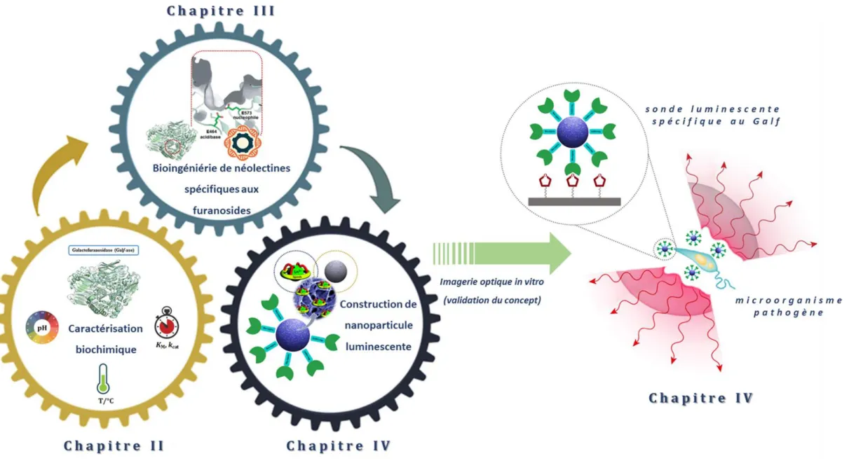
In the context of interdisciplinary approach, the NeoLect project, supported by APR IR Région Centre-Val de Loire and this doctoral thesis have been initiated. The main idea and objectives of this thesis can be outlined from its title “Design of galactofuranosyl-specific
NeoLectins: bioengineering, imaging and diagnosis”. Research work has been carried out
in collaboration between two groups, Enzymology and Glycobiochemistry group, which is part of Organic Synthesis – GlycoBio & Chemistry team at Institute of Organic and Analytical Chemistry (ICOA), and Luminescent lanthanide compounds, optical spectroscopy and bioimaging group, which is part of Chemistry, Imaging and Exobiology team at the Centre for Molecular Biophysics (CBM). Both research groups are gathered under the French National Centre for Scientific Research (CNRS).
The Enzymology and Glycobiochemistry research group of Pr. Richard Daniellou and Dr. Pierre Lafite was founded in 2012 and the group’s main research interest is towards understanding the mechanisms and structures of enzymes involved in synthesis (glycosyltransferases) or cleavage (glycoside hydrolases) of glycoconjugates. The group has well established multidisciplinary procedure and uses enzymes as biocatalysts to synthesize original carbohydrate-containing compounds, mainly thioglycosides. Since different experimental techniques, including molecular biology, biochemistry and structural studies, are employed in this laboratory thus allowing us to realize the first part of the thesis objective, characterization and bioengineering of galactofuranosidase towards neolectins.
The Luminescent lanthanide compounds, optical spectroscopy and bioimaging group, led by Prof. Stéphane Petoud, aims to provide new tools for biological analysis and imagery. This specifically includes the development of near-infrared (NIR) emitting lanthanide compounds and novel sensitizing antenna. This aspect of their expertise provided us with opportunity to realize second major objective of thesis, development of NIR luminescent probe amenable for functionalization with enzymes.
3
I. Introduction générale
En dehors de leur rôle dans les organismes vivants, les enzymes ont une longue histoired'utilisation dans la vie humaine qui remonte à la préhistoire, lorsque la culture alimentaire a commencé à émerger et que des produits fermentés par enzymes provenant de diverses sources alimentaires ont été introduits.1Depuis lors, notre dépendance à
l'égard des enzymes et des produits apparentés n'a pas changé, mais elle est plus pertinente que jamais. Aujourd'hui, alors que les préoccupations environnementales liées à la pollution et à l'industrialisation ne cessent de croître, elles offrent une alternative aux traitements chimiques traditionnels dans la production de biens.2
L'impact de la biotechnologie moderne et de la technologie de l'ADN recombinant a rendu les enzymes disponibles et économiquement viables. Elle a permis d'accéder à une toute nouvelle diversité d'enzymes dans le domaine des glycoenzymes. Parallèlement, des matériaux à base de glucides sont apparus dans un nombre croissant d'applications dans l'alimentation humaine et animale, l'industrie pharmaceutique ou d'autres industries. Pour accéder à ces structures glucidiques en forte demande, des catalyseurs naturels à base de glycoenzymes ont fourni au cours des dernières décennies une alternative à la synthèse chimique. L'utilisation des glycosides hydrolases apporte tous les avantages importants d'un catalyseur enzymatique. Elle contribue grandement au développement de nouveaux outils synthétiques et est très bénéfique pour la recherche de structures sucrières très spécifiques. Leur capacité d'ingénierie leur donne la liberté d'élargir leur répertoire pour des applications diverses. Pour mettre au point des outils aussi novateurs et polyvalents, il faut tenir compte des interactions étroites entre la communauté scientifique et l'industrie (Figure I-1.).3,4
I. Introduction générale
4
Figure I-1. Les glycosides hydrolases comme plate-forme pour des applications polyvalentes.
Un domaine d'intérêt actuel est l'élargissement du spectre de recherche aux glycoside hydrolases spécifiques des substrats de glycanes rares, de préférence formant un groupe étroit d'organismes impliqués dans des conditions pathologiques, afin de créer des caractéristiques modifiées, diverses fonctions et possibilités d'application dans le génie protéique.
Dans le cadre d’une approche interdisciplinaire, le projet NeoLect, soutenu par APR IR Région Centre-Val de Loire et cette thèse de doctorat ont été initiés. L'idée principale et les objectifs de cette thèse peuvent être résumés à partir de son titre "Mise au point de NéoLectines spécifiques des furanosides: bioingéniérie, imagerie et diagnostic ". Les travaux
de recherche ont été menés en collaboration entre deux groupes, le groupe Enzymologie et Glycobiochimie, qui fait partie de l'équipe Synthèse organique - GlycoBio & Chimie de l'Institut de Chimie Organique et Analytique (ICOA), et le groupe Composés luminescents de lanthanide, spectroscopie optique et bioimagerie, qui fait partie de l'équipe Chimie, imagerie et exobiologie du Centre de Biophysique Moléculaire (CBM). Les deux groupes de recherche sont regroupés au sein du Centre National de la Recherche Scientifique (CNRS).
Le groupe de recherche en Enzymologie et Glycobiochimie du Pr. Richard Daniellou et Dr Pierre Lafite a été créé en 2012 et le principal intérêt de recherche du groupe est de comprendre les mécanismes et structures des enzymes impliquées dans la synthèse (glycosyltransférases) ou le clivage (glycosides hydrolases) des glycoconjugués. Le
5
groupe a une approche multidisciplinaire bien établie et utilise des enzymes comme biocatalyseurs pour synthétiser des composés originaux contenant des glucides, principalement des thioglycosides. Ainsi différentes techniques expérimentales, incluant la biologie moléculaire, la biochimie et les études structurales, sont utilisées dans ce laboratoire, nous permettant ainsi de réaliser la première partie de l'objectif de la thèse, la caractérisation et la bioingéniérie de la galactofuranosidase vers les néolectines. Le groupe sur les composés luminescents de lanthanides, la spectroscopie optique et la bio-imagerie, dirigé par le Pr. Stéphane Petoud, vise à fournir de nouveaux outils d'analyse et d'imagerie biologiques. Il s'agit notamment de la mise au point de composés lanthanides émettant dans le proche infrarouge et d'une nouvelle antenne sensibilisatrice. Cet aspect de leur expertise nous a permis de réaliser un deuxième objectif majeur de thèse le développement d'une sonde luminescente NIR apte à la fonctionnalisation par enzymes.
I. General introduction
6
II. Carbohydrates – essential molecules of life
1. Natural glycofuranosesGlycans, oligosaccharide moieties that originate from a small set of monosaccharides, make general patterns that are typically found in conjugated glycoproteins and glycolipids and usually are exposed to the extracellular side of membrane where they form exopolysaccharide or glycocalyx (Figure I-2.).5–8 The most common monomeric
constituents of glycans are hexoses, given that the monosaccharides with less than four (trioses, tetroses) or more than six carbon atoms (heptoses, octoses) are relatively rare in nature.9,10
Figure I-13. The carbohydrate layer on the cell surface is composed of the glycans (oligosaccharides) attached to membrane glycolipids and membrane glycoproteins.11
Putative set of monosaccharide building blocks, i.e. glucose, mannose, galactose, fructose, ribose, xylose and arabinose, constitutes glycans in organisms.12 In practice, they exist in
a cyclic six-membered pyranose or five-membered furanose forms, among which one, the most thermodynamically stable pyranosyl moiety, is nearly always preferred. The curiosity of glycan structures is that furanoses, with the exception of (deoxy)ribofuranose backbone of DNA and RNA, are absent in mammals, including humans.13 The
hexofuranose galactofuranose (Galf), the pentofuranose arabinofuranose (Araf), and the hexulose fructofuranose (Fruf) represent three most common furanose sugars found in glycans of plants and microorganisms.14 Beside these furanoses, glucofuranose (Glcf),
fucofuranose (Fucf) and mannofuranose (Manf) are more scarcely encountered in plants or bacteria. Exploiting the differences between the human glycome and those of
7
pathogenic microorganisms is a promising target for biomedical research.15,16 Hence, the
focus is on one particular furanose, namely galactofuranose. 2. Galactofuranose
2.1. Occurrence in nature
D-Galactose is a hexoaldose, C-4 epimer of glucose, widely distributed in nature while the
existence of less common L-galactose was only found in some plant, algae and snail
glycans and it can be found in both pyranosic and furanosic configurations.17
Chemically, in the solid state and in the solutions, hexoses exist in a cyclic hemiacetal form and occupy either six-membered pyranose or five-membered furanose forms, where sterically and thermodynamically favoured is pyranose ring. Both cyclic forms of galactose, Galf and Galp, are interconverted through the linear form, which is present only to an instantaneous extent (Figure I-3.).18
Figure I-14. The equilibrium representation between cyclic forms of D-galactose in solution. The rapid interconverted of cyclic, Galf and Galp, forms through acyclic form and favorable equilibrium for Galp are indicated by the arrows.19
Nevertheless, galactofuranose is by far the most widespread hexofuranose in nature. This thermodynamically disfavoured hexofuranose is absent in mammalian glycoconjugates, but is present in other domains of life ranging from archaea and bacteria, to protozoa, fungi, and plants where it forms glyosidic linkages mainly in β-anomeric configuration, although several examples in which α-anomeric configuration occurs can be found. Distinctly, in mammals, only galactopyranose is found as a ubiquitous form. Although exceptionally it can be present as free monosaccharide, in nature, it is commonly linked to other molecules like glucose, building simple disaccharide structures (lactose or milk sugar), or as constituent of glycans in complex glycoproteins (ABO blood group antigens) and glycolipids.17
Interest for furanoses dates back to the beginning of the 20th century and the very starts of carbohydrate chemistry when the first synthetic methods for selective glycofuranoside
I. General introduction
8
synthesis were developed.20 Galactofuranose was first identified in 1937 as a component
of fungal extracellular polysaccharide, galactocarlose, produced by Penicillium
charlesii.21,22 Only year later, synthesis of the first β-D-galactofuranosides was reported.23
Over the decades, galactofuranose has been found in many naturally occurring molecules originating from variety of organisms, not necessarily pathogenic. Lately, interest for this unusual carbohydrate has not diminished due to findings of its xenobiotic and immunogenic properties. In the following sections occurrence of β-D-Galf units will be
presented. Here, the intention is not to be exhaustive and give a complete overview of all natural Galf-containing structures, but rather to focus on major structures, particularly found in prokaryotes and two different classes of eukaryotes, fungi and protozoa, and to illustrate them with representative and most studied examples. Thereby, the focus will mainly be on pathogenic organisms such are bacteria Mycobacterium tuberculosis, fungus
Aspergillus fumigatus and protozoa Leishmania major.
2.1. In bacteria
In both Gram-positive and Gram-negative bacteria, Galf is either part of a homopolymer, with a Galf disaccharide as a repetitive unit, or part of a heteropolymer, linked to another monosaccharide frequently galactopyranose, forming a regular glycans that are sometimes branched. 24 These glycans, composed of Galf units, are usually part of complex
glycoconjugates structures that are building bacterial cell wall. Some of these bacteria are highly pathogenic and presence of Galf-containing conjugates appears to be essential for their virulence. These include lipopolysaccharide (LPS) O-antigens of Escherichia coli K-12 and enteroinvasive E. coli O164 as well as the galactan-I repeating unit of O-antigen from Klebsiella pneumoniae.25
The notable example of highly complex structure composed largely of rare carbohydrates and lipids is the mycobacterial cell envelope where Galf has crucial constructive role.26
After several decades of successful chemotherapeutic treatment and vaccination prevention, Mycobacterium tuberculosis has emerged as multidrug resistant and tuberculosis become one of major causes of mortality worldwide.27 The cell envelope of Mycobacterium tuberculosis is very thick, hydrophobic structure displaying very limited
permeability. It consists of three main structural components, typical prokaryotic plasma membrane, cell wall and outer membrane, also referred as mycomembrane.28 The entire
9
complex of cell wall is noncovalently attached to plasma membrane with the bottom peptidoglycan layer, which via disaccharide linker connects to the central glycan core, highly branched arabinogalactan polysaccharide. In addition, two-thirds of arabinans are terminated by a cluster of mycolic acids that form outer membrane layer and capsular segment containing variety of loosely attached proteins, lipids and polysaccharides (Figure I-4. A).28
Arabinogalactan is cell wall core assembly, composed of arabinofuranose and galactofuranose that make two distinct structural motifs, arabinan and galactan. The galactan is linear polymer and consists of approximately 30 altering β-D-Galf-(1→5) and
β-D-Galf-(1→6) residues that bear highly branched arabinan chains which inner backbone
contains about 35 (1→5)-α-D-Araf residues (Figure I-4. B).
Figure I-15. A schematic representation of Mycobacterium tuberculosis cell wall structure. A) Depiction of the major cell wall features, including peptidoglycan, arabinogalactan and mycolic acids. B) Detailed structure of mycobacterial arabinogalactan composed of galactan and arabinan domains.28,29
Mycobacterial cell wall is essential for its viability and largely is responsible for the ability to survive and escape the hostile environment of the macrophage. It is evident that β-D
I. General introduction
10
Galf is an anchoring platform for arabinogalactan complex, therefore it remains a central focus as drug target.27,30
On the other hand, the motif β-D-Galf-(1→6)-α-D-Galf was found in exopolysaccharides
(EPS) of non-pathogenic strains such is Lactobacillus rhamnosus, isolated from human intestinal flora,23 Bifidobacterium catenulatum31 or Streptococcus thermophilus, produced
in skimmed milk.32 It is also interesting to note that the sulfated
polysaccharide-peptidoglycan complex, produced by soil bacteria Arthrobacter sp., contains a repeating trisaccharidic motif and shows antitumor activity.33
Although α-D-Galf is rare, it is sometimes present in previously mentioned structures that
are incorporated in glycan core. This is the case in the anaerobic eubacteria Clostridium
thermocellum and Bacteroides cellulosolvens, which produce a high molecular-mass
cellulose binding protein complex, cellulosome. The latter is glycosylated and within tetrasaccharide units, α-D-Galf is incorporated.24
2.2. In fungi
Fungal cell wall structural organization and composition is generally layered. It consists of the inner, relatively conserved chitin core followed by branched glucan, which is bound to the outer, more heterogeneous layer composed of proteins and/or other polysaccharides whose composition depends on the fungal species (Figure I-5. A).34
In pathogenic fungi Penicillium, Histoplasma, Cryptococcus and Aspergillus species Galf has been found as part of cell wall glycan.35
Aspergillus fumigatus causes aspergillosis, one of the main of fungal infections in humans,
especially in immunodeficient patients. It is one of the most studied fungus due to the presence of β-D-Galf units. Interestingly, single Galf is essential for immunoreactivity of
the glycan and therefore they have been studied as in vitro markers for the early diagnosis of the invasive aspergillosis.24 It is remarkable since 5% of the dry weight of A. fumigatus
consists of Galf, and it is quite abundant monosaccharide found and described in at least four different molecules, galactomannan and its secreted form, glycoproteins, and within glycosphingolipids. Galactomannan is a main exopolysaccharide structural form of the cell wall, composed of (1→2)- and (1→6)-α-D-mannopyranoside core chain with around
11
secreted and can be found as a free, soluble polysaccharide in the medium. In addition, β-Galf-(1→5)-β-Galf was identified in both O- and N-linked glycans from the peptidogalactomannan and glycoinositolphosphoceramide.36,37
Figure I-16. A schematic representation of Aspergillus fumigatus cell wall structure. A) Depiction of the major cell wall features, including chitin, glucan and galactomannan. B) Structural fragment of fungal galactomannan composed of mannan and galactan units.29,38
Other, noteworthy examples of D-Galf-containing molecules present in other fungal
species, include less common α-D-Galf found together with β-D-Galf in varianose,
extracellular polysaccharide produced by Penicillium varians.39 The same repeating units
of varianose have been found in the cell wall exopolysaccharides of P. vermiculatum and
Talaromyces flavus while Apodus deciduus contains (1→2)-Galf disaccharide only in
α-configuration.24
2.3. In protozoa
The presence of Galf in eukaryotic protozoa was reported in the early 1980s in family of
Trypanosomatidae, which includes for humans and animals parasitic genera Trypanosoma
and Leishmania. The glycoconjugates in these parasites have been in detail characterized and apparently, no other examples of protozoa except trypanosomatids Crithidia spp. and
I. General introduction
12
Protozoan parasites of the genus Leishmania have two different life cycle stages, flagellate promastigote stage in insects and amastigote stage in mammalian phagocytic cells, during which many morphological and structural changes of membrane glycocalyx occur. The related Galf glycosylated membrane macromolecules are well studied in Leishmania
major, causative agent of leishmaniosis, and include two types of molecules,
lipophosphoglycans (LPGs) and glycinositolphospholipids (GIPLs). LPG is a major glycoconjugate that covers entire surface of promastigote and contains internal hexasaccharide β-Galf(1→3)-Manp core (Figure I-6.), which is conserved among all
Leishmania species. The GIPLs are present at the membrane surface ten times more than
LPGs, and contain the same repeating unit, only externally positioned. Both glycolipid complexes LPG and GIPL are involved in the virulence and survival of the parasite, and their unconventional hexasaccharide core has become target for the search for new drugs.42,43
Figure I-17. A schematic representation of main Galf-containing glycoconjugates present at the cell surface of leishmanial promastigote.42
13 2.4. In other organisms
Beside previously mentioned groups of organisms, other eukaryotic organisms expressing different Galf-containing surface molecules include certain plants, lichens and marine organisms. Even though rare in terrestrial plants, Galf was first described in 1981 as part of the cell wall glycoproteins in the unicellular green algae Chlamydomonas
reinhardii.44 Molecularly unique, prymnesin-1 toxin, isolated from red tide alga Prymnesiou parvum, contains β-D-Galf residue linked to C90 unbranched carbon chain
(Figure I-7. A).45
Among marine organism, especially sponges of the genus Agelas sp. glycosphingolipid agelagalastatin was described to contain the trisaccharide α-Galf(1→2)-β-Galf-(1→3)-Galp and it showed antitumoral activity (Figure I-7. B).46 Steroidal glycosides and
ganglioside containing Galf were isolated from the starfish Anthenea chinensis and
Achanthaster planci respectively.25,24 The presence of Galf was also reported in the
nematode Caenorhabditis elegans, one of the model organisms in molecular and animal biology studies but no reports about the presence in the mammals, including humans, has been reported so far.47
Figure I-18. Chemical structures of Galf-containing glycoconjugates. A) The structure of toxin prymnesin-1, produced by Prymnesiou parvum. B) The structure of agelagalastatin, glycosphingolipid produced by Agelas sp..25
I. General introduction
14 3. Medical relevance & functional impact
3.1. Galactofuranose antigens – therapeutic & diagnostic target
The search for molecules unique to pathogenic microorganisms, pathogen-associated molecular patterns (PAMPs), preferably surface-exposed, conserved in pathogens, and absent in host organism, to circumvent potential risk of interference, has seemingly led to one potential target, galactofuranose.48,49
Biomolecules involving galactofuranose have attracted interest since they fulfil these requirements and their presence in many pathogens suggests to be advantageous element of survival and is considered essential for their virulence.25 Hence, galactofuranose is as a
new interesting candidate for a target in medical or biotechnological applications.
Possibilities to exploit the absence of this unusual monosaccharide in mammals, arises from its biological significance in these pathogens. Impact of galactofuranose deficiency on cell morphology and growth and its role in virulence was focus of numerous research studies, predominately done on eukaryotic pathogens of Aspergillus and Leishmania species. The modifications of the cell surface were most evident in fungi and resulted in aberrant morphological changes and growth reduction, leading to hypersensitivity to drugs and osmotic stress. The lack of Galf had variable impact on the virulence capacity of
Leishmania, Trypanosoma and Aspergillus species. While in Leishmania major Galf–
deficient mutants presented only the initial delay in infection onset, in L. mexicana infectivity was not attenuated and in the related parasite T. cruzi, Galf-containing strains were less infective than those expressing Galp. On the other hand A. fumigatus displayed doubtly attenuated virulence, which might be dose-related.19
Interestingly, the virulence effects vary within the observed organisms, indicating that Galf involvement cannot be generalized and rather should be considered separately, related to specific pathogen species. Understanding the exact role and contribution of individual Galf-containing glycoconjugates and Galf itself on morphology, survival and virulence is not unanimous as well as its role in the immune response and remains to be clarified.
Therefore, medical application of Galf-based therapeutic or mimetics is still challenging and relies on elucidation of the Galf biosynthesis pathways.
15
However, the importance of Galf as diagnostic target showed to be very useful. The presence of Aspergillus exoantigens of galactomannan (GM) origin, known to be secreted by the fungus during the growth in vitro and in vivo, is specific indicator of this invasive disease and become detection target.50 Monoclonal antibody detection methods for early
serological diagnosis of galactomannan antigens, thus invasive pulmonary aspergillosis, have been experimentally developing since 1980s. It was only until 1995 that a double-direct sandwich enzyme-linked immunosorbent assay (ELISA), that employs a rat anti-GM monoclonal antibody, EB-A2, directed against the β-Galf(1→3)-β-Galf epitopes of anti-GM was developed. Today, two GM antigen detection kits are commercially available, the Pastorex Aspergillus and the Platelia™ Aspergillus. The Pastorex, latex agglutination test,
has been for the most part replaced by the Platelia™ Aspergillus, EIA, which has been
available in Europe for more than 20 years and in the USA since 2003. Apparently, these are the only commercially available tests specifically based on the detection of Galf epitopes.51,52
In parallel, a novel experimental procedure for non-invasive detection of Aspergillus lung infection, based on antibody-guided positron emission tomography and magnetic resonance (immunoPET/MR) imaging, has been developed and tested. In 2016 the prototype version, [64Cu]DOTA-mJF5 tracer showed that a mouse monoclonal antibody
(mJF5) specifically binds to mannoprotein antigen, pathogen related only, and that the antibody-labeled, radionucleotide 64Cu and DOTA chelator complex, allows PET imaging.
This highly specific [64Cu]DOTA-mJF5 tracer allows repeated imaging and distinguishes
aspergillosis from pulmonary inflammation and from bacterial lung infections.43
Only a year later, the same team reported the development of a humanised version of the JF5 antibody (hJF5). This new, [64Cu]NODAGA-hJF5 tracer showed not only improved
imaging capabilities but also high specificity towards Galf(1→3)-β-Galf epitopes, present in a mannoprotein antigen released by Aspergillus during lung infection. This was the first time that Galf-specific, antibody-guided in vivo imaging has been used for non-invasive pre-clinical diagnosis of a fungal lung disease.54
These recent experimental techniques based on Galf specific antibodies are still developing and are greatly contributing towards more specific targeting of epitope patterns. The related imaging and diagnostic aspects are yet to be explored.
I. General introduction
16
3.2. Aspects of enzymatic biosynthesis & metabolism
The uniqueness of Galf as a component of important surface glycoconjugates of human pathogen has led to increased interest for the elaboration of its biosynthesis. The detailed mechanism of its biosynthesis and metabolism have been difficult to elucidate due to the instability of Galf itself and the fact that few of these enzymes have been isolated and studied in purified form. From the studies available so far, it is evident that two putative enzymes, UDP-galactopyranose mutase (UGM) and galactofuranosyltransferase (GalfT) catalyse two synthesis steps and give a rise to a galactofuran conjugates, while the involvement of galactofuranosidase (Galf-ase), as degrading enzyme, remains unspecified (Figure I-8.).55 In addition to the confirmation of canonical metabolism scheme and
absence of alternative pathways, identification of three classes of enzymes involved and coding genes, the efforts to understand their in vivo interaction and importance for viability or virulence, possibly in one organism, are still ongoing.
Figure I-8. Schematic representation of the Galf-glycan biosynthesis and the involvement of the three main enzymes, UDP-galactopyranose mutase (UGM), galactofuranosyltransferase (GalfT) and galactofuranosidase (Galf-ase).
UDP-Galactopyranose Mutase (UGM)
Generally, furanoses, as nucleotide activated monomer donors, are transferred to the acceptors, growing oligomers or polymers, by glycofuranosyltransferases. In vivo, these furanose donors are generated from corresponding pyranoses by the activity of pyranose mutases. The sole source of all Galf residues is uracil diphosphate (UDP)-galactofuranose (UDP-Galf) which originates directly from UDP-galactopyranose (UDP-Galp). In addition, UDP-Galp is biosynthesized in all species from glucose, originating from de novo synthesis pathway, or in some organisms it may be formed from free galactose by galactose salvage pathway. The activated UDP-Galp is interconverted, through reversible ring-contraction, into UDP-Galf by the cytosolic enzyme UDP-Galp mutase. The equilibrium of the
UGM-17
catalyzed reaction greatly favours thermodynamically more stable UDP-Galp by the 11:1 ration (Figure I-9.).56
Figure I-9. UGM-catalyzed izomerization of UDP-Galp and UDP-Galf.
It was in 1971 first established in Salmonella typhimirium that UDP-Galp is precursor of UDP-Galf. More than twenty years later, in 1996, the glf gene encoding for a UGM was first identified and cloned from E. coli.57 Soon after the existence of homologue genes and
UGMs was confirmed and characterized in several other prokaryotes, bacteria Klebsiella
pneumoniae, Mycobacterium tuberculosis and Campylobacter jejuni, as well as in
protozoan eukaryotes Leishmania major and Trypanosoma cruzi, fungus Aspergillus
fumigatus and nematode Caenorhabditis elegans.
In 2001, the first crystal structure of UGM from E. coli was reported. Currently, a total of 59 crystal structures of UGM from nine organisms have been deposited in the Protein Data Bank (PDB). Some of these structures have been crystalized in both, the active (reduced) and the inactive (oxidized) state, as well as complexed with different substrates. The structures gave insight into its tertiary structure and revealed that overall architecture of active site consists of conserved amino acid residues and that generally, UGMs are flavoenzymes working by the unique mechanism involving flavine adenine dinucleotide (FAD) cofactor in its reduced form.58,59
Since the UGM is at the centre of Galf biosynthesis, to date mutagenesis or deletion of genes encoding it continues to facilitate the characterization of the impact of Galf absence on in vivo organism integrity.
Galactofuranosyltransferase (GalfT)
After the isomerization catalyzed by UGM, the newly produced UDP-Galf is transported,
via UDP-Galf transporter, only in eukaryotes, from the cytosol into the Golgi apparatus
where the glycosylation by galactofuranosyltransferases takes place. GalfTs are the final enzymes involved in the biosynthesis of Galf-containing molecules by catalyzed reaction
I. General introduction
18
that mechanistically corresponds to the nucleophilic substitution of an acceptor on the electrophilic anomeric position of and activated sugar donor (Figure I-10.). Probably due to the high costs and difficulties in obtaining the activated donor combined with the limited access of the relevant enzymes, GalfTs were less studied than the mutases. However, at the moment only a few GalfTs of prokaryotic, exclusively bacterial, and eukaryotic origin have been cloned and characterized.
Figure I-10. Biosynthesis of Galf-glycan catalyzed by GalfT.
By far the most studied prokaryotic GalfTs are GalfT1 and GalfT2, two essential transferases for the biosynthesis of Mycobacterium tuberculosis galactan. Interestingly, they share a low sequence homology and are coded by different genes, GalfT1 is encoded by Rv3782 and GalfT2 by Rv3808c gene respectively. Both transferases have been expressed as recombinant enzymes and characterized. The subsequent studies elucidated role of each transferase and demonstrated that GalfT1 is responsible for the transfer of the two first Galf on the acceptor substrate, whereas GalfT2 pursues the polymerization of the resulting acceptor by introducing approximately 30 remaining monosaccharides. Since both use UDP-α-D-galactofuranose as the donor and the change from
α-stereochemistry in UDP-Galf to the β-α-stereochemistry of the newly synthesized glycan, indicates that catalysis follows an inverting mechanism. They are also characterized as bifunctional since are synthetizing both β-Galf-(1→5)-Galf and β-Galf-(1→6)-Galf linkages between Galf residues. GalfT2 has been more extensively studied than GalfT1therefore it is the only GalfT with determined crystal structure and confirmed presence 50,51 of only
one catalytic site.60,61
Few other well-characterized prokaryotic GalfTs include WbbI from Escherichia coli K-12 that is able to transfer β-(1→6)-Galf to α-glucose62 and WbbO from Klebsiella pneumoniae,
another bifunctional transferase that couples β-Galf-(1→6)-Galp.63
Until recently, LPG1 from Leishmania major was the first and only described eukaryotic GalfT. In the extensive study from 2018, all the all four putative transferases (LPG1,