HAL Id: tel-01241763
https://tel.archives-ouvertes.fr/tel-01241763
Submitted on 11 Dec 2015HAL is a multi-disciplinary open access archive for the deposit and dissemination of sci-entific research documents, whether they are pub-lished or not. The documents may come from teaching and research institutions in France or abroad, or from public or private research centers.
L’archive ouverte pluridisciplinaire HAL, est destinée au dépôt et à la diffusion de documents scientifiques de niveau recherche, publiés ou non, émanant des établissements d’enseignement et de recherche français ou étrangers, des laboratoires publics ou privés.
Mechanisms of lipid transport by the ORP/Osh proteins
Joachim Moser von Filseck
To cite this version:
Joachim Moser von Filseck. Mechanisms of lipid transport by the ORP/Osh proteins. Agricultural sciences. Université Nice Sophia Antipolis, 2014. English. �NNT : 2014NICE4138�. �tel-01241763�
Université de Nice Sophia Antipolis – UFR Sciences Ecole Doctorale SVS (Sciences de la Vie et de la Santé)
T
HESE
pour obtenir le titre de
Docteur en Sciences
de l’Université de Nice Sophia AntipolisMention
Interactions moléculaires et cellulaires
Mechanisms of lipid transport by the ORP/Osh proteins
Mécanismes du transport lipidique par les protéines ORP/Osh
par
Joachim M
OSER VON
F
ILSECK
Thèse dirigée par Guillaume D
RINSoutenance prévue le 16 décembre 2014
Jury: Pr Laurent COUNILLON Président
Dr Catherine JACKSON Rapporteur
Dr Guillaume LENOIR Rapporteur
Pr Felix WIELAND Examinateur
Dr Bruno ANTONNY Examinateur
5
T
ABLE OFC
ONTENTSRésumé ... 13
Preface: Studying biological membranes – Why and how? ... 19
Life necessitates boundaries. ... 19
Constant change and unchanged constants ... 19
Reducing the complexity of a biological system ... 20
Introduction ... 23
Lipids and bilayers: a physical and chemical point of view ... 25
Definition and self-organization of lipids ... 25
Thermodynamics in vesicle formation: The hydrophobic effect ... 26
Classification of biologically relevant lipid species ... 29
Different functions of lipid molecules in cells ... 29
Major building blocks of cellular membranes ... 30
Glycerophospholipids ... 30
Sphingolipids ... 32
Sterols ... 34
Lipid distribution between and inside cell membranes ... 37
Cellular lipidomics and lipid homeostasis ... 37
Biophysical aspects of lipid bilayers ... 39
Effects of lipid shape and saturation levels ... 39
Transbilayer asymmetry and anionic lipids ... 41
Special lipids: Phosphoinositides ... 41
Marking territories in eukaryotic cells ... 42
Concepts for establishing an uneven lipid distribution in eukaryotic cells ... 45
Spatial differentiation through lipid metabolism ... 45
Glycerophospholipid biosynthesis routes ... 45
Synthesis of phosphatidic acid and diacylglycerol ... 45
Functionalization of different GPL species ... 46
Phosphoinositide biosynthesis pathways ... 48
Synthesis and localization of phosphoinositide species... 48
Phosphoinositide catabolism ... 50
6
Long-chain base and ceramide synthesis ... 51
Metabolization of ceramide into sphingolipids ... 52
Sterol: biosynthetic and uptake routes ... 53
Sterol biosynthesis in eukaryotic cells ... 53
Uptake of exogenous sterol ... 54
Uptake or biosynthesis? – The feedback regulation of sterol metabolism ... 56
The origins of transbilayer asymmetry ... 57
Lipid transport between membranes ... 60
Lipid transport by vesicular trafficking ... 60
The secretory pathway ... 60
Lipid selectivity in vesicular trafficking ... 61
Non-vesicular lipid transfer between organelles ... 63
Hypotheses on the mechanisms of non-vesicular lipid transfer ... 63
Lipid transport by cytosolic carriers: Lipid transfer proteins ... 67
StAR-related lipid transfer (START) proteins: cholesterol and ceramide transporters ... 67
Glycolipid transfer proteins and FAPP proteins: glycol(sphingo)lipid transporters ... 71
Sec14p, the Sec14-homology (Sfh) proteins: PI transporters ... 73
The TULIP superfamily and SMPs: Infrastructure without lipid transport? ... 75
Mammalian Oxysterol-Binding Protein and OSBP-related proteins: Only sterol transporters? ... 81
S. cerevisiae OSBP homologs: The Osh protein family ... 85
Common features of the Osh proteins ... 85
The long Osh proteins: Osh1p, Osh2p and Osh3p ... 87
The short Osh proteins: Osh4p, Osh5p, Osh6p and Osh7p ... 88
Recent Structural aspects of Osh protein lipid binding ... 97
Hypothesis for ORP/Osh protein function and objectives ... 100
Materials and methods ... 104
Reconstitutive approach: assaying lipid transfer in vitro ... 106
Liposomes ... 107
Protein purification ... 107
Flotation assays ... 108
Fluorescence and FRET ... 108
PI(4)P detection by NBD-PHFAPP ... 108
7
FRET-based lipid transfer assays... 113
DNS-PE-based DHE transport assay ... 113
NBD-PHFAPP-based PI(4)P transport assay ... 114
NBD-PHFAPP-based PI(4)P extraction assay ... 114
NBD-C2Lact-based PS transport assay... 115
Results ... 118
Part I: Sterol homeostasis in eukaryotic cells ... 120
“A phosphatidylinositol 4-phosphate-powered exchange mechanism to create a lipid gradient between membranes” ... 124
“A Four-Step Cycle Driven by PI(4)P Hydrolysis Directs Sterol/PI(4)P Exchange by the ER-Golgi Tether OSBP” ... 170
Part II: Phosphatidylserine distribution ... 200
Introduction ... 202
Results ... 204
A brief overview of our efforts studying the Osh proteins ... 204
Osh3p-ORD, Osh6p and Osh7p do not bind or transport sterol, unlike Osh4p and Osh5p... 204
PI(4)P-binding is a conserved feature of Osh proteins ... 206
Identification of novel lipid ligands for Osh proteins ... 208
Molecular characterization of the lipid transport activity of Osh6p ... 209
Structural basis of the PI(4)P recognition by Osh6p ... 209
PS competes with saturated PI(4)P for binding Osh6p. ... 213
Osh6p exchanges PI(4)P for PS between two distinct membranes. ... 215
Discussion ... 218
Conclusion ... 222
Appendix ... 232
Building lipid ‘PIPelines’ throughout the cell by ORP/Osh proteins ... 234
8
L
IST OFI
LLUSTRATIONSFigure 1. Self-organization of amphipathic lipid molecules in different media. ... 25
Figure 2. Cellular lipids and membrane building blocks... 30
Figure 3. The five members of the GPL membrane building blocks. ... 32
Figure 4. Simple and complex sphingolipids of yeast and mammalian. ... 33
Figure 5. Chemical structure of ergosterol and cholesterol. ... 34
Figure 6. Lipid distribution in eukaryotic cells. ... 38
Figure 7. Impact of lipid shape and saturation on membrane organization. ... 40
Figure 8. Division of eukaryotic cells in two territories. ... 43
Figure 9. GPL synthesis pathways ... 46
Figure 10. PIP distribution in Mammalia ... 49
Figure 11. Crystal structure and model of the budding yeast PIP phosphatase Sac1p. ... 50
Figure 12. Short overview of the sterol biosynthetic pathway ... 54
Figure 13. Mechanism of the uptake of exogenous cholesterol in mammalian cells. ... 55
Figure 14. PS flipping on the trans-Golgi. ... 58
Figure 15. Transbilayer asymmetry at the plasma membrane. ... 59
Figure 16. The secretory pathway. ... 60
Figure 17. Lipidomic analysis of immunoisolated Golgi-derived vesicles. ... 62
Figure 18. Mechanism of non-vesicular lipid transport. ... 64
Figure 19. High and low activity sterol pools in the ER and the PM. ... 65
Figure 20. Phylogenetic analysis of the human START proteins. ... 67
Figure 21. Membrane tethering and lipid transport by START protein ... 68
Figure 22. CERT control sphingolipids homeostasis in mammalian cells. ... 69
Figure 23. STARD4 equilibrates cholesterol between organelles as cytosolic transporter. ... 71
Figure 24. Working model for FAPP2 glucosylceramide transport. ... 72
Figure 25. Lipid-binding in Sfh proteins ... 74
Figure 26. Tethering cortical ER and PM to form ER-PM MCSs. ... 76
Figure 27. The ERMES complex between the ER and the outer mitochondrial membrane. ... 78
Figure 28. Comparison of yeast NVJ and mammalian ER-late endosome contact site. ... 79
Figure 29. Localization of human ORPs. ... 81
9
Figure 31. Working hypothesis for the interplay of ORP1L and ORP5 on late endosomes... 84
Figure 32. Overall domain structure of Osh proteins. ... 85
Figure 33. Localization of the Osh proteins in S. cerevisiae ... 86
Figure 34. Crystal structure of Osh4p and analysis of mutations in ΔOsh and Sec14-ts backgrounds ... 90
Figure 35. Structural aspects of Osh4p PI(4)P recognition and PI(4)P transport assay ... 92
Figure 36. Working model for the lipid exchange function of Osh4p. ... 94
Figure 37. Osh6/7p are PS transporters ... 96
Figure 38. Ternary complex PHFAPP1/PI(4)P/Arf1 on a small bicelle. ... 109
Figure 39. Characterization of lipid recognition and fluorescence of NBD-PHFAPP ... 110
Figure 40. Characteristization of lipid recognition and fluorescence of NBD-C2Lact ... 112
Figure 41. Schematic representation of DHE transport assay with DNS-PE... 113
Figure 42. Schematic representation of NBD-PHFAPP-based PI(4)P transport assay. ... 114
Figure 43. Schematic representation of NBD-C2Lact-based PS transport assay. ... 115
Figure 44. Function of OSBP at ER-Golgi contact sites. ... 172
Figure 45. Our hypothesis on the function of Osh6p in yeast... 203
Figure 46. Osh6p and Osh7p do neither extract nor transport DHE ... 205
Figure 47. PI(4)P extraction and competition with ergosterol. ... 207
Figure 48. Screening strategies to identify a second lipid ligand for Osh proteins. ... 208
Figure 49. Structure of Osh6p in complex with PI(4)P ... 211
Figure 50. MD simulations of Osh6p in complex with PS or PI(4)P. ... 212
Figure 51. Comparison between the dynamic behavior of PI(4)P in Osh4p and Osh6p. ... 213
Figure 52. PI(4)P extraction assay. ... 214
Figure 53. Transport of PS and PI(4)P between liposomes by Osh6p. ... 215
Figure 54. Interplay of LTPs at the ER-Golgi contact site ... 227
11
L
IST OF ABBREVIATIONS25-OH: 25-hydroxycholesterol PAM: PM-associated membrane
aa: Amino acid PC: Phosphatidylcholine
ABC: ATP-binding cassette PE: Phoshpatidylethanolamine
CoA: Coenzyme A PEMT: PE methyl-transferase
CERT: Ceramide transfer protein PH: Pleckstrin homology
DAG: Diacylglycerol PI: Phosphatidylinositol
DHE: Dehydroergosterol PIK: PI Kinase
DOPS: Dioleoyl-Phosphatidylserine PI3K PI 3-Kinase
DTT: Dithiothreitol PI4K PI 4-Kinase
ER: Endoplasmic reticulum PIP: Phosphoinositide
ERMES: ER-Mitochondria Encounter Structure
PI(3)P: PI 3-phosphate PI(3,5)P2: PI 3,5-bisphosphate FAPP: Four Phosphate Adaptor Protein PI(4)P: PI 4-phosphate FFAT: Two phenylalanines in an
acidic tract
PI(4,5)P2: PI 4,5-bisphosphate
PMN: Piecemeal microautophagy at the nucleus
GPL: Glycerophospholipid
IMM: Inner Mitochondrial Membrane PS: Phosphatidyl-L-serine IPTG: Isopropyl
β-D-1-thiogalactopyranoside
PSD: PS decarboxylase PSS: PS synthase
LBPA: Lysobisphosphatidic acid PM: Plasma membrane
LDL: Low-density lipoprotein Sfh: Sec fourteen homologs
LE: Late endosome SM: Sphingomyelin
LY: Lysosome SMP: Synaptotagmin-like
mitochondrial lipid-binding proteins
LTP: Lipid transfer protein MAM: Mitochondria-associated
membrane SREBP: Sterol Regulatory Element
Binding Protein MCS: membrane contact site
NBD: 7-Nitrobenz-2-Oxa-1,3-Diazole StAR: Steroidogenic Acute Regulatory Protein NPC: Niemann-Pick type C
NVJ: Nucleus Vacuole Junction START: StAR-related lipid transfer OMM: Outer mitochondrial membrane TGN: trans-Golgi network
OSBP Oxysterol-Binding Protein TM: Transmembrane
ORP: OSBP-related protein VAP: Vesicle-associated membrane
protein (VAMP)-associated protein
Osh: OSBP homolog
P4-ATPase: Class IV P-type ATPase
13
R
ESUME
Une distribution hétérogène des lipides est essentielle à l’identité et à la fonction des diverses organelles qui constituent les cellules eucaryotes. Néanmoins, l’échange incessant de matériel entre ces organelles, notamment par les processus de transport vésiculaire, tend à annuler ces différences de composition. Ainsi, il existe des mécanismes de synthèse et de transport de lipides qui assurent à tout instant le maintien de ces compositions lipidiques, autrement dit, qui garantissent l’homéostasie cellulaire des lipides.
Alors que la plupart des lipides est synthétisée au réticulum endoplasmique (RE) et doit être transportée vers sa destination, certains sont métabolisés dans d’autres organelles, établissant un réservoir de lipides spécifiques à celles-ci. Cela est le cas, par exemple, des sphingolipides complexes, des phosphoinositides, ainsi que des lipides mitochondriaux (van Meer, Voelker et al. 2008). Les mécanismes de transport actif de lipides entre organelles peuvent être divisés en deux classes:
- Le transport vésiculaire sélectif, permettant l’intégration ou l’exclusion de certains lipides dans les vésicules de transport naissantes afin d’en augmenter ou d’en diminuer le niveau. - Le transport non-vésiculaire, dépendant de protéines de transfert de lipides (lipid
transfer proteins, LTPs), capables d’extraire un lipide d’une membrane pour le protéger du milieu aqueux et ce afin de le transporter vers une deuxième membrane et de l’y insérer.
La participation des LTPs à la formation d’un gradient lipidique, nécessaire pour maintenir l’homéostasie lipidique, est un sujet particulièrement intéressant et méconnu. Forts de ce constat, nous avons cherché à comprendre comment des LTPs appartenant à la famille ORP/Osh peuvent contribuer à cette régulation, en nous attachant plus précisément à étudier comment ces protéines parviennent, par transport de lipides à travers le cytosol, à créer et à maintenir un gradient de concentration de certains lipides entre le RE et les membranes tardives de la voie sécrétoire.
Les Oxysterol-Binding Protein (OSBP)-Related Proteins (ORP) chez les mammifères et les protéines Osh chez la levure à bourgeons Saccharomyces cerevisiae sont des transporteurs de lipides. L’accessibilité des protéines Osh ainsi que leur relative simplicité par
14
rapport à leurs homologues humains nous ont permis d’effectuer des analyses mécanistiques approfondies. Il a été décrit récemment que la protéine Osh4p peut, entre deux membranes, échanger de l’ergostérol (le stérol majoritaire chez la levure) contre un deuxième ligand lipidique, le phosphatidylinositol-4-phosphate (PI(4)P) présent sur la face trans de l’appareil de Golgi (de Saint-Jean, Delfosse et al. 2011). Le PI(4)P y est synthétisé par une des PI 4-kinases et hydrolysé sur les membranes du RE par la PI(4)P phosphatase Sac1p, assurant ainsi un gradient de concentration du PI(4)P entres ces organelles (Manford, Xia et al. 2010).
Modèle de travail.
Le contre-échange entre l’ergostérol et du PI(4)P permettrait à Osh4p d’utiliser l’énergie du métabolisme des phosphoinositides pour transporter de l’ergostérol du réticulum endoplasmique (ER) au trans-Golgi en créant ainsi un gradient de stérol.
Dans le but d’observer le transport lipidique, nous avons opté pour une approche reconstructive. L’utilisation de membranes artificielles, de protéines recombinantes purifiées à homogénéité ainsi que de lipides naturellement fluorescents et la création de sondes fluorescentes spécifiques à certains lipides, nous ont permis d’effectuer nos recherches dans des conditions de haute résolution temporelle, tout en évitant des influences extérieures.
Pour optimiser l’analyse de l’activité de la protéine Osh4p, nous avons mis au point des outils de fluorescence permettant de mesurer avec une précision inégalée le mouvement du stérol et du PI(4)P entre des membranes lipidiques artificielles. Le domaine d’homologie
15
pleckstrin (PH) de la protéine humaine FAPP1 reconnaît spécifiquement le PI(4)P et insère une partie du domaine dans la membrane lors de cette reconnaissance. En partant de la structure cristallographique du domaine PH de FAPP1 (Lenoir, Coskun et al. 2010) remplacer un résidu par une cystéine (T13C), dans la partie insérée dans la membrane, permet d’attacher une sonde fluorescente NBD sensible à son environnement. Lors de la reconnaissance d’une molécule de PI(4)P par le NBD-PHFAPP, la partie du domaine marquée au NBD est insérée dans la membrane entraînant un décalage du spectre NBD et en une augmentation de sa fluorescence.
A l’aide de cet outil nous démontrons que la protéine Osh4p peut échanger de l’ergostérol et du PI(4)P entre deux membranes par un mécanisme de contre-échange liant intimement le transport d’un des deux ligands au transport de l’autre. La protéine est capable de transporter du stérol contre son gradient de concentration en utilisant l’énergie d’un gradient de PI(4)P. L’intégration de la phosphatase Sac1 dans notre système reconstitué permet un maintien du transport de stérol grâce au maintien du gradient de PI(4)P. Le couplage entre le transport de stérol et le métabolisme des phosphoinositides dans la cellule permettrait à Osh4p d’alimenter la membrane du trans-Golgi avec du stérol synthétisé dans le RE. Il a été proposé que le transport de stérols soit maintenu par un gradient d’activité chimique entre les organelles. En utilisant des membranes artificielles à différentes activités chimiques, nous avons pu démontrer que la présence d’un tel gradient favorise le transport mais n’est pas suffisante pour un transport de stérol contre son gradient de concentration à l’échelle de temps cellulaire. Notre conclusion est qu’Osh4p possède la capacité de créer et de maintenir le gradient de stérol observé entre ces organelles grâce au métabolisme du PI(4)P.
En parallèle nous avons prouvé que ce mécanisme de contre-échange est conservé dans la protéine humaine OSBP. Plus complexe que la protéine Osh4p, elle participe à la création de zones de jonction entre deux organelles, via sa capacité à connecter la membrane du RE à celle du trans-Golgi (Levine and Munro 2002). Nous avons aussi vérifié qu’elle utilise le PI(4)P pour transporter du cholestérol du RE au trans-Golgi et que la maintenance du gradient de concentration du PI(4)P par Sac1 favorise ce transport. L’activation d’OSBP par son partenaire d’interaction VAP-A, ancré sur le RE, est néanmoins requise pour cette activité de transport. Ce contre-échange stérol/PI(4)P permet également à OSBP d’autoréguler sa capacité à former des jonctions RE-Golgi grâce à son domaine PH
16
reconnaissant le PI(4)P. La capacité de certaines protéines ayant une structure globale similaire (type CERT ou FAPP2) à peupler les zones de jonction entre le RE et le Golgi, dépendrait donc également de l’activité d’OSBP et de Sac1.
Enfin, la découverte de la phosphatidylsérine (PS) comme ligand de la protéine Osh6p (Maeda, Anand et al. 2013) nous a permis d’analyser la possibilité d’une extrapolation du mécanisme de contre-échange avec le PI(4)P. Osh6p est capable de transporter ce lipide entre membranes artificielles ainsi qu’entre le RE et la MP chez S. cerevisiae. En collaboration avec le Centre de Biologie Structurale de Montpellier, nous avons résolu la structure cristallographique de la protéine Osh6p en complexe avec du PI(4)P. La structure montre une géométrie globale conservée entre les protéines Osh, et en particulièrement celle de la liaison au PI(4)P. Utilisant notre outil fluorescent reconnaissant le PI(4)P, nous avons pu déterminer que la liaison du PI(4)P et son transport sont un aspect fonctionnel conservé des protéines Osh. Afin de mesurer le transport de la PS nous avons développé un autre outil fluorescent, selon le même principe que pour le NBD-PHFAPP, mais basé sur un domaine C2 de la Lactadherin bovine (Yeung, Gilbert et al. 2008). Cet outil, le NBD-C2Lact, permet de suivre le transport de la PS et du PI(4)P entre membranes artificielles, dans les mêmes conditions en temps réel. Ainsi nous avons pu observer que le transport par Osh6p des deux ligands entre deux membranes artificielles est accéléré sous condition de leur contre-échange et qu’il existe une sélectivité de la protéine par rapport au niveau de saturation des chaînes acyl de ses ligands. Ce contre-échange permettrait à Osh6p d’alimenter de PS, synthétisée au RE, la MP en consommant le réservoir de PI(4)P de la MP. Reste à démontrer l’effet du métabolisme du PI(4)P et la conservation de ce mécanisme de contre-échange sur le transport de PS in vivo.
En conclusion, cette étude nous permet de suggérer que l’échange de PI(4)P avec divers lipides, via certaines protéines ORP/Osh, serait un mécanisme général par lequel les cellules maintiendraient des gradients de lipides entre le RE et les compartiments tardifs de la voie sécrétoire.
19
P
REFACE
:
S
TUDYING BIOLOGICAL MEMBRANES
–
W
HY AND HOW
?
Life necessitates boundaries.
Where does life begin? Where does it end? One particularly simple yet undoubtedly correct answer to these highly philosophical questions could be: at a membrane. Delimiting themselves from their environment with a membrane is a common feature for every living being. These membranes may vary significantly in their composition between life forms, but they are all based on the same class of molecules, the lipids. Lipid membranes not only allow cells to separate themselves from their environment, but also to accumulate nutrients, energy, ions and other cytosolic factors necessary for their proper function.
Constant change and unchanged constants
Within eukaryotic cells, at a smaller level, the sub-cellular organization into organelles is also defined by their respective membranes, and that separation is likewise mandatory for their functions. These organelles are highly dynamic and in perpetual contact with each other, exchanging material and signals. Notwithstanding these dynamics, they keep their organelles functionally separated and their membrane composition constant. The mechanisms of how this lipid homeostasis between membranes is created and sustained are not yet fully understood. Our scope is to demonstrate the implication of a family of lipid transfer proteins, the Osh proteins that are found in the baker’s yeast (or budding yeast) Saccharomyces cerevisiae, in lipid homeostasis, and to extrapolate our hypothesis to higher eukaryotes in order to ultimately understand how functional separation is maintained between organelles despite current exchange of membrane material.
20
Reducing the complexity of a biological system
Trying to understand how cells conserve the properties of their membranes has been a challenge for biologists, as these processes are extremely rapid and it is particularly delicate to follow lipid transfer specifically and in real time. Our approach aims on identifying mechanisms of lipid transport between membranes by reducing the complexity of a cellular system by experimenting in vitro. Instead of using real cellular membranes, we use liposomes, artificial bilayers with a defined composition that mimic the cellular membranes. We also reduce the number of proteins implied in transport to a level where we can precisely follow their activity, giving a unique insight into the function of lipid transfer proteins without eventual interference of other cellular factors. We use fluorescence-based assays to measure the motion of various lipids in real-time. In our reduced system, no compensatory mechanisms or regulatory response can interfere with our measurements, even though they might in vivo, thus allowing us to do measure with an unprecedented precision the activity of lipid transfer proteins. Despite the advantages of this kind of approach, we are aware of its limitations, since our reconstituted system might lack so far unidentified key factors involved in lipid transfer.
23
25
L
IPIDS AND BILAYERS
:
A PHYSICAL AND CHEMICAL POINT OF VIEW
Definition and self-organization of lipids
Lipids (their name derives from the greek λίπος (lipos), meaning fat) are organic molecules that are insoluble in water due to their long (>10), non-polar carbon chains. Lipids thus cannot be hydrated and form aggregates in an aqueous environment, like oil drops in water, for example. Some lipids dispose of a water insoluble part (often referred to as “tail”) as well as of a polar moiety (referred to as “head”) that, unlike the tail part, can be hydrated. This particular feature, called amphipathicity (or amphiphilicty), has an intriguing effect: amphipathic lipids do not aggregate into lipid drops, but rather form micelles or vesicles (Figure 1).
Figure 1. Self-organization of amphipathic lipid molecules in different media.
Amphipathic lipids in water (blue), forming (a) a micelle, (b) a lipid bilayer or (c) a liposome, a vesicle in which an inner aqueous phase is separated from the outer one by a lipid bilayer. (d) represents a soap bubble in air (white), an example for an inverted vesicle; (e) shows an inverted micelle in a non-polar solvent (orange).
In micelles, the tails of the lipids interact with each other allowing the heads to interact with the aqueous environment, forming spheres of a diameter approximately twice the length of one lipid molecule. In an apolar environment, amphiphilic lipids organize into
26
inverted micelles with inverted lipid geometry. In vesicles, the internal environment is separated from the external medium by a lipid bilayer i.e. two lipid sheets. In each sheet, the polar headgroups face the aqueous phase whereas the liposoluble tails of two layers face each other. With few exceptions, biological membranes are lipid bilayers. In some cases, inverted bilayers, analogous to inverted micelles, can be formed (Figure 1).
Due to the low polarity of the membrane, polar molecules cannot cross such a bilayer. Pores and specialized transporters that are inserted in a membrane can allow or, on the contrary, prohibit exchange between the two compartments delimited by the membrane, and thus create concentration gradients between the inside and the outside.
Thermodynamics in vesicle formation:
The hydrophobic effect
When dissolved in water, lipid molecules are surrounded by a “water cage”, in which the water molecules are ordered due to their restricted participation in the formation of a network of interactions between water molecules. The water molecules implied in this “cage” formation are limited in terms of their degrees of freedom. Association of multiple lipid molecules with each other reduces the surface accessible for the surrounding water and more water molecules hence gain their full degrees of freedom. This process is called spontaneous demixing.
The criterion for spontaneity of a chemical reaction is the free enthalpy or Gibb’s free energy, defined by the enthalpy of the reaction and an entropic term:
defines the free enthalpy for any system, in which G is the free enthalpy, H the enthalpy, T the temperature and S the entropy. The free enthalpy of any isothermal change (where the temperature does not change) of that system is
.
By definition, chemical reactions are spontaneous for ΔG < 0. In the case of most amphipathic lipids, the enthalpy of association with other lipids is neglectably small
27
compared to the entropic term, mainly governed by the water “cage”. This makes the demixing a spontaneous process, yet in some cases spontaneous demixing occurs only above a critical temperature. Other physical factors facilitating spontaneous demixing of amphipaths in water such as water surface tension and dielectric constant shall be mentioned here, but not be detailed any further.
29
C
LASSIFICATION OF BIOLOGICALLY RELEVANT LIPID SPECIES
Different functions of lipid molecules in cells
Considering functional biological membranes, the overall physical properties of lipids are no longer sufficient to understand the interactions between them and the functionality of a membrane. We thus have to take a closer look at the lipids existing in cells, particularly highlighting the fact that numerous lipid species serve as building blocks for biological membranes.
In this work only eukaryotic cells are studied, hence the lipids described in the following chapters are those found in Eukaryotes. However, it is noteworthy that the three kingdoms of life (Eukaryotes, Archea and Bacteria) have different lipidomes, i.e. they use different lipid species to form their respective membranes. According to the endosymbiotic theory, (later to be eukaryotic) cells absorbed during evolution protobacteria that became cellular organelles, namely peroxisomes, mitochondria and, in plants, the plastids. These organelles therefore have a lipidome that varies significantly from the lipidome of the surrounding cell, and in order to preserve this difference, these organelles must independently produce their own lipids species. In mitochondria, phophatidylglycerol and cardiolipin are lipids that are very akin to the lipids of bacterial lipidome and indeed necessitate independent synthesis machineries.
The eukaryotic lipids can be subdivided in two major subfamilies: The lipids based on fatty acids and those based on terpenoids (Figure 2). Fatty acid-based lipids are the major building blocks of cellular membranes, and their structure and function will be detailed below, but it is nonetheless noteworthy that fatty acids are not only used as membrane building blocks. They can serve as storage for lipids (triacylglycerols), as energy source, as signaling molecules, or as precursors for eicosanoid biosynthesis. The fatty acid-based membrane building blocks are the phospholipids, which can again be subdivided in the glycerophospholipids and the sphingolipids.
30
Figure 2. Cellular lipids and membrane building blocks
Lipid classes in eukaryotic cells, the membrane building blocks are highlighted in orange.
The only terpenoid lipids that serve as membrane building blocks are the sterols. Again, it must not be forgotten that other terpenoid molecules play major roles unrelated to their function as building blocks, such as sterol metabolites (bile acids, steroid hormones, vitamine D), retinoids (vitamin A), tocopherols (vitamin E), phylloquinones (vitamin K) and ubiquinone (coenzyme Q).
Major building blocks of cellular membranes
Inside the eukaryotic kingdom, differences in lipidomes exist between species. Our laboratory examines two types of eukaryotic model systems: human (Homo sapiens) and the budding yeast Saccharomyces cerevisiae (baker’s yeast); differences between these two species will be highlighted.
Glycerophospholipids
Glycerophospholipids (GPLs) are the most important building blocks of eukaryotic cellular membranes. The common basis of those GPLs is the hydrophobic moiety diacylglycerol (DAG) in wich two acyl chains are linked in sn-1 and sn-2 position by an ester bond (or ether bond in the case of plasmalogen) to a glycerol “backbone” that is further modified in order to yield phospholipids (Figure 3). Phosphatidic acid (PtdOH or PA) is DAG phosphorylated on its sn-3 hydroxyl function. Under physiological conditions, phosphate moieties bear a negative charge; they thus participate in the total charge of GPLs attributing
31
one negative charge. Esterification of the sn-3 phosphate with different headgroups gives the remaining four GPLs: phosphatidylcholine (PtdCho or PC), phosphatidylethanolamine (PtdEtn or PE), phosphatidylinositol (PtdIns or PI) and phosphatidyl-serine (PtdSer or PS; only the L-form is found in eukaryotes). The first two, that are also the most abundant GPLs in higher eukaryotes, have a headgroup formed of choline and ethanolamine, respectively, bearing a positive charge; they are thus zwitterionic (no total net charge). In PI the headgroup is formed by a myo-inositol and in PS it is L-serine. Myo-inositol is neutral and serine zwitterionic, therefore PI and PS bear a total negative charge, just as PA. Phosphoinositides (PIPs) are PI species phosphorylated on one (or more) hydroxyl groups on the inositol ring and therefore bear additional negative charge on their headgroup. They do not serve as major building blocks, but despite their scarceness are key functional lipids for organelle identification and signaling events (van Meer, Voelker et al. 2008). In phospholipids, the headgroup can not only vary in terms of overall charge but also in terms of volume; the
headgroup size is greatest in PI and decreases from PC, PE and PS to PA, with absence of
headgroups in DAG.
The tails of lipid molecules are formed by acyl chains, which can be fully saturated,
mono- or polyunsaturated, that means they carry no, one or several double bonds. These
double bonds can be in or trans-configuration, but in lipid molecules the cis-conformation is predominant. In phospholipids generally only one chain is unsaturated. The range of acyl chain lengths in eukaryotic cells is broad, ranging from 12 to 26; however only chain lengths are always even. Most phospholipids have chain lengths between 16 and 22 carbon atoms, with C12 and C14 being minor fatty acids, and have zero to four double bonds (Schneiter, Brugger et al. 1999; Ejsing, Sampaio et al. 2009). Describing an acyl chain, the number of carbon atoms (20, for example) and double bonds (4, for example) are usually noted as C20:4.
Under normal growth conditions, S. cerevisiae produces only saturated and monounsaturated fatty acids, narrowing down the variety of saturation levels. The most abundant acyl chain lengths are C16 and C18, overall palmitoleic acid (C16:1) and oleic acid (C18:1) are the most abundant fatty acids, followed by palmitic acid (C16:0) and stearic acid (C18:0) (Ejsing, Sampaio et al. 2009).
It is noteworthy that polycarbon chains are flexible, however a double bond decreases the flexibility and particularly the kink induced by cis-double bonds also increases
32
the volume occupied by the chains. The number, length and saturation level of acyl chains thus define together with the headgroup size, the overall geometry of GPLs. Phospholipids can have a conical (bis-unsaturated DAG), cylindrical (saturated PC or saturated PS) or
inversed-conical (lyso-GPLs) shape. This has important implication for the membrane
properties (Figure 7).
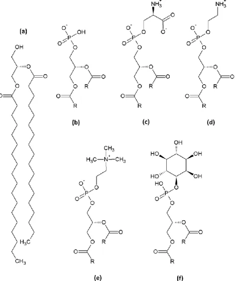
Figure 3. The five members of the GPL membrane building blocks.
(a) Diacylglycerol (DAG), (b) Phosphatidic acid (PA), (c) Phosphatidyl-L-serine (PS), (d)
Phosphatidylethanolamine (PE), (e) Phosphatidylcholine, (f) Phosphatidylinositol. In (b)-(f), acyl chains are shown as rest (R).
Sphingolipids
Sphingolipids are phospholipids in which the backbone is not a glycerol but derives from serine and palmitic acid, forming the long-chain base or sphingosine backbone (Figure
33
to form ceramide. In yeast, the long chain base and acyl chains are saturated and can be hydroxylated in C4 of the long chain base (phytosphingosine) and/or in C2 of the VLCFA, increasing the amphiphilicity of sphingolipids compared to GPLs.
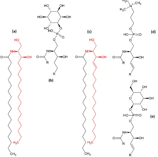
Figure 4. Simple and complex sphingolipids of yeast and mammalian.
Yeast sphingolipids (a)-(b): (a) Dihydroceramide, the yeast long-chain base dehydrosphingosine is highlighted in red. (b) inositol-phosphorylceramide (IPC). Complex mammalian sphingolipids (c)-(e):
(c) ceramide, the mammalian long-chain base dehydrosphingosine is highlighted in red, (d) a
sphingo-myelin (SM), (e) a simple cerebroside. In (b), (d) and (e) acyl chains of the fatty acid and the sphingoid backbone are shown as rest (R).
Like in GPLs, the ceramide backbone can be phosphorylated on its 1-OH function and headgroups can be added for formation of complex sphingolipids. In mammalian cells, addition of choline or ethanolamine yields sphingomyelins (SMs), whereas one or more glycosylations allow the formation of a variety of glycosphingolipids (cerebrosides and gangliosides). SMs are more abundant that cerebrosides and gangliosides. In yeast only three complex sphingolipids are synthesized, all of them with an inositol headgroup that can be glycosylated: inositol-phosphorylceramide (IPC), mannosyl-inositol-phosphorylceramide
34
(MIPC) and mannosyl-diinositol-phosphorylceramine (M(IP)2C). Complex sphingolipids have an important role in biological membranes, notably due to their affinity for sterols (Schneiter 1999).
As for GPLs, sphingolipids shape is controlled by acyl chain length and saturation and the headgroup size. Sphingolipids display the longest acyl chains, C24 and C26 (VLCFAs) are only found in sphingolipids, and are generally saturated. Ceramide displays a conical shape like DAG, whereas complex sphingolipids with their bulky headgroups display an inversed-conical shape (Schneiter, Brugger et al. 1999; Ejsing, Sampaio et al. 2009).
Sterols
Sterols, lipids from the isoprenoid lipid family, are also major building blocks for membranes in eukaryotic cells. Though both are amphiphilic, but their particular shape varies significantly from the aforementioned phospholipid species: Their polar headgroup (3-OH in cholesterol) is tiny and displays no charge; their hydrophobic moiety does not have a flexible and long shape but a planar four-ringed structure (the steroid backbone) with a short aliphatic “tail”. Their particular shape allows specific interactions with phospholipids, particularly saturated GPLs and sphingolipids, and these features make sterol an essential, yet unconventional membrane building block. Higher eukaryotes contain mainly cholesterol, whereas in budding yeast ergosterol (bis-unsaturated, methylated cholesterol) is the most abundant sterol (Mesmin, Antonny et al. 2013) (Figure 5).
Ergosterol Cholesterol
Figure 5. Chemical structure of ergosterol and cholesterol.
Ergosterol and cholesterol are the major sterol species found in budding yeast and mammalian membranes, respectively.
35
Oxidized metabolites of sterol, oxysterols, are precursors of steroid hormones and bile acids and implied in signaling (Massey 2006). 25-hydroxycholesterol (25-OH), for example, is a potent cholesterol biosynthesis inhibitor in concentrations in the nanomolar range (Olsen, Schlesinger et al. 2009). Sterol oxidation significantly alters its behavior towards membranes: Oxidation leads to more hydrophilic properties, and oxysterols in membranes are twisted into an orthogonal orientation compared to phospholipids (Olsen, Schlesinger et al. 2009). Cholesterol orientation is mainly governed by interaction between its 3-OH headgroup and surrounding phospholipid headgroups, it is inserted parallel to phospholipids. Due to their increased hydrophilicity, oxysterols diffuse more rapidly (102-fold increase compared to cholesterol) between membranes (van Amerongen, Demel et al. 1989)
37
L
IPID DISTRIBUTION
BETWEEN AND INSIDE CELL MEMBRANES
Cellular lipidomics and lipid homeostasis
Biological membranes are not at all as homogenous as they are often represented. Their composition varies significantly between species, tissues, cell types and their respective organelles. These variations can concern the protein/lipid ratio, membrane symmetry and overall lipid composition (ratio of charge and neutral GPL species, abundance of sterol and sphingolipids, acyl chain length and saturation), altogether governing membrane properties. The study of differences in lipid distribution and its dynamics has given rise to a new field in membrane biology: cellular lipidomics. This chapter will give insight into differences between of organellar membranes and the therefore emerging properties (van Meer, Voelker et al. 2008; Bigay and Antonny 2012; Holthuis and Menon 2014).
Some general features are conserved among all eukaryotic cells: The nuclear envelope is continuous with the endoplasmic reticulum (ER) and their lipid compositions are alike: > 40 % PC, 30 % PE, 10 % PI, 5 % PS, 5% sterol (Drin 2014) (Figure 6). ER and nuclear envelope are protein-rich membranes, with > 10 mg protein/mg phospholipid (Zinser, Sperka-Gottlieb et al. 1991). This ratio is only 3 mg/mg for the plasma membrane (PM), and anionic phospholipids (PS, PI) are enriched there as well as sphingolipids and sterols, and it is thicker (9.2 ± 0.4 nm for the PM and 7.5 ± 0.8 for the ER (Schneiter, Brugger et al. 1999)) and denser (Zinser, Sperka-Gottlieb et al. 1991; Schneiter, Brugger et al. 1999). The Golgi apparatus is at the crossroad between the ER and the PM, its composition changes from cis-Golgi (whose membranes are like those of the ER) to more PM-resembling trans-cis-Golgi membranes. Endosomal compartments have compositions comparable to those of the PM from which they originate, but are characterized by the presence of an specific endosomal GPL, lyso-bisphosphatidic acid (LBPA, also bis(monoacylglycero)phosphate BMP) (van Meer, Voelker et al. 2008). Budding yeast vacuolar membranes are rather loose and deprived of sterols and protein, but contain steryl esters (Schneiter, Brugger et al. 1999). Certain
38
organelles are labeled by minor pools of PIPs that have important functions. Mitochondria are surrounded by two lipid bilayers (Outer mitochondrial membrane OMM and inner mitochondrial membrane IMM) that show important differences in their lipid composition. The OMM is quite alike to the PM, but deprived of ionic phospholipids, whereas the IMM has a high protein/lipid ratio (7 mg protein / mg phospholipid), high amounts of unsaturated acyl chains and is rich in (≈ 10 mol%) a specific mitochondrial lipid, cardiolipin (Comte, Maisterrena et al. 1976; Schneiter, Brugger et al. 1999). Despite these general features, it is noteworthy that in all yeast membranes PI is more abundant at the expense of PC, particularly in the PM, which also has an elevated PS content compared to higher eukaryotes (McGee, Skinner et al. 1994; van Meer, Voelker et al. 2008).
Figure 6. Lipid distribution in eukaryotic cells.
The cellular organelles and their respective lipid composition with phospholipid concentrations expressed in percent of total phospholipid. Sterol abundance is described as ratio over total phospholipid for mammalian cells (CHOL/PL) and budding yeast (ERG/PL). Illustration from (van Meer, Voelker et al. 2008).
39
The uneven lipid distribution between and within organellar membranes allow each of them to optimize its function. Membrane composition, curvature, electrostatics and packing have to be considered as a whole in order to understand the functionalization of subcellular membranes (Bigay and Antonny 2012). It is thus important to describe how overall membrane properties arise from lipid composition.
Biophysical aspects of lipid bilayers
Effects of lipid shape and saturation levels
Lipid shape governs different aspects of membranes. The archetypal cylindrical lipids will take a lamellar organization as shown in (Figure 1b). With lipids deviating from the ideal, cylindrical shape, interaction either between headgroups of between acyl chains is decreased, as the accessible volume for the headgroups and the acyl chains, respectively, increases. The volume and charge is thus no longer homogenously distributed on a membrane, but locally displays dynamic higher or lower density. These imperfections in headgroup distribution are called lipid packing defects (Vamparys, Gautier et al. 2013) (Figure 7). Locally, such packing defects can increase membrane fluidity and facilitate protein interaction with lipids, but on a larger scale will deform the membrane: Local accumulation of conical or inversed-conical lipids will bend membranes to optimize the interactions between lipids, and thus induce membrane curvature (Bigay and Antonny 2012) (Figure 7).
Three phase states can be defined for a bilayer: a liquid-disordered phase (ld, low degree of order, fast diffusion), a solid gel phase (so, high degree of order, slow diffusion) and
liquid-ordered phase (lo, high degree of order, fast diffusion). These phases depend on the composition of the membrane and on the temperature. In liquid-ordered membranes the interaction of lipids with each other is stronger; formation of these phases is thus favored by high acyl chain saturation levels: Saturated phospholipids with a cylindrical shape are not subject to steric hindrance as conical, unsaturated phospholipids are due to their kinked acyl chains; cylindrical lipids hence display larger surfaces for lipid-lipid interaction (van Meer, Voelker et al. 2008) (Figure 7).
40
Figure 7. Impact of lipid shape and saturation on membrane organization.
See text for details. Illustration from (Holthuis and Menon 2014).
Sterols play an intriguing role in membranes depending on their phase: Their particular shape allows upon insertion in a lipid bilayer the stabilization of the membrane: It decreases the interaction between phospholipids by interacting with their acyl chains, thus preventing formation of solid gel phases. This interaction is stronger when the acyl chains surrounding the sterol molecule are saturated; lipid saturation can therefore allow segregation of sterol and vice versa. On the other hand, fluid membranes are rigidified and thickened by sterol insertion by the “condensing effect”. The acyl chain length together with the saturation level also govern membrane thickness: longer and saturated acyl chains can interact with sterols in the core of the membrane. Saturated and sterol-rich membranes are thus thicker than unsaturated membranes deprived of sterol (Munro 2003) (Figure 7).
Additionally, under certain circumstances one lipid bilayer can separate into two distinct coexisting phases (van Meer, Voelker et al. 2008). The microdomains formed by phase separation in one membrane, also referred to as ‘lipid rafts’, which are enriched in sterols and sphingolipids, have been analyzed, but whether they can play a physiological role is still discussed (Munro 2003; van Meer, Voelker et al. 2008; Toulmay and Prinz 2013). Despite the possibility of phase formation and separation in artificial membranes, the situation in biological membranes might not be so clear cut.
41
Transbilayer asymmetry and anionic lipids
Biological membranes are lipid bilayers and therefore have one cytosolic and one lumenal (in the case of the PM: extracellular or exoplasmic) leaflet. In some membranes, the two leaflets do not share the same lipid composition, i. e. they display a transbilayer
asymmetry (van Meer, Voelker et al. 2008). In the ER, anionic lipids, particularly PS, are
mainly facing the ER lumen. In the PM the extracellular face is devoid of PS and highly enriched in sphingolipids, PC and sterols whereas its cytosolic face displays opposite pattern with an elevated PS concentration thus increasing PM surface charge on its cytoplasmic face (Leventis and Grinstein 2010) (Figure 15).
Conservation of transbilayer asymmetry is mostly due to the inability of phospholipids to cross the membrane. Diffusion from one leaflet to another implies for a lipid molecule disruption of headgroup interaction and passage of the polar moiety through the hydrophobic core of the bilayer, which is the reason for the slow transbilayer movement of phospholipids, called flip-flop. GPLs and sphingolipids flip with t1/2 of hours (Holthuis and Levine 2005). Lipids without polar headgroup (such as DAG and ceramide) and neutral sterol molecules, on the other hand, can flip rapidly (t1/2 of seconds to minutes) between leaflets of one membrane (Holthuis and Levine 2005; Leventis and Grinstein 2010). The establishment of transbilayer asymmetry will be discussed below (See The origins of transbilayer asymmetry).
Special lipids: Phosphoinositides
Certain organelles are labeled by minor pools of PIPs that were initially seen as mere precursors for the formation of PI(4,5)P2 that is cleaved by phospholipase C (PLC) to yield Ins(1,4,5)P3 and DAG, two signaling molecules associated to G-protein coupled receptors at the PM. Moreover, a variety of functions of PIPs in multiple cellular processes have been unveiled. The PIPs are no major membrane constituents, but act, together with small G-proteins, as specific organellar signposts to facilitate their recognition. For example, PI(4)P marks mainly the trans-Golgi region, but it should be noted that functionally distinct pools mark also the PM and endosomal fractions. One of the most common protein domains to interact with PI(4)P are Pleckstrin Homology (PH) domains that will be detailed below (PI(4)P detection by NBD-PHFAPP). Other PI(4)P-interacting protein domains are found in the
42
clathrin adaptor proteins, such as AP-1 and GGAs, that recognize both cargo protein and PI(4)P to mediate clathrin coat formation. The γ subunit of dimeric human AP-1 (Apl4p in budding yeast) allows PI(4)P recognition by a conserved binding site inferred from homology from the crystal structure of murine AP-2, but the detailed mechanism remains to be elucidated (Collins, McCoy et al. 2002). Golgi-localized, γ-ear-containing, ARF (ADP (adenosine diphosphate) ribosylation factor)-binding proteins (GGAs, Gga1p and Gga2p in yeast) are clathrin-adaptors required for Golgi-to-endosome traffic. They all contain a GAT domain that detects both PI(4)P and Arf1-GTP by coincidence detection. The binding site for PI(4)P has been identified in a solvent-exposed three helix bundle of that domain that shows no homology with other PI(4)P-binding domains (Wang, Sun et al. 2007; Lenoir and Overduin 2013).
PI(4,5)P2 in key for PLC signaling at the PM and it plays an important role in the interaction between PM and the cytoskeleton as well as for exocytotic and endocytotic events (Tan and Brill 2014). Intriguingly, in yeast, PI(4,5)P2 deficiency phenotypes differ depending on the PI(4)P pool used for PI(4,5)P2 synthesis, indicating that there are distinct pools of PI(4,5)P2 within the PM (Audhya, Foti et al. 2000); nevertheless, both of these pools can be recognized by PH domains.
PI(3)P is mainly found on early endosomal compartments, and PI(3,5)P2 labels mainly late endosomal compartments (Behnia and Munro 2005; Mayinger 2012). PI(3)P is recognized by zinc finger motifs called FYVE (named after the proteins Fab1p, YOTB, Vac1p and EEA1 where it was first identified) domains and Phox homology (PX) domains that target proteins to the endolysosomal system. PI(3,5)P2 is synthesized from PI(3)P at the late endosomal/lysosomal system and epsin N-terminal homology (ENTH) domain containing proteins are recruited there by PI(3,5)P2 recognition (Mayinger 2012).
Certain protein domains thus allow specific recognition of different subcellular membranes highlighted by different PIPs.
Marking territories in eukaryotic cells
Combining the general trends in membrane compositions and their biophysical implications, an overall tendency can be seen in lipid distribution: The ER is rich in unsaturated lipids and sterol is scarce and therefore mainly in a liquid-disordered phase.
43
Such fluidity is thought to be important for the folding of proteins with transmembrane-spanning domains and the tabulated structure of the ER, implying high curvature. Anionic phospholipids are scarce, thus making the ER and early membranes of the secretory pathway a subcellular region mainly defined by high packing defects (Bigay and Antonny 2012).
Continuing along the secretory pathway these characteristics are reversed, with the Golgi apparatus being and intermediate compartment crucial for this change. At the PM, the lipids there have higher saturation levels and the enrichment of sterol and complex sphingolipids allows a more liquid-ordered phase state, and packing defects are scarce. The cytoplasmic face of the PM additionally is enriched in the anionic phospholipid PS, together with PIPs making late membranes as the trans-Golgi and the PM mainly governed by electrostatics (Bigay and Antonny 2012) (Figure 8).
Figure 8. Division of eukaryotic cells in two territories.
Early regions of the secretory pathway such as the ER and cis-Golgi display a high level of unsaturated lipids and its biophysics is mainly governed by membrane packing defects. Late membranes such as trans-Golgi and the PM are densely packed and rich in anionic phospholipids, therefore making it the territory of electrostatics. Illustration from (Bigay and Antonny 2012).
45
C
ONCEPTS FOR ESTABLISHING AN UNEVEN
LIPID DISTRIBUTION IN EUKARYOTIC CELLS
Despite their different compositions, organelles perpetually exchange parts of their membrane by vesicular trafficking that allows transport of proteins and nutrients in cells. The constant arrival and departure of material necessitates mechanisms to regulate its lipid homeostasis, i.e. to keep its overall lipid composition and uneven distribution constant.
The mechanisms implied in establishment differences in lipid compositions between subcellular compartments can be divided in three classes: The first class is lipid metabolism, i.e. lipids are produced or modified at different places inside the cell. The second class is lipid transport between bilayers of a single bilayer, which is required for establishing transbilayer asymmetry. The third class is vesicular or non-vesicular mechanisms that deliver lipid molecules specifically between membranes.
Spatial differentiation through lipid metabolism
Glycerophospholipid biosynthesis routes
Synthesis of phosphatidic acid and diacylglycerol
PA is synthesized on the cytosolic face of the ER from glycerol-3-phosphate by acylation of the free 1- and 2-hydroxyl groups with fatty acids activated in the form of acyl-CoA (Coenzyme A). A single acylation yields lysoPA, a subsequent, second acylation yields PA. Dephosphorylation of PA produces DAG, the precursor for GPL biosynthesis via the Kennedy pathway (see below) and for biosynthesis of triacylglycerol (TAG), a storage lipid. PA can also be nucleotidylated with cytosine-triphosphate (CTP) by phosphatidate cytidylyltransferase yielding CDP-DAG, the precursor for GPL synthesis via the de novo-pathway (see below) and cardiolipin biosynthesis in mitochondria (Henry, Kohlwein et al. 2012).
46
Functionalization of different GPL species
In order to obtain fully functional GPLs, a headgroup has to be added to the DAG precursors. In eukaryotic cells, there are two pathways of GPL synthesis: In the Kennedy
pathway, the major GPL biosynthesis pathway in higher eukaryotes, PC is synthesized by the
addition of CDP-choline to DAG and in a reaction catalyzed by CPT1 (cholinephosphotransferase 1), whereas EPT1 (ethanolaminephosphotransferase 1) assures the synthesis of PE from DAG and CDP-ethanolamine. PS is subsequently synthesized from PC and PE, by swapping the headgroups for serine by PS synthases PSS1 and PSS2, respectively. Counter-reaction exist in which PS is decarboxylated, yielding PE, that can undergo subsequent tri-methylation, yielding PC. These reactions are catalyzed by PS decarboxylases (PSD) and PE-methyl transferases (PEMT), respectively. PI is synthesized following the so called de novo pathway from CDP-DAG and myo-inositol (Daum, Lees et al. 1998; Vance and Steenbergen 2005; Gibellini and Smith 2010) (Figure 9).
Figure 9.GPL synthesis pathways
All eukaryotes use the Kennedy pathway (orange boxes), but budding yeast can also produce PS via the de novo pathway (blue box) and PE and PC by decarboxylation and successive methylation. In Mammalia and yeast, PI is synthesized from CDP-DAG and myo-inositol, like PS in yeast (not shown). Illustration modified from (Leventis and Grinstein 2010)
47
The budding yeast S. cerevisiae possesses the same synthesis machinery, but additionally, it disposes of a de novo PS synthase, Pss1p (Cho1p), which synthesizes PS directly from CDP-DAG and L-serine. Despite the fact that the Kennedy pathway machinery is fully functional in yeast, it exploits mainly the de novo pathway and synthesizes most of its PC and PE by modifying the PS headgroup. The PS synthase Pss1p hence has to ensure the production of the majority of the total GPL in yeast, with PS thus being the key intermediate for bulk GPL synthesis. The syntheses of PS and PI share a common precursor, CDP-DAG, and its limited availability therefore restrains global GPL synthesis (Leventis and Grinstein 2010). For PE biosynthesis by decarboxylation S. cerevisiae encodes two PSDs (Psd1p and Psd2p) and two PEMTs, Cho2p and Opi3p. The former catalyzes the first and the latter catalyzing mainly the two other methylation steps. Both Kennedy pathway and de novo pathway enzymes are localized to the cytosolic face of the smooth ER in yeast and human (Daum, Lees et al. 1998; Vance and Steenbergen 2005; Leventis and Grinstein 2010) (Figure 9).
Of the abovementioned enzymes, PS decarboxylases are the only not to be localized at the ER but at the IMM (mammalian PSID and yeast Psd1p) and Golgi/endosomes (yeast Psd2p) (Leventis and Grinstein 2010). It is noteworthy that for budding yeast, an efficient transfer of PS from the ER to mitochondria and Golgi/endosomes is essential, as decarboxylation of PS and methylation of PE are the major sources of PE and PC, respectively. The lipid export/import appears to be favored by the localization of the concerned enzymes: They are not homogenously distributed within the ER but are rather enriched at parts of the ER, called membrane contact sites (MCSs) that encounter other membrane-bound compartments (Helle, Kanfer et al. 2013). These particular sites and their importance in lipid transport will be discussed in detail later. Human PS synthases PSS1 and PSS2, for example, are enriched in mitochondria-associated membranes (MAM) (Stone and Vance 2000) as well as its yeast counterpart Pss1p which is additionally found in the PM-associated membrane (PAM), thus in proximity of another organelle requiring PS import (Gaigg, Simbeni et al. 1995; Pichler, Gaigg et al. 2001).
Mammalian PI synthase activity has recently been localized to highly dynamic, ER-derived structures termed PI-Producing ER-ER-derived Organelles or PIPEROsomes that would directly supply other organellar membranes with PI by ample contacts (Kim, Guzman-Hernandez et al. 2011). PI is an essential lipid for budding yeast that produces significantly more of this lipid than higher eukaryotes. The function of PI as a negatively charged building
48
block in membranes might be taken over by PS, thus the importance of PI in yeast might be linked to its function as a precursor for PIP and morevover, for sphingolipid synthesis or as basis of glycosylphosphatidylinositol (GPI)-anchored proteins (Daum, Lees et al. 1998).
Phosphoinositide biosynthesis pathways
Synthesis and localization of phosphoinositide species
PIPs are synthesized from PI by different kinases (PIK) that allow the localized and specific creation of PIP pools (Figure 10). PI(3)P is synthesized by class II and class III PI3K in the early endosomal system and regulates its dynamics during endocytosis and autophagy. In yeast, both PI(3)P and PI(3,5)P2 are localized to the vacuole (the yeast counterpart of the endosomal/lysosomal compartment in higher eukaryotes) where they are synthesized by the class III PI3K Vps34 and the PI5K Fab1, respectively, two non-essential enzymes (Mayinger 2012).
In mammalian cells, PI(4)P is synthesized from PI by four PI4K (PI4KIIIα, PI4KIIIβ PI4KIIα and PI4KIIβ, also named, PI4KA, PI4KB, PI4K2A and PI4K2B respectively)and labels the Golgi as well as the PM. Golgi and PM-localized PI4K produce functionally distinct pools of PI(4)P: PI4KIIIβ (Pik1p in yeast) creates the most considerable pool in the Golgi apparatus and thus governs Golgi function in secretion via multiple PI(4)P effectors (Audhya, Foti et al. 2000; Tan and Brill 2014). Pik1p is localized to the Golgi by interaction with Frq1p, and is rapidly detached from Golgi membranes under glucose starvation conditions, leading to an arrest of vesicular trafficking (Faulhammer, Kanjilal-Kolar et al. 2007). PI4KIIIα (Stt4p in yeast) is targeted to the PM by interaction with Sfk1p and also with Efr3p via Ypp1p, which together regulates Stt4p localization and activity (Baird, Stefan et al. 2008; Wu, Chi et al. 2014). The endosomal/lysosomal PI(4)P pool synthesized by PI4KIIα and PI4KIIβ (only one homolog in yeast, Lsb6p) is minor and non-essential compared to the others, and regulates endosomal function (Han, Audhya et al. 2002; Shelton, Barylko et al. 2003; Jovic, Kean et al. 2014). Recently, a novel highly specific and sensitive probe for PI(4)P determination based on the P4M domain of the Legionella pneumophilia SidM protein has been developed. Utilization of this probe revealed a broader distribution of PI(4)P, promising a more detailed insight into dynamics of PI(4)P distribution (Del Campo, Mishra et al. 2014; Hammond, Machner et al. 2014; Hubber, Arasaki et al. 2014).
49
PM localized PI(4)P serves as a precursor for synthesis of PI(4,5)P2 by PIP kinases (PIP5K, Mss4p in yeast), and PI(4)P for PI(4,5)P2 synthesis can originate from both the PI4KA (Stt4p) and PI4KB (Pik1p) pools. PI(4)P together with PI(4,5)P2 makes up to 90 % of cellular PIPs in yeast (Audhya, Foti et al. 2000; Audhya and Emr 2002; Tan and Brill 2014). Phosphorylation of PI(4)P and PI(4,5)P2 by class I PI3K yields PI(3,4)P2 and PI(3,4,5)P3, respectively, two short-lived regulators of cell survival and growth, which are not found in yeast due to the lack of a class I PI3K (Odorizzi, Babst et al. 2000; Mayinger 2012).
Figure 10. PIP distribution in Mammalia
PI(3)P is found on early endosomal compartments whereas PI(3,5)P2 is localized to late endosomes
and lysosomes. Two different pools of PI(4)P highlight the Golgi and the PM which also displays PI(4,5)P2. PI(5)P and the short lived PI(3,4,5)P3 are found at the PM. Illustration from (Billcliff and
50
Phosphoinositide catabolism
PIPs are hydrolyzed by more or less specific PIP phosphatases, allowing interconversion of PIP species and regulation of PIP controlled processes. Sac1p shall be the only PIP phosphatase detailed here due to its implication in PI(4)P hydrolysis; for details on other PIP phosphatases see (Billcliff and Lowe 2014).
Figure 11. Crystal structure and model of the budding yeast PIP phosphatase Sac1p.
Left: Ribbon diagram showing the structure of the cytosolic portion (residues 1-503) of Sac1p with
numbered secondary structure elements. The N-terminal domain (1-182) is shown in blue and the catalytic domain (183-503) in yellow. The catalytic motif CX5R(T/S) in the P-loop of the catalytic
domain is shown in red and protruding loops around the catalytic motif are green. Right: Surface representation of the cytosolic portion (yellow and blue) attached to its transmembrane domains (grey) by a flexible linker (spotted in orange). The catalytic site recognizes PI(4)P (green hexagon with phosphate as yellow sphere) and hydrolyses it to yield PI (green hexagon). Illustrations from (Manford, Xia et al. 2010).
Sac1p is a double-spanning transmembrane protein that is localized to the ER, but can be shuttled to the Golgi under glucose starvation conditions where it is retained by interaction with Vps74 until reestablishment of normal growth conditions (Konrad, Schlecker et al. 2002; Wood, Hung et al. 2012; Cai, Deng et al. 2014). It is the major PIP phosphatase in budding yeast and controls mainly PI(4)P levels. The crystal structure of its cytosolic portion (1-503) has been solved and it displays two domains, the N-terminal SacN domain (1-182) and the catalytic phosphatase domain (183-503) (PDB entry: 3LWT) (Manford, Xia et al. 2010) (Figure 11). The phosphatase domain is localized in a loop (P-loop) and displays a CX5R(T/S)