Identification of Plant-like Galactolipids in Chromera velia, a
Photosynthetic Relative of Malaria Parasites
*
□SReceived for publication, April 28, 2011, and in revised form, June 14, 2011 Published, JBC Papers in Press, June 28, 2011, DOI 10.1074/jbc.M111.254979 Cyrille Y. Botte´‡§1, Yoshiki Yamaryo-Botte´¶储1, Jan Janousˇkovec**, Thusita Rupasinghe储, Patrick J. Keeling**, Paul Crellin¶, Ross L. Coppel¶, Eric Mare´chal§, Malcolm J. McConville储2,3, and Geoffrey I. McFadden‡3,4
From the‡School of Botany, University of Melbourne, Parkville, 3010 Victoria, Australia, the§Unite´ Mixte de Recherche 5168, CNRS, Commissariat a` l’Energie Atomique, Institut National de Recherche Agronomique, Universite´ Grenoble 1, Institut de Recherches et Technologies en Sciences fu Vivant/Commissariat a` l’Energie Atomique, Grenoble 38054, France, the¶Department of Microbiology, Monash University, 3800 Victoria, Australia, the储Department of Biochemistry and Molecular Biology, Bio21 Institute of Molecular Science and Biotechnology, University of Melbourne, Melbourne, Parkville, 3010 Victoria, Australia, and the **Botany Department, University of British Columbia, Vancouver, British Columbia V6T 1Z4, Canada
Apicomplexa are protist parasites that include Plasmodium spp., the causative agents of malaria, and Toxoplasma gondii, responsi-ble for toxoplasmosis. Most Apicomplexa possess a relict plastid, the apicoplast, which was acquired by secondary endosymbiosis of a red alga. Despite being nonphotosynthetic, the apicoplast is oth-erwise metabolically similar to algal and plant plastids and is essen-tial for parasite survival. Previous studies of Toxoplasma gondii identified membrane lipids with some structural features of plastid galactolipids, the major plastid lipid class. However, direct evi-dence for the plant-like enzymes responsible for galactolipid syn-thesis in Apicomplexan parasites has not been obtained. Chromera
velia is an Apicomplexan relative recently discovered in Australian
corals. C. velia retains a photosynthetic plastid, providing a unique model to study the evolution of the apicoplast. Here, we report the unambiguous presence of plant-like monogalactosyldiacylglycerol and digalactosyldiacylglycerol in C. velia and localize digalactosyl-diacylglycerol to the plastid. We also provide evidence for a plant-like biosynthesis pathway and identify candidate galactosyltran-ferases responsible for galactolipid synthesis. Our study provides new insights in the evolution of these important enzymes in plas-tid-containing eukaryotes and will help reconstruct the evolution of glycerolipid metabolism in important parasites such as
Plasmo-dium and Toxoplasma.
Galactolipids, i.e. monogalactosyldiacylglycerol (MGDG)5
and digalactosyldiacylglycerol (DGDG), are the most abundant
lipid classes in the membranes of plant and algal plastids, where they represent up to 85% of the total lipid composition (1). This unique lipid composition is only otherwise found in cyanobac-teria (2), the lineage from which the plastid was derived by endosymbiosis (3). In plants and algae, MGDG and DGDG syn-theses are catalyzed by two galactosyltransferases localized in the plastid envelope (1, 4). In these organisms, MGDG is syn-thesized in a single step by MGDG synthases that transfer a -galactosyl moiety from a UDP-galactose (UDP-Gal) donor to the sn-3 position of diacylglycerol (DAG). In cyanobacteria, MGDG synthesis occurs via a two-step process in which a glu-cosyltransferase, referred to as monoglucosyldiacylglycerol (MGlcDG) synthase, transfers a glucosyl from UDP-glucose (UDP-Glc) donor onto DAG to form MGlcDG, which is subse-quently epimerized into MGDG (5). In both plants and cyano-bacteria, a DGDG synthase catalyzes the transfer of a second ␣-galactose from UDP-Gal to MGDG. As the major lipids of thylakoid and plastid membranes, MGDG and DGDG are thought to be essential for the biogenesis and function of these membranes (6). Molecular disruption of the MGDG synthetic pathway in Arabidopsis thaliana showed that galactolipids play an essential role in early embryo development and biogenesis of a functional photosynthetic apparatus (6). Furthermore, both MGDG and DGDG were found tightly associated with crystal-lized photosystems and light harvesting complexes (7), and DGDG might be required for their stability and proper activity (8 –12). Galactolipids have long been thought to be restricted to plastid localization and functions. However, several important studies showed that DGDG could also be exported and become a critical component of extraplastidic compartments. Under specific environmental conditions, such as phosphate shortage, DGDG can substitute for phospholipids in extraplastidic mem-branes such as the plasma membrane, mitochondria, and tono-plast (1). Galactolipids are therefore involved in critical func-*This work was supported in part by European Research Council FP7 Marie
Curie Actions via an International Outgoing Fellowship Marie Curie fellow-ship (to C. Y. B.), National Health and Medical Research Council of Australia, CNRS, and Agence Nationale de la Recherche ReGal grant (to E. M.). □S
The on-line version of this article (available at http://www.jbc.org) contains
supplemental Figs. S1–S5.
Author’s Choice—Final version full access.
The nucleotide sequence(s) reported in this paper has been submitted to the Gen-BankTM/EBI Data Bank with accession number(s) JF912518 and JF912519.
1Both authors contributed equally to this work.
2National Health and Medical Research Council of Australia Principal Research Fellow.
3Both authors are senior authors.
4Federation Fellow of the Australian Research Council and a Howard Hughes International Scholar. To whom correspondence should be addressed: School of Botany, University of Melbourne, Parkville, Victoria, Australia 3010. Tel.: 61-3-8344-5053; Fax: 61-3-9347-5460; E-mail: gim@unimelb.edu.au.
5The abbreviations used are: MGDG, monogalactosyldiacylglycerol (1,2-diacyl-3-O-(-D-galactopyranosyl)-sn-glycerol); DGDG,
digalactosyldiacyl-glycerol (1,2-diacyl-3-O-(␣-D-galactopyranosyl-(136)-O--D -galactopyra-nosyl)-sn-glycerol); DAG, 1,2-diacylglycerol-sn-glycerol; HPTLC, high precision thin layer chromatography; GC-MS, gas chromatography-mass spectrometry; SQDG, sulfoquinovosyl-diacylglycerol; MGlcDG, mono-glucosyl-diacylglycerol 91,2-diacyl-3-O-(␣-D-glucopyranosyl)-sn-glycerol; MURG, UDP-␣-D -(N-acetyl)-glucosamine:N-acetylmuramyl-(pentapep-tide)pyrophosphoryl-undecaprenol-4-D -9N-acetyl)-glucosamyltrans-ferase; RACE, rapid amplification of cDNA ends; MGD, monogalactosyldia-cylglycerol; DGD, digalactosyldiacylglycerol.
at INRA Institut National de la Recherche Agronomique on June 13, 2018
http://www.jbc.org/
tions of membrane biogenesis and homeostasis, as well as developmental and physiological processes in autotrophic organisms.
The Apicomplexa include a large group of protozoa that are the causative agents of major human diseases such as malaria (Plasmodium spp.) and toxoplasmosis (Toxoplasma gondii). Apicomplexa acquired a plastid, termed the apicoplast, by the secondary endosymbiosis of a red alga (13, 14). The apicoplast no longer retains photosynthetic enzymes but is essential for the viability of these organisms, probably due to the retention of other key metabolic functions (15, 16). The discovery of the apicoplast immediately raised questions regarding the possible occurrence of plastidial galactolipids, their synthesis, and putative role in these parasites. None of the genes encoding for galactolipid synthesis have been found in Apicomplexa genomes to date (17). However, the synthesis of galactolipids co-migrating with spinach MGDG and DGDG could be detected in Plasmodium falciparum and T. gondii lysates by metabolic labeling with radioactive UDP-Gal (18). Hydrolysis
with␣- and -galactosidases combined with alkaline hydrolysis
confirmed that these galactolipids contained␣- and
-galacto-ses as polar heads and esterified fatty acids as their hydrophobic tail, making them very similar to plant galactolipids (18). Fur-thermore, a lipid with chromatographic properties similar to plant DGDG was detected in total lipid extract from P.
falcipa-rumand T. gondii (19). A digalactolipid reacting against an
anti-DGDG antibody was localized within the pellicle mem-branes of T. gondii, but not in the apicoplast (20). This plant-like digalactolipid was proposed to be the same as those detected in previous labeling experiments. Lipidomic analysis performed in T. gondii showed the presence of two types of hexosyl(galacto) lipid classes as minor membrane components as follows: (i) plant-like hexosylglycerolipids and (ii) hexosylce-ramides (20, 21). Although these data are suggestive of a path-way of galactolipid biosynthesis in some Apicomplexan para-sites, many questions remain. Are Apicomplexan galactolipids generated via a plant-like or cyanobacterium-like pathway using highly divergent enzymes? Is the galactolipid biosynthetic machinery located in the plastid or other compartments?
Very recently, Chromera velia, a photosynthetic protist closely related to Apicomplexa, was discovered in corals in Syd-ney Harbor (22). C. velia contains a four membrane-bound plastid that is still photosynthetic and is therefore an interesting model to study the conversion of secondary plastids from autotrophic to heterotrophic state, as happened to the apico-plast. Here, we report the unambiguous characterization of plant-like galactoglycerolipids in C. velia via HPTLC, GC-MS, and LC/MS-MS approaches and determine their localization by immunocytochemistry. Metabolic labeling is used to char-acterize a plant-like galactolipid synthesis pathway in C. velia. Finally, we identify the candidate genes encoding galactosyl-transferases responsible for galactolipid synthesis in C. velia and discuss their importance for the evolution of this pathway in photosynthetic eukaryotes.
EXPERIMENTAL PROCEDURES
C. velia Culture and Lipid Extraction—C. velia was main-tained as described previously (14) and harvested by
centrifu-gation at 700⫻ g, and total lipids were extracted in chloroform/
methanol (2:1, v/v) and chloroform/methanol/water (1:2:0.8, v/v). After removal of insoluble material by centrifugation
(15,000⫻ g, 10 min), extracts were dried under nitrogen and
subjected to biphasic partitioning in 1-butanol and water (2:1, v/v). The organic phase was dried, and lipids were resuspended in 1-butanol. Total lipid was analyzed by HPTLC using alu-minum-backed silica gel sheets (Merck). One-dimensional HPTLCs were developed in chloroform/methanol/water (65:35:2, v/v). Glycolipids were stained and visualized with
orcinol/H2SO4.
Sugar Head Analysis by Gas Chromatography-Mass Spectrometry—Individual glycolipid species were extracted from silica scrapings from one-dimensional HPTLCs using chloroform/methanol/water (10:10:3, v/v), and the supernatant was dried under nitrogen and desalted by 1-butanol/water biphasic partitioning (2:1, v/v). The sugar head of each glyco-lipid was analyzed by GC-MS after methanolysis and trimeth-ylsilyl derivatization (40).
Mass Spectrometric Analysis of MGDG and DGDG—The
MGDG or DGDG fraction was diluted 10-fold in 10 mM
ammo-nium formate in methanol and then the lipid was flow-injected to Agilent 6410 triple quadrupole mass spectrometer (Agilent Technologies, Santa Clara, CA) at the rate of 0.2 ml/min in the
solvent of H2O/MeOH/tetrahydrofuran⫽ 14:20:66 (v/v). The
capillary voltage was set at 4000 V, and the gas temperature and flow rate were set to 250 °C and 7 liters/min, respectively. Neb-ulizer was set at 40 p.s.i. MS/MS experiments were conducted in positive mode with a fragmentation voltage of 135 V and collision energy of 15 eV.
Immunofluorescence Assay—All media and antibody dilu-tions used for immunofluorescence assays were prepared in PHEM buffer, pH 6.9 (PIPES 60 mM, HEPES 25 mM, EGTA 10
mM, MgCl22 mM). C. velia cells were fixed in 4%
paraformal-dehyde for 45 min on ice prior to permeabilization in 0.1% Triton X-100 for 10 min on ice. After being blocked with 3% BSA in PHEM for 45 min at 4 °C, cells were then incubated with polyclonal rabbit serum anti-DGDG (1:25) for 1 h at 4 °C with agitation. Labeled cells were incubated with secondary anti-rabbit Alexa488 (1:1000) for 1 h at 4 °C, followed by incubation with DAPI (1:15,000) for 15 min at 4 °C. Cells were mounted on slides and coverslips and observed with a Leica confocal microscope.
Activity Assay—C. velia was harvested by centrifugation at
700 ⫻ g and resuspended in 5 volumes (v/v) of lysis buffer
(MOPS-NaOH, pH 7.9, 50 mM, 20% glycerol (v/v), sodium
ace-tate 0.8M, DTT 10 mM) followed by sonication for three
inter-vals of 15 s on ice with 2 min resting on ice between each pulse (Branson sonifier 250, output 40%). 2.5 g of pre-frozen (in liquid
N2) spinach leaves were crushed with a mortar and a pestle in
the presence of 2.5 ml of lysis buffer. Spinach lysate was resus-pended in 5 volumes (w/v) of lysis buffer. DTT was added (10
mM) prior to centrifugation at 500⫻ g, 4 °C for 20 min. Then a
90-l aliquot was preincubated at 30 °C for 5 min with either
UDP-[14C]galactose or UDP-[14C]glucose (PerkinElmer Life
Sciences, 6.3 Ci/mmol, final concentration, 1 Ci/reaction)
added to start the enzyme reaction. After 2 h, reactions were
stopped by addition of 375l of chloroform/methanol (1:2, v/v)
at INRA Institut National de la Recherche Agronomique on June 13, 2018
http://www.jbc.org/
and vortexing. After centrifugation at 4000⫻ g for 10 min, the supernatant was pooled, and another chloroform/methanol/ water (1:2:0.8, v/v) was added for further extraction of lipids. The supernatant was recovered and mixed with the pooled supernatant and dried up under nitrogen gas. The dried mate-rial was biphasically partitioned by 1-butanol/water (2:1, v/v). The butanol phase was recovered and dried. Again the lipid was
dissolved in the 10l of water-saturated 1-butanol. Total lipid
was analyzed by HPTLC using aluminum-backed silica gel sheets (Merck). HPTLC was migrated in the solvent system of chloroform/methanol/water (65:35:2, v/v). The HPTLC was exposed to high sensitivity x-ray film, Kodak BiomaxMR film
(Eastman Kodak Co.), to detect14C-labeled lipids.
RNA Isolation, RT-PCR, RACE, and Sequencing—Short frag-ments of MGDG and DGDG synthase genes were identified in the 454-pyrosequencing of C. velia genomic DNA (14). Total
RNA was isolated using TRIzol威 reagent (Invitrogen) and
puri-fied using RNeasy MinElute cleanup kit (Qiagen). For each gene, fragments determined in the 454-data survey were linked
using RT-PCR with specific oligonucleotides. 3⬘-RACE was
used to obtain a complete 3⬘ sequence of the MGDG synthase
transcript. In DGDG synthase, 3⬘-RACE repeatedly produced a
single amplicon containing an incomplete 3⬘ portion of the
transcript, probably due to the oligo(dT) adaptor misannealing to an A-rich region upstream of the stop codon. All PCR cons were cloned and several clones from each of these ampli-cons sequenced. All sequences for each gene were
unambigu-ously assembled into a single contig. Our attempts to obtain 5⬘
ends of the transcripts were unsuccessful and lea only to non-specific amplification products. MGDG and DGDG synthase
sequences of C. velia were deposited under GenBankTM
acces-sions numbers JF912518 and JF912519.
Phylogenetic Analyses—For genomic sequence survey of C.
velia, GenBankTMprotein and EST databases were searched using multiple rounds of blastp and tblastn similarity searches using different queries (MGDG synthase from A. thaliana,
Cyanidioschyzon merolae, and Phaeodactylum tricornutum; MGlcDG synthase from Synechocystis PCC6803; DGDG syn-thase from A. thaliana and Synechocystis PCC6803; and
Esche-richia coli MURG). Nearly identical paralogs likely derived from different gene models on identical genomic loci (e.g. in
Oryzaand Populus) were limited to the most canonical model. Highly divergent sequences of some bacteria were excluded from these sequence sets. Two principal datasets from each gene were created as follows: (i) global dataset for assessing the monophyly of eukaryotic homologs and their position within bacterial genes, and (ii) eukaryotic dataset for exploring the branching patterns with eukaryotes and the position of C. velia. In global phylogenies of DGDG synthase, the relationships of both the plant-like and the cyanobacterial form (also present in
Cyanophoraand cyanidiales) were explored. All dataset were aligned in MAFFT using the local pair (LINSI) algorithm and ambiguously aligned positions were trimmed in Gblocks using
following parameters:⫺b1 ⫽ 50% ⫹1; ⫺b2 ⫽ 50% ⫹1; ⫺b3 ⫽
12 ⫺b4 ⫽ 4 ⫺b5 ⫽ h. This procedure resulted in following
matrix sizes: MGDG phylogenies insupplemental Fig. S4, A and
B, and Fig. 8A⫽ 160, 183, and 326 sites, respectively; DGDG
phylogenies insupplemental Fig. S5, and Fig. 8B⫽ 112, and 364
sites (alignments available on request). Global phylogenies
were computed in PhyML 3.0 using LG model (parameters,␥ 8,
I, SPR) with aLRT branch supports. Phylogenies from eukary-otic datasets were computed under identical conditions in PhyML 3.0 and supported by PhyML and RaxML bootstrap analyses.
RESULTS
C. velia Contains MGDG and DGDG Enriched in Long Poly-unsaturated Fatty Acid Chains—To assess the presence of plant/plastid-like galactolipids in C. velia, total lipids were ana-lyzed by HPTLC. As shown in Fig. 1, C. velia synthesized a number of abundant orcinol-positive glycolipids that had a similar HPTLC migration to the plastid MGDG and DGDG galactolipids and sulfoquinovosyldiacylglycerol (SQDG) from spinach (Spinacia oleracea) leaf extracts. MGDG and DGDG fractions from S. oleracea and the corresponding C.
veliafractions (fraction A and fraction B, respectively) were extracted from the HPTLC sheets and reanalyzed by HPTLC in a different solvent system. Again the extracted C. velia MGDG-like and DGDG-like fractions showed similar mobil-ity to the S. oleracea galactolipids. Fractions A and B were subjected to mild alkaline treatment to hydrolyze ester-linked fatty acids and release the galacto-glycerol backbone. The glycan headgroups of the C. velia glycolipids (fractions A and B) displayed similar chromatographic behavior on HPTLC to those obtained from hydrolyzed S. oleracea
MGDG and DGDG (supplemental Fig. S1). The presence of
galactolipids was confirmed by GC-MS analysis of monosac-charides released by solvolysis and trimethylsilylation. These analyses showed that the major sugar in fractions A and B was galactose (Fig. 2).
To further define the structures of the C. velia galactolipids, the HPTLC-purified glycolipids were subjected to LC/MS-MS
FIGURE 1. C. velia contains plant-like glycolipids. Total lipids from spinach and C. velia were extracted from whole leaves and whole cells, respectively. Extracted lipids were migrated by HPTLC in chloroform/methanol/water (65: 25:25, v/v) and sprayed with orcinol/sulfuric acid to allow glycolipid detec-tion. Plastidial MGDG, DGDG, and SQDG are indicated. Fr., fracdetec-tion.
at INRA Institut National de la Recherche Agronomique on June 13, 2018
http://www.jbc.org/
analysis (Fig. 3). Neutral loss scanning for m/z 179 ion and m/z 341 ion in positive mode gave the characteristic ions for an ammonium-adducted and dehydrated monohexose from MGDG and an ammonium-adducted and dehydrated dihexose from DGDG, respectively (Fig. 3, A and C). The parent ions that were detected by neutral loss scanning of m/z 179 from the S.
oleraceagalactolipids had masses of m/z 792.3 and 764.2, cor-responding to molecular species with 36:6 (36 carbon and 6
double bonds) and 34:6 fatty acyl chains, respectively (
supple-mental Fig. S2). Similarly, neutral loss scan for m/z 341 in S.
oleracea lipids detected DGDG species with masses of m/z 954.2 and 932.3, corresponding to molecular species with the same lipid composition (36:6 and 34:3). Similar analyses of the
C. veliagalactolipid fractions A and B allowed detection of a different set of parent ions indicating a distinct fatty acyl composition. Neutral loss scanning for m/z 179 and 341 revealed a series of parent ion peaks in the m/z 500 –1100 mass ranges. Parent ion masses of m/z 840.3 and 794.3
cor-responded to MGDG [M⫹ NH4]⫹ion containing 36:5 and
40:10 acyl chains, respectively (Fig. 3A). Similarly, DGDG
parent ions at m/z 956.1 and 1002.2 corresponded to [M⫹
NH4]⫹ion with 36:5 and 40:10 acyl chains, respectively (Fig.
3C). When molecular species at m/z 840.3 and 794.3 were fragmented, ions derived from loss of galactose (m/z 179),
diacylglycerol ([DAG (40:10) ⫹ H]⫹ (m/z 661.3) or [DAG
(36:5) ⫹ H]⫹ (m/z 615.4)), and monoacylglycerol ([MAG
(20:5)⫹ H ⫺ H2O]⫹(m/z 359) and/or [MAG (16:0)⫹ H ⫺
H2O]⫹(m/z 313)) were detected (Fig. 3B). These
fragmenta-tion patterns are the same as those for S. oleracea MGDG except for the difference of fatty acid species confirming the
galactolipid fraction as MGDG. Similarly, MS/MS fragmen-tation of m/z 956.1 and 1002.2 generated characteristic ions corresponding to loss of digalactose (m/z 341), a peak of
[DAG⫹ H]⫹, and peaks of MAG (20:5) and/or MAG (16:0)
(Fig. 3D). These fragmentation patterns also corresponded to those of DGDG from S. oleracea confirming galactolipid fraction B as DGDG. These data demonstrate that the C.
veliagalactolipids are equivalent to the plant galactoglycer-olipids. Furthermore, the presence of SQDG sulfolipid was confirmed by the precursor ion scan for the characteristic ion of SQDG, m/z 225 in negative mode. The major SQDG
were detected as [M⫺ H]⫺ions at m/z 817 and 843, SQDG
(34:2) and SQDG (36:3), respectively (supplemental Fig. S3).
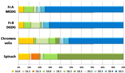
The fatty acyl chain composition of the galactolipids was fur-ther supported by GC-MS fatty acid analysis of C. velia MGDG, DGDG, and total lipids (Fig. 4). Total lipids from both S.
olera-ceaand C. velia are highly enriched in unsaturated fatty acids
(89 and 84%, respectively). MGDG and DGDG fractions from
C. veliafollowed a similar pattern in terms of their unsaturated fatty acid composition (i.e. 86 and 88%, respectively). By con-trast with spinach total lipids, which are highly enriched in lin-olenic acid (C18:3; 62%), C. velia total lipid fraction contained a large majority of eicosapentenoic acid (C20:5, 52%). A similar composition was found in both MGDG- and DGDG-like frac-tions, whereas eicosapentenoic acid contributed to 74 and 70% of the fractions, respectively. C. velia lipids predominantly
con-tained very long fatty acid chains (⬎18 carbons) as follows: 61%
in the total lipid fraction and 77% for both MGDG and DGDG fractions, and S. oleracea only contained shorter fatty acid chains.
DGDG Is Detected in the C. velia Plastid—In plants and algae, galactolipids are predominantly found within the plastid membranes. In contrast, preliminary analyses of the T.
gon-dii DGDG-like lipid indicated localization to the pellicle
membranes (20). To assess the subcellular localization of C.
veliagalactolipids, anti-DGDG serum was used to detect this glycolipid on whole cells (18, 20, 23). Cells were fixed and permeabilized prior to their incubation with the anti-DGDG serum, and fluorescence was detected by confocal micros-copy (Fig. 5). Because the plastid of C. velia is photosynthet-ically active and contains chlorophyll, it can be observed directly courtesy of the chlorophyll autofluorescence. The chlorophyll signal showed that the single plastid of C. velia occupies a large part of the cell and overlaps with the signal detected by anti-DGDG fluorescence suggesting that the galactolipid is localized within the plastid membranes, as observed in plants and algae.
C. velia Contains an Active MGDG Synthase—Plant MGDG is generated by the transfer of a galactosyl polar head onto a DAG, a reaction catalyzed by a multigenic family of galactosyltransferases called MGDG synthases. By contrast, cyanobacteria synthesize MGDG by glucosylation of DAG and epimerization of the glucosyl polar head to synthesize MGDG, which is believed to be the ancestral pathway. To determine whether C. velia uses the plant-like or cyanobac-terium-like pathway to initiate the galactolipid synthesis, we reconstituted this pathway in cell-free lysates. Whole C.
veliaand S. oleracea cells were lysed, and an organellar
frac-FIGURE 2. Identification of MGDG and DGDG in C. velia using GC-MS anal-ysis. C. velia glycolipid fractions (Fr. A and Fr. B in Fig. 1) were subjected to GC-MS after methanolysis and tetramethylsilyl derivatization. The corre-sponding chromatograms were compared with those of galactose and glu-cose standards.
at INRA Institut National de la Recherche Agronomique on June 13, 2018
http://www.jbc.org/
tion was incubated with either UDP-[14C]galactose or
UDP-[14C]glucose prior to total lipid extraction. Total lipids were
separated by HPTLC and visualized by autoradiography. Synthesis of both MGDG and DGDG was detected in S.
oleracea lysate after incubation with UDP-[14C]galactose (Fig. 6). Markedly less synthesis of MGDG and DGDG was
observed with UDP-[14C]glucose; we interpret this as
epi-merase activity converting UDP-glucose to UDP-galactose. In Chromera, however, only the lysate incubated with
UDP-[14C]galactose resulted in labeling of radioactive MGDG and
DGDG (Fig. 6). No galactolipid was detected in the lysate
incubated with UDP-[14C]glucose (Fig. 6). We conclude that
MGDG synthesis operates in a higher plant-like fashion in C.
velia.
Identification of MGDG Synthase and DGDG Synthase Genes in C. velia—The C. velia genome has not yet been sequenced. However, we made use of a C. velia genome sequence survey (14) to search for potential galactolipid synthesis genes using MGDG, MGlcDG, and DGDG synthase sequences as queries (see under “Experimental Procedures”). We identified two candidate fragments corresponding to a MGDG synthase gene, three candidate hits for the DGDG synthase, but no candidate hit for the cyanobacterial MGlcDG synthase. We tried to obtain full-length versions of the putative C. velia
FIGURE 3. LC-MS/MS analysis of C, velia MGDG and DGDG molecular species. A, total lipid extracts of C. velia were analyzed by positive ion LC-MS/MS in neutral loss scanning mode. Neutral loss scanning for m/z 179 (loss of hexose) identified parent ions at m/z 840.3 and 794.3 corresponding to MGDG [M⫹ NH4]⫹molecular species with 36:5 and 40:10 (total carbon length/degree of unsaturation) fatty acyl chains, respectively (arrows). B, MS/MS of these parent ions gave fragment ions containing the diacylglycerol ([DAG (40:10)⫹ H]⫹at m/z 661.3 or [DAG(36:5)⫹ H]⫹at m/z 615.4) and the monoacyl-glycerol ([MAG (20:5)⫹ H ⫺ H2O]⫹at m/z 359 and/or 16:0 MAG [MAG (16:0)⫹ H ⫺ H2O]⫹at m/z 313) moieties. C, DGDG species were detected by neutral loss scanning for m/z 341 in positive ion mode. Parent ions at m/z 956.1 and 1002.2 corresponded to [M⫹ NH4]⫹ion of DGDG molecular species with 36:5 and 40:10 acyl chains, respectively (arrow). D, MS/MS of the DGDG parent ions gave fragment ions containing the same diacylglycerol and monoacyl species as in MGDG.
at INRA Institut National de la Recherche Agronomique on June 13, 2018
http://www.jbc.org/
MGDG and DGDG synthases by RT-PCR and 5⬘- and
3⬘-RACE approaches. An almost complete sequence of
MGDG synthase (likely missing only a portion of its 5⬘ end)
and an incomplete sequence from DGDG synthase were obtained (Fig. 7).
Phylogenic History of MGDG and DGDG Synthases in Eukaryotes—Using sequences from bacterial glycosyltrans-ferases and eukaryotic homologs from both MGDG and DGDG
synthases, we conducted a two-step phylogenic analysis for each of the two genes: (i) global phylogeny evaluating the monophyly of eukaryotic forms and their relationships to
cya-nobacterial and bacterial sequences (supplemental Figs. S4 and
S5), and (ii) phylogeny restricted to eukaryotic homologs to
assess the evolution of eukaryotic forms in general and C. velia in particular (Fig. 8). Similarity searches with MGDG synthase queries (see under “Experimental Procedures”) confirmed that eukaryotic and cyanobacterial forms are distantly related, as suggested previously (17). In global MGDG synthase phylog-enies, all eukaryotes formed a monophyletic clade nested
within diverse bacterial glycosyltransferases (supplemental Fig.
S4, A and B), with the closest affiliation to those from chloro-flexi, firmicutes, deinococci, and spirochetes. This affiliation received absolute support when the divergent bacterial MURG sequences were excluded (data not shown). In contrast, cyano-bacterial MGlcDG synthase was related to a distinct group of bacterial glycosyltransferases (data not shown). The eukaryotic phylogeny of MGDG synthase in Fig. 8A is largely consistent with vertical inheritance within the major lineages; chloro-phytes and streptochloro-phytes form a group to the exclusion of the red algal lineage (red alga C. merolae and secondary red plas-tids). The MGDG synthase sequence from C. velia was consis-tently associated with the red algal lineage, as expected if it was vertically inherited from a secondary red algal endosymbiont (Fig. 8A).
FIGURE 4. C. velia fatty acid composition analyzed via GC-MS. Fatty acid compositions of C. velia MGDG, DGDG, and total lipid fractions were analyzed by GC-MS analysis. Each fatty acid class is as indicated. C. velia MGDG (Fr. A) and DGDG (Fr. B) fractions contained unsaturated fatty acid (i.e. 86 and 88%, respectively). Spinach total lipids contained linolenic acid (C18:3; 62%), C. velia total lipid fraction contained eicosapentenoic acid (C20:5, 52%).
FIGURE 5. C. velia DGDG localizes at the plastid. C. velia whole cells were fixed and permeabilized prior to labeling with rabbit anti-DGDG as the primary antibody followed by AlexaFluor anti-rabbit 488 as the secondary antibody. Nuclei were visualized by Hoechst staining and plastids detected by autofluores-cence of chlorophyll.
FIGURE 6. Activity assay shows that C. velia synthesizes its galactolipids in plant-like fashion. Spinach leaves and C. velia whole cell lysate were incu-bated with UDP-[14C]glucose (UDP-Glc) or UDP-[14C]galactose (UDP-Gal). Total lipids were then extracted and subjected to HPTLC, which was exposed to an x-ray film to detect possible radioactive lipids. MGDG and DGDG are indicated by arrows.
at INRA Institut National de la Recherche Agronomique on June 13, 2018
http://www.jbc.org/
Homology searches with DGDG synthase also revealed that the plant-like form that is present in the majority of eukaryotes that possess DGDG synthase does not closely resemble the cya-nobacterial form (see “Experimental Procedures”). However, in two eukaryotic lineages, the glaucophyte Cyanophora and cyanidiales red algae (represented by C. merolae and
Cya-nidium caldarium), only the cyanobacterial form was
detected (supplemental Fig. S5). Importantly, DGDG
syn-thase is encoded in the plastid genome of the cyanidiales, providing direct evidence that cyanobacterial DGDG syn-thase was ancestral to all eukaryotes, but has been substi-tuted in some lineages by an unrelated galactosyltransferase. The plant-like DGDG synthase formed a clade with three ␣-proteobacterial sequences. Unfortunately, these were too divergent to serve as a reliable outgroup (data not shown), so the phylogeny of eukaryotic DGDG synthase genes lacks an outgroup (Fig. 8B). Nevertheless, the phylogeny of
eukary-otic DGDG synthase is consistent with vertical inheritance and suggests acquisition of this novel enzyme took place prior to divergence of green and red primary plastids. The C.
velia DGDG synthase branched with high support among other red plastids, as a sister to heterokont algae, once again consistent with the conclusion that it was vertically acquired from a red algal endosymbiont.
DISCUSSION
C. velia is a close relative of parasites like malaria and T.
gondiiand has a photosynthetic plastid homologous to the non-photosynthetic plastid (apicoplast) typical of these parasites. Here, we have shown that C. velia contains abundant galacto-lipids with two acyl chains esterified to a glycerol backbone, namely MGDG and DGDG. Immunolocalization of DGDG revealed it to be concentrated within the C. velia plastid mem-branes. C. velia incorporates galactose into MGDG and DGDG
FIGURE 7. Identification of MGDG and DGDG synthase genes in C. velia. Candidate sequences for MGDG and DGDG synthases were initially identified by mining a C. velia genomic survey using sequence queries from A. thaliana, P. tricornutum, C. merolae, and Synechocystis. An almost complete sequence of the MGDG synthase (missing a part of its 5⬘ end) and an incomplete sequence of the DGDG synthase were obtained by 5⬘- and 3⬘-RACE approaches using specific primers designed from candidate hits. Corresponding protein sequence from C. velia MGDG synthase (A) and DGDG synthase (B) were aligned with homo-logues from Arabidopsis (AtMGD1), spinach (SoMGD1), and diatom (PtMGD and PtDGD).
at INRA Institut National de la Recherche Agronomique on June 13, 2018
http://www.jbc.org/
from UDP-galactose similar to plant chloroplasts, probably courtesy of plant-like MGDG and DGDG synthase enzymes encoded by nuclear C. velia genes.
Galactolipids are the principal components of the mem-branes of plastids from plants, red and green algae, and are also abundant in cyanobacteria (1). Galactolipids are believed to play a key role in photosynthesis (6, 24). Synthesis of galacto-lipids is reasonably well understood in cyanobacteria (5) and in primary plastids of plants and some red and green algae. Less is known about the galactolipid synthetic pathway in the primary plastids of glaucophytes (17) and very little in organ-isms bearing secondary plastids like C. velia. LC-MS/MS analysis confirmed that the chemical structures of C. velia galactolipids are similar to higher plant chloroplast MGDG and DGDG (Fig. 3). Indeed, fraction A (MGDG-like) and fraction B (DGDG-like) contained a monohexosyl and dihexosyl corresponding to their glycosyl polar head, as detected by the typical neutral loss of m/z 179 and 341 ions, respectively. Together with the results obtained by GC-MS and HPTLC (Figs. 1 and 2), this confirms that fractions A and B are monogalactolipid and digalactolipid, respectively. Fur-ther analysis of the LC-MS/MS-produced fragments of both fractions A and B showed that they contain a DAG backbone, confirming a structure similar to that of chloroplast MGDG
and DGDG, respectively. We also found that C. velia galac-tolipids are enriched in very long polyunsaturated fatty acid chains, especially C20:5 (Figs. 3 and 4). Overall levels of unsaturated fatty acids are quite similar between S. oleracea and C. velia (i.e. 89 and 84%, respectively). However, both MGDG and DGDG of C. velia contained a fatty acid combi-nation of C20:5 and C16:0, as opposed to the canonical C18:3 and C16:3 combination typical of most plant galactolipids (25, 26). Whether the C20 chain derives solely from de novo synthesis by a plastidial FASII pathway or through a cooper-ative synthesis involving both FASII and elongase(s) remains to be established.
MGDG and DGDG have also been identified in dinoflagel-lates; alveolate protists that also possess a secondary plastid (27). Electrospray ionization tandem mass spectrometry approaches showed that dinoflagellate galactolipids mainly contain C20:5, C18:5, and C18:4 fatty acids (28). Abundance of C20:5 fatty acid in both C. velia and dinoflagellate galactolipids is consistent with them both having obtained their plastids through a common ancestral secondary endosymbiosis of a red alga (14). Intriguingly, growth temperature modulates the level of unsaturation in dinoflagellate DGDG; low temperature favors C18:5 over C18:4, and C20:5 remained constant (28). We did not explore the effect of growth temperature on
sat-FIGURE 8. Eukaryotic phylogeny of C. velia MGDG and DGDG synthases. Maximum likelihood phylogenies of 52 eukaryotic MGDG synthase (A) and 61 DGDG synthase (B) genes. The trees display PhyML aLRT and PhyML bootstrap and RaxML bootstrap branch supports (for details see “Experimental Proce-dures”). Highly supported branches are thickened as indicated.
at INRA Institut National de la Recherche Agronomique on June 13, 2018
http://www.jbc.org/
uration levels in C. velia galactolipids, but this may be of interest in the general context of coral reef response to tem-perature variations.
We detected an abundance of galactose-containing lipids in whole cells of C. velia by HPTLC and GC-MS (Figs. 1 and 2). In plant chloroplasts, MGDG and DGDG typically represent 75% of the total lipids (1), and high levels of galactolipids in C. velia, a photosynthetic alveolate, are consistent with the importance of these lipids for photosynthesis. By contrast, previous lipido-mic analysis of the Apicomplexan parasite T. gondii concluded that hexosyl lipids were only a minor lipid class (20, 21). Because most Apicomplexa are obligate intracellular parasites and depend on their host to provide most nutrients and precur-sors for membrane biogenesis, the apparent difference (on the basis of HPTLC) in relative abundance of galactolipids between
C. veliaand T. gondii likely reflects life style difference. Galac-tolipids are definitely present in Apicomplexa, as demonstrated by metabolic labeling and immunodetection approaches, but they are a minor component and apparently do not occur in the relict plastid of these parasites (18, 20). C. velia is thus like other autotrophic organisms but is different from related Apicompl-exan parasites in that it contains a considerable amount of galactolipids that are mainly localized within its photosynthetic plastid (Fig. 5). We do not know which of the C. velia plastid membranes contain galactolipids. In plant chloroplasts, galactolipids occur in the thylakoids and the two bounding membranes. C. velia has an additional set of two bounding membranes (four in all), acquired during the secondary endosymbiotic process (29). Whether these extra mem-branes contain galactolipids will require subfractionation of
C. veliaplastids.
In the parasite T. gondii, a digalactolipid-like epitope was immunolocalized to the plasma membrane and the underlying alveoli (inner membrane complex), which together form the pellicle structure (20). Here, we show digalactolipids of the related C. velia mainly localize to the plastid. How is this differ-ence rationalized? Plants are known to relocalize DGDG from the plastid to extraplastidial membranes during phosphate starvation (23, 30 –32), allowing them to remobilize vital phos-phates from phospholipids replaced by DGDG (24). One possi-ble hypothesis is that Apicomplexa also delocalize DGDG from the apicoplast to extra-plastidial membranes. We tested this hypothesis by characterizing genes likely responsible for C.
veliagalactolipid synthesis and using these to search for similar synthesis pathways in Apicomplexa.
We identified two C. velia genes encoding putative galacto-syltransferases sharing high sequence identity with plant chlo-roplast MGDG synthase and DGDG synthase (Fig. 7). Based on the glycosyltransferase classifications (CAZy is available on line (33)), the putative C. velia MGDG and DGDG synthases (CvMGD and CvDGD, respectively) belong to the typical GT28 and GT4 families, respectively (20, 34). In Arabidopsis, there are three isoforms of MGDG synthases as follows: AtMGD1, AtMGD2, and AtMGD3. MGD1 synthesizes the bulk of MGDG required for expansion of photosynthetic membranes, and MGD2 and -3 are up-regulated under stress conditions such as phosphate deprivation (26). AtMGD1 is nucleus-en-coded but localizes to the inner membrane of the plant
chloro-plasts in plants courtesy of an N-terminal transit peptide (35). The C. velia cDNA we recovered is incomplete preventing us from predicting if CvMGD is plastid-targeted. A structural model of the plant MGD1, established using E. coli MURG as a template and validated by site-directed mutagenesis, identified residues critical for activity (17). UDP-Gal binding residues are conserved between CvMGD and plant MGD1 (Fig. 7), which is congruent with our substrate incorporation analyses. Critical DAG-binding site residues (17, 36) are also present in CvMGD, again consistent with our structural analyses showing DAG is the backbone. Plant MGDG synthase is activated by phospha-tidic acid and phosphatidylglycerol (36), and two residues pro-posed to be involved in activation of AtMGD1 (Arg-260 and Trp-287) occur in C. velia (Fig. 7), perhaps suggesting the algal enzyme also undergoes such activation. Interestingly, CvMGD has an unusual insertion, from residues Phe-98 to Glu-115, not present in plants or unicellular algae. The insertion localizes in the vicinity of the hinge separating the two subdomains on the predicted MGDG synthase structural model (17). Two
addi-tional shorter insertions flank the⬘2-␣⬘2 region of the
com-plete fold model in C. velia MGDG and could have roles in substrate discrimination.
Higher plants possess two DGDG synthase isoforms, DGD1 and DGD2. DGD2 forms a functionally related group with most algae. A structural model for A. thaliana DGDG synthase 2 (AtDGD2) identified residues critical for activity (37), and CvDGD has essential residue Val-32 (Fig. 7). AtDGD2 lacks transmembrane domains but associates with the outer plastid membrane via hydrophobic interactions, and tryptophan resi-dues oriented toward the surface of AtDGD2 are important for the enzyme activity (37). CvDGD apparently also lacks trans-membrane domains, and a hydrophobic region, particularly the critical Trp-26 and Trp-57, may mediate membrane interaction as in AtDGD2 (Fig. 7) (37). Moreover, Trp-139 and Trp-177, which retained partial activity in AtDGD2 when mutated from tryptophan to phenylalanine in AtDGD2 (37), occur as pheny-lalanine in CvDGD (Fig. 7), further supporting the authenticity of C. velia DGDG synthase.
Phylogenetic trees of MGDG and DGDG synthases (Fig. 8) show that both sequences from C. velia group with those of heterokont algae (diatoms and phaeophytes) to the exclusion of green algae and plants. These data are consistent with other evidence that the alveolate plastid is derived from a red algal endosymbiont rather than from a green lineage (14).
Considering the importance of galactolipids for oxygenic photosynthesis, it is somewhat surprising that the phylogenetic histories of enzymes involved had not been investigated in much detail. We therefore sought to provide a comprehensive picture of the galactolipid synthesis origin and evolution in eukaryotes. Our analyses revealed that enzymes of plant-like galactolipid biosynthesis are not closely related to cyanobacte-rial enzymes. This is surprising because galactolipids, although abundant in cyanobacteria and plastids, are rarely found in other bacteria (9), and it might therefore be antici-pated that eukaryotic plastid enzymes were acquired from the cyanobacterial ancestor of the plastid. However, the phy-logenies show that plant-like MGDG synthase is derived from a bacterial glycosyltransferase distinct from
at INRA Institut National de la Recherche Agronomique on June 13, 2018
http://www.jbc.org/
terial MGDG (supplemental Fig. S4), probably by horizontal gene transfer. Remarkably, however, several bacterial species from this group (e.g. Chloroflexus auranticus and Treponema sp.) have also been shown to contain MGDG galactolipids (38, 39). None of these bacterial glycosyltransferases have yet been characterized, but this coincidence leads us to propose that
they constitute a previously overlooked type of
-galactosyl-transferase that catalyzes MGDG synthesis in a fashion similar to land plants. If confirmed, this would also explain why eukaryotes could replace a two-step cyanobacterium-type MGDG synthesis with a single enzyme while retaining the galactolipid product essential for their thylakoid membranes.
Plant-like DGDG synthase was also apparently acquired by a horizontal gene transfer event, although the gene donor is unclear in this case. However, because a glaucophyte and two red algae still retain the cyanobacterial DGDG synthase (encoded by the plastid genome), two evolutionary scenarios are possible. On one hand, the plant-like DGDG synthase may have been acquired before the divergence of the green and red plastid lineage (or all primary plastids), which co-existed in par-allel with the plastid genome-encoded DGDG synthase of cya-nobacterial origin for a certain amount of time, and eventually the two forms were differentially lost resulting in the current pattern. On the other hand, the plant-like DGDG synthase may have been acquired in the ancestor of the green algal lineage and horizontally transferred to the red plastid lineage after the divergence of cyanidiales. Both scenarios are complex consid-ering the timing and course of plastid evolution, but it is possi-ble they can be distinguished by presence/absence patterns of the two enzyme forms in an expanded data base of red algae and glaucophytes.
The eukaryotic phylogenies of MGDG and DGDG synthases show that multimeric enzyme forms found in some land plants originated by an ancestral duplication preceding the angio-sperm divergence. This division is also in agreement with func-tional divergence between MGD1 and MGD2/MGD3 enzymes in A. thaliana, which have acquired distinctive roles in galacto-lipid biosynthesis depending on cellular conditions (26). Inter-estingly, our analyses also revealed the existence of many pre-viously unrecognized paralogs of both MGDG and DGDG synthases in several other eukaryotic species. For example, the diatoms and the phaeophyte Ectocarpus contain three to four paralogs of the enzymes (Fig. 8, A and B). Although the partic-ular functions of these paralogs remain unknown, it is notewor-thy that a majority of them retains conserved essential residues involved in galactolipid synthesis in land plants. It is therefore likely that presence of several MGDG and DGDG synthase iso-forms in these species reflects segregation of functions within the same biosynthetic pathway, perhaps in a fashion similar to those of A. thaliana.
CONCLUSION
Galactolipids are essential components of photosynthetic plastids and the most abundant polar lipids on earth. Here, we provide the first comprehensive study of galactolipids in a sec-ondary plastid containing alga C. velia, a close relative of plastid containing Apicomplexan parasites of medical importance. The identification of galactolipids in photosynthetic plastids of
C. velia, and their apparent absence in apicoplasts of Apicom-plexan parasites, supports the notion that an abundance of MGDG and DGDG correlates with photosynthesis. Galacto-lipid metabolism in C. velia provides an extant model for a process that was either lost or remodeled after photosynthetic loss in Apicomplexa. Because the C. velia MGDG and DGDG synthase genes characterized here have no identifiable homologs in Apicomplexan parasite genomes, we must assume that synthesis of galactolipids in Apicomplexa is achieved by an alternative means. This is fascinating given that eukaryotes apparently invented an alternative galactolipid synthesis mech-anism and abandoned the cyanobacterium-like endosymbiont mechanism during integration of the symbiont. Our data show that this pathway too was subsequently lost in preference for yet another system when photosynthesis was relinquished by Api-complexa in favor of parasitism. In any case, galactolipid syn-thesis is absent in animal cells and constitutes an attractive target against which to develop drugs to combat malaria or toxoplasmosis.
Acknowledgments—We thank Dr. Falconet for help with the immu-nofluorescence analysis. We also thank W. A. Webster for technical assistance.
REFERENCES
1. Jouhet, J., Mare´chal, E., and Block, M. A. (2007) Prog. Lipid Res. 46, 37–55
2. Joyard, J., Teyssier, E., Miege, C., Berny-Seigneurin, D., Marechal, E., Block, M. A., Dorne, A. J., Rolland, N., Ajlani, G., and Douce, R. (1998) Plant Physiol. 118,715–723
3. Keeling, P. J. (2004) Am. J. Bot. 91, 1481–1493
4. Joyard, J., Ferro, M., Masselon, C., Seigneurin-Berny, D., Salvi, D., Garin, J., and Rolland, N. (2010) Prog. Lipid Res. 49, 128 –158
5. Awai, K., Kakimoto, T., Awai, C., Kaneko, T., Nakamura, Y., Takamiya, K., Wada, H., and Ohta, H. (2006) Plant Physiol. 141, 1120 –1127
6. Kobayashi, K., Kondo, M., Fukuda, H., Nishimura, M., and Ohta, H. (2007) Proc. Natl. Acad. Sci. U.S.A. 104,17216 –17221
7. Do¨rmann, P., and Benning, C. (2002) Trends Plant Sci. 7, 112–118 8. Do¨rmann, P., Hoffmann-Benning, S., Balbo, I., and Benning, C. (1995)
Plant Cell 7,1801–1810
9. Ho¨lzl, G., Za¨hringer, U., Warnecke, D., and Heinz, E. (2005) Plant Cell Physiol. 46,1766 –1778
10. Guo, J., Zhang, Z., Bi, Y., Yang, W., Xu, Y., and Zhang, L. (2005) FEBS Lett. 579,3619 –3624
11. Ivanov, A. G., Hendrickson, L., Krol, M., Selstam, E., Oquist, G., Hurry, V., and Huner, N. P. (2006) Plant Cell Physiol. 47, 1146 –1157
12. Steffen, R., Eckert, H. J., Kelly, A. A., Do¨rmann, P., and Renger, G. (2005) Biochemistry 44,3123–3133
13. McFadden, G. I., Reith, M. E., Munholland, J., and Lang-Unnasch, N. (1996) Nature 381, 482
14. Janouskovec, J., Hora´k, A., Oborník, M., Lukes, J., and Keeling, P. J. (2010) Proc. Natl. Acad. Sci. U.S.A. 107,10949 –10954
15. Ralph, S. A., van Dooren, G. G., Waller, R. F., Crawford, M. J., Fraunholz, M. J., Foth, B. J., Tonkin, C. J., Roos, D. S., and McFadden, G. I. (2004) Nat. Rev. Microbiol. 2,203–216
16. Fichera, M. E., and Roos, D. S. (1997) Nature 390, 407– 409
17. Botte´, C., Jeanneau, C., Snajdrova, L., Bastien, O., Imberty, A., Breton, C., and Mare´chal, E. (2005) J. Biol. Chem. 280, 34691–34701
18. Mare´chal, E., Azzouz, N., de Macedo, C. S., Block, M. A., Feagin, J. E., Schwarz, R. T., and Joyard, J. (2002) Eukaryot. Cell 1, 653– 656 19. Bisanz, C., Bastien, O., Grando, D., Jouhet, J., Mare´chal, E., and
Cesbron-Delauw, M. F. (2006) Biochem. J. 394, 197–205
20. Botte´, C., Saïdani, N., Mondragon, R., Mondrago´n, M., Isaac, G., Mui, E.,
at INRA Institut National de la Recherche Agronomique on June 13, 2018
http://www.jbc.org/
McLeod, R., Dubremetz, J. F., Vial, H., Welti, R., Cesbron-Delauw, M. F., Mercier, C., and Mare´chal, E. (2008) J. Lipid Res. 49, 746 –762
21. Welti, R., Mui, E., Sparks, A., Wernimont, S., Isaac, G., Kirisits, M., Roth, M., Roberts, C. W., Botte´, C., Mare´chal, E., and McLeod, R. (2007) Bio-chemistry 46,13882–13890
22. Moore, R. B., Oborník, M., Janouskovec, J., Chrudimsky´, T., Vancova´, M., Green, D. H., Wright, S. W., Davies, N. W., Bolch, C. J., Heimann, K., Slapeta, J., Hoegh-Guldberg, O., Logsdon, J. M., and Carter, D. A. (2008) Nature 451,959 –963
23. Jouhet, J., Mare´chal, E., Baldan, B., Bligny, R., Joyard, J., and Block, M. A. (2004) J. Cell Biol. 167, 863– 874
24. Benning, C. (2009) Annu. Rev. Cell Dev. Biol. 25, 71–91
25. Moreau, P., Bessoule, J. J., Mongrand, S., Testet, E., Vincent, P., and Cas-sagne, C. (1998) Prog. Lipid Res. 37, 371–391
26. Awai, K., Mare´chal, E., Block, M. A., Brun, D., Masuda, T., Shimada, H., Takamiya, K., Ohta, H., and Joyard, J. (2001) Proc. Natl. Acad. Sci. U.S.A. 98,10960 –10965
27. Leblond, J. D., and Chapman, P. J. (2000) J. Phycol. 36, 1103–1108 28. Leblond, J. D., Dahmen, J. L., and Evens, T. J. (2010) Eur. J. Phycol. 45,
13–18
29. Kalanon, M., and McFadden, G. I. (2010) Biochem. Soc. Trans. 38, 775–782
30. Ha¨rtel, H., Dormann, P., and Benning, C. (2000) Proc. Natl. Acad. Sci. U.S.A. 97,10649 –10654
31. Andersson, M. X., Stridh, M. H., Larsson, K. E., Liljenberg, C., and Sand-elius, A. S. (2003) FEBS Lett. 537, 128 –132
32. Andersson, M. X., Larsson, K. E., Tjellstro¨m, H., Liljenberg, C., and Sand-elius, A. S. (2005) J. Biol. Chem. 280, 27578 –27586
33. Coutinho, P. M., and Henrissat, B. (1999) J. Mol. Microbiol. Biotechnol. 1, 307–308
34. Kelly, A. A., and Do¨rmann, P. (2002) J. Biol. Chem. 277, 1166 –1173 35. Mie`ge, C., Mare´chal, E., Shimojima, M., Awai, K., Block, M. A., Ohta, H.,
Takamiya, K., Douce, R., and Joyard, J. (1999) Eur. J. Biochem. 265, 990 –1001
36. Dubots, E., Audry, M., Yamaryo, Y., Bastien, O., Ohta, H., Breton, C., Mare´chal, E., and Block, M. A. (2010) J. Biol. Chem. 285, 6003– 6011 37. Ge, C., Georgiev, A., O¨ hman, A., Wieslander, Å., and Kelly, A. A. (2011)
J. Biol. Chem. 286,6669 – 6684
38. Livermore, B. P., and Johnson, R. C. (1974) J. Bacteriol. 120, 1268 –1273 39. Knudsen, E. J., Bryn, K., Ormerod, J. G., and Sirevag, R. (1982) Arch.
Mi-crobiol. 132,149 –154
40. McConville, M. J., Homans, S. W., Thomas-Oates, J. E., Dell, A., and Bacic, A. (1990) J. Biol. Chem. 265, 7385–7394
at INRA Institut National de la Recherche Agronomique on June 13, 2018
http://www.jbc.org/
and Geoffrey I. McFadden
McConville
Patrick J. Keeling, Paul Crellin, Ross L. Coppel, Eric Maréchal, Malcolm J.
Cyrille Y. Botté, Yoshiki Yamaryo-Botté, Jan Janouskovec, Thusita Rupasinghe,
Relative of Malaria Parasites
doi: 10.1074/jbc.M111.254979 originally published online June 28, 2011 2011, 286:29893-29903.
J. Biol. Chem.
10.1074/jbc.M111.254979
Access the most updated version of this article at doi: Alerts:
When a correction for this article is posted
•
When this article is cited
•
to choose from all of JBC's e-mail alerts
Click here
Supplemental material:
http://www.jbc.org/content/suppl/2011/06/28/M111.254979.DC1 http://www.jbc.org/content/286/34/29893.full.html#ref-list-1This article cites 40 references, 19 of which can be accessed free at
at INRA Institut National de la Recherche Agronomique on June 13, 2018
http://www.jbc.org/