Hepatic iron load differs strikingly between peritoneal dialysis and hemodialysis patients
Le contenu hépatique en fer diffère de façon significative entre les patients en dialyse péritonéale et les patients en hémodialyseGuy Rostoker1, 2*, Mireille Griuncelli1, Nasredine Ghali3, Séverine Beaudreuil4, Yves Cohen5 and Belkacem Issad6. 1Service de Néphrologie et de dialyse, Ramsay-Santé, Hôpital Privé Claude Galien, Quincy-sous-Sénart, France.
2Collège de Médecine des Hôpitaux de Paris.
3Service de Néphrologie et de dialyse, Centre Hospitalier Marc Jacquet, Melun, France.
4Service de Néphrologie, dialyse et de transplantation, Centre Hospitalier Universitaire Bicêtre, Kremlin-Bicêtre, France. 5Service de Radiologie, Ramsay-Santé, Hôpital Privé Claude Galien, Quincy-sous-Sénart, France.
6Service de Néphrologie et de dialyse, Groupe Hospitalier Pitié-Salpêtrière, 75013 Paris, France.
Résumé
Introduction
La surcharge martiale est l’un des sujets les plus controversés dans la prise en charge de l’anémie des patients dialysés. La supplémentation parentérale (IV) en fer est couramment prescrite aux patients en hémodialyse (HD), mais moins fréquemment aux patients traités par dialyse péritonéale (DP). De plus les cibles de ferritine sérique sont beaucoup plus faibles et physiologiques en DP qu’en HD.
Méthodes
Nous avons comparé la concentration hépatique en fer (CHF), mesurée par imagerie par résonance magnétique (IRM), à l’aide de la méthode du rapport signal-intensité (SIR) selon l’Université de Rennes, dans une cohorte de 32 patients en DP résidant en région parisienne (publiée en 2017), avec deux cohortes de patients hémodialysés français, étudiés de la même manière (119 patients publiés en 2012 et 80 patients supplémentaires publiés en 2014). Résultats
Une charge hépatique normale en fer (CHF ≤ 50 µmol/g de poids sec) a été observée chez 81,3% des 32 patients de DP (IC: 64,3-91,5%), comparativement à seulement 16% (IC: 10,4-23,7%) dans la première cohorte HD et 35% (IC: 25,4-45,9%) dans la deuxième cohorte HD (p < 0,0001 dans les deux cas ; test X2). Une surcharge légère en fer (50 < CHF ≤ 100 µmol/g) a été observée chez 5 patients de DP et une surcharge importante (CHF> 200 µmol/g) chez un seul patient de DP (qui avait reçu du fer intraveineux (IV)) (3,1% ; IC: 0-17,1%). Inversement, une surcharge en fer importante a été observée chez 30,3% des patients de la première cohorte HD (IC: 22,7-39%) et 11,3% de ceux de la deuxième cohorte HD (IC: 5,8-20,2%) (p = 0,0033 par rapport à la première cohorte ; test X2).
Conclusion
Contrairement à l’hémodialyse, la surcharge en fer est rare et généralement légère chez les patients en dialyse péritonéale.
Le
B
ulletin de la
D
ialyse à
D
omicile
journal officiel du Registr e de D ialyse Péritonéale de Langue Française RDPLF www .rdplf.or g Summary Introduction
Iron overload is one of the most controversial topics in the manage-ment of anemic dialysis patients. Parenteral iron supplemanage-mentation is commonly prescribed to hemodialysis (HD) patients but less frequently to peritoneal dialysis (PD) patients. Moreover, ferritin targets are far lower and more physiological in PD than in HD. Methods
We compared the liver iron concentration (LIC) measured by means of Signal-Intensity ratio (SIR) magnetic resonance imaging (MRI) according to Rennes University method in a cohort of 32 PD patients living in the Paris region published in 2017, with two co-horts of French HD patients studied in the same way (119 patients reported in 2012 and 80 further patients reported in 2014). Results
Normal hepatic iron load (LIC ≤ 50 µmol/g of dry weight) was observed in 81.3% of the 32 PD patients (CI: 64.3-91.5%), as compared to only 16% (CI: 10.4-23.7%) in the first HD cohort and 35% (CI: 25.4-45.9%) in the second HD cohort (p < 0.0001 for both comparisons; X2 test). Mild iron overload (50 < LIC ≤ 100 µmol/g) was found in 5 PD patients and severe overload (LIC > 200 µmol/g) in only one PD patient (who had received IV iron) (3.1%; CI: 0-17.1%). Conversely, severe iron overload was found in 30.3% of patients in the first HD cohort (CI: 22.7-39%) and 11.3% of those in the second HD cohort (CI: 5.8-20.2%) (p = 0.0033 versus the first HD cohort, X2 test).
Conclusion
Contrary to hemodialysis patients, iron overload is rare and mostly mild in peritoneal dialysis patients.
Note : cette publication est en deux langues (version française disponible à la même adress URL : https://doi.org/10.25796/bdd.v2i4.23613 )
*Corresponding author: Pr. Guy Rostoker, Associate Professor at Collège de Médecine des Hôpitaux de Paris and Service de Néphrologie et de Dialyse, HP Claude Galien, Ramsay-Santé, 20 route de Boussy-Saint-Antoine, 91480 Quincy-Sous-
INTRODUCTION
The discovery of epoetin in the eighties has represented for end-stage kidney disease (ESKD) patients and nephrologists a therapeutic revolution, enabling ane-mia to be partially corrected in most patients, thereby markedly improving their quality of life, reducing the need for blood transfusion and finally the risk of HLA (Human Leucocyte Antigen) sensitization [1]. Paren-teral iron is required to ensure erythropoiesis-stimula-ting agents (ESA) full therapeutic efficacy since iron deficiency is common in hemodialysis patients, due to massive transfer of stored iron to erythroid progenitor cells during ESA therapy, together with inadequate iron mobilization from storage sites (due to high hepcidin levels observed in ESKD), and important blood losses due to the hemodialysis (HD) technique, aggravated by iterative routine blood sampling for follow-up of uremic state, and also to occult fecal bleeding related to uremic enteropathy [2-5]. This latter blood loss is increased by the mandatory use of anticoagulation (unfractionated or low-molecular-weight heparin) to avoid clotting in the extracorporeal circuit during HD sessions [5, 6]. The use of intravenous (IV) iron therapy in HD patients has increased massively worldwide over the last fifteen years, because of its convenience and superiority over oral preparations for treating true iron deficiency, and its ability to overcome functional iron deficiency, often encountered in ESKD [1-6]. Moreover, IV iron products enable cost savings of about 20-30% on expensive ESAs [5-6]. The Kidney Disease Outcomes Quality Initiative (KDOQI)’s guideline in the USA, issued in 2006 and endorsed by the European Renal Best Practice (ERBP) statement of the European Renal Association (ERA-ED-TA) in 2009 tightened the definition of iron deficiency in ESKD (ferritin < 100 µg/L instead of 20 µg/L) and adopted higher iron-store repletion criteria for hemodia-lysis patients (250 < ferritin target < 500 µg/L) [2, 3]. The KDIGO (Kidney Disease Improving Global Outco-mes) 2012 guideline set the upper ferritin limit at 500 µg/L for hemodialysis patients, underlining the risk of functional iron deficiency during ESA treatment, as well as the ability of IV iron to avoid the use (and adverse effects) of ESAs [4]. These clinical guidelines, which are largely followed by nephrologists worldwide, have clearly contributed to the increased use of parenteral iron in hemodialysis patients over the last decade [5, 6]. Liver is the main site of iron storage in human health and disease, and liver iron concentration (LIC) has been shown to correlate closely with total body iron stores in patients with iron-overload disorders [7, 8].
Magne-tic resonance imaging (MRI) has become in the recent years the gold-standard method for non-invasive liver iron load estimation and therefore for diagnosing iron overload and finally for monitoring non-renal patients with iron-overload disorders especially under phlebo-tomy or iron-chelating agents [7, 8].
Recent radiological studies in hemodialysis patients have shown a high frequency of iron overload and also demonstrated a strong link between the infused iron dose and the risk of iron overload in this setting, thus challen-ging current guidelines with respect to the safety of IV iron at high repeated doses, as well as the reliability of iron biomarker cutoffs and methods for monitoring iron stores in dialysis patients [9-12]. These findings led to calls for a revision of iron therapy guidelines in ESKD patients [10, 12-15]. Iron overload which was thought, few years ago, to be very rare in hemodialysis patients in the actual ESAs era, is now increasingly recognized and considered to be one of the most controversial topics in anemia management of ESKD patients [9-14]. Hemo-dialysis-associated hemosiderosis was recently shown in a pool-analysis to be encountered in up to 66% of 500 patients studied by non-invasive radiological methods (Magnetic Susceptometry study, n=1; MRI studies; n=10) [16].
Compared to hemodialysis patients, peritoneal dialysis patients have less blood losses [5, 13]; moreover, ferri-tin targets advocated by current guidelines are far lower and more physiological in PD than in HD [2-4, 17]. Fi-nally, while almost all hemodialysis patients receive in-travenous iron, only few peritoneal dialysis patients are treated by parenteral iron, generally as second line. We hypothesized as rationale of this study, that a com-parison of liver iron content by MRI between peritoneal dialysis and hemodialysis patients would give an unique insight on the influence of ESKD status itself on liver iron metabolism and iron overload.
MATERIAL AND METHODS
Patients and dialysis
With the patients’ written informed consent and ap-proval from the Drug, Devices and Clinical Trials Com-mittee of Claude Galien hospital (COMEDIMS Claude Galien, 9 December 2004 and 15 February 2013 [12]), we enrolled between 17 June 2014 and 17 November 2015, 32 adult patients treated at least for 2 months with peritoneal dialysis (in one of the four following nephro-logy divisions in the Paris region: Groupe Hospitalier
journal officiel du Registr e de D ialyse Péritonéale de Langue Française RDPLF www .rdplf.or g
Pitié-Salpêtrière, Paris; Centre Hospitalier Universi-taire Bicêtre, Kremlin-Bicêtre; Centre Hospitalier Marc Jacquet, Melun ; Hôpital Privé Claude Galien, Quin-cy-sous-Sénart) and analyzed their liver iron content by means of quantitative MRI without gadolinium. Patients characteristics have been previously described in depth [18]. The inclusion and exclusion criteria of this study have also been previously published and described in depth [12, 18].
Anemia treatment of the PD patients followed ERBP guideline and comprised, if required, ESA and iron [18]. The ferritin target was set at 100 µg/L. As the first step of iron therapy, patients were advised to eat red meat. Oral iron therapy was only prescribed if dietary mea-sures failed or if the iron deficiency was severe. IV iron was only used if oral iron was ineffective or poorly to-lerated [18].
This PD cohort was compared to two historical cohorts of hemodialysis (HD) patients studied at Claude Galien hospital [12, 19]. The first HD cohort comprised 119 fit hemodialysis patients free of overt inflammation or malnutrition and undergoing chronic intermittent bipu-ncture bicarbonate hemodialysis three times a week (at the Claude Galien dialysis unit) with ultrapure dialysate and single-use biocompatible membranes. They were enrolled in a 60-month prospective cross-sectional study from 31 January 2005 to 31 January 2010 [12]. These 119 patients were the subjects of a 2012 publication highlighting the risk of iron overload in hemodialysis patients [12]. The second cohort comprised a further 80 fit hemodialysis patients studied in the same way and recruited from 1 February 2010 to 31 August 2013 at Claude Galien hospital [19]. These 80 patients, together with the 119 patients of the first HD cohort, were the subject of two publications in 2014 and 2015, focusing on the monthly potential toxic dose of IV iron in HD patients and on the predictive value of iron biomarkers for diagnosing iron overload in this setting [19, 20]. Of note, the results of our first HD cohort study led us to call for a revision of guidelines in this area and to anti-cipate the new European guideline which sets the upper ferritin target at 300 µg/L and the TSAT at 30%, with the same hemoglobin target of 10-12 g/dL [12, 17, 19]. This study is registered under International Standard Rando-mized Controlled Trial Number (ISRCTN) 80100088 [12, 18-20]. This study is also declared to the French Institute of Health Data (INDS: Institut National des Données de Santé) as an observational study (category MR-4 of Jardé law), and its database is declared to the devoted French commission (CNIL) under the succes-sive numbers 1875675 (until year 2018) and 2214279
(since year 2019).
Quantitative magnetic resonance imaging of hepatic iron stores
We used a signal-intensity ratio method for quantitative magnetic MRI based on T1 and T2* contrast imaging without gadolinium, as established by Gandon and coworkers at Rennes University in 2004 [21]. Where-ver possible, patients received their last iron dose (IV or oral) at least one week before MRI. MRI measure-ments were done centrally at the Division of Radiology of Claude Galien hospital by the same senior radiologist (YC), who was unaware of the patients’ medical history (with the exception of the dialysis technique) and bio-chemical results. As the upper 95th percentile of LIC in healthy adults is 32 µmol/g of dry liver, but as hepatic MRI accurately detects liver iron overload exceeding 50 µmol/g of dry liver, the upper limit of normal iron load was set at 50 µmol/g for these studies of dialysis patients [12,18-20]. Mild iron overload is considered by 50 < LIC ≤ 100 µmol/g of dry liver, values 100 < LIC ≤ 200 µmol/g is moderate iron overload and LIC values > 200 µmol/g severe iron overload [21]. These LIC cut-offs are based on previous and current liver biopsy data and correspond to an increasing risk of complications in iron-overload disorders such as genetic hemochroma-tosis and secondary hemosiderosis [7, 8]. Of note, the MRI method was done similarly for these three cohorts on the same apparatus, an Optima™ MR450w MRI ma-chine (GE Medical Systems, Milwaukee, Wisconsin, USA) operating at a field strength of 1.5 Tesla, by the same radiologist (YC) helped by the same technician team, using for LIC calculation the same free analytical software available on the Rennes University website.
Biological markers of iron metabolism Anemia treatment efficacy was estimated by hemoglo-bin assay and reticulocyte counts every month, as well as monthly or quarterly measurements (depending on local policy) of iron biomarkers (ferritin, transferrin, serum iron and transferrin saturation (TSAT), soluble transferrin receptors (sTfR)) and C-reactive protein. All the blood samples used to measure biological markers of iron metabolism in hemodialysis patients were obtained at the outset of the midweek dialysis session and where-ver possible, patients received their last iron dose at least one week before blood sampling [12, 19, 20]. For PD patients, the blood samples for measurement of biolo-gical markers of iron metabolism were obtained at least seven days after the last iron infusion (in the few pa-tients treated with IV iron) or one week after the
inges-journal officiel du Registr e de D ialyse Péritonéale de Langue Française RDPLF www .rdplf.or g
X2 test) (Table I). The oral route was the preferred me-thod of iron administration for PD patients (25%; CI: 13-42.3%), but was used in none of the HD patients in either cohort (Table I). Despite these differences in the use of ESA and IV iron, hemoglobin levels were similar in the PD patients and the HD patients of both cohorts (Table II).
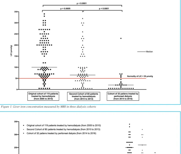
Hepatic iron load by MRI differs strikingly between peritoneal dialysis and hemodialysis patients The LIC differed between the 3 cohorts (p < 0.0001 at kruskal-Wallis test) (Table I) (Figures 1 and 2). Hepatic iron load by MRI was normal (≤ 50 µmol/g) in 81.3% of the 32 PD patients (CI: 64.3-91.5%), as compared to only 16% (CI: 10.4-23.7%) of the first HD cohort and 35% (CI: 25.4-45.9%) of the second HD cohort (p < 0.0001 versus each HD cohort; X2 test).
Iron overload by MRI was mild (50 < LIC ≤ 100 µmol/g) in 5 of the 6 PD patients with hemosiderosis (Figures 1 and 2). The sole remaining PD patient (3.1%; CI: 0-17.1%) had severe iron overload on MRI (> 200 µmol/g) (Figures 1 and 2). By comparison, MRI showed severe iron overload (> 200 µmol/g) in 30.3% of patients in the first HD cohort (CI: 22.7-39%) and 11.3% of those in the second HD cohort (CI: 5.8-20.2%) (p = 0.0033 versus the first HD cohort; X2 test) (Figure 2). None of the PD patients had moderate iron overload (100 < LIC ≤ 200 µmol/g). Interestingly, the only PD patient with severe iron overload had received IV iron (Figure 3). Iron overload by MRI was not associated with the C282Y, H63D and S65C HFE gene mutations (either ho-mozygous or heterozygous), either in the PD patients, or in the hemodialysis patients. None of the six PD patients with liver iron overload had any mutation of the HFE gene, whereas the frequency of any mutation was found low and identical in HD patients with iron overload as compared to those with normal LIC [12, 18, 23]. Of note, the lower ferritin and TSAT targets applied to the second HD cohort relative to the first HD cohort had a substantial impact on the risk of iron overload by MRI: the proportion of patients with liver iron overload (LIC > 50 µmol/g) fell from 84% in the first HD cohort to 65% in the second HD cohort (p < 0.005, X2 test) [12, 19]. Se-vere iron overload with potential clinical consequences (LIC > 200 µmol/g) also fell markedly, from 30.3% to 11.3% (p < 0.005, X2 test [12, 19]).
tion of the last iron pill [18]. Statistical analyses used the mean of three values for each iron biomarker, obtained the same month as hepatic MRI and one month before and one month after MRI for hemodialysis patients and, if available, for PD patients [12, 18-20].
Search for HFE gene mutation
To exclude a pathophysiological role of hemochroma-tosis genes, the PD patients as the HD patients with ab-normal iron load on MRI were screened for the major C282Y HFE gene mutation and minor gene mutations H63D and S65C after obtaining specific written infor-med consent for genetic analysis, in keeping with French law. Testing was performed by BIOMNIS (Lyon, France) and CERBA (Saint-Ouen-l’Aumône, France), based on allelic discrimination, using real-time Polymerase Chain Reaction (PCR)(Chimie TaqMan® ABI PRISM 7000, Roche, France) and a standardized kit [12, 18, 19].
Statistical analyses
As values did not conform to a Gaussian distribution (at Shapiro-Wilk normality test), all data are expressed as medians and ranges; percentages are given with their 95% confidence intervals calculated with the modified Wald method [22].
The different groups of patients (PD patients, first HD cohort, second HD cohort) were compared by using non-parametric analysis of variance (Kruskal-Wallis test) for continuous variables, followed by post-tests using the non-parametric Dunn test and the chi-square test for categorical variables [22].
Prism 7 software (Graphpad, San Diego, USA) was used for all tests, and p values < 0.05 were considered to de-note statistical significance [22].
RESULTS
Characteristics of the patients
The PD study cohort consisted of 32 french adults treated in the Paris region; details results for this PD cohort and for the two hemodialysis cohorts have been previously published elsewhere [12, 18, 19].
There were striking differences in anemia therapy between the peritoneal dialysis and hemodialysis pa-tients: 71.9% of the PD patients received ESA, versus 99.2% of the first HD cohort and 95% of the second HD cohort ( p < 0.0001 for PD cohort and cohort n°1 and p = 0.0018 for PD cohort and cohort n°2; X2 test) (Table I). Likewise, only 12.5% of the PD patients received IV iron, versus 95% of the first HD cohort and 85% of the second HD cohort (p < 0.0001 versus each HD cohort;
journal officiel du Registr e de D ialyse Péritonéale de Langue Française RDPLF www .rdplf.or g
journal officiel du Registr e de D ialyse Péritonéale de Langue Française RDPLF www .rdplf.or g
Table I: Characteristics and findings in 2 cohorts of hemodialysis patients and a cohort of 32 peritoneal dialysis patients
ESA: erythropoiesis-stimulating agents; IV: intravenous; MRI: Magnetic Resonance Imaging; LIC: Liver Iron Content; Values are given as median and [range]
Variables Original Cohort (HD cohort n°1)
n= 119 Second Cohort (HD cohort n°2) n= 80 PD patients (PD cohort n°3) n= 32
p value at Kruskal-Wallis test with Dunn’s post-test or at Chi2 test (comparison of cohorts 1, 2 and 3)
Age (years) [19 - 87]60 [23 - 91]70.5 [34 - 92]64.5 p = 0.0020; 1/3: p = 0.6201; 2/3: p = 0.6602; 1/2: p = 0.0013 Female sex, Percentage of patients (%) [30.4 - 47.6]38.7 [28.8 - 49.7]38.8 [30.9 - 63.6]46.9 1/3: p = 0.5233 at X 2 test; 2/3: p = 0.5639; 1/2: p = 0.8926 Dialysis vintage alysis vintage
(months) (months) [2 - 95]16 [2 - 66]8.5 [2 - 52]12.5 p = 0.0015; 1/3: p = 0.3435; 2/3: p > 0.9999; 1/2: p = 0.0011 ESA therapy, Percentage of patients (%) [94.9 - 100]99.2 [87.5 - 98.4]95 [54.5 - 84.6]71.9 1/3: p < 0.0001 at X 2 test; 2/3: p = 0.0018; 1/2: p = 0.1687 ESA dose (μg/month)/month) [0 - 566]130 [0 - 775]157.8 [0 - 150]59.1 p < 0.0001; 1/3: p < 0.0001; 2/3: p < 0.0001; 1/2: p = 0.0362
Iron therapy (IV or oral),
Percentage of patients (%) [89.2 - 97.9]95 [75.4 - 91.4]85 [22.9 - 54.8]37.5 1/3: p < 0.0001 at X 2 test; 2/3: p < 0.0001; 1/2: p = 0.0316 IV iron therapy, Percentage of patients (%) [89.2 - 97.9]95 [75.4 - 91.4]85 [4.4 - 28.7]12.5 1/3: p < 0.0001 at X 2 test; 2/3: p < 0.0001; 1/2: p = 0.0316 Oral iron, Percentage of patients (%) 0 0 [13 - 42.3]25 1/3: p < 0.0001 at X 2 test; 2/3: p < 0.0001 Charlson’s comorbidity index [2 - 16]6 [2 - 16]7 [2 - 15]5 p = 0.0186; 1/3: p > 0.9999; 2/3: p = 0.0652; 1/2: p = 0.0435 Diabetes, Percentage of patients (%) [16 - 31.1]22.7 [28.8 - 49.7]38.8 [20.3 - 51.8]34.4 1/3: p = 0.2615 at X 2 test; 2/3: p = 0.8290; 1/2: p = 0.0223 LIC at MRI (μmol/g) [5 - 340]100 [5 - 320]65 [5 - 230]20 p < 0.0001;1/3: p < 0.0001; 2/3: p < 0.0001;
1/2: p = 0.0005
Table II: Biochemical markers of iron metabolism in two cohorts of hemodialysis patients and a cohort of 32 patients treated by peritoneal dialysis
Variables Original Cohort (HD cohort n°1)
n= 119 Second Cohort (HD cohort n°2) n= 80 PD patients (PD cohort n°3) n= 32
p value at Kruskal-Wallis test with Dunn’s post-test
Hemoglobin
(g/dL) [8.4 - 15.1]12 [8.4 - 14.7]11.1 [8.7 - 16.2]11.5 p = 0.0113; 1/3: p > 0.9999, 2/3: p = 0.2496, 1/2: p = 0.0099 Serum ferritin (μg/L) [15 - 1383]265.5 [12 - 2229]145.3 [11 - 885]144 p = 0.0008; 1/3: p = 0.0225, 2/3: p > 0.9999, 1/2: p = 0.0024 Serum iron (μmol/L) [3.6 - 26.3]9.7 [4.2 - 26.3]10.6 [5.5 - 24.3]13.2 p = 0.0172; 1/3: p = 0.0141, 2/3: p = 0.1094, 1/2: p = 0.8732 Serum transferrin (g/L) [1.1 - 2.8]1.7 [1.2 - 4.5]2 [1.5 - 3.6]2.3 p < 0.0001; 1/3: p < 0.0001, 2/3: p = 0.1266, 1/2: p < 0.0001 Transferrin saturation
(TSAT)(%) [6.3 - 72.2]23.1 [6.5 - 61.2]21.6 [1.1 - 50]23.2 p = 0.3558; 1/3: p > 0.9999, 2/3: p > 0.9999, 1/2: p = 0.4743 Serum transferrin soluble
receptors (sTfR)(mg/L) [1.4 - 13]4.3 [0.5 - 12.8]5.4 [2.3 - 7.9]3.3 p = 0.0019; 1/3: p = 0.3983, 2/3: p = 0.0236, 1/2: p = 0.0122 C-reactive protein (mg/L) [0.3 - 75.9]4.3 [1 - 107.3]3.9 [1.3 - 67.6]6.7 p = 0.1551; 1/3: p = 0.2610, 2/3: p = 0.1739, 1/2: p > 0.9999
journal officiel du Registr e de D ialyse Péritonéale de Langue Française RDPLF www .rdplf.or g
Figure 1: Liver iron concentration measured by MRI in three dialysis cohorts
Differences in biological markers of iron metabolism between PD and HD patients
As suspected, the patients in the second HD cohort had lower ferritin levels, similar to those of the PD patients (Table II). The most striking differences in biological iron markers between PD and HD patients of the first cohort involved serum iron (p = 0.0141) and transferrin (p < 0.0001), despite similar levels of C-reactive protein (Table II).
DISCUSSION
We measured liver iron concentration non-invasively, by means of a validated MRI method (Signal Intensity Ratio with Rennes algorithm) [8, 21], in a cohort of 32 French PD patients, by comparison with two cohorts of French HD patients (published in 2012 and 2014). These patients were studied with the same centralized radiolo-gical method and by the same radiology team [12, 18, 19].
We observed striking differences in liver iron load between the PD and HD patients. Hepatic iron load was normal in most PD patients (81.3%) but in few HD pa-tients (16% in the first HD cohort and 35% in the second HD cohort, which had a lower ferritin target) [12, 18, 19]. Furthermore, iron overload by MRI was mild in 5
of the 6 PD patients with hemosiderosis; only one PD patient (3.1%) had severe iron overload, compared to 30.3% of patients in the first HD cohort and 11.3% of those in the second HD cohort. None of the PD patients had moderate hepatic iron overload [12, 18, 19].
MRI has made a major contribution to knowledge of iron overload disorders and to the care of non-renal pa-tients in this setting, especially by allowing “serial ra-diological biopsy” [7, 8, 24]. Quantitative MRI has also recently provided new insights into iron metabolism in hemodialysis patients, and the risk of iron overload [10-13, 16]. Liver iron determination based on signal-inten-sity-ratio MRI and the Rennes University algorithm was recently shown to accurately identify iron load in hemo-dialysis patients by comparison with liver histology, as in non-renal patients [25].
Of note, MRI has been advocated by some authors for follow-up of iron stores of dialysis patients in countries where it is fully reimbursed for diagnosis and follow-up of iron-overload disorders by the national health system such as France and many other European countries; it cost varies around 300 to 350 euros which represents about the cost of one and half dialysis session in a centre [5, 12, 13]. The case of US is more complex since the price of an MRI exam with radiologist fees is about 3000-3500 US dollars and usually not reimbursed by
journal officiel du Registr e de D ialyse Péritonéale de Langue Française RDPLF www .rdplf.or g
Figure 3: Histogram of distribution of liver iron concentration at MRI in 32 patients treated by peritoneal dialysis according to the modality of iron therapy
Medicare or Medicaid [16].
This study comparing LIC in PD and HD patients gives additional information on the respective role of ESKD and iron therapy in the pathophysiology of dialysis-as-sociated hemosiderosis: the rarity of increased liver iron content observed here in PD where IV iron is used in second (or third line) of therapy and where ferritin target is physiological as compared to HD, strongly reinforce the hypothetical role of indiscriminate use of IV iron in this setting and the lack of influence of ESKD “per se” in this clinical complication. Indeed, previous binary lo-gistic regression analyses in these two HD cohorts have shown that the main factor associated with LIC was the monthly infused iron dose (together with age, gender and hepcidin) which also represented about 33% to 37% of the variance of LIC at the Spearman correlation test (mathematically related to Pearson correlation test) [12, 19, 22]. Interestingly, in a combined statistical analysis of these two cohorts of fit HD patients devoted to iron biomarkers, ferritin was also shown to correlate to LIC (Rho = 0.52 at the Spearman correlation test) and had the best discriminatory capacity in ROC curves analysis to predict iron overload (AUC = 0.77) [20].
Moreover, in our original publication of the first HD co-hort published in 2012, we could demonstrate the culprit role of IV iron in a longitudinal follow-up of 44 hemo-dialysis patients: in 11 patients who were monitored clo-sely during parenteral iron therapy, the iron dose infused per month correlated strongly with both the overall in-crease and the monthly inin-crease in liver iron concentra-tion by MRI (respectively rho = 0.66; p = 0.0306 and rho = 0.85; p = 0.0015; Spearman test). In the 33 patients with iron overload, iron stores fell significantly after iron withdrawal or after a major reduction in the iron dose (Median LIC at first MRI: 220 µmol/g (CI: 60-340); Me-dian LIC at last MRI: 50 µmol/g (CI: 5-210); p < 0.0001, Wilcoxon’s paired test) [12]. Finally, the role of the high ferritin target in the onset of severe iron overload may also be strongly suspected in view of the reduction of occurrence of such cases in the second HD cohort where lower ferritin and TSAT targets were voluntarily applied [19].
Iron metabolism differs markedly between peritoneal dialysis and hemodialysis; in particular, PD is associated with fewer sources of iron deficiency, including blood losses directly related to the hemodialysis technique and occult gastrointestinal tract bleeding aggravated by an-ticoagulation of the hemodialysis circuits [5, 13]. This lower need for iron store replenishment explains the more conservative strategy advocated for PD patients in current guidelines, with a ferritin target of > 100 µg/L and the use of oral iron for first-line therapy [2-4, 17].
Moreover, IV iron is not recommended by the KDI-GO guideline and the ERBP position statement as an ESA-sparing agent for PD patients, contrary to HD pa-tients, but is reserved for PD patients who do not tolerate or respond poorly to oral iron, and for PD patients with high iron requirements [4, 17]. This explains why only a small fraction of PD patients in France and many other countries receive IV iron despite its efficacy [26, 27, 28]. The rationale of this study was that a comparison of li-ver iron content by MRI between peritoneal dialysis and hemodialysis patients would reflect the reciprocal in-fluence of uremic status itself and parenteral iron therapy on liver iron metabolism. The normal liver iron content observed here in most of our PD patients as compared to far fewer HD patients strongly supports and reinforces the role of excessive IV iron infusion and high ferritin targets in iatrogenic iron overload recently observed in a large proportion of hemodialysis patients (up to 66%) [9-14, 16].
The normal serum transferrin levels observed here in PD patients, versus the low levels seen in HD patients, des-pite similar levels of C-reactive protein, calls for further studies of the possible negative relationship between in-fused IV iron, excess iron stores and transferritinemia in ESKD patients, as suggested several years ago by Descombes and Fellay, who linked hypotransferrinemia to increased ferritin levels beside the classical role of inflammation and uremia per se on transferrin [29-31]. Finally, the few cases of mild iron overload in PD pa-tients treated by oral iron suggest the possibility in ESKD of iron overload related to excessive ingestion of oral iron medication as encountered in healthy subjects [7], together with the ability of erythropoietin to increase iron intestinal absorption in ESKD patients as shown in a rat model of chronic renal failure [32]. This suggests the need of a cautious follow-up of LIC of ESKD pa-tients treated by the new iron-derived phosphate binders some of which are absorbed [13].
CONCLUSION
Contrary to HD patients, iron overload by MRI is rare and mostly mild in PD patients. The normal liver iron content observed here in most PD patients strongly rein-forces the role of excessive IV iron infusion and high ferritin targets in the pathophysiology of iatrogenic iron overload recently observed in a large proportion of he-modialysis patients.
DISCLOSURE
The authors have no conflict of interest to declare.
journal officiel du Registr e de D ialyse Péritonéale de Langue Française RDPLF www .rdplf.or g
Authors’ contributions
Guy Rostoker contributed to the conception, design and
supervision of the study; data acquisition, and planning and conduct of the study in the peritoneal dialysis unit of the Hôpital Privé Claude Galien, Division of Nephro-logy and Dialysis. He supervised the statistical analysis, data interpretation and reporting of the work, and write the article.
Nasredine Ghali contributed to the study design and data
acquisition, and to the planning and conduct of the study in the peritoneal dialysis unit of the Centre Hospitalier Marc Jacquet, Division of Nephrology and Dialysis.
Séverine Beaudreuil contributed to data acquisition and
to the planning and conduct of the study in the peritoneal dialysis unit of the Bicêtre Hospital, Division of Nephro-logy, Dialysis and Transplantation.
Mireille Griuncelli contributed to data acquisition,
ana-lysis and interpretation, and to the statistical anaana-lysis, and prepared the tables and figures.
Yves Cohen contributed to the acquisition and analysis of
centralized MRI exams.
Belkacem Issad contributed to the conception, design
and supervision of the study; data acquisition, and plan-ning and conduct of the study in the peritoneal dialysis unit of the, Groupe Hospitalier Pitié-Salpêtrière, Divi-sion of Nephrology and Dialysis. He also participated in the writing of the article.
REFERENCES
1. Hörl WH. Clinical aspects of iron use in the anemia of kidney disease. J Am Soc Nephrol. 2007 Feb;18(2):382-93.
2. KDOQI; National Kidney Foundation. Clinical pract ice guidelines and clinical practice recommendations for anemia in chronic kidney disease in adults. Am J Kidney Dis. 2006 May; 47(5 Suppl. 3):Sl6-85.
3. Locatelli F, Covic A, Eckardt KU, Wiecek A, Vanhol-der R and on behalf of the ERA-EDTA ERBP Advisory Board. Anaemia management in patients with chronic kidney disease: a position statement by the Anaemia Working Group of European Renal Best Practice (ERBP). Nephrol Dial Transplant. 2009 Feb;24(2):348-54.
4. KDIGO Clinical practice Guideline for anemia in chronic kidney disease. Kidney Int Suppl. 2012;2:279-335.
5. Rottembourg J, Rostoker G. Use of intravenous iron supplementation in chronic kidney disease: interests, li-mits, and recommendations for a better practice. Nephrol Ther. 2015 Dec;11(7):531-42.
6. Macdougall IC, Bircher AJ, Eckardt KU, et Al; Conference participants. Iron management in chronic kidney disease: conclusions from a « Kidney Disease: Improving Global Outcomes» (KDIGO) Controversies Conference. Kidney Int. 2016 Jan;89(1):28-39.
7. Barton JC, Edwards CQ, Phatak PD, Britton RS, Ba-con BR. Handbook of Iron overload disorders. Cam-bridge University Press; 2010. ISBN 978-0-521-87343-7.
8. Paisant A, d’Assignies G, Bannier E, Bardou-Jac-quet E, Gandon Y. MRI for the measurement of liver iron content, and for the diagnosis and follow-up of iron overload disorders. Presse Med. 2017 Dec;46(12P-t2):e279-e287.
9. Canavese C, Bergamo D, Ciccone G, et al. Validation of serum ferritin values by magnetic susceptometry in predicting iron overload in dialysis patients. Kidney Int. 2004 Mar;65(3):1091-8.
10. Ghoti H, Rachmilewitz EA, Simon-Lopez R, et al. Evidence for tissue iron overload in long-term hemodia-lysis patients and the impact of withdrawing parenteral iron. Eur J Haematol. 2012 Jul;89(1):87-93.
11. Ferrari P, Kulkarni H, Dheda S, et al. Serum iron markers are inadequate for guiding iron repletion in chronic kidney disease. Clin J Am Soc Nephrol. 2011 Jan;6(1):77-83.
12. Rostoker G, Griuncelli M, Loridon C, et al. Hemo-dialysis-associated hemosiderosis in the era of erythro-poiesis-stimulating agents: a MRI study. Am J Med. 2012 Oct;125(10):991-999.e1.
13. Rostoker G, Vaziri ND, Fishbane S. Iatrogenic iron overload in dialysis patients at the beginning of the 21st century. Drugs. 2016 May;76(7):741-57.
14. Vaziri ND. Epidemic of iron overload in dialysis po-pulation caused by intravenous iron products: a plea for moderation. Am J Med. 2012 Oct;125(10):951-2.
journal officiel du Registr e de D ialyse Péritonéale de Langue Française RDPLF www .rdplf.or g
15. Gaweda AE, Ginzburg YZ, Chait Y, Germin MJ, Aronoff GR, Rachmilewitz E. Iron dosing in kidney di-sease: inconsistency of evidence and clinical practice. Nephrol Dial Transplant. 2015 Feb;30(2):187-96. 16. Rostoker G, Vaziri ND. Risk of iron overload with chronic indiscriminate use of intravenous iron pro-ducts in ESRD and IBD populations. Heliyon. 2019 Jul;5(7):e02045.
17. Locatelli F, Bárány P, Covic A, et al and on behalf of the ERA-EDTA ERBP Advisory Board. Kidney di-sease: Improving global outcomes guidelines on anae-mia management in chronic kidney disease: a European Renal Best Practice position statement. Nephrol Dial Transplant. 2013 Jun;28(6):1346-59.
18. Issad B, Ghali N, Beaudreuil S, Griuncelli M, Cohen Y, Rostoker G. Hepatic iron load at magnetic resonance imaging is normal in most patients receiving peritoneal dialysis. Kidney Int Rep. 2017 Jul;2(6):1219-1222. 19. Rostoker G, Griuncelli M, Loridon C, et al. Maxi-mal standard dose of parenteral iron for hemodialysis patients: an MRI-based decision tree learning analysis. PloS One. 2014 Dec;9(12):e115096.
20. Rostoker G, Griuncelli M, Loridon C, et al. Reas-sessment of iron biomarkers for prediction of dialy-sis iron overload : an MRI study. PloS One. 2015 Jul 16;10(7):e0132006.
21. Gandon Y, Olivié D, Guyader D, et al. Non-invasive assessment of hepatic iron stores by MRI. Lancet. 2004 Jan;363(9406):357-62.
22. Sheskin DJ. Handbook of parametric and nonpa-rametric statistical procedures. 4th ed. Boca Raton, USA: Chapman and Hall, Taylor and Francis Group; 2007.
23. Rostoker G, Griuncelli M, CohenY. HFE gene mutations are not risk factors for iron overload in
Eu-ropean hemodialysis patients. Hemodialysis Int. 2017 Jul;21(3):440-442.
24. Rostoker G. The changing landscape of iron over-load disorders at the beginning of the 21st century. Presse Med. 2017 Dec;46(12Pt2):e269-e271
25. Rostoker G, Laroudie M, Blanc R, et al. Signal-inten-sity-ratio MRI accurately estimates hepatic iron load in hemodialysis patients. Heliyon. 2017 Jan;3(1):e00226. 26. Wish JB. Intravenous iron: not just for hemo-dialysis patients anymore. Perit Dial Int. 2008 Mar-Apr;28(2):126-9.
27. Li H, Wang SX. Intravenous iron sucrose in peri-toneal dialysis patients with renal anemia. Perit Dial Int. 2008 Mar-Apr;28(2):149-54.
28. Issad B, Griuncelli M, Verger C, et al. What do we learn about the “Anemia Module” of the French language Peritoneal Dialysis ? Interest and Results. Bulletin De la Dialyse à Domicile. September 2019; (3):143-9 [On line] URL : https://doi.org/10.25796/bdd.v2i3.20983 [consulted 2019/11/20].
29. Mercadal L, Metzger M, Haymann JP, et al. A 3-mar-ker index improves the identification of iron disorders in CKD anaemia. PLoS One. 2014 Feb;9(2):e84144. 30. Kirschbaum B. Hypotransferrinemia of chro-nically hemodialyzed patients. Artif Organs. 1999 Dec;23(12):1047-54.
31. Descombes E, Fellay G. Hypotransferrinemia in chronic hemodialyzed (HD) patients. Artif Organs. 2000 Dec;24(12):988-9.
32. Srai SK, Chung B, Marks J, et al. Erythropoietin re-gulates intestinal iron absorption in a rat model of chro-nic renal failure. Kidney Int. 2010 Oct;78(7):660-7.
Received 10/10/19, accepted after revision 19/11/25, published 19/12/15 journal officiel du Registr e de D ialyse Péritonéale de Langue Française RDPLF www .rdplf.or g
Open Access This article is licensed under a Creative Commons Attribution 4.0 International License, which permits use, sharing, adaptation, distribution and reproduction in any medium or
format, as long as you give appropriate credit to the original author(s) and the source, provide a link to the Creative Commons license, and indicate if changes were made. The images or other third party material in this
article are included in the article’s Creative Commons license, unless indicated otherwise in a credit line to the material. If material is not included in the article’s Creative Commons license and your intended use is not permitted by statutory regulation or exceeds the permitted use, you will need to obtain permission directly from the