A new species of the primitive
dinosaur
Thecodontosaurus
(Saurischia: Sauropodomorpha)
and its implications for the
systematics of early dinosaurs
Adam M. Yates
Department of Earth Sciences, University of Bristol, Bristol, BS8 1RJ, UK
SYNOPSIS Juvenile sauropodomorph specimens from a Late Triassic/Early Jurassic fissure fill in
Pant-y-ffynnon Quarry, South Wales are redescribed and named as a new species, Thecodontosaurus caducus. T. caducus can be diagnosed by the presence of pleurocoel-like pits on the neurocentral sutures of the sixth, seventh and eighth cervical vertebrae. It is further distinguished from the type species of the genus, T. antiquus, by the primitive shape of its proximal humerus and ilium. Data from this specimen are incorporated into a cladistic analysis of basal sauropodomorph relationships. It is found that Thecodontosaurus is basal to all other sauropodomorphs, with the exception of Saturnalia from the late Carnian of Brazil. As such Thecodontosaurus is a key taxon, with a novel combination of characters that has important implications for early dinosaur phylogenetics. Thecodontosaurus provides evidence that ‘prosauropods’ are paraphyletic with respect to Sauropoda and that Herrera-sauridae lies outside the clade containing Sauropodomorpha + Theropoda.
KEY WORDS Thecodontosaurus, T. caducus, Cladistic analysis, Sauropodomorpha, Prosauropoda,
Paraphyletic
Contents
Introduction 2
Systematic palaeontology 2
SAURISCHIA Seeley, 1888 2
SAUROPODOMORPHA von Huene, 1932 2
Genus THECODONTOSAURUS Riley and Stutchbury, 1836 2
Thecodontosaurus caducus sp. nov. 2
Description 4 Skull roof 5 Palate 8 Braincase 9 Mandible 10 Vertebral column 11 Forelimb 15 Hindlimb 16 Skeletal reconstruction 20 Cladistic analysis 21 Methods 21 Results 22 Prosauropod monophyly 26 Acknowledgements 31 References 31
Appendix 1: list of anatomical abbreviations 33
Appendix 2: list of characters 34
Appendix 3: list of unambiguous synapomorphies 40
Introduction
The remains of several juvenile sauropodomorphs were found in Pant-y-ffynnon Quarry in South Wales by Professor K. Kermack and Dr P. Robinson in 1952. The material is part of an Upper Triassic assemblage found in fine-grained sand-stone that filled a fissure in the Carboniferous Limesand-stone of the Quarry. The sauropodomorph specimens include a disar-ticulated skull with associated forelimb elements and cervical series, isolated skull elements and several postcranial bones, including a partial hind limb. The skull was reconstructed and described as Thecodontosaurus sp. by D. Kermack (1984). The postcranial remains have been featured in skeletal re-constructions (Kermack 1984; Galton 1990; Upchurch 1997; Benton et al. 2000) but remain undescribed.
Thecodontosaurus was the first sauropodomorph
dino-saur to be scientifically described (Riley & Stutchbury 1836), but its anatomy has remained poorly known relative to other sauropodomorph dinosaurs. What is known is that, with an adult length of no more than 3 m (Benton et al. 2000), it is one of the smallest and most gracile members of the Sauropodo-morpha. Some authors claim that it retained an obligatory bipedal posture during locomotion (Kermack 1984; Galton 1990, 2000; Benton et al. 2000) but it certainly did not re-tain a predatory lifestyle. It had the typical sauropodomorph dental specialisations that suggest it included a high propor-tion of vegetable matter in its diet (Galton 1985a; Crompton & Attridge 1986).
The type species, T. antiquus Morris, 1843, was based on largely disarticulated bones found in a Late Triassic fissure fill deposit from Bristol in south-west England. Many of these bones were lost during the Second World War but hundreds still survive and these have been redescribed recently (Benton
et al. 2000). More bones from another locality in south-west
England are known (Whiteside 1983) and these are currently being prepared and studied.
The importance of Thecodontosaurus lies in its basal position within sauropodomorph phylogeny. It has either been thought of as the sister group of all other sauropodo-morphs (Gauthier 1986) or as the basal member of a mono-phyletic Prosauropoda (Galton 1990). The only computer-based, cladistic analysis to include Thecodontosaurus found it to be part of a monophyletic prosauropod group but was unable to resolve the position of the genus within this clade (Benton et al. 2000).
In this paper, the Pant-y-ffynon prosauropod specimens are fully described and illustrated and their relationship to
T. antiquus and other early sauropodomorphs is investigated
using cladistic analysis. The implications of this analysis for early dinosaur systematics are discussed. In particular the case for prosauropod monophyly is examined in detail.
The abbreviations for the various institutions where material discussed in this paper is held are as follows:
AM = Amherst College Museum, Massachusetts, USA.
BMNH = Natural History Museum, London, UK. BRSUG= Department of Earth Sciences, University of
Bristol, UK.
GPIT = Institut und Museum f¨ur Geologie und Pal¨aontologie der Universit¨at T¨ubingen, Germany.
HMN = Museum f¨ur Naturkunde der Humboldt Uni-versit¨at, Berlin, Germany.
PVL = Fundaci´on ‘Miguel Lillo’, Tucum´an, Argentina. SMNS= Staatliches Museum f¨ur Naturkunde, Stuttgart,
Germany.
Systematic palaeontology
SAURISCHIA Seeley, 1888
SAUROPODOMORPHA von Huene, 1932
Genus THECODONTOSAURUS Riley and
Stutchbury, 1836
TYPE SPECIES. Thecodontosaurus antiquus Morris, 1843; Late Triassic, Bristol, England.
DIAGNOSIS. Small, gracile sauropodomorphs with the fol-lowing derived character states.
1. The basipterygoid processes are elongate and slender, with the length of the process, measured from its tip to the dorsal margin of the parabasisphenoid, being equal to the height of the braincase, measured from the dorsal margin of the parabasisphenoid to the top of the supraoccipital (convergent in ‘Efraasia diagnostica’).
2. The dentary is short and deep, occupying less than 40% of the total mandibular length, and with a maximum dor-soventral depth that is greater than 20% of its length (con-vergent in Saturnalia tupiniquim).
3. The epipophyses of the cranial cervicals are flat plates that overhang the caudal margins of the postzygapophyseal facets but do not form raised ridges on the dorsal surface of the postzygapophysis.
4. The proximal and mid-caudal neural spines are positioned at the extreme caudal end of their neural arches, filling the interpostzygapophyseal space (convergent in ‘Efraasia
diagnostica’).
5. The ventral furrowing of the caudal centra is reduced so that it is only weakly present in the proximal caudals and is absent altogether from the mid and distal caudals. REMARKS. The first two characters of the diagnosis are from Benton et al. (2000) while the last three are novel. The third character listed in Benton et al. (2000), ‘caudal process of the iliac blade subquadratic’ is also present in the basal saurischian Guiabasaurus candelariensis (Bonaparte et al. 1999), Neotheropoda (e.g. Dilophosaurus wetherilli Welles, 1984) and ‘Efraasia diagnostica’ (pers. obs. SMNS 12354, 12667). Consequently it is interpreted as a plesiomorphic characteristic of Thecodontosaurus.
Thecodontosaurus caducus sp. nov.
ETYMOLOGY. Latin, caducus, fallen. Refers to the fact that the holotype is an articulated specimen preserved in a fissure fill, indicating that the animal may have fallen into the fissure and died there.
HOLOTYPE. BMNH P24, a nearly complete but disarticulated skull, both mandibular rami, a complete series of cervical vertebrae, the proximal ends of both humeri, a proximal right scapula and both coracoids from one individual (Fig. 1).
Figure 1 Thecodontosaurus caducus sp. nov., holotype, BMNH P24; skull and partial postcranial skeleton. 1A, photograph; 1B, interpretive line drawing. Solid black bones represent unrelated crocodylomorph and lepidosaur bones. For abbreviations, see Appendix 1.
Scale bars= 20 mm.
PARATYPES. BMNH P24/3, a right ischium; BMNH P39/2, a left coracoid; BMNH P59/5, a right quadrate; BMNH P64/1, a series of eight proximal–mid caudals; BMNH P65/21, a right ectopterygoid; BMNH P77/1, a series of distal caudal vertebrae, the right ilium, femur, tibia, fibula and pes; BMNH P126/1, a ?proximal pubis; BMNH P141/1, a basioccipital.
TYPE HORIZON AND LOCALITY. Late Triassic fissure deposits, Pant-y-ffynnon Quarry, near Bonvilston, South Wales. The age of the Mesozoic fissure deposits is difficult to determine. Given the faunal similarities between Pant-y-ffynnon and the Thecodontosaurus-bearing fissure fills from south-west England, they are likely to be of a similar age. The presence
Figure 2 Thecodontosaurus caducus sp. nov., holotype, BMNH P24; reconstruction of skull. 2A, lateral view; 2B, ventral view; 2C, dorsal view; 2D, ventral view of mandibular ramus. For abbreviations see Appendix 1. Scale bar= 10 mm.
of phytosaurs in the English fissure fills constrains their age to the Late Triassic (Benton et al. 2000) while palynomorphs may indicate a Rhaetian age (Whiteside 1983). A fuller ac-count is given in Benton & Spencer (1995).
DIAGNOSIS. A species of Thecodontosaurus with the follow-ing autapomorphy: pleurocoel-like pits on the neurocentral sutures of the sixth, seventh and eighth cervical vertebrae.
T. caducus can be further distinguished from T. antiquus by
exhibiting the plesiomorphic state for the autapomorphies of that species. These include a medial tubercle of the
prox-imal humerus that does not project strongly (versus strongly projecting in T. antiquus) and a preacetabular process of the ilium that projects cranially (versus a downcurved preacetab-ular process in T. antiquus).
Description
The most complete specimen in the collection is the holotype, BMNH P24. Except where mentioned, the description of
the skull, mandible and cervical vertebrae is based on this specimen.
The dorsal skull roof of this specimen is situated at the cranial end of the cervical series, while the elements from the left temporal region, mandible, palate and braincase are scattered for some distance to the left of the dorsal skull roof. All of the braincase elements have separated, indicating that suturing had not begun at the time of death, one of the many juvenile characteristics that can be seen in these specimens. The reconstruction of the skull presented here (Fig. 2) differs somewhat from that of Kermack (1984). This is partly be-cause some bones have been re-identified and partly bebe-cause missing, or damaged, parts were restored using shapes more similar to those known in other early saurischians.
Skull roof
PremaxillaThe medial surface of the right premaxilla is exposed. Its surface is damaged and the nasal process is missing. The main body of the premaxilla is quite short (8 mm long and 6 mm high) and bears four teeth. Like juvenile
Massospon-dylus carinatus (Cooper 1981) and Mussaurus patagonicus
(Bonaparte & Vince 1979), the height of the premaxillary teeth exceeds the height of the maxillary teeth. The crowns are simple subcylindrical spikes that bear a few weak serra-tions on the caudal carina (there are six such serraserra-tions on the second tooth). The first crown is the largest at 4.5 mm in height.
Maxilla (Fig. 3)
Only a fragment of the left maxilla is available for study. It is quite poorly preserved so details are hard to discern. The preserved portion, which is 25 mm long, consists of the
Figure 3 Thecodontosaurus caducus sp. nov., holotype, BMNH P24; left maxilla in lateral aspect.3A, photograph; 3B, interpretive drawing. nv.c= neurovascular canal, nv.f = neurovascular foramen. Scale bar= 5 mm.
caudal ramus and what is probably the base of the ascending ramus. The ascending ramus itself and the rostral ramus are missing. The specimen is 4 mm high at the rostral end and tapers to a point at its caudal end. The number of alveoli can-not be determined but there are six teeth present and space for four more. On the lateral surface there appear to be five relatively large, neurovascular foramina. The caudal-most foramen is smaller than the more rostral foramina. In most other sauropodomorphs (e.g. Riojasaurus incertus: Bona-parte & Pumares 1995; Plateosaurus engelhardti: pers. obs. of HMN MB. 1927.19.1, GPIT Skelett 1) the foramen at the caudal end of the row is distinctly larger than the rest of the maxillary foramina. This includes sauropods, where the caudal maxillary foramen is so enlarged it has been termed the pre-antorbital fenestra (Wilson & Sereno 1998).
A sharp edge (the ventral rim of the external antor-bital fenestra) delimits the lateral surface of the caudal ra-mus from the dorsal surface. The dorsal surface bears a short, longitudinal groove that extends from near the mid-point to a mid-point above the fourth preserved tooth. This groove lies on the floor of the antorbital fossa and would have housed the maxillary nerve and associated vascu-lature (Witmer 1997). A similar groove has been repor-ted in Plateosaurus engelhardti (Witmer 1997), Sellosaurus
gracilis? (Galton 1985b) and Massospondylus carinatus
(Gow et al. 1990). The groove becomes closed over by the jugal and the lacrimal to form a canal in ornithischians (Witmer 1997), whereas a foramen, or a series of foramina, pierce the dorsal or medial surface of the maxilla in sauro-pods and non-avian therosauro-pods (Witmer 1997). Therefore, a dorsally open canal for the maxillary nerve on the ventral surface of the antorbital fossa is potentially a synapomorphy uniting the traditional Prosauropoda into a monophyletic group.
The maxillary teeth are poorly preserved and those de-tails that are present appear to be similar to the dentary teeth. The largest crown is 3 mm high.
Nasal
Both nasals have been fractured and distorted by compression against the underlying bones. Consequently many details are lost. However, the natural edge of the caudal margin is pre-served and indicates that the suture with the frontals was con-cave, as it is in primitive dinosaurs such as Coelophysis bauri (Colbert 1989) and Lesothosaurus diagnosticus (Sereno 1991a). Some derived sauropodomorphs such as
Plateo-saurus engelhardti, have a caudally convex naso-frontal
suture. There is no indication that there was a median nasal depression as is present in some specimens of
Plateo-saurus engelhardti (Galton 1984a) and ‘Efraasia diagnost-ica’ (Galton 1985b as Sellosaurus gracilis). The rostroventral
process is missing from the main body of each nasal.
Prefrontal
The dorsal surface of the main body of the left prefrontal is exposed. It is a small, dorsally facing, elliptical plate, 11 mm long and 6 mm wide, that would have formed part of the skull roof behind the lacrimal. It is not enlarged, as it is in other basal sauropodomorphs such as Massospondylus carinatus (Cooper 1981) and Plateosaurus engelhardti (Galton 1984a). The right prefrontal is still articulated with the lacrimal. Most of the main body is missing, but a short, thin descending
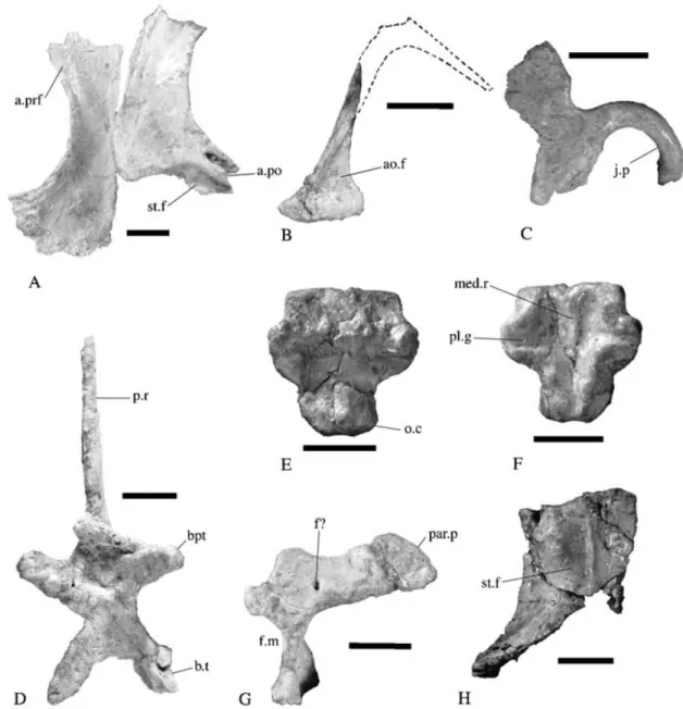
Figure 4 Thecodontosaurus caducus sp. nov., holotype, BMNH P24; elements of the skull. 4A, pair of frontals in dorsal aspect; 4B, left lacrimal in lateral aspect (dorsal outline restored from right lacrimal);4C, right ectopterygoid in dorsal aspect; 4D, parabasisphenoid complex in ventral aspect;4E, basioccipital in ventral aspect; 4F, basioccipital in dorsal aspect; 4G, right exoccipital-opisthotic complex in occipital view; 4H, left parietal in dorsal aspect. For abbreviations see Appendix 1. Scale bars= 5 mm.
process arises from the caudolateral margin and extends about halfway down the medial side of the ventral ramus of the lacrimal. The dorsal exposure of the prefrontal is much greater than that of the lacrimal.
Frontal (Fig. 4A)
Both frontals are well preserved and are visible in dorsal view. Each frontal is longer than it is wide, with the maximum width developed at the caudal end. A deep, rostromedially inclined slot, for the reception of the frontal ramus of the postorbital, is incised into the tip of the caudolateral corner of the right frontal (this region is damaged in the left frontal). There is a faintly raised area medial to this slot, present on both frontals. The supratemporal fossa extends onto the frontals, producing a sharply defined, crescentic depression
on the caudal margin of each frontal. The midsection of the frontals, which forms the roof over the orbits, is constricted transversely. The rostral end is expanded transversely, but not as greatly as the caudal. A facet for the articulation of the pre-frontal occupies the rostral third of the lateral margin. Thus, the prefrontal does not restrict the frontal contribution to the orbital margin as it does in more derived sauropodomorphs such as Plateosaurus engelhardti (Galton 1984a) and
Lufen-gosaurus huenei (Young 1941a). A row of foramina occurs
on each side of the median frontal symphysis.
Parietal (Fig. 4H)
The parietals had separated before burial, indicating that they were not fused or tightly sutured together, which is a sign of immaturity. As in Plateosaurus engelhardti the parietals comprise a rectangular rostral portion that forms the caudal
end of the dorsal skull roof and a caudolateral wing that sutures with the squamosal. The rostral end of the parietals forms a straight suture with the frontals in dorsal view. Faint ridges, on either side of the midline, mark the medial mar-gins of the supratemporal fossae. Each ridge is confluent with the sharper ridge that bounds the rostral margin of the supratemporal fossa on the frontal. Medial to the ridges, the parietals form a flat, horizontal surface. Lateral to each ridge the parietal curves ventrally to meet the lateral walls of the braincase. The caudolateral wings are more steeply in-clined than the lateral sides of the rostral end of the parietal. A scar marking the articulation with the squamosal occupies the distal half of the lateral surface of this wing. In lateral view the caudolateral wing curves ventrally so that the squamosal and, consequently, the quadrate head, would have been held below the level of the dorsal skull roof.
Lacrimal (Fig. 4B)
The bone identified by Kermack (1984) as the right squamosal is re-interpreted here as the right prefrontal and right lacrimal, exposed on their medial side. The lacrimal is an approximately L-shaped bone with a long ventral ramus (26 mm) and a short rostral ramus (8 mm). A narrow strip of the rostral ramus is exposed dorsolaterally on the skull roof, rostral to the prefrontal and lateral to the nasal. A sulcus extends up the dorsal half of the caudal face of the ven-tral ramus, medial to the venven-tral process of the prefrontal. A single lacrimal foramen is situated at the dorsal end of the sulcus. The rostral opening of the lacrimal foramen cannot be seen. The short rostral ramus sutures with the ascending ramus of the maxilla. The ventral ramus is quite narrow at its midpoint (1 mm) but flares rostrocaudally at its ventral end (6 mm). Dislocation has made it impossible to determine if the ventral end of the lacrimal contacted the caudal ramus of the maxilla.
As in other basal sauropodomorphs (e.g. Plateosaurus
engelhardti: Galton 1984a) and the basal saurischian, Her-rerasaurus ischigualastensis (Sereno & Novas 1993), the
lacrimal formed a lateral wall over the caudodorsal corner of the antorbital sinus, whereas the caudoventral corner of the sinus extended over the lateral surface of the lacrimal, to form a laterally facing fossa. In most basal sauropodomorphs (e.g.
Plateosaurus engelhardti: Galton 1984a) this fossa is small
and restricted to the rostroventral corner of the main vent-ral ramus of the lacrimal. In T. caducus, however, the fossa extends at least half way up the ventral ramus. This condi-tion is probably the plesiomorphic one, since it is also seen in neotheropods and the basal saurischians Herrerasaurus
ischigualastensis (Sereno & Novas 1993) and Eoraptor lunensis (Sereno et al. 1993).
Jugal
Both jugals are obscured below overlying bones. Only parts of the lateral side of the left jugal are visible. It is estimated to be about 30 mm in length. The suborbital portion of the jugal is a slender, medio-laterally compressed bar that is 30 mm deep in the mid-orbital region. In keeping with the relatively large size of the orbit of this juvenile, the postorbital ramus is placed far back along the jugal (the rostral end of its base is approximately 23 mm from the rostral end of the jugal). The postorbital process is triangular with a relatively broad base. The ventral margin of the jugal is gently arched
upwards. No details of the maxillary, lacrimal, postorbital and quadratojugal articulations are visible.
Postorbital
The left postorbital is visible in lateral view, while the medial side of the right postorbital is exposed. The postorbital is tri-radiate with long jugal and frontal rami and a short squamosal ramus. The frontal ramus is steeply inclined anterodorsally from its junction with the other rami to the frontal. The ra-mus also curves medially to articulate with the frontal but this curvature is not as strong as in Plateosaurus engelhardti (Galton 1984a). The frontal ramus becomes broader towards its rostral end, which is slightly forked. The supratemporal fossa extends onto the dorsomedial surface of this ramus as it does in other basal saurischians such as Herrerasaurus
ischigualastensis (Sereno & Novas 1993) and Plateosaurus engelhardti (Galton 1984a). The squamosal ramus was a
short, slender and pointed process that was probably hori-zontally oriented. The jugal ramus is an elongate strap that is mediolaterally compressed and gently bowed caudally. Bones overlying both of the postorbitals obscure the articu-lation facets for the jugals.
Quadratojugal
Neither of the two quadratojugals can be positively identified; however, a small plate of bone protruding from beneath the right quadrate head is likely to be the main body of the right quadratojugal.
Quadrate (Fig. 5)
Both quadrates of BMNH P24 and an isolated right quad-rate, BMNH P59/5, can be viewed in their medial and caudal aspect. The main body of the quadrate consists of two inae set at right angles to each other. Where the two lam-inae meet along the caudal edge, a sharp keel is formed. This keel extends dorsally to the small knob-like quadrate head. A large semi-circular lamina extends rostromedially
Figure 5 Thecodontosaurus caducus sp. nov., BMNH P59/5; right quadrate.5A, medial, 5B, caudal; 5C, mandibular condyle. Scale bar= 5 mm.
and forms the pterygoid wing, while the narrower, rostro-laterally directed lamina thickens ventrally to form the shaft that bears the quadrate condyles. The base of the pterygoid wing is long, occupying more than 70% of the length of the quadrate. This is a primitive character state that is also seen in ‘Efraasia diagnostica’ (Galton 1985b as Sellosaurus
gracilis) but not in more derived sauropodomorphs such as Plateosaurus engelhardti (pers. obs. of SMNS 12950), Col-oradisaurus brevis (from photographs of PVL 3967) or most
sauropods (e.g. Camarasaurus lentus: Madsen et al. 1995). Unlike most other early saurischians (e.g. Herrerasaurus
ischigualastensis: Sereno & Novas 1993; Liliensternus lilien-sterni: pers. obs. of HMN MB.R.2175.7.4; Sinraptor dongi:
Currie & Zhao 1993; ‘Efraasia diagnostica’: pers. obs. of SMNS 12668; Plateosaurus engelhardti: pers. obs. of GPIT Skelett 1) the quadrate foramen is not deeply incised into the lateral margin of the rostrolateral lamina. If a quadrate fora-men was present, it would have been a narrow gap between the quadrate and quadratojugal such as in Lesothosaurus
diagnosticus (Sereno 1991a) and Heterodontosaurus tucki
(Weishampel & Witmer 1990). The articular surface is nar-rowly triangular in ventral view with the long axis oriented transversely and the apex pointing laterally. An oblique sul-cus running antero-medially divides the articular surface into two condyles, of which the more medial is taller.
Squamosal
The bone lying under the caudal end of the basisphenoid and the paroccipital process of the right exoccipital–opisthotic complex appears to be the left squamosal exposed in dor-somedial aspect (identified as part of the ?opisthotic by Kermack 1984). The squamosal head is subrectangular in dorsal view unlike the triangular shape that is usual amongst dinosaurs, including other basal sauropodomorphs such as
Plateosaurus engelhardti (Galton 1984a). Like other
dino-saurian squamosals the complete bone would have consisted of four rami, however, only two of these can be seen in this specimen. The rostromedially directed parietal ramus is short, slender and distinctly raised above the dorsal surface of the squamosal head. The rostrolaterally directed postorbital ramus has broken away. Caudal to the base of the parietal ramus, the base of a slender, caudoventrally directed, quad-rate ramus can be seen. The overlying paroccipital process of the right exoccipital–opisthotic complex obscures the caudal ramus of the squamosal.
Palate
PterygoidMost of the right pterygoid is exposed in ventral and me-dial views, while only a fragment of the transverse flange remains of the left pterygoid. The pterygoid is a complex bone, consisting of three main projections: the rostral ramus, the quadrate ramus and the transverse flange. The rostral ramus was the longest of these, measuring 25.5 mm long as preserved. It is an elongate triangular plate that, in life, would have faced ventromedially and formed a large part of the palate. Its medial margin is almost straight, with the rostral end forming a median symphyseal surface. The me-dial margin is flared upwards in this region so that when the two pterygoids were in contact a low, dorsally project-ing, median crest was formed. The base of the transverse flange forms the caudolateral margin of the rostral ramus.
The pterygoid is bent downwards sharply along this line so that the transverse flange faces more or less rostroventrally. The flange itself is a short, subrectangular process that is dir-ected laterally in ventral view. Judging from the shape of the cross-section of the left pterygoid fragment, the transverse flange was gently curved about the transverse axis so that the caudodorsal surface was concave. It is 11 mm long along its caudal margin. The quadrate ramus is a short, vertical, triangular plate that is 7 mm long and is directed dorsally and laterally. It flares distally from its narrow, waist-like junction with the rest of the pterygoid, at the caudomedial corner of the rostral ramus and the transverse flange. Perhaps the most significant feature of the pterygoid is the absence of a caudomedial flange that hooks around the basiptery-goid process to contact its counterpart medially. Most dino-saurs have such a flange (e.g. Plateosaurus engelhardti: pers. obs. of HMN 24; Lesothosaurus diagnosticus: Sereno 1991a;
Sinraptor dongi: Currie & Zhao 1993) although it is reduced
to a small dorsomedially oriented hook, or is absent alto-gether, in eusauropods (Wilson & Sereno 1998).
Ectopterygoid (Figs 4C, 6)
The right ectopterygoid is visible in dorsal view, while the ventral view can be seen in an isolated right ectopterygoid (BMNH P65/21). The main body is twice as long as it is wide (10 mm long) with a sinuous medial margin. A deep concav-ity occupies the ventral surface and closely resembles the ventral pneumatic fossa of neotheropods. The jugal process is slender and strongly recurved. In this respect the ecto-pterygoid resembles that of a neotheropod more than any other sauropodomorph.
Palatine
A flat, roughly quadrangular sheet of bone exposed between the right pterygoid, the left ectopterygoid and the supraoccipital is probably the left palatine, exposed
latero-Figure 6 Thecodontosaurus caducus sp. nov., BMNH P65/21; right ectopterygoid in ventral aspect. j.p= jugal process, v.f = ventral fossa. Scale bar= 5 mm.
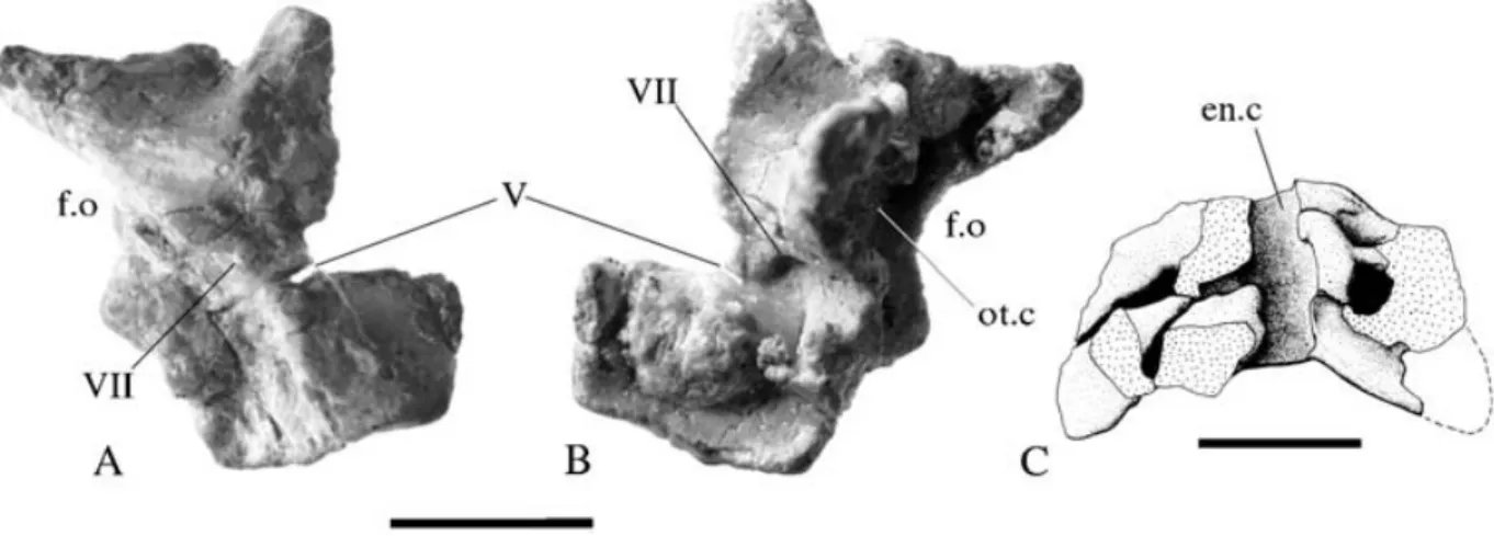
Figure 7 Thecodontosaurus caducus sp. nov., holotype, BMNH P24. 7A, right prootic in lateral aspect; 7B, right prootic in medial aspect; 7C, supraoccipital in cranial aspect. For abbreviations see Appendix 1. Scale bars= 5 mm.
dorsally. The ventro-lateral margin bears a deep, narrow sul-cus for the reception of the medial side of the maxilla. Little else can be said except that the caudal palatine margin (the rostral border of the palatine fenestra) is quite straight and not as strongly emarginate as it is in Plateosaurus engelhardti (Galton 1984a).
Braincase
Supraoccipital (Fig. 7C)
The inner surface of the crescent-shaped supraoccipital is exposed. Like Anchisaurus polyzelus (Galton 1976) and ‘Efraasia diagnostica’ (Galton 1985b), but unlike most other basal sauropodomorphs, the bone is much wider than it is high. The ventral margin is concave and would have formed the dorsal margin of the foramen magnum. The dorsal mar-gin is evenly arched and is not peaked at the midline as it is in Plateosaurus engelhardti (Galton 1984a). The caudal end of the endocranial cavity forms a deep dorsoventrally orientated median sulcus running up the midline of the in-ner face. This sulcus is flanked by two pairs of facets, the dorsal pair of which face rostrally and would have articu-lated with the prootics while the ventral pair face rostrolat-erally and would have contacted the opisthotic–exoccipital complexes. On each side, a deep, narrow channel extends lat-erally between these two facets, from the endocranial cavity to the dorsolateral margin of the supraoccipital. This channel may have allowed the passage of the vena capitis dorsalis.
Exoccipital–opisthotic complex (Fig. 4G)
The occipital face of the right complex is exposed, with lim-ited lateral exposure. A tongue-shaped paroccipital process projects laterally and slightly dorsally. A conspicuous fora-men exits from the middle of the occipital surface of the bone, at the base of the paroccipital process. This foramen is not present in the braincase of T. antiquus, neither is it present in Plateosaurus engelhardti (Galton 1984a), thus the position of this foramen on the paroccipital process may be a juvenile characteristic or a specific autapomorphy. Below the base of the paroccipital process a vertical sheet of bone des-cends to articulate with the basioccipital. The caudoventral corner of this sheet projects to form the dorsolateral corner
of the occipital condyle. This sheet forms the lateral mar-gin of the foramen magnum. Its lateral surface is pierced by two foramina for rostral and caudal rami of the hypoglossal nerve (cranial nerve XII). A deep narrow sulcus, the metotic fissure, extends caudodorsally from a point rostral to these foramina. This fissure is bordered rostrodorsally by a thin crista interfenestralis.
Prootic (Figs 7A, B)
Both prootics are preserved, the right one of which has been freed from the matrix. Each bone is roughly rectangular in lateral view. The ventral margin formed a relatively straight contact with the dorsal margin of the lateral wall of the parabasisphenoid complex, in front of the fenestra ovalis. The rostral margin bears a deep notch midway along its length. This is an incompletely closed foramen for the exit of the trigeminal nerve (cranial nerve V). Exiting though the centre of the prootic is a foramen for the facial nerve (cranial nerve VII). The facial nerve foramen lies against a crescentic ridge that extends from the ventral margin to a point halfway up the caudal margin and separates a depressed caudoventral region from the rest of the prootic. Most of the caudal mar-gin of the prootic forms the rostral rim of the fenestra ovalis. Above this rim the caudodorsal corner was produced into a short, caudally projecting, triangular process that would have overlapped the rostral face of the opisthotic–exoccipital complex. The dorsal margin, where it would have contacted the skull roof, forms a dorsally and laterally concave saddle. Medially, a tall process, standing 6 mm from the medial sur-face, arises from the centre of the bone. This process curves caudally to enclose a roughly pyramidal space that housed the inner ear. This cavity is open caudally, immediately adjacent to the rostral rim of the fenestra ovalis.
Basioccipital (Figs 4E, F)
There are two basioccipitals in the sample, one from BMNH P24, while BMNH P141/1 is an isolated specimen. Both have been freed from the matrix, permitting all aspects to be observed. The occipital condyle still bears a small noto-chordal pit, another indication of juvenility. In ventral view the parabasisphenoid contact forms a raised transverse ridge, which is the basioccipital contribution to the basal tubera.
Figure 8 Thecodontosaurus caducus sp. nov., holotype, BMNH P24; mandibular elements. 8A, left articular in ventro-medial aspect; 8B, left dentary in lateral aspect. med.p= medial process. Scale bars = 5 mm.
Unlike many sauropodomorphs, such as Plateosaurus
engel-hardti (pers. obs. of HMN MB.1927.19.1), Massospondylus carinatus (pers. obs. of a cast of SAM 1314) and Camara-saurus lentus (Madsen et al. 1995), this raised area is
un-divided by a median excavation. Dorsally there is a broad midline sulcus that forms the caudal floor of the endocra-nial cavity. Two short perilymphatic grooves extend laterally from either side of the endocranial floor, above the basal tubera. A low but sharp median ridge on the braincase floor extends from between the perilymphatic grooves to the con-tact with the basispenoid. Similar, although weaker, ridges can also be seen in Thecodontosaurus antiquus (pers. obs. of uncatalogued BRSUG material), Plateosaurus engelhardti (pers. obs. of SMNS 6014) and Massospondylus carinatus (Gow 1990).
Basisphenoid–parasphenoid complex (Fig. 4D)
The parabasisphenoid complex is exposed in ventral aspect. In ventral view the main body comprises a flat central area from which the basipterygoid processes project rostrally and the basal tubera project caudally. The elongate, peg-like basipterygoid processes extend ventrolaterally as well as rostrally. Unlike many other sauropodomorphs, such as ‘Efraasia diagnostica’ (pers. obs. of SMNS 12667),
Plateo-saurus engelhardti (Galton 1984a), ColoradiPlateo-saurus brevis
(from photographs of PVL 3967), Brachiosaurus brancai (Janensch 1935–36) and Camarasaurus lentus (Madsen et al. 1995), there is no interbasipterygoid web of bone. There is, however, a rostrally open fossa at the base of the cultriform process (the ‘blind pocket’ of Gow 1990) that is bordered caudally by a scarp-like wall that spans the interbasiptery-goid space. It is from this feature that the interbasipteryinterbasiptery-goid web of more derived taxa almost certainly evolved. The cul-triform process is a slender, laterally compressed, blade-like structure.
On the lateral surface there is a small elliptical fora-men for the internal carotid artery set in a deep fossa located between the basal tubera and the base of the basipterygoid process. Dorsal to this fossa the ventral margin of the fen-estra ovale forms a semicircular embayment in the dorsolat-eral margin of the bone. Compared to the adult braincase of
T. antiquus (Benton et al. 2000), the fenestra ovale was
relat-ively larger, which is almost certainly related to the juvenile nature of the specimen.
Figure 9 Thecodontosaurus caducus sp. nov., holotype, BMNH P24; eighth and ninth teeth from the left dentary. Scale bar= 1 mm.
Mandible
Dentary (Fig. 8B)The labial surface of the left dentary is exposed, while the lingual side of the right is partially exposed. The left dentary clearly bears 12 alveoli, all of which, except the eleventh, bear teeth (Fig. 9). The first alveolus is inset a short distance, less than the width of an alveolus, from the rostral tip. The dentary is short relative to the reconstructed length of the mandible, with the dentigerous portion occupying no more than 43% of the mandibular length (27 mm). Correlated with its brevity, the dentary is deeper, relative to its length, than in other basal sauropodomorphs. This feature is also found in the dentaries of T. antiquus (Benton et al. 2000). The labial surface of the dentary is flat and is not marked by a strong ridge below the caudal end of the tooth row, as it is in other early sauropodomorphs such as Riojasaurus incertus (Bonaparte & Pumares 1995), Anchisaurus polyzelus (Galton 1976) and Plateosaurus engelhardti (Galton 1984a). A row of neurovascular foramina exits from the lateral side of the dentary below the dentigerous margin. In lateral view the ventral margin is straight while the dentigerous margin is
gently curved ventrally at its rostral end. However, since the ventral margin is not concave, the dentary tip cannot be regarded as ventrally curved as it is in Coloradisaurus
brevis (from photographs of PVL 3967) and Plateosaurus engelhardti (Galton 1984a).
Surangular
Both surangulars are exposed medially. The left surangular is the more completely exposed of the two. It is a sheet-like bone that is 35 mm long and 6 mm deep at its deepest point. The thickened and medially inflected dorsal margin forms a gently convex surface in lateral view that was not de-veloped into a strong coronoid peak as it is in Coloradisaurus
brevis (from photographs of PVL 3967), Plateosaurus engel-hardti (Galton 1984a) and macronarian sauropods (Wilson &
Sereno 1998). A short, medial projection from the dorsal mar-gin braced the rostral end of the articular. Behind the medial process there is a slender caudally projecting process that would have covered the ventrolateral surface of the retro-articular process. The anteroventral margin is too poorly pre-served to judge the size of the external mandibular fenestra.
Angular
Only a small section of what is probably the right angular can be seen under the right prearticular and it does not offer any details for description other than that it appears to be quite narrow relative to the surangular.
Prearticular
The lateral (internal) surface of the right prearticular (identi-fied as the right angular by Kermack 1984) is exposed. It is a thin, elongate, sheet-like bone that is slightly curved laterally along its ventral margin. It is deeper caudally in the region of the glenoid socket, where it is 5 mm deep. Rostrally it forms a long, dorsoventrally shallow process that is at its narrowest at the midpoint, where it formed the ventral border of the internal mandibular fenestra.
Articular (Fig. 8A)
The ventral side of the left articular is exposed. It has two flattened surfaces, one facing ventro-laterally and the other ventro-medially, that meet along the ventral midline to form a sharp keel. The ventro-lateral surface is narrower than the ventro-medial surface, although it becomes broader at its caudal end. A weakly defined, shallow fossa occupies the expanded rostral end of the ventro-medial face. The me-dial edge of the glenoid fossa forms a deep semicircular notch along the dorsal margin of the articular in medial view. The retro-articular process is quite primitive when com-pared to other basal sauropodomorphs such as ‘Efraasia
dia-gnostica’ (Galton 1985b), Plateosaurus engelhardti (Galton
1984a), Coloradisaurus brevis (Bonaparte 1978),
Lufengo-saurus huenei (Young 1941a) and Massospondylus carinatus
(Gow et al. 1990). Unlike these taxa, which have a long, low prong-like retro-articular process, that of T. caducus is short, deep and bears a pointed medial process. The medial pro-cess is a primitive feature that can be seen in Herrerasaurus
ischigualastensis (Sereno & Novas 1993) and many
neotheropods (e.g. Allosaurus fragilis: Madsen 1976;
Ty-rannosaurus rex: Carr 1999).
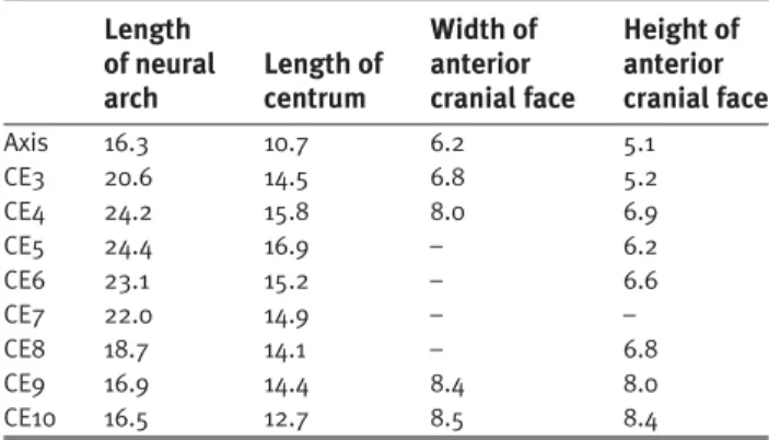
Table 1 Dimensions of the cervical vertebrae (in mm).
Length Width of Height of
of neural Length of anterior anterior arch centrum cranial face cranial face
Axis 16.3 10.7 6.2 5.1 CE3 20.6 14.5 6.8 5.2 CE4 24.2 15.8 8.0 6.9 CE5 24.4 16.9 – 6.2 CE6 23.1 15.2 – 6.6 CE7 22.0 14.9 – – CE8 18.7 14.1 – 6.8 CE9 16.9 14.4 8.4 8.0 CE10 16.5 12.7 8.5 8.4 CE= cervical vertebra.
Splenial and coronoid
No splenial or coronoid can be confidently identified, al-though they are probably included amongst a number of simple flat bones, that are poorly exposed and remain uniden-tified.
Vertebral column
Cervical vertebrae (Table 1)
Ten cervical (CE) vertebrae are preserved and, given that the tenth is quite like a dorsal vertebra in its morphology, this was almost certainly the last cervical. Although no dorsal vertebrae are preserved with which to compare the cervicals, it can be determined that the neck was elongated as it is in other saurischians. The centra of CE3–9 are all longer than the axial centrum, a condition seen in other sauropodomorphs and neotheropods.
Thecodontosaurus caducus differs from other sauro-podomorphs, except Riojasaurus incertus, in not having mid-cervical centra that are at least three times as long as wide. The cervical vertebrae show strong indications of immaturity. These are the lack of fusion between any of the individual ele-ments of the atlas–axis complex and the presence of plainly visible neurocentral sutures on the postaxial cervicals. In-deed the neural arches have parted from their centra in CE3, 6 and 7. Lack of sutural closure in the cervical vertebrae was found to be characteristic of immature crocodilians by Brochu (1996) and is almost certainly indicative of immatur-ity in dinosaurs as well.
The cervical ribs are poorly preserved, but it is clear that, like other saurischians, they are longer than their respective vertebrae, and that in life they lay parallel to the cervical column.
Atlas (Fig. 10C)
All elements of the atlas–axis complex are incompletely os-sified, remain separate from each other, and have become scattered from their original positions. Thus it is difficult to distinguish the atlantal intercentrum from the axial in-tercentrum and the odontoid. The element identified by Kermack (1984) as the atlantal intercentrum is here thought to be too rounded and not transversely wide enough to be that element, and is re-interpreted as the odontoid. The element identified as the axial intercentrum is re-interpreted here as the atlantal intercentrum. It is a low, broadly U-shaped bone
Figure 10 Thecodontosaurus caducus sp. nov., holotype, BMNH P24; elements of the atlas–axis complex.IOA, axial intercentrum in cranial aspect;IOB, axial intercentrum in left lateral aspect; IOC, right neurapophysis in medial aspect;IOD, axial neural arch in dorsal aspect. For abbreviations see Appendix 1. Scale bars= 5 mm.
in cranial view. In ventral view the cranial margin is convex, while the caudal margin is straight. The right neurapophysis can be viewed medially, rostrally and dorsally. The bone consists of two subrectangular processes, the pedicel and the prezygapophysis, and an elongate prong-like postzygapo-physis. The vertically oriented pedicel forms the lateral wall of the neural canal. The prezygapophysis joins the dorsal edge of the pedicel at a roughly right angle, to from a roof over the top of the neural canal. The slender postzygapo-physis extends caudally from the junction of the pedicel and the prezygapophysis. A thin, pointed epipophysis continues caudal to the postzygopophyseal facet, but it is not as elong-ated as it is in some basal sauropodomorphs such as
Plateo-saurus engelhardti (von Huene 1926) and ColoradiPlateo-saurus brevis (Bonaparte & Pumares 1995). In these taxa, the
at-lantal epipophyses extend as far back as the cranial margin of the axial postzygapophyses. The odontoid is small and rounded. The dorsal surface is flattened, while the ventral and cranial surfaces are strongly convex. The lateral surface bears a small, round depression.
Axis (Figs 10A, B, D)
The probable axial intercentrum is a small, crescentic ele-ment with a strongly concave dorsal margin in cranial view and a pointed cranially directed process, developed on the midpoint of the ventral margin. The element is craniocaud-ally flattened and is only 2.9 mm long at its thickest point. The axial centrum is a simple spool-shaped element that is 11 mm long and 5 mm wide. A probable juvenile characteristic is the lack of any form of parapophysis at the cranial end of the centrum. Like other sauropodomorphs (e.g. Riojasaurus
incertus: Bonaparte & Pumares 1995; Plateosaurus engel-hardti: pers. obs. of GPIT Skelett 1; Camarasaurus lentus:
Madsen et al. 1995), but in contrast to other basal dinosaurs, the axis is not ventrally keeled. The axial neural arch covers a wide neural canal that is 75% the width of the cranial face of the axial centrum. The size of the foramen magnum relative to the size of the animal decreases through ontogeny (Dodson 1975) and we can expect the width of the axial neural canal to be strongly correlated with that of the foramen magnum. Thus the relatively wide axial neural canal is probably a juvenile characteristic. The prezygapohyses are small, dorsolaterally facing facets mounted on tab-shaped processes that project from the cranial margin of the neural arch, similar to the axial prezygapophyses of neotheropods, but unlike those of
Herrerasaurus ischigualastensis (Sereno & Novas 1993) and
other sauropodomorphs (e.g. Camarasaurus lentus: Madsen
et al. 1995). In these taxa the prezygapophyses are simple
raised areas that do not project cranially. Below the prezyga-pophyses at the antero-ventral corners of the arch there are weakly developed tubercles that represent the diapophyses. In dorsal view, the lateral margins of the neural arch flare ab-ruptly outwards at the level of the postzygapophyses. Thus, like other saurischians, the postzygapophyses are set wider from the midline than the prezygapophyses. The axial neural spine is damaged dorsally but it appears to be a long, low rectangular process that extends for the full length of the neural arch. Stout epipophyses project a short distance from the caudal margin of the postzygapophyses.
Cervicals 3–5 (Figs 11A, B, C)
The first three postaxial cervical vertebrae are similar to one another. Their neural arches are low, sided and flat-topped structures. Cranially projecting prezgapophyses over-hang the cranial face of the centrum by as much as a third of the length of the centrum. The prezygapophyses meet caudally to form a U-shaped space, of which the caudal half is floored by a thin interprezygapophyseal lamina. Thus a cranially open, U-shaped fossa is developed at the cra-nial end of the dorsal surface of the neural arch. A long neural spine extends from the vertex of this fossa to the caudal margin of the arch. The spines have broken off at their bases, so their height cannot be determined. Wide, tongue-shaped postzygapophyses project posterolaterally from pos-terodorsal corners of the neural arch and overhang the caudal face of the centrum by a few millimeters. Their dorsal
Figure 11 Thecodontosaurus caducus sp. nov., holotype, BMNH P24; cervical vertebrae. 11A, centrum of CE3 in right lateral aspect; 11B, centrum of CE3 in ventral aspect; 11C, CE4 in left lateral aspect; 11D, CE10 in dorsal aspect; 11E, CE10 in ventral aspect; 11F, CE10 in right lateral aspect;11G, CE10 in caudal aspect; 11H, CE10 in cranial aspect. For abbreviations see Appendix 1. Scale bars= 10 mm.
surfaces remain flat and horizontal along their length as they do in T. antiquus (Benton et al. 2000). The caudal edge projects a short distance beyond the caudal edge of the ventrally-facing articulation facet, producing a stubby, caudally-projecting epipophysis. Such overhanging, postaxial epipophyses have been thought to diagnose the Theropoda (Sereno & Novas 1993), but they are also present in Plateosaurus engelhardti (pers. obs. of GPIT Skelett 1), which suggests that they diagnose the Saurischia and have
been subsequently lost in later sauropodomorphs. There is no development of any lamina on these neural arches. The diapophysis is not visible on CE3 while it is borne on a small tubercle on the anteroventral corner of the arch in CE4 and CE5. The neurocentral articulation was weakly su-tured in CE3 so that the two elements separated prior to burial. The centra of these vertebrae are elongate, amphi-coelous spools that increase in length from CE3 to CE5. The caudal face is set distinctly lower than the cranial face in
Figure 12 Thecodontosaurus caducus sp. nov., holotype, BMNH P24. 12A, cervical vertebrae 6, 7 and 8 in left ventrolateral aspect; 12B, interpretive line drawing of 12A, showing the pseudopleurocoels (arrowed). Cross-hatching= exposed sutural surfaces, horizontal hatching= surfaces of broken bone. For abbreviations see Appendix 1. Scale bar = 10 mm.
CE4 and CE5 producing an upward bend in this region of the neck. Poorly developed parapophyses occur on the antero-dorsal corners of the centra in CE4 and CE5, just below the diapophyses. The ventral surfaces of the centra are rounded transversely without any trace of the longitudinal keel that is commonly present in early dinosaurs (e.g. Herrerasaurus
ischigualastensis: Sereno & Novas 1993).
Cervicals 6–7 (Fig. 12)
These vertebrae are similar to CE4 and CE5, differing mainly in that the diapophyses are now borne on short, slender and
pointed processes. These processes, which arise from near the anteroventral corners of each neural arch, are strongly pendent as well as being directed slightly forwards. The centra are shorter than that of CE5 and show signs of hav-ing borne small simple pleurocoel-like pits developed on the neurocentral suture just caudal to the diapophyses. The large elliptical spaces that are present below the neural arches are artefacts caused by the separation of the neural arches from the centra and lateral rotation of the latter relative to the former. Nevertheless, a distinct sharp-edged depression de-veloped on the contact surfaces of the centra would have formed small elliptical pits just caudal to the diapophyses
when the elements were correctly articulated. Stout epipo-physes with planar dorsal surfaces are also present on these vertebrae, but unlike those of more cranial vertebrae, they do not overhang the postzgapophyseal facet.
Cervical 8 (Fig. 12)
The centrum of the eighth cervical is noticeably shorter than the centra of CE3–7 but it is still longer than the axial centrum. A small elliptical pleurocoel-like pit is located on the neurocentral suture below the transverse process. The cra-nial projection of the prezygapophysis is less marked than in the previous postaxials, while it is angled dorsally, indicating that the neck had an upward bend at this point. The transverse process, with its terminally placed diapophysis is centrally located and is directed laterally, unlike the vertebrae cranial to it. Three laminae radiate from the transverse process: the prezygapophyseal, the cranial centro-diapophyseal and the caudal centro-diapophyseal. The diapo-postzygapophyseal lamina is not expressed. ‘Efraasia diagnostica’ (pers. obs. of SMNS 12667), Plateosaurus engelhardti (pers. obs. of GPIT Skelett 1) and Massospondylus carinatus (Cooper 1981) also lack a diapo-postzygapophyseal lamina, so this condition might diagnose the Sauropodomorpha. Sauropods that do possess a diapo-postzygapophyseal lamina in all of their cer-vical vertebrae (Wilson 1999), have apparently reverted to the primitive condition.
Cervicals 9 and 10 (Figs 11D–H)
These are the last two cervical vertebrae. Like CE8, the centra of these two are shorter than those of CE3–7, but are longer than the axial centrum. The centrum of CE10 is the first to bear a sharp ventral keel. The parapophysis forms an oval tubercle halfway up the cranial margin of the centrum in lat-eral view. As they do not reach the neurocentral suture in the last cervical, it can be deduced that the parapophyses were located on the centrum of the cranial dorsals, as they are in Plateosaurus engelhardit (von Huene 1926). Both sets of zygapophyses are angled upwards in lateral view. The diapo-physis is borne on a laterally projecting, elongate, pendent transverse process. A diapo-postzygapophyseal lamina now connects the base of the transverse process with the post-zygapophysis, thus creating a posterior semiconical fossa, or chonos (Welles 1984). The postzygapophyseal facet is curved at its ventral end so that there is a narrow laterally-facing ledge that is connected with its counterpart by an interpost-zygapophyseal lamina. This structure is a weakly developed hyposphene. The tenth cervical has a space, the hypantrum, to receive the hyposphene of CE9. The neural spine of each of these vertebrae is placed caudally, so much so that it pro-jects into the interpostzygapophyseal space in dorsal view.
Caudal vertebrae (Fig. 13)
There is a series of eight proximal–mid caudals (BMNH P64/1) and 13 distal caudals (BMNH P77/1). Although these vertebrae are not very big (they probably come from a similar-sized individual as the holotype if not from the holo-type itself) the neurocentral sutures are completely closed. This indicates that, like crocodilians, the closure of the neuro-central sutures proceded from caudal to cranial (Brochu 1996). The more proximate vertebrae of the first series have short laterally projecting transverse processes, while they are reduced to mere longitudinal ridges in the eighth. By comparison with other basal sauropododomorphs
(Plateo-Figure 13 Thecodontosaurus caducus sp. nov., BMNH P77/1; caudal vertebrae.13A, mid caudal (CA20?) and associated chevron in left lateral aspect;13B, distal caudal in left lateral aspect with associated chevron in proximal aspect. For abbreviations see Appendix 1. Scale bar= 5 mm.
saurus engelhardti: pers. obs. of GPIT Skelett 1; Lufen-gosaurus huenei: Young 1941a), in which the most distal
transverse process occurs in caudal 27, it is probable that BMNH P64/1 represents caudals 20–28. The ventral sur-faces of the centra are flattened and the longitudinal sulcus bordered by two ridges, which is usually present in sauro-podomorphs, is absent. This is a derived condition shared with T. antiquus. Caudal 20 has a centrum that is 12 mm long and 6 mm high at its proximal end. Such an elongate CA20 is a primitive character state, the caudal centra of all other sauropodomorphs do not develop such proportions un-til CA27–30 (Young 1941a; pers. obs. of GPIT Skelett 1). The neural spines are proximo-distally short and placed far back on the neural arches, between the postzygapophyses. Thus, the U-shaped interpostzygapophyseal space, which is present in most dinosaurs, is filled by the base of the neural spine. This is a derived condition that T. caducus shares with
T. antiquus. The length of the associated chevrons is equal to
the height of their respective vertebrae. The proximal ends of the chevrons are bridged-over, while the distal ends are slightly expanded in the proximo-distal plane.
The distal series of caudals consists of elongate centra with reduced neural arches that lack transverse processes and neural spines. The prezygapophyses are small tongue-shaped processes that do not project far from the centra, unlike those of herrerasaurids (Novas 1993) and neotheropods (Chiappe
et al. 1996). The postzygapophyses are similar in size and
shape.
Forelimb
ScapulaOnly a fragment from the proximal ventral corner of the right scapula of the holotype remains. It does not differ signific-antly from those of other early dinosaurs.
Coracoid
The medial surface of the right coracoid of the holotype is exposed, while the holotype’s left coracoid and another isolated left coracoid (BMNH P39) have been freed from the matrix. The coracoid is an elongately oval plate that is 25 mm
Figure 14 Thecodontosaurus caducus sp. nov. 14A, proximal left humerus of BMNH P24, holotype, in caudal aspect; 14B, incomplete left coracoid of BMNH P39/2; lateral view and14C, medial aspect. For abbreviations see Appendix 1. Scale bars= 10 mm.
long and 15 mm high in P24. The long axis of the coracoid is parallel to its suture with the scapula. The glenoid region is greatly thickened compared to the cranial and caudal margins of the bone. The ventral margin is rounded and lacks a notch separating the glenoid from the pointed cranioventral corner that can be seen in some early sauropodomorphs, such as
Lufengosaurus huenei (Young 1941a). There is, however, a
laterally projecting tubercle developed at the cranioventral extremity of the bone.
Humerus (Fig. 14)
The left humerus (BMNH P19/7) could not be located, so this description is based entirely on the two proximal humeral fragments preserved in the holotype. The proximal humerus is a craniocaudally flattened structure capped by a narrow head (19 mm wide) that is gently convex in the medio-lateral plane. The medial corner of the head does not project as strongly as it does in T. antiquus. In that species the strong medial projection of the humeral head causes the margin of the humerus, underneath the medial tuberosity, to be greatly arched, as it is in crurotarsans (Sereno 1991b). An elongate deltopectoral crest extends 30 mm down the lateral margin, from the proximolateral corner. Distal to the deltopectoral crest, the shaft narrows sharply to a cylindrical structure that is 7 mm in diameter.
Hindlimb
Except where mentioned, all of the hindlimb and pelvic ele-ments described here come from a single, partially articulated specimen (BMNH P77/1).
Ilium (Fig. 15)
The ilium is closer in shape to those of other basal sauro-podomorphs than it is to the ilium of T. antiquus. In the latter species the ilium is low and elongate, especially caudally, whereas in T. caducus it is tall, short and has a rhomboidal shape. The preacetabular blade is a pointed structure that is directed cranially without any ventral curvature as it is in all other basal sauropodomorphs, except T. antiquus. The elong-ate pubic peduncle is craniocaudally flattened so that the transverse width of the articular facet for the pubis (5.5 mm) is greater than its craniocaudal length (3 mm). This articular facet is as long as it is wide in T. antiquus and most other sauropodomorphs. The lateral acetabular margin of the pu-bic peduncle forms a sharp ridge that is confluent with the supra-acetabular crest. The supra-acetabular crest appears to reach its widest point at the base of the public peduncle, a derived condition amongst archosaurs, but the closely ad-pressed femur may have crushed the supra-acetabular crest between the pubic and ischial peduncles. Nevertheless the supra-acetabular crest is widest over the pubic peduncle of T.
antiquus and other sauropomorphs that are more derived than Saturnalia tupiniquim, so it is simplest to infer that the
supra-acetabular crest of BMNH P77/1 is not damaged. The medial wall of the acetabulum is extensive, with a gently concave ventral margin. Such an incompletely perforate acetabulum is a primitive character that is rare amongst dinosaurs but is seen in Herrerasaurus ischigualastensis (Novas 1993),
Guaibasaurus candelariensis (Bonaparte et al. 1999) and Saturnalia tupiniquim (Langer et al. 1999). The ischial
ped-uncle is a short rounded process. The postacetabular blade is short and, like T. antiquus, ‘Efraasia diagnostica’ and neotheropods, it has a subrectangular caudal margin in lat-eral view. The medial shelf that joins to the last sacral rib
Figure 15 Thecodontosaurus caducus sp. nov., BMNH P77/1; right ilium.15A, lateral aspect; 15B, ventral aspect. For abbreviations see Appendix 1. Scale bar= 10 mm.
and forms the medial margin of the brevis fossa is absent, as is the brevis fossa itself. Other basal sauropodomorphs, such as Saturnalia tupiniquim, T. antiquus and ‘Efraasia
diagnostica’, have large brevis fossae. It is probable that as T. caducus matured and the connection between the sacral
ribs and the ilium began to suture firmly, the medial shelf of the postacetabular blade would have ossified. For this reason it is assumed that the absence of a brevis shelf may be more an indication of immaturity than a diagnostic character of the species.
Pubis
There is only one fragment of a possible pubis that may be-long to T. caducus (BMNH P126/1). However, the referral is dubious because the specimen comes from an individual that was distinctly larger than the other specimens. The fragment includes what might be the iliac peduncle and some of the obturator plate, including part of the margin of an obturator foramen. No other details can be gleaned from this specimen.
Ischium (Fig. 16)
Kermack (1984) misidentified an isolated, right ischium (BMNH P24/3), as the distal end of the scapula of the holo-type. It is, however, too expanded at its distal end to be a scapula. It is nearly complete and measures 40 mm along its greatest dimension. The proximal half forms a plate that, in life, would have been angled dorsolaterally from the sym-physis along its ventral margin. A thin obturator plate ex-pands ventrally at the proximal end. A short longitudinal sulcus is developed on the dorso-lateral margin where it
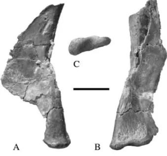
Figure 16 Thecodontosaurus caducus sp. nov., BMNH P24/3; right ischium.16A & 16B, lateral aspect; 16C, dorsal aspect. For abbreviations see Appendix 1. Scale bar= 10 mm.
curves upwards to form the iliac peduncle. Such a sulcus may diagnose Neotheropoda + Sauropodomorpha, since it is seen in other sauropodomorphs (e.g. Saturnalia tupiniquim: pers. obs. of MCP 3844 – PV; Plateosaurus engelhardti: pers. obs. of GPIT Skelett 1; Dicraeosaurus hansemani: pers. obs. of HMN material) and neotheropods (e.g. Lilliensternus
lilliensterni: pers. obs. of HMN MB.R.2175.7.4). The distal
half forms a shaft that is triangular in cross-section with a keeled ventral edge and a flat dorsal face. This is diagnostic of Saurischia (see discussion below). The distal end is ex-panded both mediolaterally and dorsoventrally. In distal view the conjoined ischial expansions would have been as wide as high, unlike Plateosaurus engelhardti, where the conjoined expansions are higher than they are wide (von Huene 1926).
Femur (Fig. 17)
The single known femur (from BMNH P77/1) is incomplete. The proximal end, from the middle of the fourth trochanter, is missing. Assuming that the position of the fourth trochanter along the femoral shaft remained constant throughout onto-geny and the femoral proportions were similar to T. antiquus, the total length of the femur is estimated to have been 72 mm. The steep distal margin of the fourth trochanter indicates that the profile was asymmetrical, like most other early sauro-podomorphs except Melanorosaurus readi (Van Heerden & Galton 1997). The distal shaft is strongly bowed cranially when viewed laterally and slightly bowed medially when viewed cranially. The sinuous nature of the femoral shaft is a plesiomorphic feature found in most early sauropodomorphs (Galton 1990). The space between the distal condyles is dis-tincty hollowed out, suggesting incomplete ossification. A broad but shallow popliteal fossa is developed at the distal end of the caudal surface while the cranial surface remains convex without any trace of an extensor groove. The tibiofib-ular crest on the caudolateral surface of the distal end is low and is only weakly separated from the fibular condyle by a poorly impressed fibular trochlea.
Figure 17 Thecodontosaurus caducus sp. nov., BMNH P77/1; distal right femur.17A, medial aspect; 17B, caudal aspect. For abbreviations see Appendix 1. Scale bar= 10 mm.
Tibia (Fig. 18)
If the estimate of the length of the femur is accurate, then the tibia is only slightly shorter than the femur. It is 70 mm long,
Figure 18 Thecodontosaurus caducus sp. nov., BMNH P77/1; right tibia. 18A, cranial aspect; 18B, lateral aspect; 18C, caudal aspect; 18D, medial aspect. fib. con= fibular condyle, cn = cnemial crest. Scale bar = 10 mm.
which is 97% of the estimated length of the femur. This is in contrast to other sauropodomorphs where the tibia is much shorter than the femur (e.g. 89% in Anchisaurus polyzelus: Galton 1976; 65% in Lufengosaurus huenei: Young 1941a; 62% in Apatosaurus louisae: Gilmore 1936). The relatively elongate tibia may be due to the small size, and juvenile nature, of the specimen, or it may be a plesiomorphic fea-ture of the species. The proximal head is similar to that of
T. antiquus (e.g. BRSMG C4531, note that BMNH 49884,
the holotype of Agrosaurus mcgillivrayi, is aberrant and un-like all other tibias assigned to T. antiquus). The triangular, proximal surface is flat and slopes both mediodistally and caudodistally. The low and simple cnemial crest projects cranially from the medial margin of the cranial face, at the proximal end. The fibular condyle forms a low, rounded, lateral projection from the centre of the lateral surface at its proximal end. The tibial shaft is straight, slender and rounded in cross-section. The distal end is only slightly expanded, and is not flared transversely so that the distal surface is square-shaped. The lateral surface of the distal end is gently concave. This concavity is confluent with the notch that separates the caudodistal flange from the facet for the ascending process of the astragulus. Although damaged distally, it is clear that the caudodistal flange was quite low and did not project much further laterally than the craniolateral corner of the distal end.
Fibula (Fig. 19)
The fibula is a slender, rod-like bone that is 65 mm long. The proximal end is mediolaterally compressed but cranio-caudally expanded. The caudal proximal corner forms a stout pointed process in lateral view, while the cranial prox-imal corner is rounded. The proxprox-imal tibial facet forms a planar surface. The shaft is narrow (3.5 mm wide at its midpoint) and has an oval cross-section, with the long axis
Figure 19 Thecodontosaurus caducus sp. nov., BMNH P77/1; right fibula.19A, lateral aspect; 19B, caudal aspect; 19C, medial aspect. Scale bar= 20 mm.
oriented craniocaudally. There is no trace of a tubercle for the tibiofibularis ligament on the cranial margin of the shaft; this is probably another feature of immaturity. Similarly the expansion at the distal end was not ossified and the fibula is shorter than the tibia.
Pes (Fig. 20)
The right pes is articulated and almost complete. It is exposed on its plantar surface, although the proximal surface of the metatarsus and the lateral and medial sides of some elements can be observed as well. It is a slender foot, when compared to other basal sauropodomorphs such as Plateosaurus
en-gelhardti (von Huene 1926) and Massospondylus carinatus
(Cooper 1981), but this is almost certainly a correlate of the specimen’s small size and juvenile nature.
Metatarsal I (Table 2)
The first is the shortest digit-bearing metatarsal. It is a flattened element that is less than 60% of the length of meta-tarsal III, the longest of the metameta-tarsals. It is gently twisted about its long axis so that the dorsal face of the compressed proximal head faces dorsomedially while the transverse axis through the distal articular end is oriented mediolaterally. The proximal head is strongly compressed and has a narrowly el-liptical head that fits against the dorsomedial articular facet of metatarsal II. The lateral side remains in contact with metatarsal II for its entire length. In plantar view the distal articular surface is set at an angle so that the medial side is higher than the lateral. This would have enabled the hallux to separate from the rest of the digits of the foot during
exten-Figure 20 Thecodontosaurus caducus sp. nov., BMNH P77/1; right pes.20A, plantar aspect; 20B, proximal aspect. For abbreviations see Appendix 1. Scale bars= 20 mm.
sion. This could be correlated with the use of the hallux as a weapon, as is suggested by the enlarged size of the ungual in this digit. A small, weakly developed ligament pit occurs on the lateral side of the distal end, while a weak extensor pit occupies its dorsal face.
Metatarsal II
This metatarsal is shorter and more robust than metatarsals III and IV. The proximal articular surface is parallelogram-shaped, with the transverse width being less than the dorsoplantar depth. The dorsal and plantar faces are straight Table 2 Dimensions of the metatarsals (in mm).
Maximum Width of Distal proximal dorsal Length width dimension proximal face
Mt I 20.1 5.7 5.6 3.2 Mt II 29.0 6.3 7.3 4.1 Mt III ∼35 5.5 7.8 3.5 Mt IV 33.3 4.3 6.8 4.2 Mt V 14.8 – 5.8 – Mt= Metatarsal