HAL Id: hal-02632676
https://hal.inrae.fr/hal-02632676
Submitted on 27 May 2020
HAL is a multi-disciplinary open access
archive for the deposit and dissemination of sci-entific research documents, whether they are pub-lished or not. The documents may come from teaching and research institutions in France or abroad, or from public or private research centers.
L’archive ouverte pluridisciplinaire HAL, est destinée au dépôt et à la diffusion de documents scientifiques de niveau recherche, publiés ou non, émanant des établissements d’enseignement et de recherche français ou étrangers, des laboratoires publics ou privés.
Isolation and identification of Pseudomonas syringae
facilitated by a PCR targeting the whole P. syringae
group
Caroline Guilbaud, Cindy E. Morris, Mohamed Barakat, Philippe Ortet,
Odile Berge
To cite this version:
Caroline Guilbaud, Cindy E. Morris, Mohamed Barakat, Philippe Ortet, Odile Berge. Isolation and identification of Pseudomonas syringae facilitated by a PCR targeting the whole P. syringae group. FEMS Microbiology Ecology, Wiley-Blackwell, 2016, 92 (1), pp.fiv146. �10.1093/femsec/fiv146�. �hal-02632676�
Version postprint
Isolation and identification of Pseudomonas syringae facilitated by a PCR targeting the whole P. syringae group
Caroline Guilbaud1, Cindy E. Morris1, Mohamed Barakat2,3,4, Philippe Ortet2,3,4, Odile Berge1
1INRA, UR0407 Pathologie Végétale, F-84143 Montfavet cedex, France
2CEA, IBEB, Lab Ecol Microb Rhizosphere & Environ Extrem, Saint-Paul-lez-Durance,
F-13108, France
3CNRS, UMR 7265 Biol Veget & Microbiol Environ, Saint-Paul-lez-Durance, F-13108, France 4Aix Marseille Université, BVME UMR7265, Marseille, F-13284, France
Key words: P. syringae identification; Population structure; P. syringae diversity;
Environmental pathogen; PCR-assisted detection, Specific PCR.
Running title: PCR for the entire P. syringae group
Correspondence:
Tel: +33 432722886/43 Fax: + 33 432722842
E-mail: odile.berge@avignon.inra.fr
FEMS Microbiology Ecology Advance Access published November 25, 2015
by guest on December 1, 2015
http://femsec.oxfordjournals.org/
Version postprint
Abstract
We present a reliable PCR-based method to avoid the biases related to identification based on the conventional phenotypes currently used in the identification of Pseudomonas syringae
sensu lato, a ubiquitous environmental bacterium including plant pathogens. We identified a
DNA target suitable for this purpose by applying a comparative genomic pipeline to
Pseudomonas genomes. We designed primers and developed PCR conditions that led to a
clean and strong PCR product from 97 % of the 185 strains of P. syringae strains tested and gave a clear negative result for the 31 non-P. syringae strains tested. The sensitivity of standard PCR was determined with pure strains to be 106 bacteria mL-1 or 0.4 ng of DNA µL
-1. Sensitivity could be improved with the touchdown method. The new PCR-assisted isolation
of P. syringae was efficient when deployed on an environmental sample of river water as compared to the isolation based on phenotypes. This innovation eliminates the need for extensive expertise in isolating P. syringae colonies, was simpler, faster and very reliable. It will facilitate discovery of more diversity of P. syringae and research on emergence,
dispersion, and evolution to understand the varied functions of this environmental bacterium.
Introduction
There is growing concern worldwide about environmentally persistent pathogens. This new dimension of research on pathogens is making considerable progress for human pathogens (Aujoulat et al., 2012) but it has received little attention for plant pathogens (Morris, et al., 2009, Vayssier-Taussat, 2014, Bartoli et al., 2015). For such studies it is essential to have reliable techniques for the isolation and/or identification of natural populations that can be present at low concentrations in substrates other than infected tissues such as rivers (Selezska, et al., 2012) or insects (Carolan et al., 2014). To promote research on the environmental persistence of plant pathogens and the role of environmental reservoirs in emergence of plant diseases, we have developed a technique to facilitate the tracking and identification of the ubiquitous plant pathogen, Pseudomonas syringae sensu lato (Morris, et
al., 2013). P. syringae sensu lato (named P. syringae or P. syringae group in the text)
currently consists of a phylogenetic lineage containing P. syringae-related species (Mulet et al., 2010) together with P. syringae populations classified into phylogroups by MLSA (multi locus sequence analysis) (Berge et al., 2014). The metapopulation of P. syringae comprises strains that are involved in various crop diseases (Lamichhane et al., 2014, Lamichhane et
al., 2015), but many strains have been frequently isolated all along the water cycle both in
and outside of agricultural zones and from a range of substrates. The environmental populations of P. syringae constitute a reservoir of plant pathogens (Monteil et al., 2013, Bartoli et al., 2015) and at the same time they belong to microbial communities and are likely to participate in the functioning and evolution of these communities. Among the various
by guest on December 1, 2015
http://femsec.oxfordjournals.org/
Version postprint
environmental impacts of P. syringae, strains of this bacterium are purported to contribute to meteorological phenomena via their ice nucleation activity that can trigger rain and snow fall from cloud droplets under specific conditions (Amato et al., 2007, Morris et al., 2014). It is important to be able to isolate the widest diversity of these populations but screening isolates is difficult because of the large genotypic and phenotypic diversity represented by this group of bacteria (Berge et al., 2014). Actually the culture approach extensively used for isolating putative P. syringae strains is still the method with the highest sensitivity but it contains biases linked to the use of phenotypic properties. Moreover, strains from the P. syringae group have extensive diversity in virulence gene repertoires like those for effectors, toxins or plant hormone production (Baltrus et al., 2011) that cannot be used to characterize the whole
P. syringae group. Some molecular tools have been proposed for the specific detection of
single pathogenic varieties so-called pathovars (Kong et al., 2004, Gervasi & Scortichini 2009, Cho et al., 2010, Gallelli et al., 2011), a group of pathovars (Tegli et al., 2010, Popovic
et al., 2014, Vaseghi et al., 2014) or one phylogroup (Clarke et al., 2010, Cottyn et al., 2011).
All these PCR-tools have in common that only few phylogroups and fewer than 50 strains were used to test their specificity. There is currently no molecular probe that targets the whole P. syringae group and this is reflected in the routinely conventional isolation of P.
syringae mainly based on phenotypic tests (Kaluzna et al., 2012). Our objective is to develop
a reliable molecular probe for the entire P. syringae group and to illustrate the effectiveness of this probe with an environmental sample of river water from one of the sites where the greatest diversity of P. syringae in a single sample has been reported (Morris et al., 2010).
Materials and methods Bacterial strains
Bacterial strains used in this study are listed in supporting Table S1. For P. syringae, 185 strains were selected that represent the diversity of the 13 phylogenetic groups of P.
syringae as recently described (Berge et al., 2014). An additional 33 reference strains from
outside the P. syringae group were used to determine the specificity of the PCR-based method (information about strains provided in Table S1). All strains were stored in nutrient broth containing 20 % glycerol at – 85°C and were cultivated on King’s medium B (KB) (King
et al., 1954) at 25°C for 48 h before use.
Sequence analysis and primer design
Primer design for the specific detection of strains of the P. syringae group required targeting discriminating regions of the genome. In an initial step, this was accomplished with an in-house pipeline designed to take as inputs the genome sequences of strains DC3000, B728A and Pph1448A (http://www.ncbi.nlm.nih.gov/) and to scan DNA sequences to constitute what
by guest on December 1, 2015
http://femsec.oxfordjournals.org/
Version postprint
we call the ORFeome-like. The ORFeome-like corresponds to the entire set of extended ORFs defined as the DNA segments occurring between two stop codons in the six reading frames and exceeding 100 nucleotides. The ORFeome-like allows the exploration of the whole genome including intergenic regions, without any further assumption about the presence or not of coding sequences. For instance, the ORFeome-like from P. syringae DC3000 includes 111175 sequences. A pairwise sequence alignment was performed based on all-against-all BLAST comparisons. The results were stored in a MySQL database, and parameters such as identity percentage, alignment length, mismatches, gap openings, e-value and bit score were saved. We filtered the data to select the sequences with an identity percentage and an alignment length greater than 98% and 250 nucleotides respectively. Analysis of the genomic regions in 24 P. syringae genomes (including DC3000, B728A and Pph1448A) representing a wide diversity in the P. syringae group, together with 11 genomes of Pseudomonas sp. that are clearly outside the P. syringae group (additional information about strains is provided in Table S1) led to the identification of a common short sequence (166 bp) in P. syringae genomes beginning at position 4342391 in the P. syringae DC3000 genome (Fig. S1). This sequence was positioned astride two contiguous housekeeping genes plsX and rpmF coding respectively for the fatty acid/phospholipid synthesis protein PlsX and the ribosomal protein L32 (Fig. S2). Primers were designed manually across discriminant positions (Fig. S1): Psy_F 5’-ATG ATC GGA GCG GAC AAG 3’ and Psy_R 5’ GCT CTT GAG GCA AGC ACT 3’ and allowed the amplification of a 144-bp DNA fragment. This PCR was named Psy-PCR.
P. syringae specific polymerase chain reaction (Psy-PCR)
Psy-PCR standard reactions were conducted in a final volume of 25 µL using the ready-to-use GoTaq reagents (Promega, France). Each reaction mix contained 14.17 μl of milliQ water, 5.0 μl of colourless GoTaq PCR buffer (5 x containing MgCl2), 1.5 µL of MgCl2 (25
mM), 0.2 μl of dNTP mix (25 mM each), 1 μl of each primer (10 µM), 0.13 μl of GoTaq DNA polymerase (5 units μl-1) corresponding to final concentrations of dNTPs 0.2 mM, MgCl
2 at
1.5 mM, each primer at 0.4 µM, 1 x GoTaq flexi buffer and 0.65 units of GoTaq G2 flexi DNA polymerase; 2.0 μl of DNA template was added to this mixture. In routine tests, DNA
template was replaced by cell suspensions adjusted to 108 colony-forming units (cfu mL-1) with a spectrophotometer (A580 = 0.06). To evaluate the sensitivity of Psy-PCR, subsequent
decimal dilutions of suspensions corresponding to 102 to 108 cfu mL-1 were tested as template. Strains used to test the specificity and the sensitivity of Psy-PCR are listed in supporting Table S1. Cell concentrations were checked on KB medium. In some cases DNA was extracted from 2 mL broth culture with a kit (QIAamp DNA minikit – Cat 51304) and used as template. DNA concentration was determined with a Nanodrop ND-1000
by guest on December 1, 2015
http://femsec.oxfordjournals.org/
Version postprint
spectrophotometer then diluted in sterile milliQ water to obtain 0.1 to 10 ng μl-1
. PCR was conducted in an Mastercycler® epGradient (Eppendorf) with initial polymerase activation for 5 min at 96°C followed by 30 cycles at 94°C for 30 s, 61°C for 30 s, 72°C for 30 s and a final extension at 72°C for 10 min. The Psy-PCR product detection was performed by
electrophoresis through 1.5 % agarose gel with ethidium bromide at 0.5 µg mL-1 (Euromedex, EU0070) and then visualized under ultraviolet light. A preliminary touchdown-PCR (TD-PCR) was also performed to test its sensitivity using a sub-collection of 29 strains (additional information about strains is provided in Table S1). TD-PCR is a simple and rapid method to optimize PCR, increasing specificity, sensitivity and yield by using a high annealing
temperature during the first cycles to enhance stringency and therefore enhancing the specificity of amplification. Hybridisation temperature is then gradually lowered during the following cycles to have a better efficiency of the PCR. TD-PCR is particularly useful for templates that are difficult to amplify but is also currently used to enhance specificity and product formation. TD-PCR was performed with the same mix but dNTPs were at 0.25 mM and primers at 0.6 µM with an initial step of 95°C for 5 min followed by 20 cycles of 94°C for 30 s, annealing temperatures starting at 62°C for 30 s then decreasing 0.5°C per cycle, and 72°C for 30 s for extension. This step was followed by 20 cycles at 94°C for 30 s, 55°C for 30 s, 72°C for 30 s and finally 72°C for 7 min.
Deployment of Psy-PCR to isolate P. syringae from environmental samples
Water was collected in September 2013 from the Tarn River (Castelbouc, Lozère, France, GPS coordinates: 44.339905, 3.465330). The microbiology of this river is of considerable importance because of the occasional presence of toxic cyanobacteria that can cause death of wild and domesticated animals when these animals drink river water during periods of blooms (Quiblier et al., 2013). Hence, this river is regularly monitored for indicators of these blooms. Upstream of the site sampled here, a wide diversity of P. syringae (phylogroups 1, 2, 7, 9 and 10 (Berge et al., 2014)) has been observed (Morris et al., 2010). Microbial isolation from the water sample was conducted following previously-described procedures (Morris et
al., 2008) based on cultivation on a modified KB medium (KBC) containing selective agents
(Mohan & Schaad, 1987). One bulk water sample was selected to test the method. Two replicates of the water sample (500 mL) were each filtered across a membrane (0.22 µm pore diameter – Millipore France GSWP 047 00) to concentrate bacteria before isolation. Each filter was agitated in 2.5 mL of filtrate to concentrate bacteria by a factor of 200. These suspensions were then plated on KBC medium and incubated for 4 days at 25°C.
The two replicates were then subjected to two different procedures to isolate colonies of P.
syringae. For one of the replicates, colonies were collected and purified with the conventional
screen based on expertise of the operator that involves recognition of phenotypes (typical
by guest on December 1, 2015
http://femsec.oxfordjournals.org/
Version postprint
colony morphology, production of a pale-blue pigment fluorescent under UV light, testing for cytochrome C oxidase activity and arginine dihydrolase activity). For the second replicate, all colonies growing on the KBC medium were screened with the Psy-PCR before purification. All these colonies were also purified by streaking on KB medium, tested again with the Psy-PCR and the phenotypes used in the conventional screen were assessed. Strains isolated through the conventional and the Psy-PCR screens were named “TAW” (for Tarn water) and “S2W” (for Strategy-2 water), respectively. To further validate the identity of Psy-PCR
positive strains, their phylogenetic context was determined based on partial sequences of the citrate synthase (cts) housekeeping gene as previously described (Berge et al., 2014). Primers Cts-FP (forward): 5’ AGT TGA TCA TCG AGG GCG C(AT)G CC 3’ and Cts-RP (reverse): 5’ TGA TCG GTT TGA TCT CGC ACG G 3’ (Sarkar & Guttman 2004, Morris et al., 2010) were used for DNA amplification and primer Cts-FS (fwd): 5’-CCC GTC GAG CTG CCA ATW TTG CTG A-3’ for sequencing. Strains that were negative for Psy-PCR were submitted to box-PCR fingerprinting (Versalovic et al., 1994) so that we could select only one representative of each box-fingerprint to identify by sequencing of their cts or 16S rRNA gene. 16S rDNA was amplified as previously described (Berge et al., 2002) using the primers Fd1 (forward): 5’ AGA GTT TGA TCC TGG CTC AG 3’ (Weisburg et al., 1991) and S17 (reverse): 5’ GTT ACC TTG TTA CGA CTT 3’ (Achouak et al., 1999). Cts and 16S rDNA sequences were deposited in the European Nucleotide Archive respectively under the accession numbers LN875503 to LN875544 and LN870360 to LN870385. Phylogenetic analysis of partial cts gene sequences was performed as described previously using P.
syringae reference strains (Berge et al., 2014). Alignment of sequences was made by using
DAMBE (version 5) and a Neighbour joining tree was built with Mega (version 4).
Results
Detection of P. syringae with Psy-PCR is specific
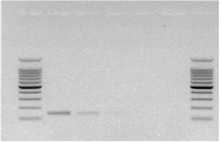
For the 33 strains outside of the P. syringae group that were tested here, no visible PCR products were obtained (Table1, Fig. 1). For the 185 P. syringae strains, a PCR product of the expected size was obtained for 97% (180) of the strains. A PCR product could not be obtained for fewer than 3 % (5) of the strains (Table 1). Strains for which a PCR product could not be obtained comprised 1 strain from each of phylogroups 7 and 11 and all of the three strains tested for phylogroup 12. For the other ten phylogroups represented in this analysis (phylogroups 1 to 6, 8 to 10 and 13), 100 % of strains were positive for the Psy-PCR test (Table 1). The small number of strains currently available for phylogroup 12 limits the scope of the findings for this group. In phylogroup 11, one strain (83.1) among the 7 tested was negative, but we obtained a positive amplification (single weak product band) by increasing cell concentrations (>108 cfu mL-1) (Fig 1).
by guest on December 1, 2015
http://femsec.oxfordjournals.org/
Version postprint
Sensitivity of Psy-PCR
The Psy-PCR tested on a range of decimal dilutions of P. syringae cultures with the standard protocol showed that for cell suspensions of concentrations less than 106 cfu mL-1,
corresponding to 2 103 cfu in a reaction mixture of 25 μL, no visible product was obtained. Using purified DNA, the minimum concentration to obtain a positive PCR was 0.4 ng µL-1 corresponding to 0.8 ng per PCR reaction.
A touchdown Psy-PCR was tested and it improved the specificity of the test, by allowing the detection of two strains from phylogroup 12 (PG12) and one strain from phylogroup 11 (PG11) that were negative with the standard protocol (Table 2). For these three strains and for one more strain from PG11,a product band was visible with a cell suspension at 106 or 107cfu mL-1 while for the other P. syringae strains the minimal concentration to obtain a visible product was between 103 to 105 cfu mL-1 (corresponding to 2 to 200 cfu per 25 μL reaction). Strain PV612 was the only strain that gave negative results for all PCR using either standard or TD-PCR. Using purified DNA, the minimum concentration needed to obtain a positive result with TD-PCR was less than 0.1 ng µL-1.
Characterization of strains giving negative results with standard Psy-PCR
We investigated why some strains (PV612, 83.1, GAW0112 and 113, see Table S1) did not amplify with the standard Psy-PCR. Primers were designed to the conserved sequences flanking the region amplified by the diagnostic primers and used with these strains. PCR products were sequenced and aligned with sequences of control strains (P. syringae and non-P. syringae Pseudomonas strains). This revealed between 1 to 3 mismatches in the sequence corresponding to the Psy-PCR reverse primer (Figure S3). A PCR with the forward primer Psy_F combined with a degenerated reverse primer (CGC YCT TKM GGC WAG CAC HC) that should amplify the sequences of these strains, was tested with the standard
protocol. This PCR gave false positive responses and worked only with an expensive Taq DNA polymerase used for multiplex PCR. A more intensive exploration is needed to elaborate a protocol that yields no false responses.
The use of Psy-PCR improves the isolation efficiency of P. syringae from environmental samples
P. syringae was isolated from Tarn river water after concentration of samples via filtration.
Using the conventional screening procedure, 24 colonies were identified as putative P.
syringae strains among the 234 colonies that grew on the three plates of KBC medium
(Table 3). Expertise of the operator was used to detect the putative P. syringae based mainly on colony morphology, pigments and the absence of cytochrome C oxidase. Some
by guest on December 1, 2015
http://femsec.oxfordjournals.org/
Version postprint
fluorescent but morphologically-typical colonies were also selected among these 24 colonies (Table 3). Phylogenetic analysis of partial cts gene sequences confirmed that 22 of these 24 strains were P. syringae and 2 strains (TAW80 and TAW100) belonged to other
Pseudomonas species (Table 3, Fig 2). Psy-PCR was positive for the 22 P. syringae strains
and negative for TAW080 and TAW100 strains. Overall for the conventional isolation method, 9.4 % of the colonies obtained on KBC medium were identified as P. syringae. For the Psy-PCR-assisted screening procedure, all 95 colonies growing on one plate of KBC medium were analyzed with Psy-PCR conducted directly with cells from the colonies. Among these 95 colonies, 13 gave positive results in a preliminary Psy-PCR (Table S2). All 95 colonies were purified, yielding 108 strains. Among them 23 were no longer culturable after one sub-culturing (S2W-13, 18, 19, 22, 30, 34, 36 to 39, 42 to 47, 58 to 60, 70, 72, 73B, 81B). The non-culturability of most environmental bacteria has been brought to light previously in fresh water in particular (Amann et al., 1995). Morphological differences after purification revealed that some of the colonies initially obtained on KBC medium were mixtures of strains. The Psy-PCR-assisted method indicated that among the 85 culturable strains 17 gave a positive response. Phylogenetic classification of positive strains confirmed these 17 strains to be P. syringae. One of these strains (S2W-16B) originated from a mixed isolate that tested negative in the first preliminary Psy-PCR (Table S2, Fig 2). One isolate (S2W-7) that tested positive in the first screen did not yield a P. syringae strain after purification (Table S2). Overall for the PCR-assisted method of isolation, 15 % of the colonies on KBC medium were identified as P. syringae in that they represented single strains of P. syringae or mixtures of P. syringae with other bacteria. Phenotypic tests of these
P. syringae strains were variable particularly for strains belonging to phylogroup 7 (P. viridiflava) that could be non-fluorescent and variable for the oxidase test (Table S2). When
comparing efficiency, Psy-PCR was better than the conventional method in particular for its higher relative accuracy and sensitivity (Table S3). The intraspecific diversity of the isolated
P. syringae strains was comparable between the two protocols (Fig. 1). Altogether strains
belonged to 5 P. syringae phylogroups among the 13 groups currently known and were dominated by phylogroups PG02 and PG07.
Discussion
We have developed a molecular probe for rapid, specific and sensitive identification of strains in the entire P. syringae group. Psy-PCR can be used directly with cells from colonies thereby permitting unbiased sorting of colonies for further characterization. This eliminates the need for expert recognition of P. syringae colonies or the bias that can be caused due to unexpected phenotypic variability in traits that have been used in the past to select putative
P. syringae strains (Bartoli et al., 2014, Berge et al., 2014). We have also shown that typical
by guest on December 1, 2015
http://femsec.oxfordjournals.org/
Version postprint
P. syringae colony morphology can be masked by the mixture of P. syringae with other
bacteria. This probe has been developed and validated on the basis of strains from the collection of P. syringae chosen for their geographical origin and isolation substrate (Table S1) and representing the full genetic diversity of the 13 phylogroups (Parkinson et al., 2011, Berge et al., 2014). This PCR tool will greatly facilitate the isolation of P. syringae especially from substrates where this bacterium is not very frequent. A direct PCR on DNA from substrates could be used to detect the presence of P. syringae especially when populations are high such as in plants or after an enrichment when populations are low (data not shown). A QRT analysis of DNA using the same DNA region could be employed to estimate the total
P. syringae population size within the samples. However, in this paper we chose to develop a
method combining the cultivation of bacteria and PCR detection. Cultivating bacteria is justified by the fact that a very efficient isolation method based on cultivation is available and able to detect low densities of this bacterium from environmental samples. By concentrating water samples by a factor of 100 x, for example, densities of cells as low as 102 cells of P.
syringae L-1 can be detected with cultivation methods (Morris et al., 2008). Direct PCR applied to the same concentrated samples would require about 10 times more bacteria per sample because of the smaller volumes of sample that are typically processed in PCR reactions than in dilution plating. In addition, obtaining isolated strains is necessary to
determine the ensemble of traits associated with a given strain (pathogenicity, ice nucleating activity, etc). Psy-PCR is a powerful, effective tool that opens up many possibilities for exploring the diversity of P. syringae and its link to disease epidemiology.
Diagnosis of plant disease and the development of molecular tools to assist this diagnosis generally focus on one or a limited number of lineages that have been isolated from the populations that are dominant in infected tissue. As a consequence, highly specific probes limited to a narrow genetic range of strains have been proposed for diagnosis. Examples for the case of phytopathogenic P. syringae sensu lato, including P. cichorii (Hseu et al., 2006),
P. savastanoi (Penyalver et al., 2000), P. s. pv. tomato (Zaccardelli et al., 2005), P. s. pv. actinidiae (Mazzaglia et al., 2011) and P. s. pv. phaseolicolae (Schaad et al., 1995). These
markers are useful to show whether a common strain is found during an outbreak or to trace pathogens in a regulatory legal framework. In contrast to this highly specific approach at the strain or the small group level, a more global method could be used in surveillance of reservoirs that are suspected to harbor a diversity of strains that are potential pathogens. This would be especially important for strains with broad host range as in the case of strains of phylogroup 1 from water (Bartoli et al., 2015) or for which host range is not readily
predictable from other phenotypes such as strains in phylogroups 7 and 8 (Bartoli et al., 2014). A more global approach has been developed for some human pathogens that are ubiquitous in the environment such as P. aeruginosa (Selezska et al., 2012) and for which a
by guest on December 1, 2015
http://femsec.oxfordjournals.org/
Version postprint
PCR that targets the species has been used (De Vos et al., 1997). Likewise a detection method based on PCR was developed for Ralstonia solanacearum validated on diverse strains of the bacterium in several countries and laboratories (Opina et al., 1997) and for
Erwinia amylovora causing fire blight in plants in the Rosaceae family, dispersed by insects
and aerosols (Buhlmann et al., 2013). For P. syringae a key to understanding the role of environmental populations in diseases of crop plants is to clearly account for their diversity, the fluctuations of their populations and their evolution. The corollary to the notion that environmental populations have roles in disease is that these environmental populations themselves play other roles in environmental processes. The most-frequently cited example for P. syringae is its potential role in rainfall due to its ice nucleation activity (Morris et al., 2014). Detection tools that do not place special importance on markers associated with epidemic potential allow this latter concept to be explored more fully.
Acknowledgements
We thank Cécilia Pierre, Gwénaëlle Alcaras and Emiliano Stopelli for their technical help, Cécile Desbiez and Claudia Bartoli for their assistance in the design of PCR primers and conditions and Jean-François Humbert for his support during field work in the Tarn region.
Conflict of interest. None declared
References
Achouak W, Normand P & Heulin T (1999) Comparative phylogeny of rrs and nifH genes in the Bacillaceae. Int J Systematic Bacteriol 49: 961-967.
Amann RI, Ludwig W & Schleifer KH (1995) Phylogenetic identification and in-situ detection of individual microbial-cells without cultivation. Microbiol Reviews 59: 143-169.
Amato P, Parazols, M, Sancelme M, et al. (2007) Microorganisms isolated from the water phase of tropospheric clouds at the Puy de Dome: major groups and growth abilities at low temperatures. Fems Microbiol Ecol 59: 242-254.
Aujoulat F, Roger F, Bourdier A, et al. (2012) From Environment to Man: Genome Evolution and Adaptation of Human Opportunistic Bacterial Pathogens. Genes 3: 191-232. Baltrus DA, Nishimura MT, Romanchuk A, et al. (2011) Dynamic Evolution of Pathogenicity
Revealed by Sequencing and Comparative Genomics of 19 Pseudomonas syringae Isolates. PLoS Pathogens 7.
Bartoli C, Lamichhane JR, Berge O, et al. (2015) A framework to gauge the epidemic
potential of plant pathogens in environmental reservoirs: the example of kiwifruit canker.
Mol Plant Pathol 16: 137-149.
by guest on December 1, 2015
http://femsec.oxfordjournals.org/
Version postprint
Bartoli C, Berge O, Monteil CL, et al. (2014) The Pseudomonas viridiflava phylogroups in the
P. syringae species complex are characterized by genetic variability and phenotypic
plasticity of pathogenicity-related traits. Environ Microbiol 16: 2301-2315.
Berge O, Guinebretiere MH, Achouak W, et al. (2002) Paenibacillus graminis sp. nov. and
Paenibacillus odorifer sp. nov., isolated from plant roots, soil and food. Int J Systematic Evol Microbiol 52: 607-616.
Berge O, Monteil CL, Bartoli C, et al. (2014) A User's Guide to a Data Base of the Diversity of
Pseudomonas syringae and Its Application to Classifying Strains in This Phylogenetic
Complex. PLoS ONE 9.
Buhlmann A, Pothier JF, Rezzonico F, et al. (2013) Erwinia amylovora loop-mediated isothermal amplification (LAMP) assay for rapid pathogen detection and on-site diagnosis of fire blight. J Microbiol Methods 92: 332-339.
Carolan K, Garchitorena A, Garcia-Pena GE, et al. (2014) Topography and Land Cover of Watersheds Predicts the Distribution of the Environmental Pathogen Mycobacterium
ulcerans in Aquatic Insects. PLoS Negl Trop Dis 8.
Cho MS, Jeon YH, Kang MJ, et al. (2010) Sensitive and specific detection of
phaseolotoxigenic and nontoxigenic strains of Pseudomonas syringae pv. phaseolicola by TaqMan real-time PCR using site-specific recombinase gene sequences. Microbiol
Res165: 565-572.
Clarke CR, Cai RM, Studholme DJ, Guttman DS & Vinatzer BA (2010) Pseudomonas
syringae Strains Naturally Lacking the Classical P-syringae hrp/hrc Locus Are Common
Leaf Colonizers Equipped with an Atypical Type III Secretion System. Mol Plant-Microbe
Interact 23: 198-210.
Cottyn B, Baeyen S, Pauwelyn E, et al. (2011) Development of a real-time PCR assay for
Pseudomonas cichorii, the causal agent of midrib rot in greenhouse-grown lettuce, and
its detection in irrigating water. Plant Pathol 60: 453-461.
De Vos D, Lim A, Jr, Pirnay J, et al. (1997) Direct detection and identification of
Pseudomonas aeruginosa in clinical samples such as skin biopsy specimens and
expectorations by multiplex PCR based on two outer membrane lipoprotein genes, oprI and oprL. J Clin Microbiol 35: 1295-1299.
Gallelli A, L'Aurora A & Loreti S (2011) Gene sequence analysis for the molecular detection of Pseudomonas syringae pv. actinidiae: developping diagnostic protocols. Journal of
Plant Pathology 93: 425-435.
Gervasi F & Scortichini M (2009) Detection of Pseudomonas avellanae from hazelnut twigs by taqman real-time PCR. J Plant Pathol 91: 573-578.
Hseu SH, Shentue H & Lin CY (2006) Development of specific PCR primers for identification of Pseudomonas cichorii. Plant Pathol Bul 15: 275-285.
by guest on December 1, 2015
http://femsec.oxfordjournals.org/
Version postprint
Kaluzna M, Janse JD & Young JM (2012) Detection and identification methods and new tests as used and developed in the framework of COST 873 for bacteria pathogenic to stone fruits and nuts Pseudomonas syringae pathovars. J Plant Pathol 94: S1.117-S111.126. King EO, Ward MK & Raney DE (1954) Two simple media for the demonstration of
pyocyanin and fluorescin. The J lab and clinical medicine 44: 301-307.
Kong H, Patterson CD, Zhang W, et al. (2004) A PCR protocol for the identification of
Pseudomonas syringae pv. tagetis based on genes required for tagetitoxin production Biol Control - 30: 83- 89.
Lamichhane JR, Messean A. & C.E. M (2015) Insights into epidemiology and control of diseases of annual plants caused by Pseudomonas syringae. J Gen Plant Pathol 81: 331-350.
Lamichhane JR, Varvaro L, Parisi L, et al. (2014) Disease and frost damage of woody plants caused by Pseudomonas syringae: seeing the forest for the trees. Adv Agronomy 126: 235-295.
Mazzaglia A, Renzi M & Balestra GM (2011) Comparison and utilization of different PCR-based approaches for molecular typing of Pseudomonas syringae pv. actinidiae strains from Italy. Can J Plant Pathol 33: 8-18.
Mohan SK & Schaad NW (1987) An Improved Agar Plating Assay for Detecting
Pseudomonas-syringae pv syringae and P-S pv phaseolicola in Contaminated Bean
Seed. Phytopath 77: 1390-1395.
Monteil CL, Cai RM, Liu HJ, et al. (2013) Non agricultural reservoirs contribute to emergence and evolution of Pseudomonas syringae crop pathogens. New Phytol 199: 800-811. Morris CE, Monteil CL & Berge O (2013) The Life History of Pseudomonas syringae: Linking
Agriculture to Earth System Processes. Annu Rev Phytopathol, Vol 51 51: 85-104. Morris CE, Bardin M, Kinkel LL, et al. (2009) Expanding the Paradigms of Plant Pathogen
Life History and Evolution of Parasitic Fitness beyond Agricultural Boundaries. Plos
Pathog 5 e1000693.
Morris CE, Conen F, Huffman JA, et al. (2014) Bioprecipitation: a feedback cycle linking Earth history, ecosystem dynamics and land use through biological ice nucleators in the atmosphere. Glob Chang Biol 20: 341-351.
Morris CE, Sands DC, Vanneste JL, et al. (2010) Inferring the Evolutionary History of the Plant Pathogen Pseudomonas syringae from Its Biogeography in Headwaters of Rivers in North America, Europe, and New Zealand. Mbio 1: 00107–00110.
Morris CE, Sands DC, Vinatzer BA, et al. (2008) The life history of the plant pathogen
Pseudomonas syringae is linked to the water cycle. Isme J 2: 321-334.
Mulet M, Lalucat J & Garcia-Valdes E (2010) DNA sequence-based analysis of the
Pseudomonas species. Environ Microbiol 12: 1513-1530.
by guest on December 1, 2015
http://femsec.oxfordjournals.org/
Version postprint
Opina N, Tavner F, Hollway G, et al. (1997) A novel method for development of species and strain-specific DNA probes and PCR primers for identifying Burkholderia solanacearum (formerly Pseudomonas solanacearum). Asia Pac J Mol Biol Biotechnol 5: 19-30. Parkinson N, Bryant R, Bew J, et al. (2011) Rapid phylogenetic identification of members of
the Pseudomonas syringae species complex using the rpoD locus. Plant Pathol J 60: 338–344.
Penyalver R, Garcia A, Ferrer A, et al. (2000) Detection of Pseudomonas savastanoi pv.
savastanoi in olive plants by enrichment and PCR. Appl Environ Microbiol 66:
2673-2677.
Popovic T, Balaz J & Stankovic S (2014) A Method for the Rapid Detection and Identification of Halo Blight Pathogen on Common Bean. Arch Biol Sci 66: 1393-1399.
Quiblier C, Wood S, Echenique-Subiabre I, et al. (2013) A review of current knowledge on toxic benthic freshwater cyanobacteria - Ecology, toxin production and risk management.
Water Res 47: 5464-5479.
Sarkar SF & Guttman DS (2004) Evolution of the core genome of Pseudomonas syringae, a highly clonal, endemic plant pathogen. Appl Environ Microbiol 70: 1999-2012.
Schaad NW, Cheong SS, Tamaki S, et al. (1995) A Combined Biological and Enzymatic Amplification (Bio-Pcr) Technique to Detect Pseudomonas syringae pv phaseolicola in Bean Seed Extracts. Phytopathology 85: 243-248.
Selezska K, Kazmierczak M, Musken M, et al. (2012) Pseudomonas aeruginosa population structure revisited under environmental focus: impact of water quality and phage pressure. Environ Microbiol 14: 1952-1967.
Tegli S, Cerboneschi M, Libelli IM, et al. (2010) Development of a versatile tool for the simultaneous differential detection of Pseudomonas savastanoi pathovars by End Point and Real-Time PCR. Bmc Microbiol 10: 156.
Vaseghi A, Bakhshinejad B, Safaie N, et al. (2014) PCR amplification of the hrcV gene through specific primers for detecting Pseudomonas syringae pathovars. World J
Microbiol & Biotechnol 30: 413-421.
Vayssier-Taussat M (2014) Lyme and associated tick-borne diseases: global challenges in the context of a public health threat. Front Cell Infect Microbiol 4.
Versalovic J, Schneider M, de Bruin FJ, et al. (1994) Genomic fingerprint of bacteria using repetitive sequence-based polymerase chain reaction. Meth Mol Cell biol 5: 25-40. Weisburg WG, Barns SM, Pelletier DA, et al. (1991) 16S ribosomal DNA amplification for
phylogenetic study. J Bacteriol 173: 697 - 703.
Zaccardelli M, Spasiano A, Bazzi C, et al. (2005) Identification and in planta detection of
Pseudomonas syringae pv. tomato using PCR amplification of hrpZ(Pst). Eur J Plant Pathol 111: 85-90.
by guest on December 1, 2015
http://femsec.oxfordjournals.org/
Version postprint
by guest on December 1, 2015
http://femsec.oxfordjournals.org/
Version postprint
Table 1. Specificity of standard Psy-PCR based on 185 strains from the 13 P. syringae phylogroups and on 31 non-P. syringae strains. Additional information about strains is provided in Table S1
P. syringae phylogroups or other
species* N° of strains Positive Psy-PCR reaction % positive strains P. syringae PG01 24 24 100 P. syringae PG02 47 47 100 P. syringae PG03 19 19 100 P. syringae PG04 4 4 100 P. syringae PG05 2 2 100 P. syringae PG06 1 1 100 P. syringae PG07 32 31 97 P. syringae PG08 5 5 100 P. syringae PG09 10 10 100 P. syringae PG10 25 25 100 P. syringae PG11 7 6 86 P. syringae PG12 3 0 0 P. syringae PG13 6 6 100
Total P. syringae strains 185 180 97
Pseudomonas aeruginosa group 1 0 0
Pseudomonas fluorescens group 16 0 0
Pseudomonas lutea group 2 0 0
Pseudomonas stutzeri group 2 0 0
Pseudomonas putida group 1 0 0
Pectobacterium sp. 3 0 0 Agrobacterium sp. 1 0 0 Erwinia sp. 3 0 0 Burkholderia 1 0 0 Dickeya 1 0 0 Pantoea sp. 1 0 0 Escherichia coli 1 0 0
Total Non-P. syringae strains 33 0 0
* the P. syringae phylogroups and Pseudomonas groups refer to the current classifications (Mulet, et al., 2010, Berge, et al., 2014)
by guest on December 1, 2015
http://femsec.oxfordjournals.org/
Version postprint
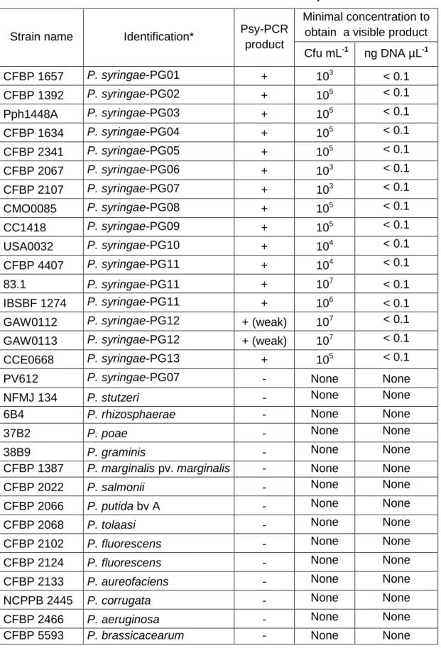
Table 2. Specificity and sensitivity of touch-down Psy-PCR. Fourteen strains from the 13 P. syringae phylogroups and 14 non-P. syringae strains were used. For all tested strains, decimal intervals of concentrations from 102 to 109 cells mL-1 were tested. Additional information about strains is provided in Table S1.
Strain name Identification* Psy-PCR
product
Minimal concentration to obtain a visible product Cfu mL-1 ng DNA µL-1 CFBP 1657 P. syringae-PG01 + 103 < 0.1 CFBP 1392 P. syringae-PG02 + 105 < 0.1 Pph1448A P. syringae-PG03 + 105 < 0.1 CFBP 1634 P. syringae-PG04 + 105 < 0.1 CFBP 2341 P. syringae-PG05 + 105 < 0.1 CFBP 2067 P. syringae-PG06 + 103 < 0.1 CFBP 2107 P. syringae-PG07 + 103 < 0.1 CMO0085 P. syringae-PG08 + 105 < 0.1 CC1418 P. syringae-PG09 + 105 < 0.1 USA0032 P. syringae-PG10 + 104 < 0.1 CFBP 4407 P. syringae-PG11 + 104 < 0.1 83.1 P. syringae-PG11 + 107 < 0.1 IBSBF 1274 P. syringae-PG11 + 106 < 0.1
GAW0112 P. syringae-PG12 + (weak) 107 < 0.1
GAW0113 P. syringae-PG12 + (weak) 107 < 0.1
CCE0668 P. syringae-PG13 + 105 < 0.1
PV612 P. syringae-PG07 - None None
NFMJ 134 P. stutzeri - None None
6B4 P. rhizosphaerae - None None
37B2 P. poae - None None
38B9 P. graminis - None None
CFBP 1387 P. marginalis pv. marginalis - None None
CFBP 2022 P. salmonii - None None
CFBP 2066 P. putida bv A - None None
CFBP 2068 P. tolaasi - None None
CFBP 2102 P. fluorescens - None None
CFBP 2124 P. fluorescens - None None
CFBP 2133 P. aureofaciens - None None
NCPPB 2445 P. corrugata - None None
CFBP 2466 P. aeruginosa - None None
CFBP 5593 P. brassicacearum - None None
by guest on December 1, 2015
http://femsec.oxfordjournals.org/
Version postprint
* the P. syringae phylogroups refer to the current P. syringae classification (Berge, et al., 2014)
by guest on December 1, 2015
http://femsec.oxfordjournals.org/
Version postprint
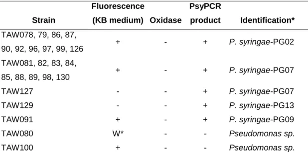
Table 3. Characteristics and identification of putative P. syringae strains isolated from Tarn river water with the conventional procedure based on phenotype screening Strain Fluorescence (KB medium) Oxidase PsyPCR product Identification* TAW078, 79, 86, 87, 90, 92, 96, 97, 99, 126 + - + P. syringae-PG02 TAW081, 82, 83, 84, 85, 88, 89, 98, 130 + - + P. syringae-PG07 TAW127 - - + P. syringae-PG07 TAW129 - - + P. syringae-PG13 TAW091 + - + P. syringae-PG09 TAW080 W* - - Pseudomonas sp. TAW100 + - - Pseudomonas sp.
* Identification was performed using the phylogenetic analysis of partial cts sequences (see Fig. 2). P. syringae phylogroups refer to the current P.
syringae classification (Berge, et al., 2014).W : weak
by guest on December 1, 2015
http://femsec.oxfordjournals.org/
Version postprint
FIGURES
Fig. 1: Detection of P. syringae strain with PSY-PCR.
Lane 1 shows positive response, lane 2 and 3 show a single weak product band (obtained for example with strain 83.1 or with strains of PG12 with Touch-Down-PCR), lane 4 shows a negative response.and lane 5 is the control (water). A 100 bp molecular weight ladder was used.
by guest on December 1, 2015
http://femsec.oxfordjournals.org/
Version postprint
Fig. 2 : Phylogenetic tree of P. syringae strains isolated from Tarn river water.
Strains were screened based on expertise of the operator (“TAW” strains for TArn Water) or based on Psy-PCR-assisted screening (“S2W” strains for Strategy-2 Water). Tree was
constructed with Neighbor-joining method based on cts partial sequences.
PG 1 PG 9 PG 7 PG 8 PG 10 PG 5 PG 4 PG 3 PG 2 PG 6 Pseudomonas sp. PG 13 PG 12 P. graminis -rhyzospharae PG 11 TAW99 B728A PG2d TAW87 TAW86 TAW78 TAW79 S2W-79 MAFF302273PT PG2d S2W-81A S2W-6A TAW90 TAW92 TAW126 S2W-9 TAW96 CC0094 PG2d USA0035 PG2f Cit7 PG2a TA0001 PG2b AI0042 PG2e CFBP1392 PG2b CC1631 PG2c PSS508 PG2c TAW97 TA0034 PG2c PSS642 PG2c S2W-2A S2W-2B S2W-3 S2W-6B 1448A PG3 ATCC11528 PG3 0893 23 PG3 ES4326 PG5 TA0011 PG1b CC1416 PG1b DC3000 PG1a M302278PT PG1a CC1629 PG4 CFBP2067 PG6 USA0102 PG10a CC1586 PG10c CVB0092 PG10d CCE0153 PG10f USA0032 PG10e TA0018 PG10b TA0019 PG10b TA0022 PG10b TA0003 PG10b TA0005 PG10b TA0008 PG10b TA0009 PG10b TA0014 PG10b S2W-66B S2W-76B TA0006 PG9b TAW91 CC1524 PG9a CMW020 PG9c CMO0085 PG8 GAW203 PG8 FMU107 PG7b S2W-16B TAW98 PV612 PG7a TAW85 TA0002 PG7a TAW82 S2W-67B S2W-66A S2W-65 S2W-12B S2W-12A TA0020 PG7a TA0043 PG7a TAW130 TAW81 TAW83 TAW84 TAW88 TAW89 TAW127 83.1 PG11 S2W-86 CFBP1477 PG11 CFBP4407 PG11 GAW0112 PG12a GAW0113 PG12b TAW129 CLA0302 PG13b UB0246 PG13a 6B4 Ps rhizosphaerae 38B9 Ps graminis S2W-68B S2W-71B 13B3 Ps graminis TAW80 S2W1-84 Pf-05 Ps protegens TAW100 PAO1 Ps aeruginosa 99 99 61 99 91 51 98 98 96 99 90 53 89 99 89 99 99 99 46 99 75 91 88 56 64 35 25 22 38 89 70 67 99 97 67 93 88 91 82 93 87 63 94 75 84 83 71 95 53 61 21 37 78 25 19 46 46 65 73 92 72 83 50 84 90 89 23 82 78 83 0.01 by guest on December 1, 2015 http://femsec.oxfordjournals.org/ Downloaded from