HAL Id: hal-02094488
https://hal-amu.archives-ouvertes.fr/hal-02094488
Submitted on 9 Apr 2019
HAL is a multi-disciplinary open access
archive for the deposit and dissemination of
sci-entific research documents, whether they are
pub-lished or not. The documents may come from
teaching and research institutions in France or
abroad, or from public or private research centers.
L’archive ouverte pluridisciplinaire HAL, est
destinée au dépôt et à la diffusion de documents
scientifiques de niveau recherche, publiés ou non,
émanant des établissements d’enseignement et de
recherche français ou étrangers, des laboratoires
publics ou privés.
Whipple’s Disease: Diagnostic Value of rpoB Gene PCR
from Peripheral Blood Mononuclear Cells.
Kathleen Weigt, Alexandra Wiessner, Annette Moter, Florence Fenollar,
Didier Raoult, Kristina Allers, Thomas Schneider
To cite this version:
Kathleen Weigt, Alexandra Wiessner, Annette Moter, Florence Fenollar, Didier Raoult, et al..
Whip-ple’s Disease: Diagnostic Value of rpoB Gene PCR from Peripheral Blood Mononuclear Cells..
Molec-ular Diagnosis and Therapy, Adis, 2018, 22 (4), pp.459-469. �10.1007/s40291-018-0339-7�.
�hal-02094488�
S H O R T C O M M U N I C A T I O N
Whipple’s Disease: Diagnostic Value of rpoB Gene PCR
from Peripheral Blood Mononuclear Cells
Kathleen Weigt1 •Alexandra Wiessner2• Annette Moter2•Florence Fenollar3•Didier Raoult3• Kristina Allers1•Thomas Schneider1•Verena Moos1
Published online: 7 June 2018
Ó Springer International Publishing AG, part of Springer Nature 2018
Abstract
Introduction Chronic infection with Tropheryma whipplei, known as Whipple’s disease (WD), classically affects the gastrointestinal tract, but any organ system may be affec-ted, and isolated manifestations occur. Reliable diagnosis based on a combination of periodic acid–Schiff (PAS) staining, T. whipplei-specific immunohistochemistry (IHC), and polymerase chain reaction (PCR) from duode-nal biopsies may be challenging in cases without classical gastrointestinal infection, so the need for additional diag-nostic materials is urgent.
Objective Our objective was to evaluate additional diag-nostic possibilities for WD.
Methods We analyzed samples from 20 patients with WD and 18 control subjects in a prospective observational pilot study. In addition to WD diagnosis by PAS staining, T. whipplei-specific IHC and PCR of duodenal or extra intestinal tissues, whole EDTA blood, peripheral blood mononuclear cells (PBMCs) and PBMC fractions enriched with or depleted of cluster of differentiation (CD)-14?cells were examined using T. whipplei rpoB gene PCR. Results Tropheryma whipplei DNA was detected in 35 of 60 (58.3%) preparations from 16 of 20 patients with WD,
most of whom lacked gastrointestinal signs and charac-teristic PAS-positive duodenal macrophages.
Conclusion This study provides evidence for the potential suitability of blood, particularly PBMCs, as material to assist in the diagnosis of WD via rpoB gene real-time PCR. Thus, PCR from blood preparations can be helpful for diagnostic decision making in atypical cases of WD.
Key Points
RpoB gene polymerase chain reaction (PCR) can be an additional tool to assist in the diagnosis of Whipple’s disease.
RpoB gene PCR from blood fractions, especially peripheral blood mononuclear cells (PBMCs), enables the surveillance of treatment efficacy and thus appears to have advantages over
immunohistochemistry (IHC) and periodic acid– Schiff (PAS) staining.
1 Introduction
The rare systemic chronic infection with Tropheryma whipplei [1], known as Whipple’s disease (WD), has an estimated incidence of 1:1,000,000 [2, 3]. The classical clinical features of WD are polyarthritis, diarrhea, weight loss, and fever [2]. However, as T. whipplei can affect almost any organ, WD cases with non-classical symptoms can be frequent. The standard for diagnosis of WD is
& Verena Moos
verena.moos@charite.de
1 Department for Gastroenterology, Infectious Diseases, and
Rheumatology, Charite´-University Medicine Berlin, Campus Benjamin Franklin, Hindenburgdamm 30, 12203 Berlin, Germany
2 Biofilmcenter, German Heart Institute Berlin, Berlin,
Germany
3 URMITE, UMR IRD 198/CNRS 6236, Medical Faculty,
Mediterranean University, Marseille, France https://doi.org/10.1007/s40291-018-0339-7
usually based on periodic acid–Schiff (PAS) staining of macrophages in the duodenal lamina propria [2] and should be confirmed by an independent specific method such as polymerase chain reaction (PCR) [4–7]. However, no systematic data are available concerning the sensitivity or specificity of these routine tests, and no ‘‘gold standard’’ is available. Improved diagnostic methods and clinical awareness means that non-classical WD, often associated with isolated infection in the joints, heart valves, or central nervous system [8–10], is increasingly being recognized [10,11]. In cases without the gastrointestinal involvement typical of classical WD (i.e., in the absence of gastroin-testinal symptoms and positive PAS staining from the duodenum), confident diagnosis depends on methods that are not routinely available (such as T. whipplei-specific immunohistochemistry [IHC]) or analysis of additional biopsies of the affected organ or heart valve. It appears that PCR and IHC of duodenal tissue can identify more patients and has similar WD detection rates in cases without PAS-positive duodenal macrophages [4, 12]. However, the sensitivity and specificity of PCR and IHC has not been evaluated systematically. Therefore, an additional diag-nostic tool that is less invasive would be helpful in inconclusive cases.
Relman et al. [7] proposed the detection of T. whipplei DNA in blood as a promising alternative. However, detection rates using PCR from the whole blood of patients with active WD targeting repeated sequences of T. whipplei [4] or from peripheral blood mononuclear cells (PBMC) targeting T. whipplei-specific 16S rDNA [13] have so far been low. These low rates may have been due to the genes
targeted for detection, DNA being prepared by methods only optimized for the isolation of eukaryotic DNA, or insufficient amount of material (e.g., 200 ll of fluid in Fenollar et al. [4]). Here, we aimed to assess the value of a previously validated more sensitive rpoB gene PCR [14] for the detection of T. whipplei DNA in whole blood and PBMCs following DNA isolation optimized for mycobac-teria [14]. In addition, given that Raoult et al. [15] reported the immunodetection of T. whipplei in circulating mono-cytes, we evaluated whether the enrichment of cluster of differentiation (CD)-14? monocytes is advantageous for the detection of T. whipplei DNA.
Therefore, we conducted a prospective observational pilot study to evaluate the relevance of rpoB gene real-time PCR [14] from EDTA blood, purified PBMCs, and PBMC fractions enriched with or depleted of CD14?monocytes in patients with chronic T. whipplei infection to assist in the diagnosis of WD.
2 Material and Methods
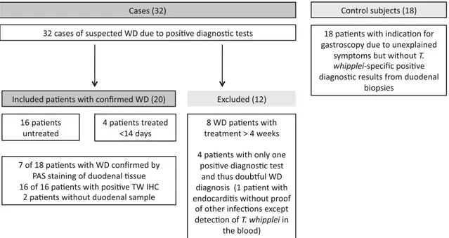
2.1 PatientsWe included 32 patients with symptoms compatible with WD (e.g., treatment-resistant seronegative rheumatoid arthritis, gastrointestinal symptoms) and 18 control sub-jects with other indications for control gastroscopy from May 2014 until May 2017 (Fig.1). Cases were identified as previously described [12], with at least two positive results from routine PAS staining, T. whipplei-specific IHC [16]
Fig. 1 Details of the analysed cohort with excluded cases, confirmed Whipple disease, and control subjects. WD Whipple’s disease, PAS periodic acid–Schiff, PCR polymerase chain reaction, TW IHC T. whipplei-specific immunohistochemistry
from duodenal or extra intestinal tissues (e.g., lymph node, cardiac valve), or rpoB gene real-time PCR [14] from tis-sues or fluids (e.g., cerebrospinal fluid, synovial fluid). Every positive rpoB–PCR result was confirmed by con-ventional T. whipplei-specific 16S rRNA amplification based on a slightly modified protocol published by von Herbay et al. [17], using primers TPU5 [18] and whip1 and whip2 [17], subsequent gene sequencing, and comparison with all currently available sequences from public data-bases (EMBL and GenBank) [14]. Patients with charac-teristic PAS-positive macrophages in duodenal samples and gastrointestinal signs were defined as having classical WD. We recruited 18 people with indications for gas-troscopy but without positive diagnostic tests for T. whip-plei as control subjects (Table1). All control subjects had received a diagnosis other than WD and responded to appropriate treatment; no further indication of WD was found during follow-up (Table1). A total of 12 patients were excluded because they had only a single positive test, meaning the WD diagnosis was doubtful, or they had received treatment for [ 4 weeks. Among the 20 patients with confirmed WD enrolled in this study (see Table1and Fig.1 for details), 16 were previously untreated and four (patients 12–14 and 17) had received antibiotic treatment up to 14 days before analysis. For six patients (patients 1, 3, 5, 10, 13, and 17), follow-up examination was performed 3 months after initiation of antibiotic treatment (see Table1for details). All patients and control subjects gave informed consent, and one patient with classical WD (pa-tient 5, see Table1) is described in a published case report [19].
2.2 Sampling of Blood, Isolation of Peripheral Blood Mononuclear Cells (PBMCs) and Sorting of Cluster of Differentiation (CD)-141 and CD142 PBMCs
Blood samples were stored at room temperature and pro-cessed within 24 h after sampling. Peripheral blood was collected in EDTA tubes (Vacutainer, BD Biosciences) for whole blood analysis, and PBMCs were isolated from heparinized blood as previously described [20]. CD14? monocytes were isolated from PBMCs using CD14 MicroBeads (1:10), LS columns, and a MidiMACS magnet according to the manufacturer’s protocol (Miltenyi Biotec, Bergisch Gladbach, Germany) [21]. The throughputs of the CD14 MACS sort depleted of CD14?monocytes served as CD14-fractions of PBMCs. Since we hypothesized that T. whipplei more reliably persist in CD14? monocytes, the fraction depleted of CD14?cells was intended to serve as a kind of negative control with lower detection rates. CD14? purity of the CD14? fractions was estimated using flow cytometry with a CD14 (61D3) antibody from eBiocience
(Frankfurt, Germany). While CD14?fractions of patients with WD (n = 16) and of control subjects (n = 11) revealed a similar mean percentage of total CD14?(WD: 91.03 ± 7.15%; controls: 96.26 ± 5.67%), CD14highwere significantly lower (WD: 77.75 ± 16.08%; controls: 94.68 ± 7.65%; p = 0.019) and CD14low were signifi-cantly higher in patients with WD than in control subjects (WD: 11.46 ± 9.10%; controls: 1.59 ± 2.04%; p = 0.003). CD14- fractions were analyzed for only five patients with WD and revealed a contamination of 8.56 ± 4.64% of CD14lowand 0.70 ± 0.44% of CD14high monocytes.
2.3 DNA Extraction
Total genomic DNA was extracted from 1 ml frozen EDTA blood or, corresponding to the cell count of 1 ml blood, from 2 9 106cells isolated from heparinized blood (PBMCs and cell fractions enriched with or depleted of CD14?monocytes, respectively). DNA was isolated using the AMPLICOR Respiratory Specimen Preparation Kit (Roche Diagnostics, Mannheim, Germany) according to the manufacturer’s instructions, with the following modi-fication for EDTA blood: twice the recommended volume of ‘‘respiratory specimen wash solution’’ was used in a washing step. For all preparations, elution volumes were identical.
To reduce PCR-inhibitory components in DNA extrac-ted from EDTA blood, an additional purification included column-based enrichment via QIAamp DNA Blood Mini Kit (Qiagen, Hilden, Germany).
2.4 RpoB Gene Real-Time Polymerase Chain Reaction (PCR) Assay
Total genomic DNA was analyzed for T. whipplei DNA by validated rpoB gene real-time PCR as previously described [14]. Primers TwrpoB-F4: CTCGGTGTTGATGTT-GATCCAA and TwrpoB-R: GCACCGCAACCTCGGA-GAAA [22] were used to amplify a 109-bp segment of the T. whipplei rpoB gene from 5 ll of isolated DNA. Real-time detection of the amplicons was achieved using LightCycler hybridization probes TwrpoB-HP1: ACGAGGTCGGATATTATCGC-[FL] (50–30) and Twr-poB-HP2: [Red 640]-ACAATTCGTTATCTCGCGGCC (50–30) [14]. Oligonucleotides were synthesized using TIB MOLBIOL (Berlin, Germany), and real-time PCR was performed in a LightCycler instrument, version 2.0 (Roche Diagnostics) as previously described [14].
As standard curve, extracted DNA from a serial dilution of T. whipplei strain Twist ATCC VR-1528 was used, as previously described [14]. The sensitivity of the real-time PCR assay was determined at 17.4 microorganisms per
Table 1 Patients characteristics and summary of histology and PCR results No. Sex Age (year s) Clinical symptoms Diagnosis Treatment Follow- up (months) Histology PCR Duo PAS Duo TW IHC Other material Duo CSF Other material EDTA blood PBMC CD14 ?
CD14-Combined blood results
WD patients M (55%) 58.9 Sensitivity (%) 38.9 (7/ 18) 100 (16/ 16) 100 (12/ 12) 53.3 (8/ 15) 41.2 (7/ 17) 61.1 (11/ 18) 60 (9/ 15) 80 (8/ 10) 80 (16/20) F (45%) Specificity (%) 100 100 100 100 100 100 100 100 100 Controls M (50%) 54.4 PPV (%) 100 100 100 100 100 100 100 100 100 F (50%) NPV (%) 62.1 100 100 41.7 41.2 70.8 68.4 77.8 81.8 WD patients (n = 20) 1 d F 3 7 Joint pain, lymphadenopathy, untreated WD Doxycycline/ hydroxychloroquine 40 -? a nd ?? nd ?? b,c ? nd ? 2 M 54
Lymphadenopathy, seronegative polyarthritis, untreated
WD Ceftriaxone and TMP/SFX 38 -? nd ?? nd -nd ? nd ? 3 d M 6 2 Lymphadenopathy, untreated WD Doxycycline/ hydroxychloroquine 37 -? a
Lymph node PAS
? ?-Lymph node ? ?? ? ? ? 4 M 62
Seronegative polyarthritis, untreated
WD Doxycycline/ hydroxychloroquine 30 -? a nd ?-Synovial fluid ? -nd -5 d F 7 4
Seronegative polyarthritis, diarrhoea,
weight loss, orbital inflammation, CWD, untreated WD Ceftriaxone and TMP/SFX 30 ?? nd ? nd Vitreous homour ? ?? ? ? ? 6 F 48
Seronegative polyarthritis, diarrhoea,
weight loss, CWD, untreated WD Ceftriaxone and TMP/SFX 36 ?? nd nd ? nd ?? b,c ? b,c nd ? 7 M 39
Seronegative polyarthritis, weight
loss, untreated WD Ceftriaxone and TMP/SFX 24 -? a
Lymph node PAS
? , IHC ? ?? Lymph node ? -? ? 8 M 73
Seronegative polyarthritis, untreated
WD Doxycycline/ hydroxychloroquine 24 -? a nd ? nd nd -? -c ?? 9 F 79
Seronegative polyarthritis, untreated
WD Doxycycline/ hydroxychloroquine 20 -? a nd ?? Synovial fluid ? , bone marrow ? -? b ? 10 d M 6 9
Seronegative polyarthritis, diarrhoea,
weight loss, CWD, untreated WD Ceftriaxone and TMP/SFX 20 ?? nd ?-nd nd ?? ? ?
Table 1 continued No. Sex Age (year s) Clinical symptoms Diagnosis Treatment Follow- up (months) Histology PCR Duo PAS Duo TW IHC Other material Duo CSF Other material EDTA blood PBMC CD14 ?
CD14-Combined blood results
11
F
6
2
Seronegative polyarthritis, untreated
WD
Doxycycline/ hydroxychloroquine
18
-?
a
Skin biopsy PAS
? , IHC ? nd -nd -12 M 6 8 T. whipplei induced endocarditis, treated for \ 14 days WD Ceftriaxone and TMP/SFX 36 nd nd
Cardiac valve PAS
? , IHC ? nd nd Cardiac valve ? --? nd ? 13 d M 4 1 Weight loss, diarrhoea, CWD, treated for \ 14 days WD Ceftriaxone and TMP/SFX 33 ? nd nd ?? nd ?? ? ? ? 14 M 6 0
Seronegative polyarthritis, treated
for \ 14 days WD Ceftriaxone and TMP/SFX 30 ? a ? a nd nd -Synovial fluid ? ?? ? ? ? 15 M 7 6
Seronegative polyarthritis, untreated
WD Doxycycline/ hydroxychloroquine 17 -? a nd nd -Synovial fluid ? --c -16 F 5 5
Seronegative polyarthritis, weight
loss, untreated WD Ceftriaxone and TMP/SFX 20 -? a Colon PAS ? ?? Colon ? -nd nd -17 d M6 0 T whipplei induced endocarditis, treated for \ 14 days WD Ceftriaxone and TMP/SFX 18 nd nd Colon IHC ? ,
cardiac valve IHC
? nd nd Colon ? , cardiac valve ? nd ? nd nd ? 18 F 3 6
Seronegative polyarthritis, diarrhoea,
CWD, untreated WD Ceftriaxone and TMP/SFX 13 ? nd nd nd ? nd nd ? nd nd ? 19 F 5 3
Seronegative polyarthritis, untreated
WD Ceftriaxone and TMP/SFX 12 -? nd ?-nd -? nd nd ? 20 F 6 9
Seronegative polyarthritis, diarrhoea,
CWD, untreated WD Ceftriaxone and TMP/SFX 9 ?? nd nd nd nd ? nd nd nd ? Control subjects (n = 18) 1 M 52 Abdominal pain,
prepyloric polypoid-ulcerated area,
untreated Gastritis PPI 15 --nd nd nd nd nd -2 M 34 Prolonged abdominal pain, weight loss, untreated Gastritis, pancreatitis PPI 15 --nd nd nd nd nd -3 F 59 Diarrhoea, stomach cramps, untreated IBS, allergy to soja Soja-free diet 15 --nd nd nd nd nd
-Table 1 continued No. Sex Age (year s) Clinical symptoms Diagnosis Treatment Follow- up (months) Histology PCR Duo PAS Duo TW IHC Other material Duo CSF Other material EDTA blood PBMC CD14 ?
CD14-Combined blood results
4 M 62 Abdominal pain, untreated Gastritis PPI 15 --nd nd nd nd nd -5 F 52 Epigastric pain, reflux oesophagitis diarrhoea, untreated Gastroesophageal reflux PPI 15 --nd nd nd nd nd -6 M 49 Abdominal pain, untreated Gastroesophageal reflux PPI 14 --nd --nd -7 M 74 Diarrhoea, abdominal pain, untreated Alcohol induced pancreatitis Alcohol-free diet, analgetics, pancreas enzymes 42 --nd -nd nd -nd -nd -8 F 57 Diarrhoea, abdominal pain, joint pain, untreated IBS, sjo ¨rgrens syndrome COX-2 and peristalsis inhibitors 32 --nd --nd -nd -9 M 63 Lymphadenopathy, fever, untreated Sarcoidosis Immuno-suppressio n 3 0 --nd --nd -10 F 6 0 Joint pain, untreated Rheumatoid arthritis Immuno-suppressio n 2 4 --nd nd -nd -nd -11 F 5 3 Reflux oesophagitis,
hypertensive gastropathy, untreated Gastroesophageal reflux PPI 30 --nd -nd nd nd --nd -12 F 2 5 Weight loss, diarrhoea, stomach cramps, untreated Celiac disease Gluten-free diet 30 --nd -nd nd nd --nd -13 M 4 4 Allergic rash, struma
lymphomatosa Hashimoto, untreated
Gastritis PPI 29 --nd -nd nd nd --nd -14 F 5 9 Diarrhoea, joint pain, epigastric pain, untreated Duodenitis PPI 25 --nd nd nd nd --nd nd -15 M 4 6 Joint pain, untreated Rheumatoid arthritis Immuno-suppressio n 2 0 --nd --nd --nd nd -16 F 7 9 Epigastric pain, diarrhoea, untreated IBS, ischemic colitis Immuno-suppressio n, peristaltic inhibitors 30 --nd -nd nd nd -nd nd -17 M 5 3
Enteropathy, diarrhoea, untreated
HIV enteropathy Anti-retroviral treatment 30 --nd -nd nd nd -nd nd
-5-ll suspension (cycle threshold [CT] 38.0 ± 0.5). At higher dilutions, the positive results were not consistent in triplicate samples. A high (CT 31.0) and a low (CT 33.0) positive control were included in every PCR run to ensure sensitivity of the actual experiment. To assure optimal amplification in a specific blood sample, an inhibition control at the concentration of the low positive control was also included.
2.5 Histology and Immunohistochemistry
Tissue specimens were fixed in formalin and embedded in paraffin, and thin sections were subjected to routine PAS staining. Immunostaining was performed as previously described [16, 23] with rabbit-anti-T. whipplei [16], visu-alized by donkey-anti-rabbit Biotin (Dianova, Hamburg, Germany), Streptavidin-alkaline phosphatase and Fast red (both DakoCytomation, Glostrup, Denmark). Nuclei were counterstained with Meyer’s Hematoxilin (DakoCytomation).
3 Results
Among the 20 patients with WD, only six experienced typical clinical symptoms and revealed characteristic PAS-positive macrophages in the duodenal lamina propria and were thus defined as having classical WD (Table 1, Fig.1). One further patient revealed atypical PAS-positive cells in the duodenal submucosa (Table 1, patient 14). Although the specificity of the PAS staining of duodenal biopsies as the classical diagnostic method for WD for our cohort was 100%, the sensitivity (38.9%) and negative predictive value (62.1%) was very low. T. whipplei-specific IHC of the duodenum and rpoB gene PCR from duodenal tissue revealed positive results for all included patients with WD and none of the control subjects (Table1). Among 11 patients with atypical WD, nine presented with only a faint positive T. whipplei-specific IHC within the duodenal submucosa (Table1, Fig. 2a, b). Two patients were ini-tially identified via histological analysis of lymph nodes excised to exclude malignant disease (Fig.2c, d), and two patients with isolated T. whipplei-induced endocarditis were diagnosed via analysis of cardiac valves (Table1, Fig.2e, f). In addition, PCR from cerebrospinal fluid was positive for 8 of 15 patients (Table1).
The overall detection rate for T. whipplei in blood samples was 35 of 60 samples and evidenced bacterial DNA in 16 of 20 patients (EDTA: 7/17 patients tested; PBMC: 11/18; enriched for CD14?: 9/15; and depleted of CD14?: 8/10) (Table1). Upon analysis of the different blood fractions, rpoB gene PCR revealed T. whipplei DNA in EDTA blood of 7 of 17 patients with WD (sensitivity of
Table 1 continued No. Sex Age (year s) Clinical symptoms Diagnosis Treatment Follow- up (months) Histology PCR Duo PAS Duo TW IHC Other material Duo CSF Other material EDTA blood PBMC CD14 ?
CD14-Combined blood results
18 F 5 8 Weight loss, difficulties in swallowing, untreated Gastritis PPI 15 --nd nd nd nd nd -nd nd -M male, F female, CWD classical Whipple’s disease, Duo duodenum, PAS periodic acid-Schiff, TW T. whipplei , EDTA EDTA-blood, IHC immunohistochemistry , PBMC peripheral blood mononuclear cells, PCR polymerase chain reaction, CD cluster of differentiation, CSF cerebrospinal fluid, PPV positive predictive value, NPV negative predictive value, TMP/SMX trimethoprim/sulfamethoxa zole, IBS irritable bowel syndrome, PPI proton pump inhibitors, nd not done, -negative, ? positive aOnly faintly in the submucosa bThawed PBMC used for rpoB PCR c\ 2 9 106 cells used for rpoB PCR dAdditionally data of three month follow-up examination available, as mentioned in the text
41.2%). The detection rate improved to 61.1% sensitivity when investigating PBMCs (Table1). Similar results were obtained investigating CD14? monocytes. However, the highest sensitivity for T. whipplei DNA (80%) and the most reliable negative predictive value (77.8%) was found in the CD14?-depleted cell fraction (Table1), originally inclu-ded for some patients as a negative control.
An interim analysis after inclusion of 15 patients with WD indicated that enrichment of CD14? cells did not improve the T. whipplei detection rate compared with
PBMCs, as initially hypothesized. Despite the good results with the CD14?-depleted PBMCs, we chose PBMCs to assist in WD diagnosis for subsequent patients, resulting in fewer specimens per patient. This decision was based on the lowest CT values for rpoB gene PCR analysing PBMCs, indicating a more reliable level of T. whipplei DNA. While mean CT values from EDTA blood samples were as high as 38.02 ± 2.02, the analysis of PBMCs resulted in significantly lower mean CT values of 32.75 ± 2.33 (p = 0.0016; Mann–Whitney test). CT values
Fig. 2 Immunohistochemical diagnosis of atypical Whipple’s disease (WD) from duodenal biopsy (a, b), lymph node (c, d), and heart valve (e, f). Panels demonstrate exemplary presentations of periodic acid– Schiff (PAS) staining in purple (a, c, e) and Tropheryma whipplei-specific
immunohistochemistry in bright red (b, d, f): a conventional PAS staining of the duodenum reveals no hint of WD; b T. whipplei-specific
immunohistochemistry identifies numerous infected cells within the duodenal submucosa; c positive PAS staining in a lymph node with lymphangiectasia (positive areas are marked by arrowheads); d corresponding clearly positive T. whipplei-specific immunohistochemistry; einflammatory infiltrate with PAS-positive inclusions in a heart valve (positive areas are marked by arrowheads); f T. whipplei-specific
immunohistochemistry demonstrates infection with T. whipplei; magnification for all pictures 9100
of PBMCs enriched for CD14? (35.22 ± 2.65) and PBMCs depleted of CD14? (34.76 ± 3.68) were also increased compared with the whole PBMC fraction. Since CT values [ 38 are at the limit of rpoB gene PCR sensi-tivity [14], high CT values as observed for EDTA blood and CD14?-depleted and enriched PBMC fractions may produce inconsistent results. In addition, sorting for CD14 is not routine in laboratories, bears a greater risk of con-tamination, and is associated with additional effort and costs.
Within 14 days of antibiotic treatment, T. whipplei DNA was still detected by rpoB gene PCR (Table1, patients 12–14, and 17). However, at the 3-month follow-up examination, none of the six specimens tested revealed a positive PCR, indicating a rapid reduction in bacterial DNA in vivo (patients 1, 3, 5, 10, 13, and 17; data not shown). In addition, no T. whipplei DNA was detected in any of the 44 blood preparations (EDTA blood, unsorted PBMCs, or PBMCs enriched with or depleted of CD14?) from the 18 control subjects. Therefore, although the sen-sitivity of the assay depends on the material used for DNA isolation, rpoB gene PCR of blood samples revealed a specificity and a positive predictive value (PPV) of 100% each in our cohort, confirming this method as a supporting tool in WD diagnosis.
4 Discussion
This study has shown the potential suitability of blood and particularly PBMCs as material for rpoB gene real-time PCR to assist in the diagnosis of WD. Awareness of the possibility of isolated WD, atypical—possibly early—WD without gastrointestinal symptoms, and PAS-positive duo-denum is increasing [4,9,10,12]. Indeed, specific IHC can confirm T. whipplei infection in tissues even in the absence of PAS staining (Fig.2) [12], but this method is only accessible at specialized centers as the antibodies are not commercialized. PCR from duodenal specimens is also highly indicative of WD [4,12]; however, healthy luminal carriage of T. whipplei without invasive infection may also result in positive PCR [4]. In cases with no gastrointestinal infection, sampling of affected tissue may be virtually impossible. Consequently, alternative diagnostic materials to assure diagnosis by T. whipplei-specific PCR from multiple physiologically sterile sites are urgently needed. We demonstrated that detection of T. whipplei DNA in whole blood and PBMCs by rpoB gene PCR seems to be highly indicative of WD, even in the absence of intestinal infection and duodenal PAS staining. In our cohort, PAS staining of duodenal biopsies identified only 38.9% of patients, whereas the combined analysis of PBMCs and PBMC fractions enriched with or depleted of CD14?
monocytes enabled detection of bacterial DNA in 16 of 20 patients with confirmed WD (80%). The feasibility of PCR with PBMCs from patients following short-term treatment was demonstrated by positive results in four patients with WD (two with isolated T. whipplei-induced endocarditis) who received appropriate antibiotic treatment for up to 2 weeks prior to blood analysis.
PCR from PBMCs identified more patients, possibly indicating a higher bacterial load, and revealed an enrich-ment of T. whipplei DNA, indicated by lower CT values as compared with PCR from EDTA blood, and thus seems superior. However, detection of T. whipplei DNA did not increase after enrichment of CD14? monocytes, and T. whipplei DNA was also detected in PBMC depleted of CD14? monocytes. Even though the PBMC fraction depleted for CD14? cells exhibited the best diagnostic results in our cohort, we recommend PBMCs as the most suitable material to assist in the diagnosis of WD. PBMCs enabled a higher detection rate at lower CT values of PCR compared with EDTA blood, and the depletion of CD14? monocytes necessitates an intensive work-up of the mate-rial, which carries a greater risk of contamination. In addition, CD14? depletion might be difficult to introduce into routine diagnostic laboratories.
Obvious advantages of using PBMCs to assist in the diagnosis of WD include the simple accessibility of blood, the feasibility for outpatients, and the ability to standardize sampling and cell numbers for semi-quantitative PCR results. The rpoB real-time PCR sensitivity was determined for approximately 17 target organisms per 5 ll of T. whipplei culture used for DNA extraction (3480 organisms/ ml) [14]. Although the rpoB real-time PCR on PBMCs has not yet been validated, we have already found that 2 9 106 PBMC—a comparatively low number—enabled the detection of T. whipplei. On the basis of our experience, 2 9 106cells correspond to the average amount of PBMCs in 1 ml of peripheral blood of healthy donors. Thus, min-imal blood is needed for T. whipplei screening, which is an advantage for patients with diminished health. Another benefit of rpoB gene PCR from PBMCs could be the rapid clearance of T. whipplei DNA after initiation of treatment, which—unlike PAS and IHC of duodenal specimens—may allow monitoring of treatment efficacy or possibly early detection of relapse. The lack of benefit from enriching CD14?monocytes for detection of T. whipplei DNA hints at a small proportion of infected monocytes only and suggests that T. whipplei are not necessarily only associ-ated with classical CD14high monocytes, as previously presumed, but also with other cells. Classical CD14high monocytes have been demonstrated to eliminate T. whip-plei in vitro [24]; thus, T. whipplei DNA might have been degraded more effectively in the PBMC fractions enriched for CD14? monocytes. In contrast, the persistence of T.
whipplei in monocytes is enabled by interleukin (IL)-16 and has been demonstrated to be associated with an upregulation of CD16 [24]. CD16 is only expressed on non-classical CD14low monocytes, which appear to be elevated during acute infection [25]. The percentage of CD14highmonocytes in the CD14?monocyte fractions of patients with WD was significantly lower as compared with controls (77.75 % in WD patients versus 94.68% in controls). However, non-classical CD14low monocytes were significantly enhanced in the CD14? fraction of patients with WD; when they were included in the analysis, the purity of the CD14?fractions was similar for patients and controls. The non-classical CD14lowmonocytes in the untreated patients with WD included in our cohort might be activated by the pathogen itself or by enhanced lipopolysaccharides in the serum [26]. Thus, the high percentage of CD14low monocytes in patients with WD might be more difficult to access for the antibodies used for depletion, resulting in contamination of the CD14-depleted fraction with CD14lownon-classical monocytes that might be preferentially loaded with T. whipplei.
As classical CD14high monocytes and dendritic cells were both removed from the PBMC fraction by depleting CD14? cells, T. whipplei-positive cells are potentially lymphocytes [27], CD16?CD14low non-classical mono-cytes [24], or basophilic granulocytes. The variable posi-tive results in different cell preparations for some patients possibly indicates day-to-day variability in blood samples or a low level of infected cells in the preparations.
The rpoB gene PCR recognized T. whipplei DNA in PBMCs and enriched CD14? monocytes, respectively, from two patients with T. whipplei-induced endocarditis (Table1, patients 12 and 17, diagnosed from excised heart valve). One additional patient with endocarditis revealed positive PCR results from PBMCs but was excluded because heart valve replacement was not conducted, meaning a second diagnostic finding to confirm WD was not possible (see Fig.1). Thus, this study indicates rpoB gene PCR for the detection of T. whipplei DNA in isolated PBMCs is a promising non-invasive material for decision making in the diagnosis of WD, especially for atypical WD when sampling of infected tissue is challenging (e.g., heart valves).
Further studies to determine diagnostic benefits should include more patients with isolated T. whipplei endo-carditis, healthy patients with T. whipplei in stool samples, and recurrent testing of blood samples and greater cell numbers to determine the intraindividual variability of the results. Importantly, identification of cell populations associated with T. whipplei is of particular interest to understand the pathways of the pathogen in the human body. Until further studies evaluate these issues, this
approach should be restricted to specialized laboratories for inconclusive cases.
Compliance with Ethical Standards
Funding This work was supported by Deutsche Forschungsgemein-schaft SFB633, KFO104, SCHN 616/6-2, European commission QLG1-CT-2002-01049 and Charite´ doctorate grants. The German Consiliary Laboratory for T. whipplei is supported by the Robert Koch Institute. The funders had no role in the study design, the data collection and interpretation, or the decision to submit the work for publication.
Conflict of interest KW, AW, AM, FF, DR, KA, TS, and VM have no conflicts of interest.
Informed consent Informed consent was obtained from all individ-ual participants included in the study (ethical commission of the Charite´; EA4/122/10).
References
1. Fenollar F, Puechal X, Raoult D. Whipple’s disease. N Engl J Med. 2007;356(1):55–66.
2. Schneider T, Moos V, Loddenkemper C, Marth T, Fenollar F, Raoult D. Whipple’s disease: new aspects of pathogenesis and treatment. Lancet Infect Dis. 2008;8(3):179–90.
3. La Scola B, Fenollar F, Fournier PE, Altwegg M, Mallet MN, Raoult D. Description of Tropheryma whipplei gen. nov., sp. nov., the Whipple’s disease bacillus. Int J Syst Evol Microbiol. 2001;51(Pt 4):1471–9.
4. Fenollar F, Laouira S, Lepidi H, Rolain JM, Raoult D. Value of Tropheryma whipplei quantitative polymerase chain reaction assay for the diagnosis of Whipple disease: usefulness of saliva and stool specimens for first-line screening. Clin Infect Dis. 2008;47(5):659–67.https://doi.org/10.1086/590559.
5. Hinrikson HP, Dutly F, Nair S, Altwegg M. Detection of three different types of ‘Tropheryma whippelii’ directly from clinical specimens by sequencing, single-strand conformation polymor-phism (SSCP) analysis and type-specific PCR of their 16S–23S ribosomal intergenic spacer region. Int J Syst Bacteriol. 1999;4:1701–6.
6. Moos V, Schneider T. Changing paradigms in Whipple’s disease and infection with Tropheryma whipplei. Eur J Clin Microbiol Infect Dis. 2011;30(10):1151–8.
7. Relman DA, Schmidt TM, MacDermott RP, Falkow S. Identifi-cation of the uncultured bacillus of Whipple’s disease. N Engl J Med. 1992;327(5):293–301.
8. Fenollar F, Celard M, Lagier JC, Lepidi H, Fournier PE, Raoult D. Tropheryma whipplei endocarditis. Emerg Infect Dis. 2013;19(11):1721–30.
9. Geissdorfer W, Moos V, Moter A, Loddenkemper C, Jansen A, Tandler R, et al. High frequency of Tropheryma whipplei in culture-negative endocarditis. J Clin Microbiol. 2012;50(2):216–22.
10. Lehmann P, Ehrenstein B, Hartung W, Dragonas C, Reischl U, Fleck M. PCR analysis is superior to histology for diagnosis of Whipple’s disease mimicking seronegative rheumatic diseases. Scand J Rheumatol. 2016. https://doi.org/10.1080/03009742. 2016.1183038.
11. Lagier JC, Fenollar F, Raoult D. Whipple’s disease and Tro-pheryma whipplei infections in internal medicine. When to think about it? How to treat? La Revue de medecine interne/fondee par
la Societe nationale francaise de medecine interne. 2014;35(12):801–7. https://doi.org/10.1016/j.revmed.2014.04. 016.
12. Gunther U, Moos V, Offenmuller G, Oelkers G, Heise W, Moter A, et al. Gastrointestinal diagnosis of classical Whipple disease: clinical, endoscopic, and histopathologic features in 191 patients. Medicine. 2015;94(15):714.
13. Marth T, Fredericks D, Strober W, Relman DA. Limited role for PCR-based diagnosis of Whipple’s disease from peripheral blood mononuclear cells. Lancet. 1996;348(9019):66–7.
14. Moter A, Schmiedel D, Petrich A, Wiessner A, Kikhney J, Sch-neider T, et al. Validation of an rpoB gene PCR assay for detection of Tropheryma whipplei: 10 years’ experience in a National Reference Laboratory. J Clin Microbiol. 2013;51(11):3858–61.https://doi.org/10.1128/JCM.01703-13. 15. Raoult D, Lepidi H, Harle JR. Tropheryma whipplei circulating in
blood monocytes. N Engl J Med. 2001;345(7):548.
16. Lepidi H, Costedoat N, Piette JC, Harle JR, Raoult D. Immunohistological detection of Tropheryma whipplei (Whipple bacillus) in lymph nodes. Am J Med. 2002;113(4):334–6. 17. von Herbay A, Ditton HJ, Maiwald M. Diagnostic application of
a polymerase chain reaction assay for the Whipple’s disease bacterium to intestinal biopsies. Gastroenterology. 1996;110(6):1735–43.
18. Lane DJ, Pace B, Olsen GJ, Stahl DA, Sogin ML, Pace NR. Rapid determination of 16S ribosomal RNA sequences for phy-logenetic analyses. Proc Natl Acad Sci USA. 1985;82(20):6955–9.
19. Blessin UB, Fischer A, Schneider T, Moos V, Muller T, Wey-landt KH, et al. More than meets the eye. Gut. 2018;67(1):69. https://doi.org/10.1136/gutjnl-2016-312390.
20. Schinnerling K, Moos V, Geelhaar A, Allers K, Loddenkemper C, Friebel J, et al. Regulatory T cells in patients with Whipple’s disease. J Immunol. 2011;187(8):4061–7.https://doi.org/10.4049/ jimmunol.1101349.
21. Schinnerling K, Geelhaar-Karsch A, Allers K, Friebel J, Conrad K, Loddenkemper C, et al. Role of dendritic cells in the patho-genesis of Whipple’s disease. Infect Immun. 2015;83(2):482–91. 22. Drancourt M, Carlioz A, Raoult D. rpoB sequence analysis of cultured Tropheryma whippelii. J Clin Microbiol. 2001;39(7):2425–30. https://doi.org/10.1128/JCM.39.7.2425-2430.2001.
23. Moos V, Schmidt C, Geelhaar A, Kunkel D, Allers K, Schin-nerling K, et al. Impaired immune functions of monocytes and macrophages in Whipple’s disease. Gastroenterology. 2010;138(1):210–20. https://doi.org/10.1053/j.gastro.2009.07. 066.
24. Desnues B, Raoult D, Mege JL. IL-16 is critical for Tropheryma whipplei replication in Whipple’s disease. J Immunol. 2005;175(7):4575–82.
25. Strauss-Ayali D, Conrad SM, Mosser DM. Monocyte subpopu-lations and their differentiation patterns during infection. J Leukoc Biol. 2007;82(2):244–52. https://doi.org/10.1189/jlb. 0307191.
26. Epple HJ, Friebel J, Moos V, Troeger H, Krug SM, Allers K, et al. Architectural and functional alterations of the small intestinal mucosa in classical Whipple’s disease. Mucosal Immunol. 2017;10(6):1542–52. https://doi.org/10.1038/mi.2017. 6.
27. Fredricks DN, Relman DA. Localization of Tropheryma whip-pelii rRNA in tissues from patients with Whipple’s disease. J Infect Dis. 2001;183(8):1229–37.