Cretaceous Crocodyliforms from the Sahara
Paul C. Sereno 1, †, Hans C.E. Larsson2, ‡
1 Department of Organismal Biology and Anatomy, University of Chicago, Chicago, Illinois 60637, USA 2 Redpath Museum, McGill University, Montreal, Quebec H3A 2K6, Canada
† urn:lsid:zoobank.org:pub:A979ECDE-871F-4AFC-9ABA-63A0FD6DC323
‡ urn:lsid:zoobank.org:author:F1B7E0C5-C76A-44ED-852C-58BF7AB961DB Corresponding author: Paul C. Sereno ([email protected])
Academic editor: Hans Sues | Received 13 November 2009 | Accepted 16 November 2009 | Published 19 November 2009 urn:lsid:zoobank.org:pub:A979ECDE-871F-4AFC-9ABA-63A0FD6DC323
Citation: Sereno PC, Larsson HCE (2009) Cretaceous Crocodyliforms from the Sahara. ZooKeys 28: 1–143. doi: 10.3897/zookeys.28.325
Abstract
Diverse crocodyliforms have been discovered in recent years in Cretaceous rocks on southern landmasses formerly composing Gondwana. We report here on six species from the Sahara with an array of trophic adaptations that signifi cantly deepen our current understanding of African crocodyliform diversity during the Cretaceous period. We describe two of these species (Anatosuchus minor, Araripesuchus wegeneri) from nearly complete skulls and partial articulated skeletons from the Lower Cretaceous Elrhaz Formation (Aptian-Albian) of Niger. Th e remaining four species (Araripesuchus rattoides sp. n., Kaprosuchus saharicus
gen. n. sp. n., Laganosuchus thaumastos gen. n. sp. n., Laganosuchus maghrebensis gen. n. sp. n.) come
from contemporaneous Upper Cretaceous formations (Cenomanian) in Niger and Morocco.
Keywords
crocodyliforms, Metasuchia, Notosuchia, Crocodylia
Introduction
Crocodyliforms were particularly diverse during the Cretaceous period and long have been a focal point for paleobiogeographic hypotheses regarding the timing of the break-up of Gondwana (Buff etaut and Taquet 1979; Buff etaut and Rage 1993; Sereno
www.pensoftonline.net/zookeys
Copyright Paul C. Sereno , Hans C.E. Larsson. This is an open access article distributed under the terms of the Creative Commons Attribution License, which permits unrestricted use, distribution, and reproduction in any medium, provided the original author and source are credited.
at al. 2003; Turner 2004). South America has the most complete fossil record of Cre-taceous crocodyliforms. More than a dozen genera are known from Late CreCre-taceous rocks in Argentina and Brazil, which are characterized by a broad range of skull shapes pertaining to terrestrial carnivores, piscivores and herbivores (Ortega et al. 2000; Mar-tinelli 2003; Candeiro et al. 2006; Candeiro and MarMar-tinelli 2006; Fiorelli and Calvo 2008; Marinho and Carvalho 2009). A similar range of Cretaceous crocodyliforms, although less taxonomically diverse, has been described recently from other south-ern landmasses, namely Madagascar (Buckley and Brochu 1999; Buckley et al. 2000; Turner 2006; Turner and Buckley 2008), Indo-Pakistan (Wilson and Gingerich 2001; Prasad and Broin 2002) and Australia (Salisbury et al. 2006).
In this paper, we provide an initial description of a range of Cretaceous crocodyli-forms from continental Africa that rivals the record from South America in taxonomic and morphological diversity (Table 1). Th ese African crocodyliforms, discovered in fossiliferous horizons in Morocco and Niger (Fig. 1), off er new insights into the evolu-tion of crocodyliform trophic and locomotor adaptaevolu-tions and have signifi cant impact on the understanding of Cretaceous paleobiogeography on southern landmasses.
Table 1. Fossil material described in this report.
Taxon Number Material Country, Formation, Age
Anatosuchus minor
(holotype)
MNN GAD603 Juvenile skull Niger, Elrhaz Formation, Lower Cretaceous (Aptian-Albian)
MNN GAD17 Skull and partial skeleton
MNN GAD18 Partial dentary
Araripesuchus wegeneri
MNN GAD19 Cranium
Niger, Elrhaz Formation, Lower Cretaceous (Aptian-Albian)
MNN GAD20–24 Partial skulls and skeletons on block MNN GAD25 Partial skeleton MNN GAD26 Juvenile dentary
Araripesuchus rattoides
(holotype)
CMN 41893 Right dentary Morocco, Kem Kem Beds, Upper Cretaceous (Cenomanian)
UCRC PV3 Dentary section
Araripesuchus sp. MNN GAD27 Large dentary Niger, Elrhaz Formation, Lower Cretaceous (Aptian-Albian)
Kaprosuchus
saharicus (holotype)MNN IGU12 Skull
Niger, Echkar Formation, Upper Cretaceous (Cenomanian)
Laganosuchus
thaumastos (holotype)MNN IGU13 Lower jaws
Niger, Echkar Formation, Upper Cretaceous (Cenomanian)
Laganosuchus
maghrebensis (holotype)UCRC PV2 Dentary section
Morocco, Kem Kem Beds, Upper Cretaceous (Cenomanian)
Fossil evidence from Africa
Circum-Sahara. Th e earliest discoveries of Cretaceous crocodyliforms in Africa were made in Cenomanian-age rocks in the eastern Sahara in Egypt. Stromer described a “blunt-snouted” skull as Libycosuchus brevirostris (Stromer 1914) and a much longer, “duck-faced” skull as Stomatosuchus inermis (Stromer 1925, 1936) (Fig. 2). Th e holo-type skull of Libycosuchus, one of the few fossil vertebrates from Stromer’s Egyptian collection to survive World War II, has since been widely interpreted as a basal noto-suchian (Price 1959; Gomani 1997; Ortega et al. 2000; Carvalho et al. 2004; Pol and Apesteguia 2005; Fiorelli and Calvo 2008), following initial comments by Price (1955). Stomatosuchus, given its unusual morphology and the loss of the holotype and only known remains, has not been placed with confi dence within crocodyliform phylogeny, although often compared and sometimes allied with the South American Cenozoic eusuchian clade “Nettosuchidae” (Steel 1973). Its fl attened, U-shaped skull is nearly two meters in length, its lower jaws are slender, and its teeth are small and closely set, as described by the only authors to examine the original material (Stromer 1925, 1936; Nopsca 1926). Discovery of a closely related genus from Niger and
Mo-Figure 1. Map showing location of fi eld areas in Morocco and Niger. Principal Cretaceous outcrops
yielding Cretaceous crocodyliforms shown in red. A Exposures of the early late Cretaceous (Cenomanian) Kem Kem Beds in eastern Morocco (left) and late Early Cretaceous (Aptian-Albian) exposures at Gad-oufaoua and early late Cretaceous exposures (Cenomanian) at Iguidi in Niger (right). B Exposures of the Upper Cretaceous Kem Kem Beds in eastern Morocco on the slope below the cliff edge, which is held by the overlying Cenomanian-Turonian limestone. C Aerial view of Gadoufaoua and the peneplain exposure of the Lower Cretacoeus Elrhaz Formation in central Niger. Th e Elrhaz Formation consists of low-lying patches of purplish outcrop exposed among the dune fi elds of the Ténéré Desert.
A B C NIGER NIAMEY Nig er 0 Kilometers 800 Gadoufaoua AÏR MASSIF River Iguidi Agadez 20o 15o 10o 5o RABAT Casablanca Marrakech Errachidia 30o 10o 5o 35o MOROCCO 0 400
Kem Kem Beds
Kilometers
rocco, described below, represents the fi rst new information available for this highly specialized crocodyliform clade.
Th e remaining Cretaceous crocodyliform taxa know from the circum-Sahara come from Lower Cretaceous (Aptian-Albian) and Late Cretaceous (Cenomanian) horizons, which are best exposed and explored in Morocco and Niger (Fig. 1). Well preserved crocodyliforms were fi rst discovered from Aptian-Albian horizons at Gadoufaoua, a richly fossiliferous area along the western edge of the Ténéré Desert in Niger. Two spe-cies were initially described, the giant Sarcosuchus imperator (Broin and Taquet 1966; Buff etaut and Taquet 1977) and a new species, Araripesuchus wegeneri Buff etaut and Taquet 1979; Buff etaut 1981), which was assigned to a genus originally described from northeastern Brazil (Price 1959). Recovery and study of more complete skulls and partial skeletons of Sarcosuchus has clarifi ed its phylogenetic position among pho-lidosaurid crocodyliforms (Sereno et al. 2001). Th e generic assignment of A. wegeneri
Figure 2. Skull and cervical vertebra of the crocodyliform Stomatosuchus inermis. A Cranium in
ventral view. B Right lower jaw in medial view. C Right lower jaw (reversed) in dorsal view. D cervical vertebra in anterior view. E Right quadrate in ventral view. F Skull reconstruction in dorsal view. G Skull reconstruction in lateral view. Scale bar for A-E equals 50 cm. A-E from Stromer (1925); F and G from Stromer (1936).
and its associated biogeographic signifi cance have remained controversial (Ortega et al. 2000), given the fragmentary nature of the holotype (a partial snout with only a few teeth bearing complete crowns). Th e much more complete remains described below, however, leave no doubt about its assignment to Araripesuchus, a genus that may reside at the base of Notosuchia (Price 1955, 1959; Sereno et al. 2003; Pol and Apesteguia 2005; Fiorelli and Clavo 2008).
More recently two additional crocodyliforms were described from Aptian-Albian horizons at Gadoufaoua in Niger. Stolokrosuchus, a narrow-snouted crocodyliform based on a nearly complete skull (Larsson and Gado 2000), has been interpreted as close to Peirosaurus (Price 1955) among basal neosuchians (Larsson and Gado 2000; Fiorelli and Calvo 2008). Anatosuchus, a blunt-snouted notosuchian based on a juve-nile skull (Sereno et al. 2003), is reconsidered below in the light of a well preserved adult skull and partial skeleton.
Well preserved crocodyliforms have also been described from Cenomanian ho-rizons in Morocco and Algeria. Hamadasuchus, originally based on a partial dentary (Buff etaut 1994), is now known from complete cranial remains with generalized skull proportions (Larsson and Sues 2007). Elosuchus (Broin 2002), a narrow-snouted croc-odyliform originally based on fragmentary remains from Algeria referred to Th
oraco-saurus (Lavocat 1955), is now also known from well preserved cranial remains from
Niger and has been considered a close relative of Stolokrosuchus (Broin 2002). Ceno-manian horizons in Morocco and Niger have yielded molariform teeth (Larsson and Sidor 1999) and other specimens that suggest a diverse array of specialized crocodyli-forms was present during the Late Cretaceous on Africa similar to that known from South America (Montefeltro et al. 2009). Below we describe three new species from these horizons.
Post-Cenomanian crocodyliforms from circum-Saharan Africa are limited to iso-lated elements collected from a small exposure of “Senonian” beds in Niger (Buff etaut 1976). Th e genus Trematochampsa was erected on the basis of an isolated right lacrimal, and several additional species have been assigned to Trematochampsa from distant lo-cales in Madagascar (Buff etaut and Taquet 1979) and Argentina (Chiappe 1988). Th e validity of the original genus and species has long been questioned (Gasparini et al. 1991), and a new genus (Miadanasuchus) was recently erected for material from Mada-gascar (Rasmusson Simons and Buckley 2009). Restudy of the collection from Niger will be needed to resolve its taxonomic affi nities.
Eastern Africa. Two blunt-snouted notosuchians with multicusped teeth have been described from continental eastern Africa based on partial skeletons with well pre-served skulls. Th e fi rst, Malawisuchus, comes from Lower Cretaceous beds in east-ern Malawi (Gomani 1997). Its molariform, multicusped posterior maxillary crowns closely resemble those of the Brazilian notosuchid Candidodon (Carvalho 1994; Zaher et al. 2006) and engaged opposing crowns in anteroposterior (proal) jaw movement (Gomani 1997), as in the notosuchians Mariliasuchus (Zaher et al. 2006) and
Th e second is a new blunt-snouted species (O’Connor et al. 2008) discovered re-cently in Lower Cretaceous horizons in southwestern Tanzania (Roberts et al. 2004). Th e dentition is markedly heterodont with incisiform, caniniform and molariform teeth that may have accommodated fore-aft jaw movement similar to that described above. Methods
Preparation. Fossil material was prepared using pin vice, pneumatic air scribe, and air-powered abrasives. To reduce color distractions in photographic images, some fossils were molded in silicone and cast in matt-grey epoxy.
Imaging. Computed tomography was undertaken for several of the skulls and one postcranial skeleton. Th e skull of Kaprosuchus saharicus (MNN IGU12) was scanned by a Philips Brilliance 64-slice scanner at 80 Kv in the University of Chicago Hospitals. Th e cranium of Araripesuchus wegeneri (MNN GAD19), a dentary section of
Araripe-suchus rattoides (UCRC PV3), and the skull and partial skeleton of AnatoAraripe-suchus minor
(MNN GAD17) were scanned at the High-Resolution X-ray Computed Tomography Facility at Th e University of Texas at Austin.
Anatomical terms. We employ traditional, or “Romerian”, anatomical and directional terms over veterinarian alternatives (Wilson 2006). We use “anterior” and “posterior” as directional terms, for example, rather than the veterinarian alternatives “rostral” or “cranial” and “caudal”. For the dentition, we use “mesial” and “distal” rather than “anterior” and “posterior” to accommodate reorientation of the crown along an arched dental arcade.
For crocodyliform skull shape, we employ fi ve terms from the literature (Langston, 1973; Busbey 1994; Brochu 2001) that have been used to describe the rostrum, the most variable aspect of the crocodyliform skull: (1) generalized, (2) blunt-snouted, (3) narrow-snouted, (4), duck-faced (= platyrostral); and (5) deep-snouted (= ziphodont or oreinirostral). Despite their utility, these skull shape categories do not neatly divide crocodyliform skull shape in multivariate space (Brochu 2001).
For tooth identifi cation, we use tooth number and a letter abbreviation for dentary (d), premaxillary (pm), and maxillary (m) teeth (e.g., “pm4” = fourth premaxillary tooth). For tooth form, we avoid the term “ziphodont” in order to separate tooth shape and the ornamentation of the carina. For tooth shape, we employ the terms “incisi-form,” “caniniform” and “postcaniniform” in species with diff erentiated dentitions, as defi ned below. For tooth ornamentation, we use the term “denticle” to identify subconical projections along the carina that are directed apically and “serration” for subrectangular projections that are directed at a right angle to the carina.
Taxonomic terms. We use a small number of suprageneric taxa to tag specifi c clades within Crocodylomorpha (Fig. 3). Phylogenetic defi nitions were proposed for these
taxa with the aim of stabilizing their meaning (Sereno et al. 2001). Th ese defi nitions specify Crocodylomorpha and Crocodylia as stem and node-based taxa, respectively. Th e latter comprises the crown clade, as specifi ed by the species Gavialis gangeticus and Crocodylus niloticus. Two node-stem triplets (Sereno 2005) are positioned at two important nodes between Crocodylomorpha and Crocodylia. Th ese include Croco-dyliformes, composed of stem-based Protosuchia and Mesoeucrocodylia, and Metas-uchia, composed of Notosuchia and Neosuchia. Data concerning the historical usage for these six taxa and their phylogenetic defi nitions are available online (Sereno 2005; Sereno et al. 2005).
Th e crocodyliforms in this paper would be widely regarded as metasuchians, their position within that clade comprising the central phylogenetic question. Th e taxo-nomic framework outlined here specifi es a split within Metasuchia, the fundamental phylogenetic question being whether the new crocodyliforms are closer to Notosuchus
terrestris or Crocodylus niloticus. Th is is a heuristic taxonomic framework, given the
cur-rent state of fl ux in basal metasuchian phylogeny. Institutional and collection abbreviations:
AMNH American Museum of Natural History, New York, New York,
USA
CMN
Canadian Museum of Nature, Ottawa, Ontario, Canada
FMNH
Field Museum of Natural History, Chicago, Illinois, USA
CROCODYLIA NEOSUCHIA METASUCHIA MESOEUCROCODYLIA CROCODYLIFORMES CROCODYLOMORPHA
suchian outgroupsTerrestrisuchus gracilisPROT OSUCHIA
Hsisosuchus chungkingensisNOT OSUCHIA
Theriosuchus pusillusGavialis gangeticusCrocodylus niloticus
Figure 3. Higher level taxonomic framework. Phylogenetic taxonomic framework employed in the
present work (following Sereno et al. 2001). Taxa surrounding two important junctions within Crocody-lomorpha are stabilized with node-stem triplets, in which a node-based taxon (Crocodyliformes, Metas-uchia) is composed of two subordinate stem-based taxa (Protosuchia + Mesoeucrocodylia; Notosuchia + Neosuchia). Dots and arrows indicate node-based and stem-based defi nitions, respectively. Tone indicates extant crocodylians.
LH Las Hoyas collection, Museo de Cuenca, Cuenca, Spain
MCNA Museo de Ciencias Naturales y Antropológicas (J. C. Moyano) de Mendoza, Mendoza, Argentina
MNN
Muséum National du Niger, Niamey, République de Niger
TMM
TMM, Texas Memorial Museum, Austin, Texas,USA
UCRC
University of Chicago Research Collection, Chicago, Illinois, USA
Results
Systematic Paleontology Systematic hierarchy:
Crocodylomorpha Hay, 1930 sensu Walker, 1970 Crocodyliformes Hay, 1930
Mesoeucrocodylia Whetstone & Whybrow, 1983 Metasuchia Benton & Clark, 1988
Notosuchia Gasparini, 1971
Anatosuchus minor Sereno et al., 2003 Figs. 4–10, 12, 13
Tables 2–6
Sereno et al. (2001, fi gs. 1, 2)
Holotype. MNN GAD603; nearly complete skull with lower jaws of a subadult indi-vidual; margins of the skull are eroded away. Th e holotype was previously catalogued as “GDF603” (Sereno et al. 2003).
Type locality. Gadoufaoua, Agadez District, Niger Republic (N 16° 46’, E 9° 22’) (Fig. 1A, C).
Horizon. Elrhaz Formation, Tegama Series; Lower Cretaceous (Aptian-Albian), ca. 110 Mya (Taquet 1976). In association with a diverse dinosaurian fauna (Taquet 1976; Sereno et al. 1998, 1999, 2007; Taquet and Russell 1999; Sereno and Brusatte 2008) and the crocodyliforms Sarcosuchus imperator (Broin and Taquet 1966; Sereno et al. 2001), Araripesuchus wegeneri (Buff etaut and Taquet 1979), and Stolokrosuchus
lapparenti (Larsson and Gado 2000). At a single fi eld locality (G109), specimens were
recovered that are referable to Anatosuchus minor (MNN GAD18) and Araripesuchus
wegeneri (MNN GAD19).
Referred material. MNN GAD17 (Figs. 4–8, 12, 13), nearly complete skull with lower jaws lacking only the anterolateral corner of the snout in articulation with a postcranial skeleton lacking the right pectoral girdle and forelimb, most of both hind limbs, sacrum, and tail; MNN GAD18 (Fig. 9), mid-section of the left dentary pre-serving alveoli 7–14 and the anterior tip of the left splenial.
Revised diagnosis. Small-bodied metasuchian (< 1.0 m) with low transversely ex-panded snout that forms the broadest portion of the cranium, broad-based anteriorly projecting pointed internarial bar, lenticular-shaped external nares, elevated narial bridge which expands transversely behind the external nares, prominent median edentulous dentary margin, laterally projecting vascularized dentary shelf on parasagittal portion of dentary ramus, enlarged neurovascular foramina located along the anterior snout mar-gin, anterior snout margin smooth, vertical and sharply defi ned on the premaxilla and maxilla, oval splenial fenestra on the anterior transverse portion of the lower jaw, six pre-maxillary teeth, prepre-maxillary and anterior pre-maxillary tooth row that angles ventrolater-ally toward the corner of the snout at approximately 25°, largest upper and lower teeth positioned along the bend in the L-shaped tooth row (m4, d12), three pairs of cervical osteoderms that decrease in size posteriorly, large manus (30% skull length), elongate poorly recurved manual unguals on digits I-III, and manual digit IV with six phalanges.
Th e initial description was based on an immature skull embedded in a hematit-ic concretion (MNN GAD603). Th e concretion was discovered on the surface with prominent edges of the skull, such as the anterior end of the snout, trimmed by erosion (Sereno et al. 2003). Th e likeness drawn between Anatosuchus and the South American genus Comahuesuchus was based on a few seemingly unique features, such as a diastema between the premaxillary tooth rows, which we can now say arose in the immature skull of Anatosuchus as an artifact of erosion. Th e revised diagnosis is based mainly on a referred adult skull and partial articulated postcranium (MNN GAD17) that pre-serves an intact portion of the paravertebral shield (Fig. 4). Th is well preserved skull was found embedded in sandstone, the right corner of the snout, right limbs, sacrum and tail lost to erosion. Th e additional information available for both Anatosuchus and
Comahuesuchus confi rms Martinelli’s (2003) view that these genera are not closest
rela-tives among known notosuchians.
Figure 4. Skeleton of the crocodyliform Anatosuchus minor. Skull and partial postcranial skeleton (MNN GAD17) in dorsal view. Scale bar equals 10 cm. Pink tone indicates restored snout margin. Abbre-viations: co1, cervical osteoderm 1; do1, 5, 12, dorsal osteoderm 1, 5, 12; f, femur; fi , fi bula; h, humerus;
Dorsal skull roof. In A. minor the snout becomes relatively broader and longer dur-ing growth. In the juvenile holotype specimen MNN GAD603, the width of the skull across the rounded anterior corner of the snout is subequal to that across the suborbital ramus of the jugal (Sereno et al. 2003). Preorbital length, in addition, is subequal to that of the remainder of the skull. In mature individuals, in contrast, the anterior snout corner is the broadest region of the skull, and preorbital length is approximately 20% greater than the posterior portion of the skull (MNN GAD17; Figs. 5, 6; Tables 2, 3). Th e following description is based primarily on this specimen.
Th e premaxilla is a broad bone housing six recurved teeth. Th e base of the in-ternarial process is broad, unlike that in Araripesuchus, but similar in this regard to
Simosuchus (Buckley et al. 2000). It extends anteriorly at approximately 30° above
the horizontal, and tapers to a point, where it joins at a sharp angle the nearly hori-zontal internarial process of the nasal (Figs. 5, 6). Th e external nares, as a result, are dorsoventrally compressed and appear as a narrow slit in lateral view. In dorsal view, the external nares are elliptical, the fl oor of the narial passage broadly exposed to each side of the tapering internarial process of the nasal. Th e fl oor of the narial passage, which is formed by the premaxilla, is raised and slightly extended anterolaterally by a short tongue-shaped fl ange (Figs. 5B, 6B, 7A). Th e anterior half of the external nares projects beyond the fi rst premaxillary tooth, a narial structure that projects anteriorly more prominently than in any other crocodyliform.
Th e narial fossa is clearly demarcated as a smooth subtriangular surface located lateral to the external nares and restricted to the premaxilla. In glancing light, a subtle division of the surface is visible. A teardrop-shaped fossa within the narial fossa is the largest surface, its tip emerging from under the lip of the rim of the external naris. In ventral view, the anterior projection is smooth and incorporates into the narial fossa the alveolar margin dorsal to premaxillary teeth 1–3. Th e lateral margin of the narial fossa is delimited by a shallow trough from the smooth, highly vascularized, verti-cal alveolar margin, which extends laterally toward the premaxilla-maxilla suture. No
Table 2. Dimensions (mm) of the holotype skull of Anatosuchus minor (MNN GAD603).
Measurement Length
Cranium, preserved length 97.0
Snout, maximum transverse width 50.3
Snout, minimum transverse width 45.0
Cranium, width across quadrate condyles 44.4
Pterygoid mandibular processes, maximum transverse width 38.6
Choana, maximum anteroposterior length 17.0
Foramen magnum, maximum transverse width 9.6
Foramen magnum, maximum dorsoventral depth 6.0
Lower jaw, maximum length (anterior tip to end of retroarticular process) 97.0
Table 3. Dimensions (mm) of the referred skull of Anatosuchus minor (MNN GAD17). Paired structures
are measured on left side except as indicated.
Structure Measurement Length
Dorsal skull roof
Cranium, maximum length 142.4
Cranium, width across posterior tip of squamosals 48.6
Cranium, width across quadrate condyles 57.1
Snout, maximum transverse width 94.2
Snout, minimum transverse width 75.4
External naris, anteroposterior length 15.1
External naris, maximum transverse width 6.7
Narial fossa, maximum transverse width 37.0
Antorbital fossa length 23.6
Antorbital fenestra length 12.4
Antorbital fenestra, maximum height 6.5
Interorbital skull roof, minimum width 15.3
Orbital anteroposterior diameter 36.6
Orbital dorsoventral diameter 30.1
Jugal orbital ramus, depth at mid-length 7.2
Jugal lower temporal bar, minimum depth 3.6
Postorbital bar, minimum anteroposterior diameter 3.2
Laterotemporal fenestra length 12.8
Laterotemporal fenestra depth 7.3
Supratemporal fossa, anteroposterior length 18.7
Supratemporal fossa, transverse width 14.7
Palate
Quadrate shaft length 13.4
Quadrate condyles, transverse width 14.51
Pterygoid mandibular processes, maximum transverse width 53.6
Choana, maximum anteroposterior length 13.5
Lower jaw
Lower jaw, maximum length (to end of retroarticular process) 136.3
Lower jaw, anterior end, transverse width 82.6
Lower jaw, mid-section end, transverse width 81.4
Lower jaw, retroarticular processes, transverse width 57.7
Symphysis (dentary and splenial) 16.9
External mandibular fenestra, length 15.31
External mandibular fenestra, depth 7.81
Retroarticular process, length 15.3
Retroarticular process, transverse width at mid-length 7.4 1Measurement from right side.
other crocodyliform known thus far closely approaches the form and orientation of the external nares in A. minor.
Th e remainder of the external surface of the premaxilla can be divided into the alveolar margin and the ramus that tapers between the nasal and maxilla. Th e alveolar
margin faces primarily anteriorly, has a vertical orientation, and is gently transversely convex (Fig. 7A, B). As in Araripesuchus wegeneri, two large neurovascular foramina are situated between the narial fossa and the premaxilla-maxilla foramen. Th e ventral margin is scalloped to match the position of the lateral three premaxillary teeth (Fig. 7B) as occurs in Simosuchus, but unlike the straight margin in Araripesuchus. Th e dorsal margin meets the dorsal surface of the snout at nearly a right angle along a rugose edge. Small foramina and grooves for impressed vessels are visible on the dorsal surface of the snout near the narial fossa and alveolar margin. Th at texture becomes deeply pitted as the premaxilla tapers to a point on the lateral aspect of the nasal bridge.
Figure 5. Skull of the crocodyliform Anatosuchus minor. Partial skull in articulation with the atlas and
the anterior portion of the axis (MNN GAD17). A Lateral view. B Dorsal view. C Ventral view. Pink tone indicates restored snout margin. Scale bar equals 5 cm.
Figure 6. Skull of the crocodyliform Anatosuchus minor. Drawings matching the skull (MNN
GAD17) in Fig. 5. A Lateral view. B Dorsal view. C Ventral view. Pink tone indicates restored snout margin; parallel lines indicate broken bone surface; dashed line indicates missing bone; grey tone indicates matrix. Scale bar equals 5 cm. Abbreviations: a, angular; antfe, antorbital fenestra; antfo, an-torbital fossa; apap, articular surface for palpebral; ar, articular; bo, basioccipital; bs, basisphenoid; C2, cervical vertebra 2 (axis); ch, choana; cqp, cranioquadrate passage; d, dentary; d1, dentary tooth 1; ec, ectopterygoid; Ef, Eustachian foramen; emf, external mandibular fenestra; en, external naris; f, frontal;
fl , fl ange; fo, foramen; gef, groove for ear fl ap; j, jugal; jfo, jugal fossa; l, lacrimal; m, maxilla; m1, 2, 4, 17, maxillary tooth 1, 2, 4, 17; n, nasal; nfo, narial fossa; oc, occipital condyle; ot, otoccipital; p,
parietal; pap, palpebral; pat, proatlas; pf, prefrontal; pl, palatine; pm, premaxilla; pm1, 6, premaxillary tooth 1, 6; pmmf, premaxilla-maxilla foramen; po, postorbital; popr, paroccipital process; pos, preotic siphonium; pra, prearticular; pt, pterygoid; q, quadrate; qj, quadratojugal; sa, surangular; sp, splenial;
In ventral view, the premaxilla is divided between the transversely convex surface of the internarial bar, the raised edges of the alveoli that scallop the alveolar margin, and the fl at palatal surface, which is only partially exposed (Figs. 5C, 6C).
Th e maxilla is the most expansive bone in the skull and forms most of the snout. Its external surface is composed of a narrow alveolar margin and broader posterodorsal and posteroventral rami that extend above and below the antorbital opening, respec-tively. Like the premaxilla, the alveolar surface is vertical (Fig. 7B). It faces anterolater-ally, borders the premaxilla-maxilla foramen, and gives passage to one additional large neurovascular foramen. Th e dorsal edge protrudes over this foramen before curving posteroventrally to join the scalloped ventral margin near the overhanging corner of
Figure 7. Skull of the crocodyliform Anatosuchus minor. Detailed views of the skull (MNN GAD17). A Left snout margin in anterolateral view. B Left maxillary teeth in anterolateroventral view. C left
an-torbital region in lateral view. D Posterior portion of the skull in left lateral view. Scale bar for A, C and D equals 2 cm; scale bar for B equals 1 cm. Abbreviations: a, angular; antfe, antorbital fenestra; antfo, antorbital fossa; apap, articular surface for a palpebral; d, dentary; emf, external mandibular fenestra; en, external naris; fl , fl ange; fo, foramen; fov, fenestra ovalis; gef, groove for the ear fl ap; j, jugal; jfo, jugal fossa; l, lacrimal; m, maxilla; m1, 4, maxillary tooth 1, 4; n, nasal; nf, narial fossa; om, orbital margin;
pm, premaxilla; pm1, 3, 4, 5, 6, premaxillary tooth 1, 3, 4, 5, 6; pmmf, premaxilla-maxilla foramen; po,
postorbital; ppr, posterior process; psi, preotic siphonium; q, quadrate; qj, quadratojugal; rp, retroarticular process; sa, surangular; sq, squamosal.
the snout adjacent to the fourth maxillary tooth. Several large foramina are present just above this edge on the corner of the snout (Fig. 7B).
Th e dorsal surface of the maxilla remains lightly textured along a band near the sharp anterior margin of the snout from the narial fossa to the anterolateral corner. Th is same low texture is present across the posteroventral ramus lateral to the antorbital depression, a muted textural pattern that resembles that seen in Simosuchus. In both taxa most of the maxilla below the antorbital opening is only lightly textured. In
Arar-ipesuchus, by contrast, the comparable region of the maxilla above m3 and m4 is more
deeply sculpted with pits (Figs. 14A, 15A). As in most crocodyliforms, in A. minor a row of neurovascular foramina runs above the alveolar margin along the posteroventral ra-mus, although these are smaller than those at the anterior end of the snout. Th e maxilla forms the smooth and elongate anterior wall of the antorbital fossa, which is pierced by a foramen (Fig. 7C). Th e posterodorsal ramus of the maxilla is deeply pitted and slightly elevated as it passes over the antorbital depression to join the lacrimal and prefrontal.
Th e nasal extends from the tip of the internarial bar anteriorly to a subquadrate process posteriorly. Th e texture is reduced on the nasals immediately posterior to the external nares. Nonetheless, shallow sculpting is present, and the nasals do not contrib-ute to the smooth narial fossa, which is isolated on the premaxilla as in Araripesuchus (Fig. 16A), Simosuchus (Buckley et al. 2000) and other crocodyliforms. Th e elevated nasal bridge is narrowest in width at mid-length along the snout, after which it broad-ens slightly to equal interorbital width (Figs. 5B, 6B). A narrow median trough is present from mid-snout to the subrectangular interdigitating ends of the nasals.
Th e L-shaped lacrimal has anterior and ventral rami, which join near a laterally prominent process for articulation with a missing anterior palpebral (Fig. 7C). Th e lacrimal foramen is tucked under this process within the orbit. Th e anterior ramus is deeply pitted and joins the maxilla along a subrectangular suture. Th e ventral ramus is smooth and divided into an orbital margin and medially inset posterior margin of the antorbital fossa.
Th e palpebrals are disarticulated in both known skulls. In the adult skull, however, they have fallen into orbital and temporal spaces, where they are partially exposed. A pair of articular fossae, the anterior on the lacrimal and prefrontal and the posterior on the postorbital, supported anterior and posterior palpebrals, respectively, as in many crocodyliforms (Fig. 7C, D). Th e prefrontal-frontal suture courses anteriorly, extend-ing parallel to the inset of the fossa for the anterior palpebral. Th e prefrontal narrows in mid-section, where it contacts the lacrimal, and then extends anteriorly to contact the maxilla, eff ectively separating the nasal and lacrimal. Th e prefrontal pillar angles ven-tromedially and slightly posteriorly, tapering strongly from the skull roof to the palate. Th e frontal and parietal are fused to their opposites and joined to each other by an interdigitating frontoparietal suture in both the adult and subadult skulls. Th e deeply pitted frontals have a median crest. Th e fl at skull table formed by the parietals is also deeply pitted and separates the supratemporal fossae to a greater degree than in
Simo-suchus (Buckley et al. 2000). During growth in A. minor, interorbital width expands
in a subadult (Sereno et al. 2003) whereas the former is nearly twice the latter in an adult (Figs. 5B, 6B).
In the adult skull the frontal forms the anteromedial rim and distinctive corner of the supratemporal fossa, which is not the case in the subadult skull. Th at corner, in addition, is invaded by diverticulae from the supratemporal fossa. Although there is a similar corner in the rim of the fossa in Araripesuchus wegeneri, the rim is not undercut by pneumatic invagination. Simosuchus, on the other hand, has diverticulae resembling the condition in A. minor that undercut the anterior rim of the supratemporal fossa, a condition that has arisen a few times among crocodyliforms.
Th e frontal contributes to the rim of the supratemporal fossa and reaches the fossa in dorsal view. Frontal participation in these supratemporal structures seems to oc-cur with maturity, given the exclusion of the frontal in a subadult skull (Sereno et al. 2003). Th e posterior margin of the skull table is scalloped to each side of a short posteromedian projection formed by the supraoccipital, which joins the parietals along a shallow V-shaped suture. Simosuchus, in contrast, is shown with a nearly straight pos-teromedian margin. In this case, notching of the posterior margin of the parietals by the supraoccipital may have been obliterated by coossifi cation.
Th e right side of the skull has rotated slightly posterolaterally, an asymmetry best seen in dorsal view (Figs. 5B, 6B). Because there is no pattern of postmortem distortion of the skull, this asymmetry appears to be pathological rather than preservational in ori-gin. Th e articular notch for the posterior palpebral on the right side is shifted posterola-terally, altering the shape of the supratemporal fossa. Th e right fossa has a convex lateral margin and its maximum parasagittal length is about 10% longer than the left side.
Th e postorbital is notched by an articular facet for a small posterior palpebral. Th e surface of the postorbital between the facet and the supratemporal fossa varies, remaining textured with pits in some species, such as A. gomesii (Price 1959) and A.
tsangatsangana (Turner 2006), and smooth in others such as A. patagonicus (Ortega et
al. 2000). In A. wegeneri that surface between the palpebral facet and supratemporal fossa is smooth and convex (Figs. 14B, 15B).
Th e squamosal is distinctly triradiate in dorsal view, the anterior process that con-tacts the postorbital the most slender. Th e dorsal surface of the anterior process is deeply pitted and depressed to form a shallow arcuate fossa (Figs. 5B, 6B). Th e poste-rior process is off set below the skull table and has a more subdued texture.
Th e jugal approaches, but does not contact, the posteroventral corner of the antor-bital fossa (Fig. 7C). Th e anterior ramus is moderately expanded dorsoventrally toward its anterior end and is deeply pitted, with an oval fossa located beneath the orbit (Fig. 7D). Th e relatively slender postorbital process is inset at its base, the location for a very small siphonal opening. Th e posterior ramus is also relatively slender under the laterotemporal fenestra, where it terminates in a shallow inset articulation on the quadratojugal.
Th e L-shaped quadratojugal is partially fused to the quadrate near the quadrate condyle, where it approaches, but does not contribute to, the jaw articulation. Th e su-ture with the quadrate shaft is relatively straight, and surface texsu-ture is low and limited to the anterior portion of the bone.
Palate. Th e confi guration of palatal sutures, shape and position of the suborbital fe-nestra, form of the mandibular rami of the pterygoid and ectopterygoid, position of the choanae, and form of the choanal septum (Figs. 5C, 6C) correspond well with those of Araripesuchus (Price 1959) (Figs. 14C, 15C) and diff er markedly from the palatal confi guration described in Simosuchus (Buckley et al. 2000). In these regards,
A. minor is less derived than Simosuchus.
Th e premaxillary portion of the palate is restricted to a broad-based triangle near the anterior margin. Th e premaxilla-maxilla suture, however, is exposed only near the alveolar margin. Th e premaxilla-maxilla foramen may communicate with the palate as in A. wegeneri; a foramen is present at the anterior margin of the maxilla just posterior to the premaxilla-maxilla suture, as is the case on one side of a cranium of A. wegeneri (Figs. 14C, 15C). Furthermore, as in another skull of that species (Fig. 20B), this pala-tal foramen appears to be associated with the tip of the fourth dentary crown (MNN GAD17, GAD603)
Th e maxilla and palatine form the majority of the palate in A. minor (Figs. 5C, 6C). Th e median one-third appears to preserve its natural arching toward the midline, whereas the lateral one-third on each side lies closer to the horizontal. Neither the vomer nor pterygoid are exposed in the midline as in Simosuchus (Buckley et al. 2000). A slit-shaped foramen opens on the maxilla. Canted along an anterolateral-posterome-dial axis, opening anterolaterally, and associated with a small palatal fossa, the foramen is far from the alveolar margin and may not correspond to maxillary foramina associ-ated with the alveolar margin in other notosuchians.
Th e pterygoid and ectopterygoid form the posterior portion of the palate, includ-ing the posteroventrally projectinclud-ing mandibular rami. Th e distal end of this process is modestly expanded as in Araripesuchus and lies in its natural position adjacent to the adductor fossa of the lower jaw. Th e ectopterygoid overlaps the ventral aspect of the pterygoid on the lateral edge of the palate.
Th e suborbital fenestra, which is best exposed in the subadult skull (Sereno et al. 2003), is subequal in size to the paired choanae and located farther anteriorly. Th e pal-atine-pterygoid suture, preserved on the right side, courses across a broad palatal border lateral to the choanae. In the midline of the adult skull, the posterior one-half of the very thin choanal septum is exposed, the remainder covered from view by extraneous bone pieces. Th e choanae are located as far posterior on the pterygoids as possible, butting against a posterior palatal ridge formed by the pterygoids. During growth the sigmoid curve of the posterior palatal ridge in the subadult becomes a broad arch in the adult (Figs. 5C, 6C). Unlike in some other species of Araripesuchus (A. gomesii, A. wegeneri), there is no development of a pair of parasagittal fl anges extending from the posterior palatal ridge.
Th e quadrate angles posteroventrally from the recessed otic region toward the quadrate condyles. In the otic region, a large opening constitutes the fenestra ovalis and confl uent cranioquadrate passage. Anterior to this opening is the preotic sipho-nium, ventral to which is a circular fossa (Fig. 7D) as in Araripesuchus wegeneri.
A sharp vertical crest on the quadrate contributes to the posterior skull margin, joining the paroccipital process with the rim of the medial condyle. In posterior view,
a foramen aërum opens on the posterior aspect of the quadrate shaft just above the medial condyle (Fig. 8). In lateral view, the posterior margin of the quadrate angles anteroventrally as in Simosuchus (Buckley et al. 2000) rather than posteroventrally as in nearly all other crocodyliforms. Th e quadrate condyles are relatively fl at and separated by a marked V-shaped cleft (Fig. 7D).
Braincase. Th e braincase is well preserved and exposed in the holotype and referred skulls (Figs. 5C, 6C). Th e supraoccipital forms a small median pitted triangle on the dorsal skull roof. On the occiput, the supraoccipital forms a short vertical nuchal keel with broad fl anges extending to either side, more closely resembling that in Simosuchus than in Araripesuchus. Th e proatlantal elements are fused together forming an inverted chevron that is preserved in articulation with the protruding dorsal rim of the foramen magnum (Figs. 5B, 6B). Th e paroccipital processes project to each side, arching ven-trolaterally to a sharp edge that connects the squamosal above and quadrate condyles below (Fig. 8). Th e ends of the paroccipital processes are marked by a series of stria-tions or ridges as in Araripesuchus and extant crocodylians.
Th e ventrally defl ected occipital condyle is formed almost exclusively by the
basi-occipital. Th e remainder of the bone angles anteroventrally at approximately 45° and
forms most of the braincase fl oor posterior to the palate. A small posterior Eustachian
Figure 8. Skull of the crocodyliform Anatosuchus minor. Detailed view of the jaw articulation and
retroarticular process in posteromedial view (MNN GAD17). Scale bar equals 1 cm. Abbreviations: ar, articular; fa, foramen aëreum; lco, lateral condyle; mco, medial condyle; popr, paroccipital process; pt, pterygoid; q, quadrate; ri, ridge; rp, retroarticular process; sq, squamosal.
foramen is located in the midline just anterior to the occipital condyle. Farther ante-riorly, a median crest rises (larger in the subadult skull), followed by a large anterior Eustachian foramen. Th is circular foramen opens posterodorsally between the basioc-cipital and basisphenoid. Th e lateral edges of the basiocbasioc-cipital curl against the medial edge of low basal tubera formed by the anterior extremity of the exoccipital.
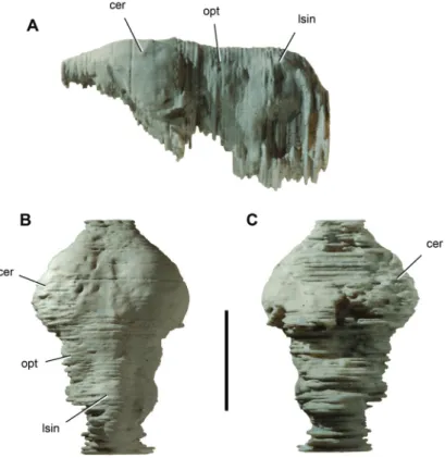
A large lateral Eustachian foramen opens posterodorsally on the anterior side of each basal tuber between the otoccipital (exoccipital + opisthotic) and basisphenoid. As in Araripesuchus, four foramina are present adjacent to the occipital condyle, the largest an anteroventrally opening foramen for the internal carotid. Along the lateral edge of the braincase, a pair of low crests is present running anteromedially from the quadrate to the pterygoid. In lateral view, the otoccipital extends from the very large cranio-quadrate passage anteriorly to the paroccipital process posteriorly, just separating the squamosal and quadrate (Figs. 5A, 6A). Th e basisphenoid has only a narrow, V-shaped ventral exposure. It fl oors a narrow depression between the pair of lateral crests and a small median patch between the basioccipital and the posterior margin of the palate. Endocast. An endocast, generated from the computed-tomographic scan of cranium MNN GAD17 (Fig. 10), closely resembles that for Araripesuchus (Fig. 22). In both the cerebral hemispheres are spade-shaped as seen in dorsal view and measure
ap-Figure 9. Dentary of the crocodyliform Anatosuchus minor. Pencil drawing of mid-section of the left
dentary including alveoli 7–14 (MNN GAD18). A Dorsal view. B Ventral view (reversed). Parallel lines indicate broken bone; double-dash pattern indicates matrix. Scale bar equals 1 cm. Abbreviations: ad7,
12, alveolus of dentary tooth 7, 12; asp, articular surface for splenial; d14, dentary tooth 14; fo, foramen; Mc, Meckel’s canal; sh, shelf.
proximately one-half of total endocast length. In general the forebrain in the endocast compares more closely with that reported for Sebecus (Hopson 1979) than the more rounded, symmetrical cerebral hemispheres in Alligator (Fig. 11) or Caiman (Hopson 1979). A sagittal venous sinus fl anked by shallow longitudinal depressions outlines the medial aspect of each hemisphere. In lateral view, the cerebral hemispheres are compressed dorsoventrally. In A. minor the posterior portion of the hemisphere is a little deeper than in Araripesuchus wegeneri. In ventral view, the absence in A. minor of the ventromedian fossa between the hemispheres observed in A. wegeneri may be an artifact of the quality of the scan. Swellings for optic lobes are visible posterior to the cerebral hemispheres. Although not well preserved in A. minor, the dorsal surface of the cerebellar region is near the height of the cerebral hemispheres.
Dentition. Th ere are 6 premaxillary teeth, 19 maxillary teeth, and 21 dentary teeth, as established on the basis of the exposed teeth and a computed-tomographic scan of skull MNN GAD17. In a subadult skull (MNN GAD603), there are 6 premaxillary teeth,
Figure 10. Endocast of the crocodyliform Anatosuchus minor. Endocast (UCRC PVC2) prototyped
from a computed-tomography scan of skull MNN GAD18. Th e endocast lacks a portion of the pituitary fossa and right and left labyrinths. A Lateral view. B Dorsal view. C Ventral view. Scale bar equals 2 cm. Abbreviations: cer, cerebrum; lsin, longitudinal sinus; opt, optic lobe.
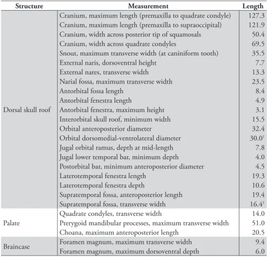
Figure 11. Endocast of Alligator mississippiensis. Endocast (UCRC PVC6) prototyped from a
com-puted-tomography scan of a recent skull (TMM M-983). A Lateral view. B Dorsal view. C Ventral view. Scale bar equals 1 cm. Abbreviations: asc, anterior semicircular canal; cer, cerebrum; lsc, lateral semicircular canal; lsin, longitudinal sinus; opt, optic lobe; pit, pituitary fossa; psc, posterior semicircular canal.
15 maxillary teeth, and an unknown number of dentary teeth. Sereno et al. (2003) orig-inally reported 5 premaxillary teeth in the subadult skull, although it is now clear that the fi rst premaxillary tooth was broken away on both sides based on comparison with the adult skull. Premaxillary tooth number thus appears to be stable in the fi nal 30% of growth in the skull, while maxillary and probably dentary tooth counts increase by a comparable percentage. Th e lower jaws and tooth rows become much more U-shaped during maturation. Th e diagnostic breadth of the snout and transverse orientation of the anterior ends of each dentary emerge late in post-hatching growth. On the other hand, the characteristic inclination of the anterior dentition from the midline to the
Table 4. Length (mm) of crowns in the right upper jaw of Anatosuchus minor (MNN GAD603).
Paren-theses indicate estimated measurement. Abbreviations: m, maxillary; pm, premaxillary.
Tooth Length pm1 2.3 pm2 2.7 pm5 3.7 pm6 3.8 m1 3.8 m2 5.5 m3 7.3 m4 (8.6) m5 5.5 m12 (3.8) m17 3.6
corner of the snout changes very little; the tooth row in anterior view of both subadult and adult skulls is angled at approximately 25° from the horizontal.
Upper and lower crowns are subconical with the base of the crown very slightly ex-panded from the root. Th e crowns curve lingually. Th ere is no distinct neck or marked constriction between root and crown. All but the fi rst premaxillary crown have unorna-mented mesial and distal carinae and very fi ne interweaving striae, which can be seen under strong magnifi cation on the labial side of premaxillary and maxillary crowns. Tooth wear is not nearly as pronounced as in Araripesuchus. Th ere are no wear facets and only a few crown tips with thinned enamel or exposed dentine from apical abrasion.
Six premaxillary teeth are one or two more than common among crocodyliforms. Pm1–3 project ventrally unopposed by dentary teeth, the fi rst of which projects be-tween pm3 and pm4. Th e tip of d4 projects dorsally into a fossa bebe-tween pm6 and m1 (MNN GAD17, GAD603), a typical dental confi guration among crocodyliforms. If the teeth at the junction of premaxilla, maxilla and dentary teeth are regarded as ho-mologous with those in other crocodyliforms, additional premaxillary teeth must have been added to the original plesiomorphic tooth count of four or fi ve teeth, beginning at the medial end of the tooth row.
Th e crown of pm1 is approximately 20% smaller than the crowns of pm2–6, lacks carinae, and is positioned lateral to the midline. Th e alveolar margins of opposing premaxillae are separated in the midline by a subtriangular gap, such that the opposing fi rst premaxillary crowns are separated by a median diastema approximately twice that between ipsilateral premaxillary crowns.
Premaxillary teeth 2–6 are very similar in size and crown detail. Th e alveoli of all premaxillary teeth are raised as rugose cylinders. Th e inner set of alveoli (pm1–3) are separated by concave intercrown festoons, whereas the raised rim of the alveolus in the outer set (pm4–6) are linked together by a rugose alveolar ridge. Th e festooning of the inner set, thus, is the result of the concave margin between alveoli; festooning in the
outer set and in the maxillary series, by contrast, is the result of the dorsally concave labial rim of the alveoli (Fig. 7B).
Th e mesial premaxillary crowns (pm1–3) are functionally distinctive. Th ey oppose a prominent edentulous edge of the dentary, which is 9 mm in transverse width in the adult skull. As confi rmed by computed tomography, the fi rst dentary tooth is posi-tioned 11 mm from the dentary symphysis. Th at tooth (d1) projects toward the base of the fourth premaxillary alveolus. Successive dentary crowns (d2–4) project toward small circular fossae between pm5 and pm6 and into a large palatal opening, respectively. Th e palatal opening is visible on both available skulls and possibly connected with the nearby premaxilla-maxilla foramen. Given that a similarly positioned fossa in
Araripesu-chus receives the tip of the caniniform fourth dentary tooth, the dental and palatal
rela-tionships in A. minor appear to be modifi ed from that observed in other notosuchians. Th e maxillary teeth have crowns that are more closely spaced than the premaxil-lary teeth with alveoli that begin to coalesce toward the distal end of the tooth row. Th e fi rst maxillary crown is approximately 20% larger than the sixth premaxillary crown. Crown size reaches its maximum in m4 at the depressed corner of the snout, distal to which it gradually decreases (m5–20). A caniniform crown is not diff erenti-ated. All maxillary crowns curve lingually with carinae that are shifted lingually. Were the crown to be split by a plane through the carinae, the labial portion would com-prise most of crown volume.
Th e dentary teeth are more poorly exposed. Crown shape seems similar to that in the maxilla and they equal opposing maxillary crowns in size. Crown size reaches its maximum in d11–13 at the depressed corner of the snout (Fig. 8A), distal to which it gradually decreases (d14–21). A caniniform crown is not diff erentiated, and the den-tary series ends mesial to the maxillary series; tooth d21 opposes m14 or m15, leaving at least m16–20 free of opposing dentary crowns. Th e diff erential between upper and lower tooth rows in A. minor is greater than that in Araripesuchus.
Lower jaw. Th e lower jaw broadens signifi cantly during growth, gaining its distinc-tive U-shape with maturity. Th is shape is similar to that in the lower jaws of mature individuals of Simosuchus as seen in dorsal view (Buckley et al. 2000). Th e lower jaw in A. minor, however, is anteroposteriorly nearly twice as long as its maximum width; in Simosuchus jaw length and width are subequal. Th e profi le of the lower jaw diff ers from that in either Simosuchus or Araripesuchus. With jaws abducted, the anterior por-tion of the lower jaws fi ts within the snout and is obscured in lateral view (Figs. 5A, 6A). Th e lateral ramus of the dentary gradually increases in depth to a point ventral to the postorbital bar and dorsal to the external mandibular fenestra, after which it tapers rapidly to an elongate, narrow retroarticular process.
Th e dentary has an immobile interdigitating symphysis with its opposite in the midline. Th e medial 9 mm of the dentary projects anterodorsally at about 45° with an articular edge for the premaxillary palate that protrudes to the height of adjacent dentary crowns. In ventral view, the process has a gently convex articular edge in con-tact with the premaxillary palate. In cross-sectional views derived from the
computed-tomographic scan, the edentulous margin appears to narrow to a sharp cutting edge. Th is masticatory structure has no parallel among other crocodyliforms (Figs. 5C, 6C).
Lateral to the median process, the dentary decreases in width and twists into a subhorizontal plane as it approaches the corner of the snout. As it turns the corner, it becomes broader transversely than deep, a very unusual proportion and quite diff er-ent from Simosuchus (Buckley et al. 2000). Much of the additional width is due to the highly vascularized dentary shelf, which extends lateral to the scalloped alveolar margin (Fig. 9). In ventral view, Meckel’s canal lies in a groove along the medial edge, lateral to which is a broad articular surface for the splenial (Fig. 9B).
Th e dentary extends posteriorly, its deep posterodorsal ramus forming the anterior por-tion of the coronoid process and anterodorsal margin of the external mandibular fenestra. Th ere is a small triangular posteroventral ramus that terminates on the angular ventral to the external mandibular ramus, as evident in several species of Araripesuchus (Price 1959).
Th e splenial contributes to the median symphysis anteriorly (Figs. 5A, 6A). Its posterior margin at the symphysis is damaged in the adult skull. In the subadult skull there is some development of a posteromedian thickening; it seems likely there was a posteromedian splenial “peg” in the adult as in many other notosuchians. In
Simosu-chus the posteromedian eminence is formed by the dentary, as the splenial approaches
but fails to reach the symphysis. Th e splenial extends laterally from the symphysis as a thin sheet of bone with a near horizontal orientation, similar to that of the dentary. Th at orientation is maintained around the corner of the lower jaw, after which a verti-cal ramus expands across the medial side of the dentary. A large oval foramen opens on the transverse ramus of the splenial and continues as a groove medially toward the posterior margin of the symphysis.
Th e surangular extends from the jaw articulation anterodorsally along the top of the coronoid process, a ramus that is swollen laterally with pitted ornamentation ex-cept where it bounds the external mandibular fenestra (Fig. 7D). It appears to form the lateralmost portion of the jaw articulation, after which it continues as a slender unornamented process between the articular and angular to the tip of the long retroar-ticular process (Fig. 7D). Th e angular also has raised pitted ornamentation except for the portion contributing to the margin of the external mandibular fenestra (Fig. 7D). It extends as a slender unornamented process to the tip of the retroarticular process.
Th e articular forms the saddle-shaped glenoid for the quadrate condyles (Fig. 8). Th e surface is transversely convex to accommodate the cleft between the condyles and gently concave anteroposteriorly, the medial socket situated farther ventrally than the lateral socket. Th ere is no anterior or posterior lip to the glenoid. Th e shape of the quadrate condyles and accommodating surface on the articular is similar to that in
Araripesuchus. In posterior view, there is a prominent attachment crest ventral to the
jaw joint. Th e articular extends to the tip of the slender, dorsoventrally fl attened retro-articular process, which is twisted to face dorsomedially.
Axial skeleton. Th e axial skeleton is preserved in articulation from the proatlas to the fi fteenth dorsal vertebra. Th is is one of the most complete presacral series available for any
notosuchian. Th e axial column is well exposed immediately posterior to the skull and par-tially exposed, mainly in right lateral view, more posteriorly. Because this is one of the rare specimens that also shows the relationship between the osteoderms and vertebrae, we left all bones in place during preparation and obtained a computed-tomographic scan to ob-serve details hidden from view. A subadult specimen of Araripesuchus gomesii is the other notable basal metasuchian preserving a complete cervicodorsal column (Hecht 1991).
Extant crocodylians have a proatlas, 8 cervical vertebrae and 16 dorsal vertebrae (Mook 1921). Th e ribs for C3–7 are short, overlapping, and parallel the vertebral column. Th e rib for C8 angles posteroventrally and is transitional to longer, broad-er-shafted dorsal ribs. Th ere are typically 16 dorsal vertebrae in extant crocodylians (Chiasson 1962). Hecht (1991: 346) suggested there were “about seven cervicals” and 17 dorsal vertebrae (thoracic and lumbar) in the subadult specimen of Araripesuchus
gomesii. Th e vertebra that would be the eighth cervical, however, is partially covered
by the scapula. Its rib is transitional in form between the short cervical and long dorsal rib, which is typical of the eighth cervical rib in extant crocodylians (Mook 1921). A similar vertebral formula and transitional rib has recently been reported in
Araripesu-chus tsangatsangana (Turner 2006). Th e axial column in A. minor also appears to have
8 cervical vertebrae and probably 16 dorsal vertebrae. Only 15 dorsal vertebrae are preserved, but a sixteenth may be inferred from the position of the sacral vertebrae, which is based on the position of the associated hind limb (Fig. 4). Cervical centra are amphiplaytan and lack hypapophyses. Dorsal centra become amphicoelous.
Th is vertebral formula diff ers from that described recently in the notosuchian
Noto-suchus. Th is genus may posses as many as 10 cervical vertebrae, 19 dorsal vertebrae, and
3 sacral vertebrae (Pol 2005; Fiorelli and Calvo 2008). Th e cervicodorsal column, thus, has 29 rather than 24 vertebrae and the sacrum 3 rather than 2 vertebrae.
A proatlas is preserved in articulation with the occiput in A. minor. It is an inverted V-shaped median element with a dorsal keel similar to that in extant crocodylians (Mook 1921). Th e proatlas in A. minor appears to be somewhat larger relative to the
atlas, which is composed of separate, paired neural arches and an intercentrum. Th e
transverse width of the proatlas is greater than that of the atlantal neural arches. Th e axis has a low subrectangular neural spine that projects only slightly posterior to the centrum as in extant crocodylians (Mook 1921; Chiasson 1962). Cervical vertebrae three through eight have tall anteriorly tilted neural arches and vertical neural spines as described in the Notosuchus (Pol 2005). Th e neural spine in C3 is subrectangular, about twice as tall as long. Th e neural spine in C7 is considerably taller and narrower, about fi ve times as tall as long. Tall neural arches may characterize notosuchians (Pol 2005).
Th e dorsal vertebrae are somewhat longer relative to their width in A. minor than in Araripesuchus gomesii (AMNH 24450; Hecht 1991). Th e broadest width in both taxa occurs in the posterior dorsal vertebrae, which have long transverse processes (Fig. 4). In A. minor maximum width across the transverse processes is approximately twice centrum length, whereas in A. gomesii maximum width is about three times centrum length. In both genera, the parapophysis migrates out onto the transverse process ante-rior to the diapophysis (D9–11), eventually coalescing to form a single rib articulation
(D12), as in extant crocodylians. Similar elevation and fusion of the parapophysis does not appear to occur in Notosuchus (Pol 2005; Fiorelli and Calvo 2008).
Th e straight ribs of the atlas and axis are preserved on the left side (Fig. 12). Th e shorter triradiate ribs of C3–8 are preserved on the right side in articulation with each other. After they clear the paravertebral shield, the shafts of the anterior dorsal ribs bend ventrally and expand slightly to form a fl ange along their anterior margin as in
A. gomesii (Hecht 1991) and A. tsangatsangana (Turner 2006). In the posterior dorsal
ribs, the capitulum and tuberculum lie in the same plane and eventually coalesce into a single head. Gastralia are preserved ventrally between the girdles (Fig. 4). Th ere do not appear to be any ventral osteoderms in A. minor.
Parasagittal rows of osteoderms are preserved above the cervicodorsal column, with each pair joining its opposite in the midline along an interdigitating suture (Fig. 12; Table 5). Articulation between successive rows of osteoderms is limited to overlap by the posterior edge of a given osteoderm with the anterior edge of the successive ipsilat-eral osteoderm. As in Araripesuchus (Hecht 1991; Turner 2006), there is no develop-ment of anteromedial processes as is common among basal crocodylomorphs, and the
Figure 12. Pectoral girdle and forelimb of the crocodyliform Anatosuchus minor. Left pectoral girdle,
forelimb and anterior portion of the paravertebral shield (MNN GAD17) in dorsal view. Scale bar equals 5 cm. Abbreviations: C2, axis; co1, 3, 4, cervical osteoderm 1, 3, 4; do1, 5, dorsal osteoderm 1, 5; h, hu-merus; l, left; r, right; ra, radius; rC1, atlantal rib; rC2, axial rib; sc, scapula; ul, ulna.
Table 5. Dimensions (mm) of the skeleton of Anatosuchus minor (MNN GAD17). Measurements of
indi-vidual bones are from the left side, except for dorsal osteoderm 12 (preserved only on the right side). Paren-theses indicate estimated measurement. Ungual length is measured along longest chord from base to tip.
Bone Measurement Length
Axial skeleton
Cervical vertebral series, length (75.0)
Dorsal vertebral series, length (268.0)
Cervical osteoderm 1, maximum length 16.0
“ “ 2, “ “ 11.5
“ “ 3, “ “ 9.6
“ “ 4, “ “ 9.9
Dorsal osteoderm 1, maximum length 11.2
“ “ 2, “ “ 12.6 “ “ 3, “ “ 14.4 “ “ 4, “ “ 15.3 “ “ 5, “ “ 16.6 “ “ 6, “ “ 17.3 “ “ 7, “ “ 18.6 “ “ 8, “ “ 18.9 “ “ 9 , “ “ 18.3 “ “ 10, “ “ 19.2 “ “ 11, “ “ 18.2 “ “ 12, “ “ 18.7
Scapula Maximum lengthNeck, minimum dorsoventral height 68.215.2
Coracoid Distal width (23.0)
Humerus Maximum lengthMinimum shaft diameter 80.87.5
Radius
Maximum length 69.3
Maximum proximal width 13.6
Maximum distal width 13.4
Minimum shaft diameter 4.1
Radiale Maximum lengthMaximum proximal width 23.013.8
Maximum distal width 10.8
overlap within each parasagittal column of osteoderms is a narrow smooth articulation limited to the edges of the dorsal series.
No osteoderms are positioned over the proatlas, atlas or axis (Fig. 12). Four paired cervical osteoderms are associated with C3–8 and 12 osteoderms are positioned over D1–12. Osteoderms distal to the twelfth were weathered away. Th e fi rst cervical osteo-derm is the largest of the cervical series and articulates over the neural spines of C3–5. It has a trapezoidal shape with a broader anterior end and a low keel that is most
promi-nent on the posterior one-half of the osteoderm. As in the other cervical osteoderm rows, there is some asymmetry in the paired plates. Th e keel in the fi rst cervical osteo-derm row is laterally displaced on the left but centered on the right side. Th e second cer-vical osteoderm is smaller and articulates with the neural spine of C6. Its shape is similar to the fi rst cervical osteoderm, the keel now reduced to a swelling along the rounded posterolateral corner on the left side or centered on the right side. Th e third cervical os-teoderm is the smallest among all preserved and articulates with the neural spine of C7. It is subtriangular on the left and subquadrate on the right and does not have a keel. Th e fourth and fi nal cervical osteoderm is slightly larger than the third cervical osteoderm and has a shape reminiscent of many of the succeeding dorsal osteoderms. Th e later-ally displaced keel is low and set back from the anterior margin of the plate. Th e lateral corners of the plate are rounded, the anterolateral corner more so than the posterolateral corner. Th ere is no overlap between the last cervical and fi rst dorsal osteoderm. Th e cervical osteoderms would allow considerable lateral and dorsoventral fl exibility of the cervical series as may have been needed during foraging on land or subaquatic feeding.
Th e dorsal osteoderms have a one-to-one relationship with underlying dorsal ver-tebrae as described in extant crocodylians (Ross and Mayer 1984) (Figs. 4, 12). Each
Bone Measurement Length
Manus
Metacarpal 1 length 13.0
Phalanx I-1 length 9.3
Phalanx I-3 (ungual) length 18.6
Phalanx II-1 length 11.1
Phalanx II-2 length 8.0
Phalanx II-3 (ungual) length 19.6
Phalanx III-1 length 10.0
Phalanx III-2 length 6.9
Phalanx III-3 length 6.0
Phalanx III-4 (ungual) length 17.0
Phalanx IV-1 length 9.7
Phalanx IV-2 length 6.7
Phalanx IV-3 length 5.3
Phalanx IV-4 length 5.1
Phalanx IV-5 length 4.2
Phalanx IV-6 length 3.4
Pes
Phalanx II-3 length 12.5
Phalanx III-2 length 11.8
Phalanx III-3 length 8.3
Phalanx III-4 (ungual) length 8.0
Phalanx IV-2 length 10.2
Phalanx IV-3 length 6.9
dorsal osteoderm contacts the neural spine of its respective vertebrae, extends poste-riorly across the interspinous gap, and rests on the anterior portion of the successive neural spine. Th is is well exposed in the middle of the dorsal series, where the right column of osteoderms is displaced ventrally against the transverse processes, exposing the natural articulation between the neural spines and the left column of osteoderms. Th e junction between the osteoderms appears to be positioned so as to coincide func-tionally with the joints between the centra to enhance mobility of the trunk (Salisbury et al. 2006).
Th e fi rst dorsal osteoderm closely resembles the last cervical osteoderm but is slightly larger and extends over the leading edge of the successive osteoderm. Each dorsal osteoderm has a smooth beveled leading edge approximately 1.75 mm broad for articulation with the next anterior osteoderm. Th e sculpted pitting is reduced in a narrow parallel band of slightly greater width adjacent to the leading articular surface. Dorsal osteoderms 2–12 are more fl exed than more anterior osteoderms, the portion of the plate lateral to the keel defl ected ventrally. Th e keel remains parallel to the mid-line across the series. Osteoderm length gradually increases until about the middle of the series (Table 5). Osteoderm shape remains very similar throughout the series, the rounding of the anterolateral corner somewhat less in posterior dorsal osteoderms. Appendicular skeleton. Th e left pectoral girdle and forelimb and portions of the left tibia, fi bula and pedal phalanges are preserved in association with the adult skull (Figs. 3, 11; Table 5). Th e left scapula has broad proportions comparable to those in
Arar-ipesuchus gomesii (Hecht 1991). Th e blade does not appear to fl are as strongly distally
as in A. tsangatsangana (Turner 2006). Th e distal end of the blade is tucked under the edge of the anterior dorsal osteoderms as in extant crocodylians (Fig. 12). Th e elongate coracoid is exposed distally near its contact with the interclavicle.
Th e humerus has a straight shaft and gracile proportions, with shaft diameter less than 10% of its length (Turner 2006) (Table 5). Th e deltopectoral crest is directed anteriorly, and the fossa for the olecranon process is well developed distally as in
Arar-ipesuchus (Hecht 1991; Turner 2006). Th e proximal end of the radius is strongly fl ared,
measuring more than twice mid-shaft diameter. Flaring of the proximal end of the radius to this degree is also present in Araripesuchus (Fig. 25B) and Notosuchus (Pol 2005). Th e radius is shorter than the ulna, because the ulna extends along the lateral side of the radiale. Th e ulna in A. minor is only partially exposed, its shaft noticeably curved. Th e diff erential in length between the radius and ulna is about 10%, as pre-served in articulation in Araripesuchus (Fig. 25B). Th e radiale is a very robust bone in
A. minor, its shaft just slightly less robust than the mid-shaft of the radius (Fig. 13A).
Th e broad lateral facet for the ulna on the proximal end confi rms the off set in the joint between the forearm bones (radius, ulna) and the proximal carpals (radiale, ulnare). From the radiale, it is clear that this off set is also present in A. tsangatsangana (Turner 2006) and Notosuchus terrestris (Pol 2005). Very little of the ulnare is not exposed in A.
minor, but the bone would have been considerably smaller than the radiale. Th e off set