HAL Id: insu-03190875
https://hal-insu.archives-ouvertes.fr/insu-03190875
Submitted on 6 Apr 2021
HAL is a multi-disciplinary open access
archive for the deposit and dissemination of
sci-entific research documents, whether they are
pub-lished or not. The documents may come from
teaching and research institutions in France or
abroad, or from public or private research centers.
L’archive ouverte pluridisciplinaire HAL, est
destinée au dépôt et à la diffusion de documents
scientifiques de niveau recherche, publiés ou non,
émanant des établissements d’enseignement et de
recherche français ou étrangers, des laboratoires
publics ou privés.
Distributed under a Creative Commons Attribution - NonCommercial| 4.0 International
License
evolution of the biosphere during Earth’s middle-age
Emmanuelle J. Javaux, Kevin Lepot
To cite this version:
Emmanuelle J. Javaux, Kevin Lepot. The Paleoproterozoic fossil record: Implications for the evolution
of the biosphere during Earth’s middle-age. Earth-Science Reviews, Elsevier, 2018, 176, pp.68-86.
�10.1016/j.earscirev.2017.10.001�. �insu-03190875�
Contents lists available atScienceDirect
Earth-Science Reviews
journal homepage:www.elsevier.com/locate/earscirev
Invited review
The Paleoproterozoic fossil record: Implications for the evolution of the
biosphere during Earth's middle-age
Emmanuelle J. Javaux
a,⁎, Kevin Lepot
baUniversity of Liège, Department of Geology, Palaeobiogeology-Palaeobotany-Palaeopalynology, 14, allée du 6 Août B18, quartier AGORA, 4000 Liège, (Sart-Tilman),
Belgium
bUniversité de Lille, CNRS, Université Littoral Côte d'Opale, Laboratoire d'Océanologie et de Géosciences UMR8187, Cité Scientifique, SN5, 59655 Villeneuve d'Ascq,
France
A R T I C L E I N F O
Keywords: Paleoproterozoic Microfossils Iron formations Prokaryotes Eukaryotes CyanobacteriaA B S T R A C T
The Paleoproterozoic (2.5–1.6 Ga) Era is a decisive time in Earth and life history. The paleobiological record (microfossils, stromatolites, biomarkers and isotopes) illustrates the biosphere evolution during a time of tran-sitional oceanic and atmosphere chemistries. Benthic microfossil assemblages are recorded in a variety of oxygenated, sulfidic, and ferruginous environments representative of the spatial heterogeneities and temporal variations characteristic of this Era. The microfossil assemblages include iron-metabolizing and/or iron-tolerant prokaryotes, sulfur-metabolizing prokaryotes, cyanobacteria, other undetermined prokaryotes, and eukaryotes. The undetermined microfossils represent a majority of the assemblages and thus raise a challenge to determine the nature and role of microorganisms in these changing environments. Despite the early evolution of the eu-karyotic cellular toolkit, early eueu-karyotic crown group diversification may have been restrained in the Paleoproterozoic by ocean chemistry conditions, but it increased during the late Mesoproterozoic–early Neoproterozoic despite the continuation of similar conditions through the (miscalled)“boring billion”, then amplified significantly (but perhaps within lower taxonomic levels), with the demise of euxinic conditions and increase in ecological complexity.
The emerging picture is one of a changing and more complex biosphere in which the three domains of life, Archaea, Bacteria and Eukarya, were diversifying in various ecological niches marked by the diversification of identified microfossils, stromatolites, increasing abundance of preserved biomarkers, and appearance of mac-roscopic problematic fossils or trace fossils.
1. Introduction
The Paleoproterozoic (2.5–1.6 Ga: billion years) Era is a decisive time in Earth and life history. Drastic environmental changes affected ocean and atmosphere chemistry, including step-wise and possiblyfluctuating oxyge-nation (Lyons et al., 2014), the Huronian glaciations (three ice ages, be-tween 2.45 and 2.22 Ga;Barley et al., 2005), large organic carbon deposits during the Lomagundi-Jatuli event (Melezhik et al., 2013) at ~2.3–2.06 Ga, an alternation of enhanced magmatic activity (maxima at ~2.7 Ga and ~1.9 Ga) and more quiescent periods (2.45–2.2 Ga) (Condie et al., 2009) (Fig. 1). These environmental changes may have had profound sequences on life's diversification by modifying the physico-chemical con-ditions and diversity of ecological niches. For example, decrease in sub-marine volcanism may have limited theflux of Fe2 +, H
2and H2S, hence
limiting major sinks acting against O2accumulation between 2.45 and
2.22 Ga (Kump and Barley, 2007).
Life also deeply marked Earth's planetary evolution during this Era (Fig. 1). The Archean–Proterozoic transition started with the so-called
great oxidation event (GOE) between 2.48 and 2.32 Ga (Bekker et al., 2004; Hannah et al., 2004), when molecular oxygen accumulated suf-ficiently in the ocean and atmosphere to be detected widely by a variety of geological and geochemical proxies (Bekker et al., 2004; Sessions et al., 2009). These proxies provide a minimum age for the advent of oxygenic photosynthesis by cyanobacteria on the planetary scale but earlier local oxygenated oases may have occurred in photic zones where oxygenic cyanobacteria first developed. Possible origin of oxygenic photosynthesis (by cyanobacteria) has been suggested as early as 3.7 Ga based on carbon and Pb isotopic memory of past U/Th ratios (Rosing and Frei, 2004), but see Buick (2008); 3.2 Ga based on thick and widespread deposits of kerogenous black shales (Buick, 2008); 2.9 Ga based on S-isotopes (Ono et al., 2006). More direct tracers linked with geochemical reactions requiring dioxygen can be tracked back as far as
http://dx.doi.org/10.1016/j.earscirev.2017.10.001
Received 21 September 2017; Received in revised form 3 October 2017; Accepted 3 October 2017
⁎Corresponding author.
E-mail addresses:[email protected](E.J. Javaux),[email protected](K. Lepot).
Earth-Science Reviews 176 (2018) 68–86
Available online 04 October 2017
0012-8252/ © 2017 The Authors. Published by Elsevier B.V. This is an open access article under the CC BY-NC-ND license (http://creativecommons.org/licenses/BY-NC-ND/4.0/).
3.0–2.8 Ga ago based on chromium isotopes (Crowe et al., 2013; Frei et al., 2009); 2.95 Ga based on Mo isotopes (Planavsky et al., 2014); and 2.5 Ga based on molybdenum and rhenium data (Anbar et al., 2007). However, some molecular phylogenies suggest that oxygenic photosynthesis appeared close to the GOE (Shih et al., 2017) and al-ternative hypotheses for earlier “whiffs” in dioxygen have been sug-gested as other biotic or abiotic processes are possible (Fischer et al., 2016). Recently, the biomarker evidence for cyanobacteria (and eu-karyotes) in Archean rocks (Brocks et al., 2003; Eigenbrode et al., 2008) was reassessed as contamination (French et al., 2015; Rasmussen et al., 2008). Some Paleoproterozoic eukaryote biomarkers have also been discussed as possible contaminants (Brocks et al., 2008; Flannery and George, 2014) and“steranes fading out beyond 800 Ma indicates the absence of a reliable record” (Brocks et al., 2017). Biomarkers for green and purple sulfur bacteria (anoxygenic photosynthesizers) occur in the 1.64 Ga Barney Creek Formation (Brocks et al., 2005). Bacterial sulfate reduction is indicated by S isotopes as early as 3.5 Ga (Shen et al., 2009; Ueno et al., 2008). Limitation of nickel concentration, a key co-factor in several enzymes of methanogens, may have led to decreasing atmo-spheric methane levels and favored the rise of O2in the atmosphere
from 2.7 Ga onwards (Konhauser et al., 2009). Enzymatic N-fixation (diazotrophy) is suggested as early as 3.2 Ga (Stüeken et al., 2015). Early cyanobacteria could only fix nitrogen at relatively low O2
con-centrations or rely on other nitrogen fixers (Tomitani et al., 2006). Cyanobacteria likely had to invent a new N2-fixation machinery that
could operate in the presence of the rising O2, possibly leading to the
advent of heterocystous cyanobacterial taxa probably as early as the GOE (Schirrmeister et al., 2013), and possibly supported by Paleopro-terozoic microfossils (Tomitani et al., 2006), but see Butterfield
(2015b). Active N-cycling was favored by the advent of an aerobic ni-trogen cycle possibly as early as 2.7 Ga (Garvin et al., 2009; Thomazo
et al., 2011), thereby favoring all heterotrophic microorganisms unable tofix N2. An increase in primary production and sulfate export to the
ocean, both favored by the new oxidative subaerial weathering condi-tions (Bekker and Holland, 2012), stimulated heterotrophic bacterial sulfate reduction (Canfield, 1998). In combination with N cycling, in-crease in phosphate extracted during oxidative weathering and active N recycling by heterotrophy stimulated primary photosynthetic produc-tion (Bekker and Holland, 2012; Papineau et al., 2013) during the Lo-magundi-Jatuli event (~ 2.3–2.06 Ga). However, increase in primary production led to deposition of organic matter that represented a co-lossal food source for oxygen-respiring bacteria and heterotrophic sul-fate-reducing bacteria, possibly leading to a drop in oxygen and sulfate by ~ 2.05 Ga (e.g.Canfield et al., 2013; Scott et al., 2014).
The redox state and composition of bottom waters during the Paleoproterozoic may have been periodically and/or locally anoxic, dysoxic or euxinic (Johnston et al., 2009) (Fig. 1). Archean oceans were mostly ferruginous; this condition recurred transiently in bottom to shallow waters around 1.9 Ga, possibly due to a return of intense sub-marine volcanism (Rasmussen et al., 2012) and associated with the post-Lomagundi decline in oxygen (see review inLyons et al., 2014). During the so-called “boring billion” (~1.9–1.8 to ~0.8 Ga), these conditions changed into sulfidic (H2S-rich) anoxic conditions (euxinia)
in continental shelf and epicontinental sea settings (Canfield, 1998;
Poulton et al., 2010) where intense primary production fed sulfate-re-ducing bacteria. Euxinia with comparatively low sulfate concentrations may also have occurred in the lower part of the photic zone, at least sporadically, in an oxygen-minimum zone-type setting (OMZ), while the surface waters of the photic zone were moderately oxygenated (10− 4to 0.1 PAL: present atmospheric level of oxygen) (Brocks et al., 2005; Lyons et al., 2014; Shen et al., 2002, 2003). However, recent studies suggests that ferruginous conditions in deep waters may have
Fig. 1. Summary of changing physico-chemical conditions in the Archean and Proterozoic ocean and atmosphere, and fossil, biomarker and isotopic evidence for the record of Eukaryotic supergroups, and Bacteria and Archaea metabolisms (black stars: large meteoritic impacts; double-headed arrows: supercontinent formation and break-up; black triangles: widespread glaciations; GOE: great oxidation event; LJE: Lomagundi-Jatuli event). References cited in the text and inJavaux (2011).
been more widespread than previously documented during Earth's middle age (1.8–1.0 Ga), and that both euxinic and ferruginous strati-fied waters may have been common below the oxygenated surface-mixing zone (Planavsky et al., 2011; Poulton et al., 2010). The extent of euxinic conditions may have varied in time and space, occurring in restricted basins, or in oceanic wedges close to continental margins with high productivity. Below the surface layer where oxygenic pho-tosynthesis occurred, euxinic conditions may have been maintained until about 750 Ma, when the return to a ferruginous ocean coeval with the break-up of the supercontinent Rodinia prior to the Sturtian gla-ciation, is documented by S, C and Fe data (Canfield et al., 2008;
Johnston et al., 2010; Sperling et al., 2013). The composition of the bottom and/or OMZ waters may have drastically influenced the nature and distribution of the photosynthetic primary producers. Cyano-bacteria may have performed anoxygenic (rather than oxygenic) pho-tosynthesis using H2S above euxinic layers, or may have been
out-competed by anoxygenic photosynthetic bacteria metabolizing H2S or
Fe2 + above ferruginous water (Johnston et al., 2009). Moreover, planktonic cyanobacteria may have suffered from Fe2 + toxicity
(Swanner et al., 2015) in offshore environments where surface water
was directly in contact with ferruginous deeper water, although this Fe2 +toxicity may have been alleviated in waters saturated with silica
(Mloszewska et al., 2015) or through intracellular Fe-biomineralization (Lepot et al., 2017). In this context, oxygenic photosynthesis may have been dominated by benthic (terrestrial and coastal) cyanobacteria from the late Archean possibly until the end of the“boring billion” (Lalonde and Konhauser, 2015). Nevertheless, cyanobacteria may have known an increase in diversification associated with or preceding the GOE and later during the Paleoproterozoic (Schirrmeister et al., 2013). Similarly, the chemical composition of seawater, including sulfate, iron (Ratti et al., 2011), and trace-metal concentrations (Anbar and Knoll, 2002) during the Paleoproterozoic and “boring-billion” may have regulated the evolution and distribution of eukaryotes and their competition with cyanobacteria in the primary photosynthetic biomass (Knoll et al., 2007). As detailed below, the available microfossil record and the molecular biomarker record suggest that the primary photosynthetic production displayed a large dominance of (cyano)bacteria over eu-karyotes, although this view could be biased by the difficulty to identify early eukaryote microfossils and the poor preservation of biomarkers in Paleoproterozoic rocks (Knoll et al., 2007).
2. The Paleoproterozoic microfossil record
Paleoproterozoic rocks are important targets not only for doc-umenting changing redox conditions, but also for micropaleontological investigations. To date, they host the oldest microfossils diagnostic of cyanobacteria (Hofmann, 1976; Knoll and Golubic, 1992) and of eu-karyotes (see reviews inJavaux, 2011; Knoll et al., 2006). The Paleo-proterozoic record of biological activities (e.g. see below and reviews in
Hofmann and Schopf, 1983; Javaux et al., 2012; Knoll, 2003; Schopf, 1992) also includes unidentified microfossils, possible microfossils of iron-oxidizing bacteria, morphologically diverse stromatolites, macro-scopic carbonaceous compressions, microbial mat rip-ups, putative fossil or trail impressions, problematic pyritized macrofossils, biological isotopic fractionation of C, S, and N, isotopic fractionation of Fe, and biomarkers. Together, these proxies indicate the presence of eu-karyotes, cyanobacteria, green and purple sulfur bacteria, genic archaea and/or bacteria, sulfate-reducing bacteria, methano-trophic bacteria and/or archaea, iron-oxidizing bacteria, anoxygenic photosynthetic bacteria, and denitrifying bacteria (Fig. 1).
2.1. Identifying early microfossils
Microfossils can be made of various altered derivatives of initial biomolecules (e.g. resilient biopolymers such as cell wall poly-saccharides), upon which secondary molecules (e.g. derivatives from
more labile proteins, lipids) may have grafted during diagenesis (de Leeuw et al., 2006). Some originally organic ultrastructures may be replaced by diagenetic minerals, e.g. by pyrite (Cosmidis et al., 2013; Wacey et al., 2013). Minerals formed in vivo by/on the organism and their diagenetic/metamorphic byproducts may also be associated with microfossils (Crosby et al., 2014; Lepot et al., 2017). Preservation and composition of organic microfossils, of their diagenetic mineral re-placements, and of biominerals depend on the original biological composition of the cellular structures (and ultrastructures) and the conditions of preservation, diagenesis, and degree of metamorphism (Alleon et al., 2016a; Alleon et al., 2016b; Schopf et al., 2005). Micro-to nano-scale characterization of organic structures and associated biogenic/diagenetic/metamorphic minerals is required to demonstrate that microfossils are endogenous (i.e. not contaminants), syngenetic (displaying the same age as the rock), and of biogenic morphology (Foucher et al., 2015; Javaux et al., 2012; Laflamme et al., 2011; Lepot, 2011; Oehler, 2014; Wacey et al., 2016). Raman microspectrometry provides information of the organization of aromatic groups in fossil organic matter at the micrometer scale, which is strongly correlated to organic-matter maturity (Schopf et al., 2005); hence, correlation of Raman-derived organic-matter maturity with independent maturity parameters (e.g. mineral assemblages, O-isotopes) provide crucial constraints on the syngenicity of the organic matter (e.g.Alleon et al., 2016b; Javaux et al., 2010). Microscopy is crucial to distinguish i) fossilization of biogenic/anatomical structures, ii) late endolithic con-tamination (Westall and Folk, 2003) and iii) mineral microstructures associated with migrated organic matter mimicking microfossils (Brasier et al., 2015; Brasier et al., 2005; Lepot et al., 2009b; Van Zuilen et al., 2002). Methods of microscopy include multiplane optical ima-ging (Brasier et al., 2005), scanning and transmitted electron micro-scopy on acid-extracted microfossils (Cloud and Hagen, 1965; Javaux et al., 2004; Moczydłowska and Willman, 2009), microscale Raman imaging (Schopf and Kudryavtsev, 2009), Transmission Electron Mi-croscopy performed on Focused Ion Beam sections (Lepot et al., 2017; Moreau and Sharp, 2004; She et al., 2013; Wacey et al., 2012), and nanotomography using Focused Ion Beam ablation coupled to electron microscopy (Wacey et al., 2016). Complex ultrastructures such as tri-laminar walls, structured wall reticulation and wall processes (e.g.
Javaux et al., 2004; Moczydłowska and Willman, 2009b) represent diagnostic evidence for microfossils that so far have not been re-produced abiotically. When the morphology of putative microfossils is simple, such as spheres and tubular sheaths, microfossil demonstration is more complex. In abiotic mimics, the distribution of organic matter is templated by the host mineral (Buick, 1990). In contrast, in micro-fossils, the micro/nanostructure of the mineral matrix appears tem-plated by the organic matter and the taphonomic sequence of the mi-croorganism (Kempe et al., 2005; Lekele Baghekema et al., 2017; Lepot et al., 2017; Moreau and Sharp, 2004; Wacey et al., 2012). Microscopy must be complemented by macro-scale characterization of the geolo-gical context and of past/recent alteration of the rocks to constrain paleoenvironments, taphonomic (post-mortem) conditions, and pos-sible abiotic processes mimicking microfossils.
Once a microstructure has passed the tests of endogenicity, syn-genicity, and biosyn-genicity, the next challenge consists in taxonomic identification. Paleontologists have to rely on information other than the genome and internal cellular organization to identify the biological affinities of early microfossils. Using a combination of morphology and microchemistry may permit differentiation between prokaryote and eukaryote microfossils. Size is not a good criterion to differentiate fossil prokaryotes from eukaryotes since extant pico-eukaryotes (1–2 μm in diameter) and large prokaryotes (such as 100μm long cyanobacterial akinetes, or bacterial cells such as Thiomargarita reaching up to 600μm in diameter) are documented in nature (see review in Javaux et al., 2003). The analyses of wall ultrastructures may be used for recognition of eukaryotic origin (Arouri et al., 2000; Javaux et al., 2003; Moczydłowska and Willman, 2009; Talyzina and Moczydłowska,
2000b; Willman and Cohen, 2011). Wall ultrastructure (e.g. tri-laminar, or perforated by canals), specific wall ornamentation, and the presence of excystment structures (openings through which cells are released from the cyst) can clarify the eukaryotic affinities of microfossils, sometimes even at the level of specific clades. Excystment structures, if observed in a large population, may be differentiated from occasional tearing, and point to an eukaryotic affinity. However, some cyano-bacterial envelopes can reach large sizes (a few tens of micrometers in diameter) and open in a fashion resembling eukaryotic excystments (Waterbury and Stanier, 1978); hence other criteria, such as a complex wall ultrastructure, should be used to confirm a eukaryotic affinity. In contrast, due to their small size and to post-mortem morphological convergence (Schopf, 1975), it is often difficult to provide morpholo-gical/ultrastructural constraints on the nature of putative prokaryotic microfossils. Recent studies using transmission electron microscopy or nanotomography have provided new morphological information on the smallest Paleoproterozoic microfossils (Brasier et al., 2015; Lepot et al., 2017; She et al., 2013; Wacey et al., 2012).
Micro-analyses of organic molecules such as micro-FTIR (e.g.Arouri et al., 1999; Marshall et al., 2005; Steemans et al., 2010) and micro-Raman spectroscopy (e.g.Javaux and Marshall, 2006; Marshall et al., 2005) can show inter-specific differences in the functional group sig-nature of single microfossils. For example, FTIR sigsig-natures such as abundant long polymethylenic chains point to preservation of algaenan from chlorophyte or eustigmatophyte microalgae (Arouri et al., 1999; Marshall et al., 2005). Nano-analyses of functional groups can be per-formed using Scanning (transmission) X-ray microscopes (SXM/STXM). STXM revealed the preservation of N- and O-bearing functional groups in Paleoproterozoic microfossils (Alleon et al., 2016b) and SXM re-vealed S- bearing molecules in Neoproterozoic microfossils (Lemelle et al., 2008). Both N- and S-groups have been proposed as relicts of proteins (Alleon et al., 2016b; Lemelle et al., 2008). However, het-eroatoms such as N- and S- may graft diagenetically onto organic matter (Lepot et al., 2009a) and it cannot be confirmed that the former
pro-teins (or other organic precursors) come from the actual microbe pre-cursors to the studied microfossils rather than from ambient soluble molecules. The former hypothesis could be confirmed by demonstration of heterogeneities in N/S/O groups between morphospecies (achieved in Neoproterozoic microfossils using NanoSIMS;Oehler et al., 2006) or between ultrastructures of a single microfossil (achieved with STXM on Phanerozoic fossils;Bernard et al., 2007). In parallel, isotopic micro-analyses (House et al., 2000; Lepot et al., 2013; Williford et al., 2013) of organic matter in microfossils reveal inter-specific and/or intra-cellular heterogeneities, indicating that different types of microfossils and/or ultrastructures in a single cell may originate from distinct metabolisms and/or biosynthetic processes. Isotopic microanalyses provided new clues that Leiosphaerida crassa and Myxococcoides sp. associated in the same Neoproterozoic rock represent algae and cyanobacteria, respec-tively (Williford et al., 2013). One limitation of this spatially-resolved molecular/isotopic approach is our limited knowledge of the changes occurring to the morphology, ultrastructure and chemistry of extant (micro)organisms during fossilization. This approach thus requires comparative actualistic studies of taphonomic processes affecting di-verse organisms in a range of natural and experimental environmental conditions (e.g.Alleon et al., 2016a; Chalansonnet et al., 1988; Javaux and Benzerara, 2009; Kleinteich et al., 2017; Lepot et al., 2014; Li et al., 2013; Picard et al., 2016; Schiffbauer et al., 2012; Storme et al., 2015). Other important challenges in identifying early microfossils include the limitations of actualism (early microorganisms may not have been ancestors of extant clades but stem clades, and thus not comparable) and the possibility of morphological and chemical convergence be-tween unrelated clades. Even when precise identification failed, fossils document steps in biological and biochemical innovations (Butterfield,
2009; Brasier et al., 2015; Javaux, 2007, 2011; Javaux and Knoll, 2017; Javaux et al., 2001; Knoll et al., 2006). Original biological properties (e.g. morphology, chemistry, division pattern, spatial distribution
patterns) may be well preserved, altered or erased by fossilization processes that vary depending on the physico-chemistry of the fossili-zation environment and the composition of the organism. A good un-derstanding of taphonomic biases is essential for deciphering the ori-ginal biology.
2.2. Paleoproterozoic eukaryotes
Eukaryotes have developed some fundamental biological innova-tions since at least the late Paleoproterozoic, such as genetically-pro-grammed excystment structures, reproduction by budding or binary division, multicellularity, and complex ornamentation requiring a so-phisticated cytoskeleton and endomembrane system (Javaux and Knoll, 2017; Javaux et al., 2001), and even macroscopic size (Zhu et al., 2016). The molecular clocks are discussed below using the demon-strable, possible and putative records of fossils and biomarkers.
2.2.1. Molecular clues?
Due to contamination and maturity issues, reliable sterane bio-markers for crown-group eukaryotes are only found in Neoproterozoic and younger rocks (Brocks et al., 2008; Brocks et al., 2017; Flannery and George, 2014; French et al., 2015; Knoll et al., 2007; Rasmussen et al., 2008; Summons and Walter, 1990). However, biomarkers in-dicate the presence of green algae (Brocks et al., 2017) as well as de-mosponges (Love et al., 2009) and possible toxic protists (Brocks et al., 2016) in the Neoproterozoic, implying an earlier origin of stem and crown-group eukaryotes, as suggested by molecular clocks possibly as early as the Paleoproterozoic GOE (Eme et al., 2014; Gold et al., 2017; Parfrey et al., 2011; and review inJavaux and Knoll, 2017).
2.2.2. Oldest eukaryotic microfossils
The oldest unambiguous eukaryotic microfossils are large (100–300 μm) organic-walled vesicles (acritarchs) ornamented with concentrically striated walls (Valeria lophostriata) (Fig. 2a, b). One sphaeromorph from the Changzhougou Fm, China (Fig. 4a in Lamb et al., 2009) shows concentric striations and resembles Valeria lophos-triata. The Changzhougou Fm, is younger than the 1673 ± 10-Ma U-Pb age of zircons in a granite-porphyry dyke that cuts underlying rocks, and older than the 1625.3 ± 6.2-Ma ash bed in overlying beds (Li et al., 2013). This species also occurs in the overlying ~ 1.65 Ga Chuanlinggou Formation (Changcheng Supergroup, China) (Lamb et al., 2009; Peng et al., 2009; Zhang, 1986), in the > 1.65-Ga Mal-lapunyah Formation (McArthur Supergroup, Australia, Fig. 1A 7,8) (Javaux, 2007) and in many younger Proterozoic siliciclastic succes-sions. Rhythmic ridge patterns at the surface of Valeria lophostriata have been suggested to represent an in vivo growth pattern (Pang et al., 2015) or a taphonomic pattern (Hofmann, 1999). These striations are in fact wall ornamentations, as shown by scanning Electron Microscopy (SEM) images evidencing that they are 1-μm-spaced ridges located on the inner side of the vesicle and not taphonomic features (Javaux et al., 2004). B-Z-type reactions or Turing reaction-diffusion processes may have formed the striations in vivo, possibly as a mechanism to guide biologically programmed excystment through medial split (Pang et al., 2015) although medial split is known in many other unornamented and ornamented vesicles. These spheroidal fossils often show excystment structure by medial split, with the separated half-vesicles usually rolling up over themselves (Fig. 2b).
2.2.3. Eukaryotic microfossil diversification
Recently, the age of a moderately diverse microfossil assemblage from the Beidajan Formation in the Ruyang Group, in China,first dated to about 1.3 Ga (Xiao et al., 1997), has been revised to the late Paleo-proterozoic–early Mesoproterozoic (Pang et al., 2013). Detrital zircons indicate that Ruyang deposition began no earlier than 1744 ± 22 Ma (Hu et al., 2014), and xenotime Pb-Pb ages of about 1411 ± 27 Ma for the overlying Luoyu Group constrain deposition from above (Lan et al.,
2014). A U-Pb zircon date from an ash bed within the Luoyu Group potentially constrains Ruyang deposition to be older than 1611 ± 8 Ma, but the abundance of detrital grains in the dated bed and the wide range of ages for individual zircons (Su et al., 2012) suggest that the published date may provide only a maximum age
constraint on Luoyu deposition (Lan et al., 2014). This 1744 ± 22 to 1411 ± 27 Ma assemblage includes sphaeromorphs, largefilaments, segmented oval vesicles, but also unambiguous eukaryotes such as the Valeria lophostriata described above, and Dictyosphaera delicata orna-mented with a reticulated wall and showing a circular excystment
Fig. 2. Paleoproterozoic microfossils: eukaryotes, sphaeromorphs and cyanobacteria.
a, b. Valeria lophostriata, > 1.65 Ga Mallapunyah Fm, Australia, an early eukaryote with a wall ornamented with concentric striations. c. Leisphaeridia sp., a sphaeromorph from the 1.9 Ga Kondopoga Fm, Karelia, Russia. Scale bar in c = 100μm for b, c, e, 50 μm for a, d, f. d. Dictyosphaera delicata, a protist from the 1.7 to 1.4 Ga Ruyang Group, China.
e. Shuiyousphaeridim macroreticulatum (left), a protist, and Leiosphaeridia minutissima, a sphaeromorph from the 1.7 to 1.4 Ga Ruyang Group, China. f. Tappania plana, a protist from the 1.5 Ga Roper Group, Australia.
g. Archaeoellipsoides, interpreted as possible cyanobacteria akinetes, Mesoproterozoic Billyakh group, Siberia. This specimen is 80μm long (photo courtesy of AH Knoll). h. Cyanobacteria trichome, Mesoproterozoic Billyakh Group, Siberia. This specimen is 85μm long (photo courtesy of AH Knoll).
i. Eoentophysalis, cyanobacterial colonies from silicified stromatolites, 1.9 Ga Belcher Group, Belcher Islands, Canada. Ellipsoidal envelopes around cells are 6–10 μm in diameter (photo courtesy of A.H. Knoll).
structure (pylome). Acanthomorph (process-bearing) acritarchs in-clude: the complex acanthomorph acritarch Tappania plana bearing heteromorphic processes and neck-like extensions, and showing evi-dence of budding, a large acanthomorph densely covered with small processes (Gigantosphaeridium fibratum), and Shuiyousphaeridium mac-roreticulatum (Fig. 2e), another complex acritarch of the assemblage, bearing furcated processes and a multilayered wall made of imbricated polygonal organic plates (Agić et al., 2015; Javaux et al., 2001; Javaux
et al., 2004; Schiffbauer and Xiao, 2009; Yin, 1997; Yin et al., 2005). Tappania plana is also reported in the 1.45 Ga Roper Group, Australia (Javaux and Knoll, 2017; Javaux et al., 2001) (Fig. 2f), in the 1.45 Ga Lower Belt Group, Montana (Adam et al., 2017), the ~ 1.3 Ga Kamo Gp, Siberia (Nagovitsin et al., 2010), the Sarda Formation of the Bahraich Group, Ganga Basin (Prasad and Asher, 2001), and possibly in corre-lative beds of the Kheinjua Group, Vindhyan Basin (Prasad et al., 2005) in northern India. U-Pb zircon dating of ash beds from the Deonar “Porcellanite” Formation just below the Vindhyan succession con-taining unambiguous Tappania indicates that these fossils are ≤1631 ± 1 Ma old (Ray et al., 2002). This also constrains the age of morphologically complex acritarchs including acanthomorphs (Shuiyousphaeridium echinulatum, Cymatiosphaeroides kullingii) reported from the overlying Chitrakoot Formation, Semri Group, Vindhyan Su-pergroup, India (Sharma et al., 2016; Singh and Sharma, 2014) al-though ages up to 1.650 ± 89 Ga have been reported (Bengtson et al., 2009). Some Paleoproterozoic microfossils have been interpreted as possible green algae based on morphological comparison with modern algae (Moczydłowska et al., 2011; Agić et al., 2015). These inter-pretations remain to be confirmed by other types of analyzes, because of the possibility of morphological convergence (Knoll, 2014).
2.2.4. Other possible eukaryote microfossils
In the absence of wall ornamentation, as explained above, detailed studies of the wall ultrastructure may permit the identification of eu-karyotic microfossils in some cases (Cohen et al., 2009; Javaux et al., 2003; Javaux et al., 2004; Javaux and Marshall, 2006; Moczydłowska and Willman, 2009b; Talyzina and Moczydłowska, 2000a), especially when ultrastructure analyses are combined with wall chemistry (Javaux et al., 2003; Javaux et al., 2004; Javaux and Marshall, 2006; Marshall et al., 2005). In Paleoproterozoic and younger rocks, large, smooth organic-walled vesicles (up to a few 100μm unornamented acritarchs, also called leiospheres or sphaeromorphs) are common and may display a medial split, but the absence of any wall ornamentation prevents their attribution to the eukaryotic domain. They could represent early pro-tists or large envelopes of prokaryotes such as those of some cyano-bacteria, which can open in a fashion that could be confused with a eukaryotic medial split (Waterbury and Stanier, 1978). Large sphaer-omorphs also occur in the Mesoarchean (Javaux et al., 2010) but do not show medial split or complex wall ultrastructure and cannot be related to a particular clade or domain. Multilayered wall ultrastructures and medial splits were reported in 1.8 Ga old large Changcheng sphaer-omorphs, possibly relating those to early eukaryotes (Lamb et al., 2009; Peng et al., 2009). Rare large (up to > 300μm) carbonaceous vesicles (acritarchs) and fragments of organic sheaths of unknown biological affinities also occur in siltstones from the upper members of the ~ 1.9 Ga Kondopoga Formation, Karelia, Russia (Javaux et al., 2012) (Fig. 2c). Poorly preserved ambiguous acritarchs from Karelia werefirst mentioned by (Timofeev, 1982). Sphaeromorphs have also been re-ported in siltstones and silty black shales from the FB2 subunit of the the ~ 2.1 Ga old Francevillian Group of Gabon (El Albani et al., 2014). Large spheroidal unicells from 2.1–1.88 Ga stromatolitic cherts of the Vempalle Formation, India (revised age in Chakrabarti et al., 2014) have also been suggested as putative eukaryotes (Schopf and Prasad,
1978), however size alone is not a sufficient criteria and other analyses
are necessary to confirm this hypothesis (seeSection 2.1). 2.2.5. Putative multicellular eukaryotes
Recently, phosphatized microfossils from the ~ 1.6 Ga Chitrakoot Formation have been interpreted as possible crown-group red algae (Bengtson et al., 2017), based on putative pyrenoids preserved in large tubes Rafatazmia and on multicellular thallus-like Ramathallus showing possible cellular differentiation. If the age and affinity of these micro-fossils are confirmed, they would evidence the acquisition of the chloroplast by eukaryotes 500 Myr earlier than evidenced by the 1.1 Ga red algae Bangiomorpha (Butterfield, 2000). Recent molecular clock studies suggest an origin of the common ancestor of the Archaeplastida (red, green and Glaucophyte algae) around 2.1–1.6 Ga but crown groups originated later between 1.3 and 0.9 Ga (Sánchez-Baracaldo et al., 2017). Branching and/or fibrous structures reported in ~ 2.8–2.7 Ga lacustrine rocks have been proposed as fossils of early algae (Kaźmierczak et al., 2016). However, we interpreted these Ar-chean microstructures as altered and metamorphosed biotite crystals, which commonly display similar microstructures and chemical com-positions (Morad, 1990). The carbonaceous particles detected by
Kaźmierczak et al. (2016) in thesefibrous/branching clays may re-present amorphous kerogen commonly embedded in Archean clays (Lepot et al., 2009a). Finally, the palynological extract ofKaźmierczak
et al. (2016)is transparent, and likely represents a preparation con-taminant or a mineral rather than black and opaque Archean (i.e. me-tamorphic) organic matter.
2.2.6. Possible macrofossils
Other possible Paleoproterozoic eukaryotes include macroscopic carbonaceous compressions and trace fossils. Centimetric carbonaceous compressions preserved on bedding plane surfaces include spherical Chuaria, sausage-shaped Tawuia and coiledfilamentous Grypania (Xiao and Dong, 2006). Grypania spiralis from the 1.87 Ga Negaunee Iron Formation, Michigan (Han and Runnegar, 1992; redated bySchneider et al., 2002) is ~ 1 mm in diameter, up to 90 mm in length, and forms coils with a diameter up to 30 mm across.Samuelsson and Butterfield (2001)have questioned the eukaryotic nature of these earlier structures from Michigan, while observations by Knoll (in Knoll et al., 2006) suggest that it was an organism, and not a colony or composite of much smaller prokaryotic filaments. Younger forms also called Grypania spiralis from the ~ 1.45 Ga Vindhyan Supergroup, India, are septate and much longer (about 500 mm), and are more convincingly eukaryotic. Reassessment of Paleo- and Mesoproterozoic, macroscopic, coiled fila-mentous compressions suggests a cyanobacterial affinity for some of them, while others might be dubiofossils and‘tissue-grade organisms’ (Sharma and Shukla, 2009) or macroalgae (Xiao and Dong, 2006). Morphometric studies combined with the detection of highly aliphatic organic matter have been interpreted as a possible algal affinity (Sharma et al., 2009), however, an aliphatisation process during diag-enesis is also possible (de Leeuw et al., 2006), and the Chuaria wall biopolymer also contain aromatic moieties (linked or not to the ma-turity of the biopolymer).Tang et al. (2017) recently suggested that Tonian specimens of Chuaria represent multicellular vegetative stage rather than an inert cyst. Carbonaceous compressions with linear to lanceolate shapes up to 30 cm long and with an attachment structure have been reported in the earliest Mesoproterozoic, 1.56 Ga old Gaoyuzhuang Formation in North China (Zhu et al., 2016). Their large size and complexity indicate the evolution of benthic macroscopic eu-karyotes; however they are not preserved in situ and their algal or other affinity is unknown.
2.3. Cyanobacteria
2.3.1. Oscillatoriales-likefilaments
Broad (15–25 μm) filaments of Syphonophycus transvaalensis in the latest Archean 2.52 Ga Gamohaan Formation of South Africa probably represent non-heterocystous cyanobacteria similar to modern Oscillatoriales (Klein et al., 1987). This is supported in particular by their thick (~ 2μm) sheath that is common in cyanobacteria but not in other types of bacteria reviewed inLekele Baghekema et al. (2017)and their alternating vertical and horizontal disposition in mats (Butterfield, 2015b; Knoll and Golubic, 1992). However, it is possible that thick sheaths may also have occurred in non-cyanobacterial bacteria in the past, in which case additional morphological/microchemical criteria supporting cyanobacteria would be necessary. Similar Oscillatoriales-like microfossils occur in the 2.1–1.88 Ga (Chakrabarti et al., 2014) stromatolitic cherts of the Vempalle Formation, India (Schopf and Prasad, 1978). Moreover, the 1.62 Ga Dahongyu Formation also hosts broad (25–36 μm in diameter) filaments with barrel-shaped cells named Oscillatoriopsis; these have been interpreted as oscillatoriales cyano-bacteria (Shi et al., 2017), althoughKnoll et al. (1988)proposed that similar Oscillatoriopsis of the ~ 1.8 Ga Duck Creek Formation could re-present S-oxidizing bacteria (Beggiatoa) as well.
2.3.2. Archaeoellipsoides
Sausage-shaped vesicles (Archaeoellipsoides,Fig. 2g) interpreted as putative akinetes (resting cysts) of nostocales cyanobacteria occur in the 2.1 Ga Francevillian Series in Gabon (Amard and Bertrand Sarfati, 1997). These may represent some of the oldest remains of cyano-bacterial cells, although their small size (usually < 5μm in diameter) and morphological similarity to unicellular fossil (cyano)bacteria make their akinete origin uncertain (Butterfield, 2015b). Sausage-shaped microfossils with larger diameter (~ 10–30 μm) occurring in cherts from the 1.65 Ga McArthur Group have been more confidently attrib-uted to cyanobacterial akinetes (Tomitani et al., 2006). However, the fact that other microorganisms such as giant bacteria and green algae may display the same morphology and that these putative Proterozoic akinetes have not been found attached as part of trichomes of cells has led to question this attribution (Butterfield, 2015b). Importantly, the modern akinete-forming cyanobacteria are also the only cyanobacterial group that form specialized cells (heterocysts) enabling aerobic N2
-fixation. These akinete-forming filamentous cyanobacteria perform oxygenic photosynthesis in oxygenated, planktonic to continental (freshwater, sometimes soil/rock surface) habitats (Golubic and Seong-Joo, 1999). If confirmed, Proterozoic akinetes in marine deposits may
represent planktonic heterocystous cyanobacteria. In turn, inferring their ability to fix N2 in oxygenated environments (Tomitani et al.,
2006) requires the assumption that ancient akinete-forming cyano-bacteria were also capable of forming N-fixing heterocysts like their modern counterparts (Butterfield, 2015b).
2.3.3. Coccoidal/unicellular cyanobacteria
The picoplanktonic cyanobacteria that dominate today's marine cyanobacterial primary production have not been found in the Proterozoic fossil record, possibly due to their dominantly simple uni-cellular spheroidal morphologies or preservation bias (Golubic and Seong-Joo, 1999). In contrast, benthic colonies of coccoidal (spher-oidal) microfossils of the Paleoproterozoic include the oldest un-ambiguously identified microfossils of cyanobacteria. Among these, Eoentophysalis belcherensis (Fig. 2i) of the 1.9 Ga Belcher Group, from the Belcher Islands, Canada (Hofmann, 1976), are the oldest undisputed cyanobacteria. The Belcher Group succession comprises chert lenses and nodules in silicified stromatolites grown in tidal and shallow
subtidal waters on a carbonate platform (Golubic and Hofmann, 1976; Hofmann, 1976). The cherts contain three-dimensionally preserved fi-lamentous and coccoidal (spheroidal) microfossils, including fossilized colonies of microscopic cells displaying superficial darkening suggested as the result of pigmentation. The distribution and pattern of division of these later microfossils (Eoentophysalis belcherensis), colonies of coc-coidal cells dividing by binaryfission in three planes inside preserved external envelopes and darker (possibly desiccated or pigmented) colony surfaces, permit relating them to the living genera of cyano-bacteria Entophysalis (Golubic and Hofmann, 1976; Hofmann, 1976). Eoentophysalis (Fig. 2i) has been recorded in other Paleoproterozoic and younger successions (Knoll and Golubic, 1992).
At the end of the Paleoproterozoic, the 1.62 Ga Dahongyu Formation of northern China displays additional morphospecies diag-nostic of cyanobacteria. These include endolithic colonies forming pseudofilaments interpreted as fossil counterparts to the extant hyel-lacean (pleurocapsales) cyanobacteria (Zhang and Golubic, 1987). These also include the coccoid morphosphecies Gloeodiniopsis sp. in-terpreted as chroococcales cyanobacteria based on large (19–68 μm), thin and occasionally multi-layered wall, thin and ghost-like outer sheath, division pattern in single envelope and envelope splitting pat-tern (Shi et al., 2017). Other possible chroococcales cyanobacteria (Gloeocapsomorpha) have been reported in the underlying 1.68–1.62 Ga Chuanlinggou Formation (Timofeev, 1966, reviewed inMendelson and Schopf, 1992, but we could not access this publication in Russian for verification).
2.3.4. Cyanobacterial diversification?
These Paleoproterozoic cyanobacteria may represent the onset of an important diversification in (identified) cyanobacteria occurring soon after in the Mesoproterozoic as attested for example by microfossils (e.g.Fig. 2h) of Siberia (Sergeev et al., 2002). However, a number of unidentified microfossils in Paleoproterozoic rocks may represent a hidden diversity among cyanobacteria, as discussed below.
2.4. Gunflint-type microfossil assemblages
Gunflint-type microfossil assemblages occur in shallow water (stromatolitic and oncoid-rich) cherts and non-stromatolitic cherts (possibly of relatively deeper water). The type and best preserved as-semblage was found in the 1.88 Ga Gunflint Iron Formation, Canada (Alleon et al., 2016b; Awramik and Barghoorn, 1977; Barghoorn and Tyler, 1965; Fralick et al., 2002). The Gunflint-type microfossil
as-semblage is usually defined by the association of its dominant micro-fossils (Fig. 3) including: 1) filaments (Gunflintia and other un-determined filaments that could represent cyanobacteria, or Fe−/S-oxidizing bacteria; Barghoorn and Tyler, 1965; Cloud, 1965; Knoll et al., 1988), 2) Huroniospora (cyanobacteria or other microorganisms, possibly heterotrophs:Barghoorn and Tyler, 1965; Strother and Tobin, 1987), 3) Eoastrion (Fe-/Mn-oxidizing bacteria or dubiofossils:Cloud, 1965; Krumbein, 2010) and 4) Kakabekia (possibly ammonia-metabo-lizing bacteria:Siegel and Siegel, 1970). These assemblages may hold key information on the Paleoproterozoic biosphere. Indeed, they are absent in older rocks and scarce in younger rocks, but widespread in the Paleoproterozoic, including occurrences in:
the 2.1 Ga Francevillian cherts of Gabon (Amard and Bertrand Sarfati, 1997), the 1.88 Ga Ferriman Group of Canada (Edwards et al., 2012; Knoll and Simonson, 1981), the 1.8 Ga Tyler Formation of Mi-chigan (Cloud and Morrison, 1980), the 1.88 Ga Frere Formation, Australia (Rasmussen et al., 2012; Walter et al., 1976b), the 1.8–1.65 Ga McArthur Group, Australia (Muir, 1983; Oehler, 1977), and the 1.77–1.65 Ga Dahongyu Formation (Yun, 1984).
The problematic nature of microfossils in Gunflint-type assemblages are discussed below.
2.4.1. Cyanobacteria
Animikiea septata consists of sheaths 7–10 μm wide with surface striations similar to those formed by barrel-shaped cells (Awramik and Barghoorn, 1977; Barghoorn and Tyler, 1965) that could represent cyanobacteria as well as S-oxidizing Beggiatoa (Knoll et al., 1988). Megalytrum diacenum (Knoll et al., 1978) and Corymbococcus hodgkissii (Awramik and Barghoorn, 1977) consist of multiple cells embedded in a common spheroidal sheath consistent with chroococcales or other co-lonial bacteria. All these likely cyanobacterial microfossils are scarce and dispersed and may be allochthonous (Knoll et al., 1988; Strother and Tobin, 1987).
2.4.2. Eosphaera and Leptotrichos: possible eukaryotes?
Eosphaera tylerii in the Gunflint Iron Formation consists of spheres ~ 30μm in diameter displaying a thin outer wall and a thicker inner wall (Barghoorn and Tyler, 1965; Brasier et al., 2015). The space be-tween these concentric walls is interspersed with sub-spherical vesicles 1–7 μm in diameter. An assignment to red algae (Tappan, 1976) has been refuted by Awramik and Barghoorn (1977). An assignment to volvocacean green algae has been proposed (Kaźmierczak, 1979), though contested byBrasier et al. (2015)on the basis that they miss an internal sphere. This question should be addressed with comparative nanostructural studies such as those performed byBrasier et al. (2015)
on Devonian putative volvolcacean microfossils (Kaźmierczak, 1979) and on artificial volvocacean fossils, but also by microchemical ana-lyses. Alternatively,Brasier et al. (2015)proposed that Eosphaera could represent a symbiotic consortium. Abundant siderite microspheres found in banded iron formations (BIFs) have been proposed as siderite-replaced Eosphaera (Kaźmierczak, 1979), but these microstructures can also form abiotically (Koëhler et al., 2013).
Leptotrichos occurs in non-stromatolitic (Knoll et al., 1978) andflat stromatolitic (Yun, 1984) facies. It consists in 5–30 μm solitary coccoids with internal organic granules that may represent the remains of eu-karyotic intracellular ultrastructures or a (cyano)bacterial degradation pattern (Knoll et al., 1978; Williford et al., 2013).
2.4.3. Gunflintia minuta
G. minuta are filaments less than ~3 μm in diameter and several hundred micrometers in length (Fig. 3a-d). G. minuta“Type 1” (Lepot et al., 2017) are empty tubes (Fig. 3b), interpreted as fossils of poly-saccharide sheaths empty of their trichomes (chains) of cells (Barghoorn and Tyler, 1965).“Type 2” G. minuta (Lepot et al., 2017) are chains of rod-shaped quartz grains coated with organic matter (Fig. 3d) that could correspond to trichomes (Awramik and Barghoorn, 1977; Licari and Cloud, 1968) or to a taphonomic alteration of Type 1 G. minuta (Brasier et al., 2015). However, the locally degraded Type 1 G. minuta only show depletion, constriction and granularization of or-ganic matter but not cell-like structures (Fig. 3c), and Type 2filaments display a sharp distribution of average segment length (~ 3.5μm) consistent with rod-shaped cells (Lepot et al., 2017). The filament curvature has been compared to those of various modern cyanobacteria (Boal and Ng, 2010). Similar mechanistic studies in non-cyanobacterial filamentous bacteria are necessary to use curvature as a potential taxonomic constraint.
2.4.4. Otherfilaments
Diversefilamentous forms occur as uncommon to rare microfossils in Gunflint-type assemblages. Animikiea has been used to regroup the uncommon, thin empty sheaths broader than ~ 3μm (Fig. 3b); this microfossil type is morphologically equivalent to Siphonophycus mi-crofossils observed in Paleoproterozoic and younger rocks (Schopf, 1992). Accordingly, we suggest synonymization of the trichome-free Animikiea as Siphonophycus pending comparison of sheath thicknesses with TEM. Even less common are Animikiea-like microfossils displaying an organic-rich narrow central canal (Fig. 3e) that could possibly re-present degraded cellular content or the inner boundary of a sheath several micrometers thick. Archaeorestis comprises enigmatic tubular forms with cyst-like internal spherical structures (Barghoorn and Tyler, 1965). Gunflintia grandis is a segmented filamentous microfossil, with
cellular structures 4μm or more in diameter and a much smaller length/diameter ratio than G. minuta cells (Barghoorn and Tyler, 1965; Lepot et al., 2017). Filaments ~ 5 in diameter with a thick (~ 2μm) organic sheath and narrow (~ 1μm) central canal from the Gunflint-type assemblage of Gabon suggest (c.f.Section 2.3.1) cyanobacteria (Lekele Baghekema et al., 2017).
2.4.5. Huroniospora
Huroniospora is a genus that comprises all the simple smooth-walled spheres 1–16 μm in diameter (Lanier, 1989; Strother and Tobin, 1987), excluding the more complex spheres listed inSections 2.4.1 and 2.4.2. Microscopy (Fig. 3a) and TEM distinguished thick- (110–600 nm) and
thin- (40–60 nm) walled Huroniospora (Lepot et al., 2017). Regular re-ticulation has been used to distinguish the morphospecies Huroniospora macroreticulata (Barghoorn and Tyler, 1965; Moreau and Sharp, 2004) and structures evoking cell appendages, budding, reticulation or cell opening have been used to argue against a cyanobacterial origin (Strother and Tobin, 1987). However, TEM revealed that all these wall textures result of the growth of quartz crystals in or through the cell wall (Lekele Baghekema et al., 2017; Lepot et al., 2017; Wacey et al., 2012).
2.4.6. Kakabekia and Eoastrion
Kakabekia umbellata is a rare umbrella-shaped microfossil (Fig. 3f) that resembles an extant microorganism for which in vitro studies suggest the use of ammonia as energy source (Siegel and Siegel, 1970). In our view, up-to-date molecular, metabolic and ultrastructural char-acterization (as well as naming) of this possible recent counterpart on new cultures/isolates appears necessary to support this comparison. Eoastrion comprises uncommon to abundant radiate (star-shaped) structures that resemble micro-colonies of Metallogenium, a Mn- and Fe-oxidizing bacterium (Cloud, 1965). However, the highly diverse mor-phology of Eoastrion (Fig. 3g-i) could also be explained by taphonomic processes (Krumbein, 2010) or migration of organic matter, as dis-cussed below.
2.4.7. Abiotic microfossil mimics
The simple shape of putative Archean microfossils is considered problematic (Brasier et al., 2005; Buick, 1990; Knoll et al., 2016). Common minerals, including silica, may form abiotic structures such as tubes, spheres, stars, and spirals (García Ruiz et al., 2002; Livage, 2009) that could drape in bitumen to mimic microfossils. Gunflint-type mi-crofossils have been used as a reference for the identification of
Fig. 3. Microfossils from a stromatolite of the Gunflint Iron Formation. Multiplane photomicrographs taken with 100× objectives (NA = 0.9) and produced by combination of images recorded at multiple focal depths. Microscopes: Olympus BX60 (a, d, i), Nikon Ni-E (all others). a. Thick-walled Huroniospora (arrowhead) are distinguished by a dark black organic wall creating an opaque microstructure. Thin-walled Huroniospora (arrow) in contrast appear completely transparent. b. Sheaths offilamentous microfossils. Type 1 Gunflintia minuta includes the narrower empty sheaths, whereas Animikiea (arrowed) includes empty sheaths broader than ~ 3μm. c. Locally degraded sheath of Type 1 Gunflintia minuta displaying regions depleted in organic matter (arrow) and constricted regions with granularization of organic matter (arrowhead). d. Type 2 Gunflintia minuta with cell-like segmentation. e. Unnamed filament with central carbonaceous canal. f. Kakabekia, an umbrella-shaped microfossil. g-i. Various Eoastrion, a group of star-shaped microfossils. j. Quartz with concentric zones coated by organic matter. Hemispherical structure and sphere coalescence (arrows) argue for an abiotic (botryoidal) growth. k. Filamentous, star-shaped organic microstructure in a void now cemented by quartz (sample top direction is on the left).
microfossils in older rocks, but their simple morphologies could also possibly be reproduced abiotically. Recent experiments have shown that organic molecules may assemble into tubular and spherical struc-tures during crystallization of elemental sulfur (Cosmidis and Templeton, 2016). However, several features argue that Gunflint-type
assemblages hold at least some true microfossils. First, the complex microstructures of the scarce septatefilaments and chroococcales-like microfossils with cell division patterns (Awramik and Barghoorn, 1977; Knoll et al., 1978; Lanier, 1989) and the cellular microstructure of Type 2 G. minuta have yet to be reproduced with abiotic experiments. Moreover, the micro/nanostructure of quartz grains in Gunflintia and Huroniospora suggests that silicification was templated by organic mi-crostructures rather than the contrary (Lekele Baghekema et al., 2017; Lepot et al., 2017; Moreau and Sharp, 2004; Wacey et al., 2012). Second, the distribution of G. minuta and Huroniospora in Gunflint stromatolites displays some patterns that have not been, to our knowledge, reproduced by in vitro growth of abiotic mineral templates. G. minuta sometimes occurs asfilament bundles wrapping sand grains (Lanier, 1989) similar tofilamentous phototrophs in modern stroma-tolites (Reid et al., 2000). Huroniospora sometimes occurs inside gran-ular microstructures (Lanier, 1989) similar to endolithic cyanobacteria of the same modern stromatolites (Stolz et al., 2001). Finally, the pre-servation of Gunflint-type microfossils upon acid demineralization of the rocks (Cloud and Hagen, 1965; Lekele Baghekema et al., 2017) argue for preservation of resilient biopolymers and morphologies.
Nevertheless, all rocks hosting Gunflint-type microfossils have been thermally altered at least into the stage of oil window (Alleon et al., 2016b; Lekele Baghekema et al., 2017; Schopf et al., 2005) and bitumen could have migrated. Indeed, Gunflint stromatolites locally show con-centric zonations of organic matter in botryoidal quartz (Fig. 3j) that most likely formed by displacement of organic matter on growth zones of opal/quartz spheres (Brasier et al., 2005; Buick, 1990; Oehler, 1976). Gunflint stromatolites display void-filling quartz with radiating organic structures (Fig. 3k) closely resembling some Eoastrion (Fig. 3i). Eoas-trion and void-filling radial structures could have formed through radial migration of organic matter. Radial migration of organic matter during silicification experiments (Fig. 5J inOehler, 1976) created structures similar to some Gunflint Fm dubiofossils (Fig. 1ofTyler and Barghoorn, 1954). Nanoscale investigation of Francevillian (~ 2.1 Ga) Eoastrion found no diagnostic cellular ultrastructure and revealed radial branches that cut across chlorite crystals suggesting organic matter migration (Lekele Baghekema et al., 2017).
Altogether, the conclusion that cyanobacteria-like microstructures
as well as Gunflintia and Huroniospora are true microfossils is supported by micro/nanostructures and ecological distribution in stromatolites. Such evidences have, so far, been lacking for Eoastrion and Kakabekia.
2.4.8. Fe-mineralization
Gunflint-type microfossils are commonly pyritized, i.e. their organic matter is replaced by pyrite with preservation of the general mor-phology with C and N remnants confirming the microfossil precursors (Wacey et al., 2013). Thin- and thick-walled pyritic Huroniospora (Lepot et al., 2017;Fig. 4a–c) suggests that pyrite replacement may preserve
the original thickness of the precursor organic wall. S-isotope micro-analyses showed that pyritization has proceeded through bacterial sulfate-reduction (Wacey et al., 2013). Small hollow pyritic vesicles interpreted as microfossils of heterotrophic sulfate-reducing bacteria occur attached to the pyritized microfossils upon which they could have fed (Wacey et al., 2013). Siderite is also sometimes associated with pyrite (Fig. 4a–c), suggesting that siderite may also replace organic
matter through microbial and/or thermal iron reduction (e.g. Fadel et al., 2017).
Iron oxides (Fe2O3and Fe3O4) are often associated with microfossils
in Gunflint-type assemblages.Cloud (1965)proposed that they could result of microbial Fe-oxidation based on the morphological similarity between Type 1 G. minuta and the Fe-oxidizing chemotrophic Lepto-thrix. Fe isotope ratios are consistent with Fe-oxidation by chemo-trophic bacteria, cyanobacteria, or abiotic reactions (Mulholland et al., 2015; Planavsky et al., 2009). Trace-element patterns suggest micro-aerophilic/anaerobic conditions in the Gunflint Formation stromato-lites, hence suggesting Fe-oxidizing chemotrophs (Planavsky et al., 2009). However, these patterns may reflect diagenesis rather than
seawater compositions (Petrash et al., 2016). Alternatively, Fe-rich groundwater may have encrusted these microfossils in iron post mortem (Shapiro and Konhauser, 2015), although Fe-isotopes support Fe-deposition from seawater (Planavsky et al., 2009). Pyrite- or side-rite-replaced microfossils (Fadel et al., 2017; Wacey et al., 2013) may also oxidize/weather into such iron oxide-rich, organic-poor micro-fossils (Licari and Cloud, 1972; Shapiro and Konhauser, 2015).
In a Gunflint Formation stromatolite preserved of groundwater oxidation, Fe2 +-minerals (pyrite, siderite and greenalite) were
sys-tematically observed within thick-walled Huroniospora (Fig. 4d), Type 2 G. minuta, and Gunflintia grandis, but absent in Animikiea, Type 1 G. minuta and thin-walled Huroniospora (Lepot et al., 2017). Francevillian thick-walled Huroniospora displayed a similar relationship with Fe-mi-nerals (Lekele Baghekema et al., 2017). The morphospecies-specific and
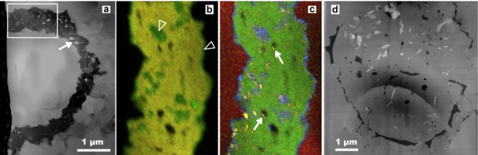
Fig. 4. Fe-minerals associated with Huroniospora. Scanning transmission electron microscope images of Focused Ion Beam ultrathin sections. a. Brightfield image of the wall of a Huroniospora replaced by porous (arrow) pyrite (in black). b–c. STEM-EDXS (13 nm probe) elemental maps of the boxed region in (a). Pyrite [Fe + S: yellow in (b), Fe: green in (c)] comprises most of the mineralized wall structure, while Ca-rich siderite [Fe0.92Ca0.08CO3; Fe without S: green in (b) and Ca: blue in (c)] occurs at the margins and inside some pores of the
pyritic wall. Some pores are empty (black), while others are partlyfilled with organic C [yellow in (c), possibly epoxy contamination]. Quartz [Si in red in (c)] is found inside and outstide the fossil. d. Dark-field image of a Huroniospora displaying thick organic walls (in black) in quartz (grey). Abundant Fe-minerals (Ca-poor siderite and greenalite, in white) occur inside this microfossil. Panel (d) reproduced fromLepot et al. (2017).
intra-microfossil only localization of these Fe-minerals (Fig. 4d) con-trasts strongly with post-mortem Fe-mineralization of walls by pyrite (Fig. 4a–c andWacey et al., 2013) or siderite (Fig. 4a–c andFadel et al., 2017). These Fe-minerals have been interpreted as the products of in-situ reductive recrystallization of intracellular Fe3 +-bearing bio-minerals. The combined C-isotopes signature (Williford et al., 2013), intracellular Fe-biomineralization, and microfossil morphology is best explained by cyanobacteria (Lepot et al., 2017). Evidence for chemo-trophic Fe-oxidizing bacteria or photoferrochemo-trophic bacteria are still lacking in stromatolitic Gunflint-type assemblages.
2.5. Chemotrophic Fe-oxidizing bacteria
Phosphatic stromatolites of the ~ 1.7 Ga Jhamarkotra Formation (Aravalli Group, India), display twisted Fe-oxide filaments similar to the Fe-biomineralized extracellular stalks of some chemolithoauto-trophic bacteria (Crosby et al., 2014). Indeed, the freshwater iron-oxi-dizing bacteria Gallionella ferruginea and the marine Mariprofundus ferrooxydans both use O2 (in microaerophilic conditions) to oxidize
Fe2 +; both are able of chemolithoautotrophy, and Gallionella can also
grow on organic compounds in addition to CO2(Emerson et al., 2010).
They deposit Fe3 +-oxides onto twisted extracellular polysaccharide
stalks that, together, withstand experimental diagenetic conditions (Picard et al., 2015).
2.6. Assemblages of deep-water cherts
Microfossil assemblages occur in chert nodules from the ~ 1.8 Ga Duck Creek Formation (Knoll et al., 1988; Schopf et al., 2015; Wilson et al., 2010) and the ~ 2.45–2.21 Ga Kazput Formation of the Turee
Creek Group (Fadel et al., 2017; Schopf et al., 2015; Van Kranendonk et al., 2012), both from Western Australia. Their occurrence in massive dolomites and stratigraphic association suggest deposition in quiet, possibly relatively deep water (Barlow et al., 2016). Accordingly, unlike the stromatolitic Gunflint-type mats, these assemblages are not lami-nated but form cobweb-like structures. Some chert nodules of the Duck Creek Formation include the typical Gunflint-type assemblage with Huroniospora, Eoastrion, Gunflintia, and rare broad segmented filaments (Knoll et al., 1988). The cobweb-forming assemblages are, however, mostly composed of filamentous microorganisms. Some filaments ∼8 μm in diameter displayed, under the optical microscope, micro-structures suggesting elongated (12–15 μm) cells (Schopf et al., 2015). Dark, apparently unsegmentedfilaments ≤1 μm in diameter have been interpreted as filament-shaped single cells, unknown among cyano-bacteria (Schopf et al., 2015); however, at the resolution limit of optical microscopy, these are difficult to distinguish from collapsed sheaths of trichome-forming microorganisms (e.g. the narrower sheaths in
Fig. 3b). The preservation of elongated cells in broadfilaments and the single-cell nature of the narrowerfilaments require confirmation with nanoscale analyses. These morphological features, combined with the presence of pyrite bearing the signature of bacterial sulfate-reduction have been used to propose that the cobweb-forming assemblages re-present sulfur-cycling consortia (sulfuretum). Indeed, sulfate-reducing bacteria (Fukui et al., 1999), in addition to cyanobacteria (Wood et al., 2008), may formfilaments with large, elongated shape. In the inferred sulfuretum, the broad (> 12μm) filaments (Knoll et al., 1988) and the Huroniospora coccoids (Schopf et al., 2015) may represent sulfur-oxi-dizing bacteria such as Beggiatoa/Thioploca and Thiovulum, respectively, instead of cyanobacteria. In contrast,Wilson et al. (2010)proposed that the Duck Creek Formation assemblage was dominated by Fe-oxidizing bacteria. In a Turee Creek Group assemblage associated with a banded iron deposit, Fadel et al. (2017)observed a distinct cobweb-forming microfossil assemblage without segmented filaments or pyrite that would support SRB, and with siderite displaying isotopic signatures of Fe-deposition through oxidation, which is also consistent with chemo-trophic or photochemo-trophic (oxygenic or anoxygenic) Fe-oxidation.
2.7. Problematic fossil record
To our knowledge, the early Paleoproterozoic has a poor microfossil record. Rounded graphitic particles from the 2.5 Ga Hutuo Fm, China have been interpreted as possible highly metamorphosed acritarchs (Schiffbauer et al., 2007), but their biogenicity remains difficult to prove.
Other problematic traces consisting of U-shaped ridges in sandstone of the 2 to 1.8 Ga Stirling Range Formation, Australia,first reported as animal traces (Bengtson et al., 2007), have been reinterpreted as pos-sible traces of giant protists such as amoebae, similar to observations in recent sediments (Bengtson and Rasmussen, 2009; Matz et al., 2008; Pawlowski and Gooday, 2009).
Recently discovered pyritic macrostructures from the 2.1 Ga Francevillian of Gabon have been interpreted as possible fossils of multicellular organisms, because of their large (~ 25 mm) size (El Albani et al., 2010). The term“multicellularity” implies only the oc-currence of several cells, without evidence supporting either a prokar-yotic or eukarprokar-yotic affinity (for reviews on multicellularity, see
Butterfield, 2009; Knoll, 2011). These pyritic macrostructures only show traces of C and have been interpreted as organic masses pyritized by (likely heterotrophic) sulfate-reducing bacteria, as indicated by S-isotopes. So far they have been discriminated from abiotic pyrite con-cretions such as those described inSeilacher (2001)by details of their morphology showing soft deformation (folding), internal texture (mass of octahedral pyrite grains, different from the fibrous cone-in-cone texture of the“pyrite suns” ofSeilacher, 2001), and distribution within the black shale beds draping them, deposited below an oxic water column in shallow water (Canfield et al., 2013; El Albani et al., 2010; El Albani et al., 2014) or under deeper water (> 200 m) (Parize et al., 2013). The identity of these macrostructures remains unknown and their biogenicity is questionable (e.g.Anderson et al., 2016) although the latter case study does not seem comparable.
2.8. Stromatolites
A general trend of increasing morphological diversity, size, and global distribution is observed in the stromatolitic record of carbonate rocks. From the Archean and early Proterozoic, through the mid-Proterozoic, stromatolite construction evolved from a texture mostly dominated by (bio)chemical carbonate precipitation with sparry mi-crofabrics forming small (decimeter scale) cones and planar stromato-lites (Allwood et al., 2009; Grotzinger and Knoll, 1999) to a more mixed andfine-grained texture of (bio)precipitation and trapping and binding of particles forming large domes (Buick, 1992; Coffey et al., 2013; Lepot et al., 2008; Lepot et al., 2009a), conic forms with a central column (Conophyton, e.g. Walter et al., 1976a), and digitate forms (Grey, 1994a, 1994b; Melezhik et al., 1997). Their biogenicity is most likely if stromatolites show complex morphologies, laterally and vertically variable laminae and organic bearing microfabrics, as these features are difficult to explain by abiotic mineral precipitation alone (see discus-sions of biogenicity criteria inBosak et al., 2013; Grotzinger and Knoll, 1999). These changes might reflect changes in hydrodynamic and
physico-chemical conditions, larger areas of shallow carbonate plat-forms, but also diversification of building microbial communities. Mi-crostructural fabrics in Proterozoic stromatolites can sometimes pro-vide a record of microbial community diversity despite diagenetic alteration (Knoll et al., 2013). Complex assemblages of stromatolites populate the 2.4–2.2 Ga Turee Creek group of Western Australia. Their complex morphologies (domical: e.g.Fig. 5a, columnar, thrombolitic, stratiform) may reflect changes in environments and microbial com-munities associated with the GOE (Barlow et al., 2016; Martindale et al., 2015). Two major events are noted in the Paleoproterozoic evolution of stromatolites on the Fennoscandian shield and include a maximum in diversity and abundance of stromatolites between 2330 and 2060 Ma ago, linked to cratonization, and a decline between 2060
and 1900 Ma ago, linked to oceanization and consequent decline of niches suitable for benthic cyanobacteria (Melezhik et al., 1997). Highly diverse and abundant stromatolites are also found in the late Paleoproterozoic of Western Australia (Grey, 1994a, 1994b). Paleo-geographical differences in abundance and diversity patterns are re-ported between the Fennoscandia, India and China on one side, and Australia and northern America on the other side (Melezhik et al., 1997).
Late Paleoproterozoic stromatolites of the Aravalli Supergroup of India display abundant phosphatized granules intimately associated with organic matter, which have been proposed as possible phospha-tized cells or phosphatized extracellular polymeric substances (Papineau et al., 2016). Stromatolites bearing Gunflint-type microfossil assemblages are siliceous and have been debated as resulting of primary silicification by hydrothermal fluids (Maliva et al., 2005) or as diage-netic cherts formed by replacement of primary carbonates (Petrash et al., 2016; Sommers et al., 2000). While the latter process is common in carbonate platform stromatolites (Fig. 5b), it remains difficult to
distinguish from primary silica in stromatolites samples where carbo-nates are essentially represented by late-diagenetic rhombs (Fig. 5c). In our view, the question of the original mineralogy of many Paleopro-terozoic siliceous stromatolites remains unanswered.
Microbially Induced Sedimentary Structures (MISS) preserved in siliciclastic deposits, are recorded from the Eoarchean to the present in intertidal environments (Noffke et al., 2013b), although the micro-organisms responsible for their occurrence have changed from anae-robic to aeanae-robic microorganisms (mostly cyanobacteria today). For a recent review of stromatolites and MISS, which is beyond the scope of this review, seeBosak et al. (2013)andNoffke et al. (2013a). 3. Implications for early biosphere evolution
The Paleoproterozoic microfossil assemblages include unambiguous eukaryotes difficult to relate to modern clades, cyanobacteria, iron-oxidizing and other undeterminedfilamentous and coccoidal prokar-yotes, possible sulfur-oxidizing and/or sulfate-reducing bacteria, and heterotrophic decomposers (possibly including some sulfate-/iron-re-ducing bacteria, some mixotrophic sulfur-/iron-oxidizing bacteria, some methanogens and other fermenters) (Fig. 1). There are no un-ambiguous microfossils of the domain Archaea and their biomarker record (for the clade performing anaerobic oxidation of methane) is known from the Carboniferous (Birgel et al., 2008). The extremely light carbon-isotopic values at ~ 2.1 Ga (Weber and Gauthier-Lafaye, 2013) and between 2.8 and 2.6 Ga (or possibly as early as 3.5 Ga;Ueno et al., 2006) indicate the activities of methanogenic archaea and subsequent (archaeal and/or bacterial) methanotrophy (Eigenbrode et al., 2008;
Hayes, 1994). However,Slotznick and Fischer (2016) suggested that methanotrophy was not responsible for the notably lowδ13Corgvalues
(in the 2.72 Ga Tumbiana Formation, Australia), which may have formed through the reductive acetyl co-enzyme A (CoA) pathway. Be-cause of the chimeric nature of the eukaryotic cell, which contains bacterial and archaeon genes, most models for the origin of eukaryotes involve the contribution of an archaea (see review inLopez-Garcia and Moreira, 2015; Spang and Ettema, 2016), thus implying a preceding origin of the domain, although this is discussed (Eme and Doolittle, 2015; Gouy et al., 2015). Most major clades of the domain Bacteria had probably diversified already in the Paleoproterozoic, since cyano-bacteria are not among the early diverging cyano-bacteria on phylogenetic reconstructions and had appeared at least by the time of the GOE, ~ 2.5 Ga ago (Pace, 1997; Schirrmeister et al., 2013).
This middle age biosphere diversified in a variety of new ecological niches. Mildly oxygenated shallow water of carbonate and siliciclastic platforms were colonized by cyanobacterial mats and stromatolites, probably other aerobe prokaryotes and by eukaryotes. Shallow-water stromatolites bearing Gunflint-type microfossil assemblages between 2.1 and 1.7 Ga display an enrichment in non-clastic, non-pyritic iron. This iron enrichment was likely associated with the transient return to ferruginous conditions associated with an increase of volcanic activity around 1.9–1.8 Ga (Rasmussen et al., 2012), and a post-GOE drop in oxygen as early as 2.1 Ga (Canfield et al., 2013; Lyons et al., 2014). Whether these assemblages developed in restricted or open-marine basins is unclear. In these conditions, oxygen produced by cyano-bacteria may have been used by, and possibly nearly titrated, through iron oxidation and organic-matter respiration. Accordingly, observed intra-microfossil Fe-mineralization is consistent with Fe-tolerant cya-nobacteria in the Gunflint Iron Formation (Lepot et al., 2017). The small amount of available sulfate was reduced by sulfate-reducing bacteria to sulfide (Wacey et al., 2013) that would have precipitated immediately with excess Fe2 +, hence limiting sulfur-oxidizing
meta-bolism (such as anoxygenic photosynthesis) that is usually observed below cyanobacterial mats in modern stromatolites (Vasconcelos et al., 2006). Photoferrotrophs and Fe-oxidizing chemo(auto)trophs may also have contributed to the Gunflint-type assemblages, but morphological evidence of Fe-using chemoautotrophy appears only in 1.7 Ga stroma-tolites without other Gunflint-type microfossils (Crosby et al., 2014). Some non-stromatolitic, cobweb-forming microfossil assemblages from chert nodules in dolomites may have formed in distinct environmental conditions, possibly below the photic zone and/or within the sediment, where it could have derived energy from redox cycling of sulfur (Schopf et al., 2015) or iron (Fadel et al., 2017; Wilson et al., 2010). Although they share the assemblage of microfossils with simple morphological features (filaments, spheres and star shapes), Gunflint-type and
Fig. 5. Paleoproterozoic stromatolites. a. Outcrop picture of a meter-scale bulbous carbonate stromatolite from the Kazput Formation of the 2.45–2.21 Ga Turee Creek Group (seeBarlow et al., 2016; Martindale et al., 2015for details on the locality); hammer = 30 cm. b. Outcrop picture of a stromatolite from the ~ 1.8 Ga Duck Creek Formation (Wilson et al., 2010). Orange zones are mostly composed of carbonates. In black zones, carbonate laminae have been partly to completely replaced by chert. c. Thin section photomicrograph (5 × objective) of a stromatolite from the Gunflint Iron Formation at the Schrieber Beach locality. The stromatolites are essentially composed of quartz (white) with organic matter laminae (brown) forming bulges (arrowheads) or cones (top left corner). Carbonates are mainly present as dispersed, coarse ankerite [(Fe,Ca,Mg)CO3] rhombohedra (arrows) of late diagenetic origin (Lepot et al.,