HAL Id: hal-01347068
https://hal.sorbonne-universite.fr/hal-01347068
Submitted on 20 Jul 2016
HAL is a multi-disciplinary open access
archive for the deposit and dissemination of
sci-entific research documents, whether they are
pub-lished or not. The documents may come from
teaching and research institutions in France or
abroad, or from public or private research centers.
L’archive ouverte pluridisciplinaire HAL, est
destinée au dépôt et à la diffusion de documents
scientifiques de niveau recherche, publiés ou non,
émanant des établissements d’enseignement et de
recherche français ou étrangers, des laboratoires
publics ou privés.
Co-expression of Foxa.a, Foxd and Fgf9/16/20 defines a
transient mesendoderm regulatory state in ascidian
embryos
Clare Hudson, Cathy Sirour, Hitoyoshi Yasuo
To cite this version:
Clare Hudson, Cathy Sirour, Hitoyoshi Yasuo. Co-expression of Foxa.a, Foxd and Fgf9/16/20 defines
a transient mesendoderm regulatory state in ascidian embryos. eLife, eLife Sciences Publication, 2016,
5, pp.e14692. �10.7554/eLife.14692�. �hal-01347068�
*For correspondence: clare. [email protected] (CH); yasuo@ obs-vlfr.fr (HY)
†These authors contributed
equally to this work Competing interests: The authors declare that no competing interests exist. Funding:See page 14
Received: 25 January 2016 Accepted: 24 June 2016 Published: 28 June 2016 Reviewing editor: Alejandro Sa´nchez Alvarado, Stowers Institute for Medical Research, United States
Copyright Hudson et al. This article is distributed under the terms of theCreative Commons Attribution License,which permits unrestricted use and redistribution provided that the original author and source are credited.
Co-expression of Foxa.a, Foxd and Fgf9/
16/20 defines a transient mesendoderm
regulatory state in ascidian embryos
Clare Hudson*
†, Cathy Sirour, Hitoyoshi Yasuo*
†Laboratoire de Biologie du De´veloppement de Villefranche-sur-mer, Observatoire
Oce´anologique, Sorbonne Universite´s, UPMC Univ Paris 06, CNRS,
Villefranche-sur-Mer, France
Abstract
In many bilaterian embryos, nuclear b-catenin (nb-catenin) promotes mesendoderm over ectoderm lineages. Although this is likely to represent an evolutionary ancient developmental process, the regulatory architecture of nb-catenin-induced mesendoderm remains elusive in the majority of animals. Here, we show that, in ascidian embryos, three nb-catenin transcriptional targets, Foxa.a, Foxd and Fgf9/16/20, are each required for the correct initiation of both the mesoderm and endoderm gene regulatory networks. Conversely, these three factors are sufficient, in combination, to produce a mesendoderm ground state that can be further programmed into mesoderm or endoderm lineages. Importantly, we show that the combinatorial activity of these three factors is sufficient to reprogramme developing ectoderm cells to mesendoderm. We conclude that in ascidian embryos, the transient mesendoderm regulatory state is defined by co-expression of Foxa.a, Foxd and Fgf9/16/20.DOI: 10.7554/eLife.14692.001
Introduction
The mesoderm, endoderm and ectoderm arise during embryonic development by a process termed germ layer segregation. In many species, at least part of the endoderm and mesoderm derive from transient ‘mesendoderm’ precursors, as is the case in ascidian embryos (Kimelman and Griffin, 2000;Rodaway and Patient, 2001). However, the precise nature of this induced regulatory state is not well understood. In ascidians, the first animal-vegetal (A-V) oriented cell division generates the eight-cell stage embryo and segregates the mesendoderm and some neural lineages into two pairs of vegetal founder lineages (the A- and B-line) and the ectoderm (epidermis and neural) into two pairs of animal lineages (a- and b-line) (Conklin, 1905; Nishida, 1987). This study focuses on the A-line mesendoderm lineages. From the 8- to 16-cell stage, the two A4.1 blastomeres divide medio-laterally to generate the two pairs of neuro-mesendodermal NNE cells, for notochord/neural/endo-derm (Figure 1a). NNE cells then divide along the A-V axis to generate NN cells (notochord/neural) and E cells (mostly endoderm) at the 32-cell stage (Figure 1a). Subsequently, NN cells segregate into notochord and neural lineages at the 64-cell stage. At this stage, while the medial E cell gener-ates two endoderm precursors, the lateral-most E cell is subject to an inductive interaction resulting in the generation of one endoderm and one mesoderm (the trunk lateral cell lineage) precursor (Shi and Levine, 2008). Later, during neural plate patterning, a muscle precursor is also generated from the lateral borders of the NN-lineage-derived neural plate (Nicol and Meinertzhagen, 1988;
Nishida, 1987). Thus, as in other species, ascidian germ layer segregation can be viewed as a pro-gressive process with part of the neural tissue arising from bipotential neuro-mesodermal progeni-tors (Henrique et al., 2015; Tzouanacou et al., 2009). The earliest cell divisions of the ascidian
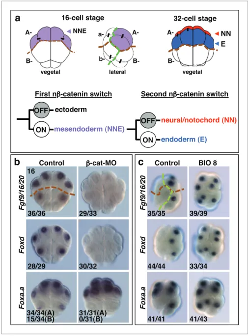
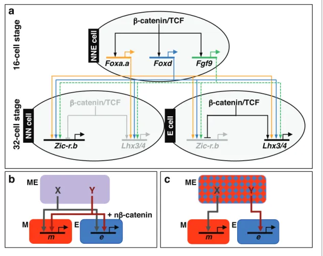
Figure 1. Foxa.a, Foxd and Fgf9/16/20 are candidate NNE lineage specification factors. (a) Schematic drawings of embryos at the 16- and 32-cell stages. In this and all subsequent figures, where shown, a green dashed line separates the animal (ectoderm) from the vegetal (mesendoderm) hemispheres and a brown dashed line separates A- (A4.1) and a- (a4.2) lineages from B- (B4.1) and b- (b4.2) lineages. Different embryonic founder lineages are indicated on the drawings. NN and E cells are indicated in red and blue, respectively. Below the embryo drawings is a schematic representation of the two rounds of nb-catenin-driven binary fate decisions that segregate firstly the mesendoderm lineages from the ectoderm lineages at the 16-cell stage and secondly segregate the mesoderm (NN) lineages from the endoderm (E) lineages at the 32-cell stage (Hudson et al., 2013). (b, c) Embryos analysed at the 16-cell stage for the marker indicated to the left of the panels following the treatment indicated above the panels [(b) vegetal pole view; (c) lateral view, vegetal pole to the right]. The numbers on the bottom-left corner of each panel indicate the proportion of embryos that the panel represents. The posterior most cells (at the bottom of the panels) are transcriptionally quiescent cells that will generate the germ line (Shirae-Kurabayashi et al., 2011). For Foxa.a expression in (b) control embryos showed expression in all four A-line (NNE) cells in 34/34 embryos, and in B-line cells, in 15/34 embryos, as indicated, whereas b-catenin-MO (b-cat-b-catenin-MO) injected embryos showed expression in NNE cells (31/31), but not B-line (0/31). Expression of Foxa.a in the four a-line precursors (not visible in the image) was not affected by b-catenin-MO injection.
embryo along the A-V axis at the 8- and 32-cell stages can be considered as the earliest steps of germ layer segregation.
b-catenin is a transcriptional co-activator which acts in a complex with TCF DNA-binding proteins to mediate the canonical Wnt signalling pathway (Valenta et al., 2012). The b-catenin/TCF complex promotes endoderm or mesendoderm in a wide range of organisms, and this process is therefore likely to represent an ancestral mechanism (Darras et al., 2011;Henry et al., 2008;Hudson et al., 2013; Imai et al., 2000; Logan et al., 1999; McCauley et al., 2015; Miyawaki et al., 2003;
Momose and Houliston, 2007;Wikramanayake et al., 1998, 2003). We have previously shown that the earliest steps of germ layer segregation in ascidian embryos are mediated by two rounds of nuclear(n)-b catenin-dependent binary fate decisions. The first nb-catenin-driven binary fate deci-sion takes place at the 8- to 16-cell stage. During this process, the b-catenin/TCF complex is differ-entially activated between mesendoderm and ectoderm progenitors, resulting in segregation of these lineages (Figure 1a) (Hudson et al., 2013;Oda-Ishii et al., 2016;Rothba¨cher et al., 2007). The second step takes place at the 32-cell stage and controls the segregation of NNE mesendoderm cells into endoderm (E cell) and notochord/neural (NN cell) lineages (Hudson et al., 2013). During this step, the b-catenin/TCF complex is again differentially activated between E and NN cells (Figure 1a). Therefore, cells in which nb-catenin remains active during the two steps (ON + ON) are specified as endoderm lineage, cells in which nb-catenin remains inactive during the two steps (OFF + OFF) are specified as ectoderm lineage and cells in which nb-catenin is active during the first step but inactive during the second step (ON + OFF) are specified as notochord-neural lineage (Hudson et al., 2013). These two rounds of nb-catenin-driven switches result in transcriptional acti-vation of the lineage specifiers, Zic-related.b (Zic-r.b, formally ZicL) and Lhx3/4 (formally Lhx3), in NN and E cells, respectively (Imai et al., 2002c;Satou et al., 2001). One of the key features of these reiterative nb-catenin-driven binary fate decisions is that the same asymmetric cue (nb-catenin) is interpreted differently during each step (Bertrand and Hobert, 2010). Thus, in the NNE lineage, it is likely that the transient regulatory state induced by the first nb-catenin input in NNE cells confers a distinct transcriptional response to the second nb-catenin input on E cells.
In this study, we characterise the NNE lineage specification factors, which are induced by the first nb-catenin input and address how these mesendoderm factors feed into the gene regulatory net-work of the NN and E lineages.
Results
Foxa.a, Foxd and Fgf9/16/20 are nb-catenin transcriptional targets in
NNE cells
Following the first nb-catenin activation at the 16-cell stage, Foxa.a, Foxd, Fgf9/16/20, cadherinII and bCD1 (b-catenin downstream gene 1) are induced in the NNE cells, with at least Foxd and Fgf9/16/20
being direct targets of the b-catenin/Tcf7 complex (Imai, 2003; Imai et al., 2002a,
2002b,2002c;Kumano et al., 2006;Oda-Ishii et al., 2016;Rothba¨cher et al., 2007;Satou et al., 2001). Consistent with a recent study (Oda-Ishii et al., 2016), we confirmed that in b-catenin–inhibited (b-catenin-MO injected) embryos analysed at the 16-cell stage, Foxd and Fgf/9/16/20 expression was lost (Figure 1b). In addition to the mesendoderm lineages, Foxa.a is also expressed in the a-line ante-rior ectoderm lineages in a nb-catenin-independent fashion (Figure 1b,c) (Lamy et al., 2006). In b-cate-nin–inhibited embryos, Foxa.a expression persisted in NNE and a-lineage cells, probably due to transformation of vegetal cells into animal cells that has been reported previously (Figure 1b) (Imai et al., 2000;Oda-Ishii et al., 2016). Conversely, ectopic stabilisation of nb-catenin resulted in activation of all three genes in ectoderm lineages at the 16-cell stage (Figure 1c). This was achieved by treating embryos with BIO, a chemical inhibitor of the upstream inhibitory regulator of b-catenin, GSK-3, from the eight-cell stage (Meijer et al., 2003). Thus, our results confirm that Foxd, Foxa.a and Fgf9/
16/20 are transcriptional targets of nb-catenin in vegetal cells, although Foxa.a also has a
Foxa.a, Foxd and Fgf9/16/20-signals are required for the correct
initiation of both NN and E gene expression
It is likely that these gene products, activated by the first nb-catenin signal in NNE cells, act together with the second differential nb-catenin signal to activate the distinct gene regulatory networks between NN and E cells. Consistent with this idea, Foxa.a has been shown to be required for both NN lineage and endoderm gene expression (Imai et al., 2006), with Foxd specifically required for NN lineage, but not endoderm fates, and Fgf9/16/20 contributing to notochord induction from the NN lineage (Imai et al., 2002a,2002b;Yasuo and Hudson, 2007). However, we found that inhibit-ing any one of these factors prevented the correct initiation of gene expression in both NN (Zic-r.b)
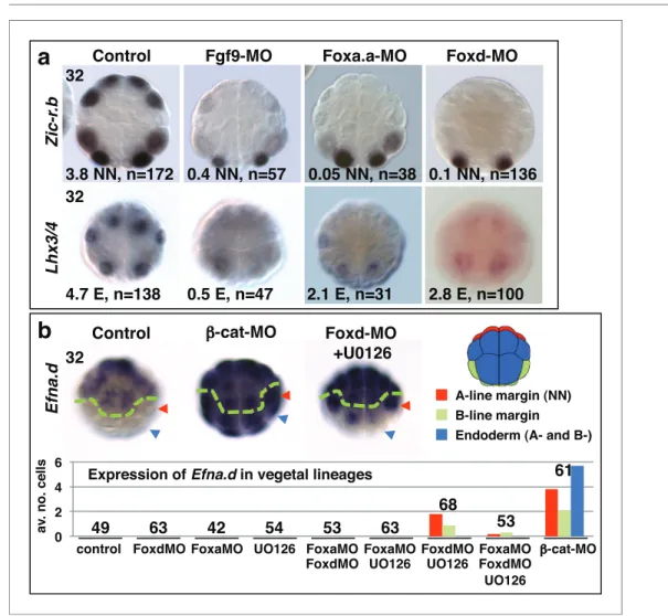
Figure 2. Foxa.a, Foxd and Fgf9/16/20 are required for initiation of NN and E gene expression. (a) Embryos analysed at the 32-cell stage. The marker analysed is indicated on the left of the panels and the treatment indicated above the panels. The average number of NN (Zic-r.b) or E (Lhx3/4) cells expressing detectable levels of each gene is indicated. This remaining expression was generally weaker than control level expression. ‘n=’ represents the number of embryos analysed. (b) Expression of Efna.d under the conditions indicated. Embryos are shown in notochord-side view, animal pole up. The graph shows the average number of cells expressing Efna.d in different vegetal lineages at the 32-cell stage, as indicated by the key. All embryos showed ectoderm expression. The number of embryos analysed is indicated above the bars on the graph.
DOI: 10.7554/eLife.14692.003
The following figure supplements are available for figure 2:
Figure supplement 1. Foxa.a, Foxd and Fgf9/16/20 are required for initiation of NN and E gene expression.
DOI: 10.7554/eLife.14692.004
Figure supplement 2. Endoderm formation under various conditions.
and E (Lhx3/4) lineages (Figure 2a,Table 1). We inhibited these factors using Morpholino anti-sense oligonucleotides (Foxd-MO, Foxa.a-MO, Fgf9-MO) and analysed Zic-r.b and Lhx3/4 expression at the 32-cell stage, when NN and E cell lineages become segregated. FGF signals are frequently mediated by the MEK/ERK signalling pathway, leading to transcriptional activation via ETS family transcription factors, as is the case in ascidian embryos (Bertrand et al., 2003; Kim and Nishida, 2001; Miya and Nishida, 2003; Yasuo and Hudson, 2007). We confirmed that Fgf9/16/20 is responsible for the broad activation of ERK at the 32-cell stage in most vegetal lineages, including NN and E lineages, as well as two neural lineages in the ectoderm (Figure 2—figure supplement 1f). Treatment of embryos from the 16-cell stage with the MEK inhibitor U0126, also inhibits this ERK1/2 activation (Kim and Nishida, 2001;Picco et al., 2007). Inhibition of Fgf9/16/20, MEK or ETS1/2 (ETS1/2-MO) gave similar results, although inhibition of ETS1/2 gave only a weak down-regu-lation of Zic-r.b expression at the 32-cell stage, perhaps indicating the involvement of additional transcription factors that are also known to mediate FGF signals in Ciona embryos (Figure 2a;
Table 1) (Bertrand et al., 2003;Gainous et al., 2015). Maintenance of Foxa.a, Foxd and Fgf9/16/20 expression at the 32-cell stage is independent of each other (Figure 2—figure supplement 1a), as was shown previously for Foxd and Fgf9/16/20 in Ciona savigni embryos (Imai et al., 2002a).
In FGF-inhibited embryos, Zic-r.b expression recovered at the 64-cell stage (Figure 2—figure supplement 1a) (Imai et al., 2006;Kumano et al., 2006). Zic-r.b expression at the 32- and 64-cell stages can be mediated by separate enhancer elements (Anno et al., 2006). In addition, in the NN-cell lineage, FGF-signalling is required for notochord fate, but has to be attenuated for neural fate (Minokawa et al, 2001; Picco et al., 2007; Yasuo and Hudson, 2007). Thus, an FGF-independent expression of Zic-r.b at the 64-cell stage, at least in neural fated cells, is not unexpected. In Foxa.a-and Foxd- inhibited embryos, Zic-r.b continues to be repressed in the NN-cell lineages at the 64-cell stage (Figure 2—figure supplement 1a) and later (Imai et al., 2006), consistent with a requirement for Foxa.a and Foxd for both NN cell lineage-derived structures, the notochord and caudal central nervous system (CNS) (Imai et al., 2002b,2006).
Endoderm gene expression was continuously reduced up to at least the early gastrula stage, fol-lowing inhibition of any one of the NNE factors (Figure 2—figure supplement 1a–e). However, using alkaline phosphatase activity as an indicator of endoderm formation (Whittaker, 1977), a com-plete loss of endoderm at larval stages was observed only in Foxa.a-inhibited embryos, consistent with previous studies (Figure 2—figure supplement 2) (Imai et al., 2002b,2006). In Foxd and FGF-signal inhibited embryos, a large domain of alkaline phosphatase activity could be detected, sug-gesting that endoderm fate recovers in these embryos (Figure 2—figure supplement 2). Simulta-neous repression of Foxd and FGF-signalling, however, resulted in both a stronger repression of early endoderm gene expression as well as an almost complete absence of alkaline phosphatase activity at larval stages (Figure 2—figure supplement 1–2). Thus, for eventual endoderm formation, the embryo is able to compensate for loss of either Foxd or FGF-signals but is not able to compen-sate for loss of both.
As well as promoting vegetal ‘mesendoderm’ fates, nb-catenin also represses the ectoderm gene programme in vegetal cells (Hudson et al., 2013; Imai et al., 2000; Oda-Ishii et al., 2016;
Rothba¨cher et al., 2007). In b-catenin knock-down embryos, ectopic expression of the early ecto-derm gene Efna.d (formally ephrin-Ad) is observed in both NN and E cells at the 32-cell stage (Figure 2b) (Hudson et al., 2013) as well as in NNE cells at the 16-cell stage (Oda-Ishii et al., 2016). Double inhibition of Foxd and FGF-signals also resulted in ectopic expression of Efna.d in NN cells, but never in E cells (Figure 2b). Thus, NNE factors repress the ectoderm genetic programme in NN
Table 1. Expression of Zic-r.b in NN cells and Lhx3/4 in E cells of 32-cell stage embryos, following inhibition of Fgf-signalling components.
Control U0126 ETS1/2-MO
Zic-r.b NN cell 4.0 cells (n = 163) 0.75 cells (n = 58) 3.4 cells* (n = 74) Lhx3/4 E cell 3.9 cells (n = 153) 2.1** cell (n = 45) 0.7 cells (n = 92) * 44/74 embryos exhibited weaker levels of Zic-r.b expression compared to controls.
**Remaining expression was weaker than control levels of expression.
cells. A lack of derepression in E cells is probably due to the presence of nb-catenin in E cells at the 32-cell stage (Hudson et al., 2013), suggesting that nb-catenin can repress the ectoderm genetic programme both via and independently of the NNE factors.
Combinatorial activity of Foxa.a, Foxd and Fgf9/16/20 induces a
mesendoderm state
Our data so far show that Foxa.a, Foxd and Fgf9/16/20-ERK1/2 are individually required for the cor-rect initiation of the genetic programmes of both NN and E cell lineages. Indeed, co-expression of these three factors takes place only in mesendoderm lineages, the NNE and B-line mesendoderm lineages of the 16- and 32-cell stage embryo (Figure 1;Figure 2—figure supplement 1a). At the 32-cell stage, the E cells continue to express these three genes, while NN cells express only Foxa.a and Fgf9/16/20. However, we have previously shown that Foxd transcripts preferentially segregate into the NN cells during the NN-E cell division, before they rapidly disappear (Hudson et al., 2013). Thus, NN cells also contain Foxd transcripts early in their cell cycle. We conclude that Foxa.a, Foxd and Fgf9/16/20 are co-expressed only in mesendoderm lineages. We next addressed whether these three factors were sufficient to induce a mesendoderm regulatory state.
As well as in vegetal cells, Foxa.a is also expressed in a-line anterior animal cells and ERK1/2 is activated in one pair of a-line cells (the a6.5 pair) at the 32-cell stage (Figure 3a) (Hudson et al., 2003;Lamy et al., 2006). Thus, a6.5 cells possess two of the three mesendoderm lineage specifiers and yet they do not adopt an NNE-like lineage. Consistent with the notion that coexpression of
Foxa.a, Foxd and Fgf9/16/20 represents a NNE regulatory state, reintroduction of the remaining
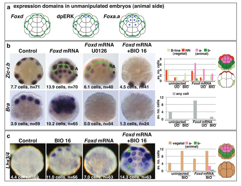
fac-tor, Foxd, by mRNA injection, was able to convert a-line cells to a mesendoderm state (Figure 3). Injection of Foxd mRNA resulted in ectopic expression of Zic-r.b in a-line cells at the 32-cell stage (Figure 3b) (Imai et al., 2002c). Similarly, expression of Bra, a marker of notochord precursors, was induced at the 64-cell stage (Figure 3b). The broad ectopic expression of Zic-r.b and Bra in the a--lineage was clearly not restricted to the a6.5 cells. The most likely reason for this was that Foxd mRNA injection led to weak activation of Fgf9/16/20 and strong inhibition of Efna.d expression in the ectoderm cells (Figure 3—figure supplement 1). Efna.d is a known antagonist of FGF-signals in
Ciona and its inhibition results in widespread activation of ERK1/2 in ectoderm lineages (Ohta and Satou, 2013;Picco et al., 2007; Shi and Levine, 2008). Consistent with this, when Foxd mRNA-injected embryos were treated with U0126, the ectopic expression of Zic-r.b was reduced and Bra expression was completely suppressed, mimicking the effect of MEK inhibition on endogenous Zic-r.
b and Bra gene expression (Figure 3b). Thus, injection of Foxd mRNA is sufficient to convert Foxa.a/ ERK1/2-positive a-line cells into mesoderm.
Injection of Foxd mRNA, however, was not sufficient to induce endoderm gene expression in ectoderm cells (Figure 3c). This was expected since the two-step nb-catenin binary fate decision model predicts that segregation of the endoderm lineage from the NNE lineage requires a second round of nb-catenin activation (Figure 1a) (Hudson et al., 2013). Accordingly, when Foxd-mRNA injected embryos were treated from the late 16-cell stage with BIO in order to mimic the second input of nb-catenin activation, both Zic-r.b and Bra were repressed and Lhx3/4 ectopically activated in the a-line ectoderm cells (Figure 3b–c). Thus, the NNE-like state induced in animal cells by Foxd mRNA injection behaves in the same way as NNE state of unmanipulated embryos.
Taken together, these experiments provide strong evidence that the combinatorial activity of
Foxa.a, Foxd and Fgf9/16/20-ERK1/2 represents a NNE mesendoderm regulatory state downstream
of the first round of nb-catenin input. To further test this model, we addressed whether co-expres-sion of Foxa.a, Foxd and Fgf9/16/20 was sufficient to rescue mesoderm in b-catenin-knockdown embryos. b-catenin-MO injected embryos would express only Foxa.a among the three genes (Figure 1b). We have shown that injection of Foxd mRNA results in induction of low levels of Fgf9/
16/20 expression, together with a strong suppression of Efna.d expression (Figure 3—figure supple-ment 1). Thus, injection of Foxd mRNA should be sufficient to recapitulate a Foxd/Foxa.a/Fgf9/16/
20 overlap. Consistent with this, injection of Foxd mRNA was able to rescue expression of
NN-line-age genes (Zic-r.b and Bra) in b-catenin-MO embryos and, as expected, this recovery depended on an intact FGF-signalling pathway (Figure 4). We conclude that co-expression of Foxd+Foxa.a+Fgf9/
16/20 is sufficient to induce a mesendoderm regulatory state, which can then be further
Foxa.a, Foxd and Fgf9/16/20 act synergistically to reprogramme
developing ectoderm cells to a mesendoderm state
We next addressed whether ectopic expression of Foxd, Foxa.a and Fgf9/16/20 was able to reprog-ramme developing ectoderm to a mesendoderm state (Figure 5,Figure 5—figure supplement 1). The upstream regulatory sequences of the Fucosyltransferase-like (FT) gene become active in ecto-derm cells from the 64-cell stage, when the ectoecto-derm genetic programme is already underway
Figure 3. Creation of an ectopic Foxa.a, Foxd, FGF-signal overlap leads to ectopic mesendoderm formation. (a) Schematics show endogenous ectodermal expression of Foxa.a, Foxd (no expression) and activation of ERK (dpERK), indicated by blue dots. (b–c) Treatment is indicated above the panels and marker analysed to the left. All embryos in animal pole view except control Bra (vegetal pole view). Numbers show the total average number of cells per embryo expressing each marker. n = total number of embryos analysed. The graphs show the average number of cells expressing each maker in the lineages indicated on the keys, following the treatments indicated of the x-axis. No Zic-r.b expression was detected in endoderm lineages. In (c), the green arrowheads highlight the eight a-lineage cells. For (b), representative panels of uninjected/UO-treated and uninjected/BIO-treated embryos are not shown. The numbers of these experiments are: for Zic-r.b- U0126 alone n = 40 (average number of cells 2.6), BIO-16 alone n = 40 (average number of cells 2.9) and for Bra- U0126 alone, n = 39 (average number of cells 0.0); BIO-16 alone n = 31 (average number of cells 0.0).
DOI: 10.7554/eLife.14692.007
The following figure supplement is available for figure 3:
Figure supplement 1. Foxd mRNA injection leads to repression of Efna.d and upregulation of Fgf9/16/20 in ectodermal cells at the 16-cell stage.
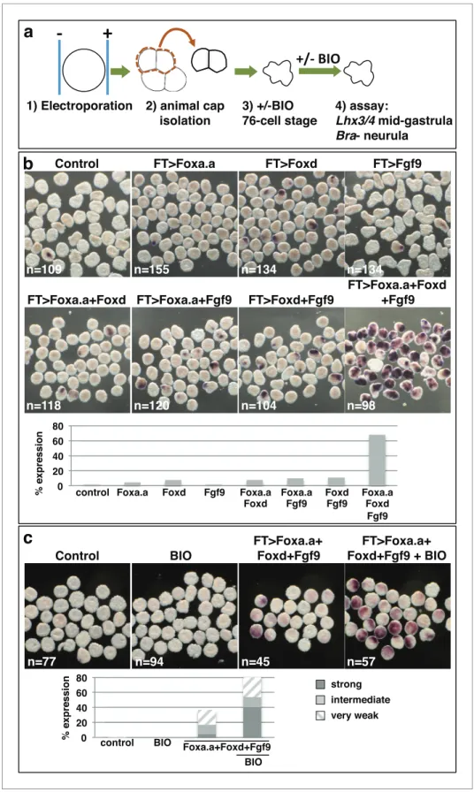
(Figure 5—figure supplement 1) and when these cells no longer express Foxa.a, Foxd or Fgf9/16/20 (Imai et al., 2004;Pasini et al., 2012). Using FT promoter-driven constructs (pFT>Foxa.a, pFT>Foxd and pFT>Fgf9/16/20), we expressed Foxa.a, Foxd and Fgf9/16/20 in different combinations in ecto-derm lineages. To simplify the analysis and to rule out the possibility that signals from the vegetal cells may influence the experimental outcome, animal hemispheres of electroporated embryos were iso-lated by micro-dissection at the eight-cell stage. Isoiso-lated explants were cultured until the neurula stage when they were assayed for Bra expression (Figure 5a–b). Bra was chosen for this assay for its meso-derm (notochord)-specific expression. We observed a clear combinatorial effect between Foxa.a, Foxd and Fgf9/16/20 on the reprogramming of ectoderm to mesoderm, with strong induction of Bra seen only when all three constructs were co-electroporated (Figure 5b). This reprogramming was accompa-nied by a strong downregulation of ectoderm gene expression and ectopic expression of Zic-r.b in the ectoderm cells of whole embryos (Figure 5—figure supplement 1). Furthermore, ectoderm explants could be reprogrammed to adopt an endoderm state (Figure 5c). To achieve this, ectoderm explants
Figure 4. Foxd mRNA injection rescues mesoderm in b-catenin-MO injected embryos. (a–b) Treatment is indicated above the panels and marker analysed to the left of the panels. The total average number of cells per embryo is indicated, ‘n=’ indicates the total number of embryos analysed for each treatment. The graphs show the average number of cells expressing each marker in the lineages indicated by the keys, following the treatments indicated.
Figure 5. Reprogramming the ectoderm lineage to mesendoderm. (a) Experimental scheme. Embryos were electroporated and the ectoderm lineage (animal cap) isolated at the eight-cell stage. Ectodermal explants were cultured until the mid-gastrula stage for Lhx3/4 expression or until the neurula stage for Bra expression. Optionally, explants were treated with BIO, when control sibling embryos reached the 76-cell stage, for
approximately 1 hr prior to fixation (Lhx3/4 only). (b–c) Expression of Bra (b) and Lhx3/4 (c) in isolated ectodermal Figure 5 continued on next page
from embryos electroporated with the triple combination (pFT>Foxa.a, pFT>Foxd and pFT>Fgf9/16/
20) were treated with a pulse of BIO from the 76-cell stage to mimic the second round of nb-catenin
activation that normally drives the segregation of NN and E lineages (Figure 5c). The 76-cell stage was chosen, as at this stage ectopic expression driven by the pFT constructs is readily detectable ( Fig-ure 5—figFig-ure supplement 1a), the ectoderm programme is downregulated (Figure 5—figure supple-ment 1b) and ectopic Zic-r.b is not yet detected (Figure 5—figure supplement 1c). This stage thus represented the best approximation of the NNE state of normal embryos. We confirmed that BIO-treatment was able to induce nuclear translocation of b-catenin in isolated explants at the 76-cell stage (Figure 5—figure supplement 2). Endoderm induction was assayed by detection of Lhx3/4 expression at the mid-gastrula stage, that is approximately 1 hr after the onset of BIO-treatment. Coupling these three factors with BIO-treatment resulted in strong induction of Lhx3/4 expression. We conclude that the combinatorial activity of Foxa.a, Foxd and Fgf9/16/20 is sufficient to reprogramme developing ectoderm cells to adopt a mesendoderm state.
Discussion
In this study, we have identified Foxa.a, Foxd and Fgf9/16/20 as the mesendoderm lineage specifiers of the NNE cell. Transcriptional activation of Foxa.a, Foxd and Fgf9/16/20 is induced by the first nb-catenin switch (Figures 1a,6a). Co-expression of these three factors is sufficient to reprogramme ectoderm cells to adopt a mesendoderm state. This ectopic mesendoderm state can be further con-verted into either mesoderm or endoderm by modulating nb-catenin activation.
A model for ascidian germ layer segregation
We propose the following model to summarise the initial stages of germ layer segregation in ascid-ian embryos (Figure 6a). At the 8- to 16-cell stage of development, nb-catenin, activated specifically in vegetal cells by as yet unknown mechanisms, promotes Foxa.a, Foxd and Fgf9/16/20 expression and represses ectoderm gene expression (Hudson et al., 2013;Imai et al., 2000;Oda-Ishii et al., 2016;Rothba¨cher et al., 2007). Foxa.a, Foxd and Fgf9/16/20, are co-expressed exclusively in mes-endoderm lineages at the 16- to 32-cell stage of development (Imai et al., 2002a,2002b; Oda-Ishii et al., 2016), where they are required, individually, for the correct initiation of both NN and E cell lineage gene expressions at the 32-cell stage (Figure 2a). The NNE factors are also required to repress ectoderm gene expression: co-inhibition of Foxd and Fgf9/16/20 resulted in ectopic ecto-derm gene expression in NN cells (Figure 2b) and Foxd overexpression alone was able to repress ectoderm gene expression (Figure 3—figure supplement 1). However, our data also suggest that nb-catenin can repress ectoderm gene expression independently of these three factors (Figure 2b). Recently, it has been shown that this can take place via a physical interaction between b-catenin/ Tcf7 and Gata.a, preventing this key regulator of ectoderm lineage from binding to its DNA target sites (Oda-Ishii et al., 2016;Rothba¨cher et al., 2007).
Following inhibition of Foxa.a, Foxd or Fgf9/16/20, both endoderm and mesoderm development is perturbed at later stages of development, although there is a redundancy between Foxd and FGF-signalling for the eventual recovery of endoderm (Figure 2andFigure 2—figure supplement 1,Figure 2—figure supplement 2) (Imai et al., 2002a,2002b,2002c,2006;Kumano et al., 2006).
Figure 5 continued
explants, following the treatments indicated above the panels. ‘n=’ represents the number of explants analysed. Graphs shows the percentage of explants with any level of Bra expression or level of Lhx3/4 expression indicated by the key, under various conditions (Foxa.a= pFT>Foxa.a; Foxd = pFT>Foxd; Fgf9= pFT>Fgf9/16/20; control = unelectroporated).
DOI: 10.7554/eLife.14692.010
The following figure supplements are available for figure 5:
Figure supplement 1. Reprogramming of ectoderm cells to mesendoderm fates.
DOI: 10.7554/eLife.14692.011
Figure supplement 2. Confirmation that BIO-treatment of ectoderm explants at the 76-cell stage results in nuclear localisation of b-catenin.
It is likely that these factors play on-going roles during mesoderm and endoderm lineage progres-sion. For example, ERK1/2 activity is detected in both notochord and endoderm until the early gas-trula stage (Nishida, 2003;Yasuo and Hudson, 2007), Fgf9/16/20 is required at the 64-cell stage for induction of notochord and repression of neural gene expression in the notochord lineage (Imai et al., 2002a;Kim and Nishida, 2001;Minokawa et al., 2001;Yasuo and Hudson, 2007) and
Foxa.a is continuously expressed in notochord and endoderm, suggesting an on-going role for Foxa.
a in both of these lineages (Imai et al., 2004).
Importantly, creating ectopic zones of co-expression of these three factors in distinct embryological settings, revealed their strong synergistic ability to induce a mesendoderm state, which can be further programmed to an NN or E-like lineage by modulation of nb-catenin levels (Figures 3–5andFigure 3— figure supplement 1,Figure 5—figure supplement 1,Figure 5—figure supplement 2). We conclude that Foxa.a, Foxd and Fgf9/16/20 are crucial for the mesendoderm ground state that canalises the daughter lineages to adopt either E or NN fates depending on the status of the second nb-catenin input.
It is important to bear in mind that the germ layers are still not fully segregated at the 32-cell stage. While this manuscript has focused on the mesendoderm fates that arise from the NNE lineage, this line-age also produces neural tissue. NNE cells divide into E cells and NN cells. In addition to notochord, the NN cell generates the posterior part of the CNS, including the equivalent of the ‘spinal cord’ of
Figure 6. Gene regulatory model for segregation of NNE into NN and E lineages. (a) Each factor induced by nb-catenin activation at the 16-cell stage feeds into both the NN and E lineage genes. The dashed line for Fgf9/16/20 represents a signalling molecule (most likely mediated, at least in part, by Ets1/2 transcription factor (Table 1). Differential gene expression between NN and E cells is mediated by the second nb-catenin-driven switch. (b–c) Schematic regulatory architectures during mesendoderm segregation. ME = mesendoderm lineage; M = mesoderm lineage; E = endoderm lineage; e = endoderm gene; m = mesoderm gene; X, Y = genes expressed in mesendoderm cells. (b) Ascidian and nematode mesendoderm regulatory architecture. (c) ‘Mixed-lineage’ mesendoderm regulatory architecture.
vertebrates (reviewed in [Hudson, 2016]). The binary cell fate decision between neural and notochord takes place at the 64-cell stage (Minokawa et al., 2001;Picco et al., 2007). The lateral neural progeni-tors that arise from the NN-cell lineage also produce a muscle cell during neural plate patterning, fol-lowing another neuromesodermal binary fate decision (reviewed in [Hudson and Yasuo, 2008]). Bipotential neuromesoderm progenitors are not an ascidian novelty (Henrique et al., 2015;
Tzouanacou et al., 2009). For example, in the zebrafish tailbud, bipotential neuromesodermal progen-itor cells generate notochord and floorplate (ventral spinal cord) (Row et al., 2016) and in both human and mouse embryonic stem cells and zebrafish tailbud stem cells, bipotential neuromesodermal pro-genitors generate paraxial mesoderm and posterior neural tube (Gouti et al., 2014;Martin and Kimel-man, 2012). Even in the classical mesendoderm model, that is the C. elegans EMS cell, the MS (mesoderm) lineage also gives rise to some neurons (Sulston and Horvitz, 1977;Sulston et al., 1983). The lateral E cells of Ciona are also not yet fate-restricted to endoderm fate. At the 64-cell stage of development, the lateral E cell divides into one endoderm and one trunk lateral cell (mesenchyme) pre-cursor, following induction of trunk lateral cell fate (Shi and Levine, 2008). Thus, as in other species, ascidian germ layer segregation is an progressive process (Tzouanacou et al., 2009) and NNE specifi-cation should thus be considered as its first step.
Regulatory architectures of mesendoderm
We have shown that, in ascidian embryos, individual mesendoderm lineage specifiers are required for the initiation of both mesoderm and endoderm GRNs (Figure 6). Furthermore, we have shown that the combinatorial activity of just three NNE factors is sufficient to reprogramme developing ectoderm cells to a mesendoderm state. The mesendoderm regulatory state in ascidian embryos is similar to the situation in the C. elegans EMS cell in which the MED1/2 GATA factors feed into both E (endoderm) and MS (mesoderm) lineage specification, such that MED1/2 directly activates both MS and E target genes (Broitman-Maduro et al., 2005;Maduro et al., 2001,2015;McGhee, 2013). Similarly, Foxa.a and Foxd can bind to the upstream sequences of both Zic-r.b (NN lineage) and
Lhx3/4 (E lineage), suggesting that this genetic interaction is direct (Kubo et al., 2010). While there is little doubt that mesendoderm transiently forms during embryogenesis of many animal models, and that both mesoderm and endoderm are induced by similar upstream regulators (b-catenin in invertebrates, b-catenin and Nodal in vertebrates), in most cases the transcriptional nature of the mesendoderm state does not appear to be similar to that of ascidians or nematodes. In particular, the existence of mesendoderm lineage specifiers (that is individual factors required for the initiation of both mesoderm and endoderm GRNs) have not been described in the majority of model organ-isms. For example, in sea urchins and anamniote vertebrates, mesendoderm has been described as a mixed regulatory state with simultaneous activation of mesoderm and endoderm GRNs, prior to the lineage segregation of these fates (Peter and Davidson, 2010;Rodaway and Patient, 2001). This type of ‘mixed-lineage’ regulatory architecture is also described in other systems and displays characteristics of multi-lineage priming, whereby the GRNs of two lineages are simultanously acti-vated prior to lineage segregation (Figure 6c) (Graf and Enver, 2009;Nimmo et al., 2015). If this were the scenario for the ascidian mesendoderm regulatory state, one would expect individual NNE factors to be required for, and be able to induce, only one or other of the two subsequent lineages (NN or E), but not both. A ‘mixed-lineage’ regulatory architecture is therefore not consistent with our data describing the NNE mesendoderm regulatory state (Figure 6).
These two regulatory architectures are, however, unlikely to be mutually exclusive. In sea urchin and sea stars, for example, genes interacting with both mesoderm and endoderm GRNs have been identified (http://sugp.caltech.edu/endomes/) (Davidson et al., 2002; McCauley et al., 2015). It cannot be ruled out that mesendoderm lineage specifiers, acting upstream of both endoderm and mesoderm GRNs, are more broadly utilised, but are simply difficult to uncover due to the sheer complexity of early embryos and their GRNs (Ben-Tabou de-Leon and Davidson, 2009;
Kiecker et al., 2016;Tremblay, 2010). It is also possible that the regulatory architecture of nema-tode and ascidian mesendoderm resulted from an adaption to a lineage-based mode of develop-ment with small numbers of cells, perhaps enabling these rapidly developing embryos to bypass the need for cross-repression and prolonged stabilisations of the endoderm and mesoderm GRNs. In summary, it is not yet clear whether an obligate mesendoderm state (that is a state with mesendo-derm lineage specifiers) is present in the majority of bilaterian developmental programmes, although this seems to be the case in nematode and ascidian embryos.
Materials and methods
Overexpression and knockdown tools
Morpholinos (MOs) were purchased from GeneTools (Philomath, Oregon) and have been reported previously: b-catenin-MO (Hudson et al., 2013); Foxa.a-MO and Foxd-MO (Imai et al., 2006); Fgf9/
16/20-MO and Fgf8/17/18-MO (Yasuo and Hudson, 2007), ETS1/2-MO (Bertrand et al., 2003). Foxa.
a-MO was injected at 0.85 mM and ETS1/2-MO at 0.75 mM. All other morpholinos were injected at
0.5 mM. U0126 was used at 2 mM and BIO (GSK-3 inhibitor IX) at 2.5 mM (both were purchased from Calbiochem (Merck, Darmstadt, Germany)). Since a full-length cDNA clone for Ciona intestinalis Foxd is not available in gene collection plates (Gilchrist et al., 2015;Satou et al., 2002), we synthesised the
Ciona savignyi Foxd mRNA from pRN3-Cs-Foxd (Imai et al., 2002b) using mMESSEGEmMACHINE kit (Thermo Fisher Scientific, Waltham, MA). The Ciona savigni Foxd used in this study corresponds to Genbank accession number AB057738.1. Foxd mRNA was injected at 75ng/ml. In order to generate
pFT>Foxa.a, pFT>Foxd and pFT>Fgf9/16/20, we first constructed Gateway (Invitrogen, a brand of
Thermo Fisher Scientific) pENTR clones containing ORFs of these genes. ORFs of Cs-Foxd, Foxa.a and
Fgf9/16/20 were PCR-amplified using the following primer pairs and templates: Foxa.a-attB1
(aaaaag-caggctaccATGATGTTGTCGTCTCCACC) and Foxa.a-attB2 agaaagctgggtTTAGCTTGCTGGTACG-CAC) on cicl044j20 template; FGF9-attB1 (aaaaagcaggctaccATGTCTATGTTAACCAACATGTTAGG) and FGF9-attB2 (agaaagctgggtTCAGTAGAGTCGGCCAGAGTAC) on citb007k01; CsFoxd-attB1 (aaaaagcaggctaccATGACTGTGGACTCTTGTACAG) and CsFoxd-attB2 (agaaagctgggtCTAAATAAG TTTATACGGGAATGG) on pRN3-Cs-Foxd. The Fucosyltransferase-like driver has been reported previ-ously (Pasini et al., 2012). The promoter region was PCR-amplified using the following pair of primers to generate a destination vector pSP1.72BSSPE-pFT::RfA-venus (Roure et al., 2007): pFT-attB3 (ggggacaagtttgtataataaagtaggctGGCATCATAACGTACAACCTG) and pFT-attB5 (ggggaccactttgta-tacaaaagttgggtTGCAGCGGTAGAGTTTACTATTATC). pFT>Foxa.a, pFT>Foxd and pFT>Fgf9/16/20 were then generated by LR reaction between corresponding pENTR clones and pSP1.72BSSPE-pFT:: RfA-venus.
Embryological experiments
Adult Ciona intestinalis were purchased from the Station Biologique de Roscoff (France). Blastomere names, lineage and the fate maps are previously described (Conklin, 1905;Nishida, 1987). Ascidian embryo culture and microinjection have been described (Sardet et al., 2011). All microinjections were carried out in unfertilised eggs. The electroporation protocol was based onChristiaen et al., 2009. Up to 60 mg of circular plasmid DNA was made up to 250 ml at 0.6 M mannitol. DNA/mannitol solution was mixed with 100 ml of eggs in artificial sea water supplemented with 0.5% BSA (to help prevent sticking). Electroporation was carried out at 50V for 16 ms using a BTX (Harvard Apparatus, Holliston, Massachusetts) ECM 830 and electroporated embryos transferred to agarose-coated dishes. For data shown inFigure 5—figure supplement 1a, 50 mg of pFT>Foxd was used. Other-wise, each FT construct was used at 20 mg to give a maximum total of 60 mg. pFT>tdTomato was used as a control electroporation at 60 mg. In all experiments, embryos that failed to develop were discarded and all other embryos scored. All data were pooled from at least two independent experi-ments (i.e., on different batches of embryos).
The experimental design of the BIO treatment of electroporated ectodermal explants, shown in
Figure 5c, is as follows. Embryos were electroporated with three plasmids, pFT>Foxa.a + pFT>Foxd + pFT>Fgf9/16/20. Ectoderm explants were isolated at the eight-cell stage from control (unelectro-porated) and electroporated embryos. Each sample of explants was split into two groups and one group of each treated with BIO from the 76-cell stage. BIO treatment was continued until fixation, when sibling embryos reached the mid-gastrula stage. Explants remained in BIO for approximately 1 hr at 20
˚
C.In situ hybridisation, gene naming, alkaline phosphatase staining and
dpERK and b-catenin immunofluorescence
All gene markers used for in situ hybridisation have previously been described (Hudson et al., 2013;
Imai et al., 2004) (http://ghost.zool.kyoto-u.ac.jp). According to recent nomenclature guidelines, we used Zic-r.b (previously called ZicL) to describe the five copies of ZicL gene named Zic-r.b – Zic-r.f,
Lhx3/4 (previously Lhx3) and Efna.d (previously ephrin-Ad) (Stolfi et al., 2015). The in situ hybridisation and alkaline phosphatase staining protocols are previously described (Hudson et al., 2013). All single embryo panels, except those inFigure 3c, were mounted in 50–80% glycerol and photographed on an Olympus (Tokyo, Japan) BX51 using a Leica DFC310FX camera. All multi-embryo panels as well as sin-gle-embryo panels inFigure 3cwere taken of embryos in PBT on a Leica (Leica microsystems, Vienna, Austria) Macroscope Z16 APO with a Canon (Tokyo, Japan) EOS 60D camera. The dpERK and b-catenin immunofluorescence protocols are described previously (Haupaix et al., 2014;Hudson et al., 2013). Immunostained embryos were mounted in Vectashield-DAPI (Vector laboratories, Burlingame, CA), analysed on a Leica SP5 confocal microscope and processed with Image J.
Acknowledgements
We thank U Ro¨thbacher and P Lemaire (FT promoter), H Nishida (b-catenin antibody), Y Satou (pRN3-Cs-Foxd and Foxa.a-MO) and N Satoh (Gene Collection Plates) for tools, Evelyn Houliston and David McClay for critical reading of the manuscript and helpful discussions and Jenifer Croce and David McClay for interesting discussions on sea urchin mesendoderm GRNs.
Additional information
Funding
Funder Grant reference number Author
Centre National de la Recherche Scientifique
Hitoyoshi Yasuo Universite´ Pierre et Marie Curie Hitoyoshi Yasuo Fondation ARC pour la
Recherche sur le Cancer
#1144 Hitoyoshi Yasuo
Agence Nationale de la Recherche
ANR-09-BLAN-0013-01 Hitoyoshi Yasuo Fondation ARC pour la
Recherche sur le Cancer
PJA 20131200223 Hitoyoshi Yasuo
The funders had no role in study design, data collection and interpretation, or the decision to submit the work for publication.
Author contributions
CH, HY, Conception and design, Acquisition of data, Analysis and interpretation of data, Drafting or revising the article, Contributed unpublished essential data or reagents; CS, Acquisition of data, Analysis and interpretation of data
Author ORCIDs
Clare Hudson, http://orcid.org/0000-0003-1585-8328
Cathy Sirour, http://orcid.org/0000-0002-0114-8985
Hitoyoshi Yasuo, http://orcid.org/0000-0002-0817-3796
References
Anno C, Satou A, Fujiwara S. 2006. Transcriptional regulation of ZicL in the Ciona intestinalis embryo.
Development Genes and Evolution 216:597–605.doi: 10.1007/s00427-006-0080-9
Ben-Tabou de-Leon S, Davidson EH. 2009. Experimentally based sea urchin gene regulatory network and the causal explanation of developmental phenomenology. Wiley Interdisciplinary Reviews: Systems Biology and
Medicine 1:237–246.doi: 10.1002/wsbm.24
Bertrand V, Hobert O. 2010. Lineage programming: navigating through transient regulatory states via binary decisions. Current Opinion in Genetics & Development 20:362–368.doi: 10.1016/j.gde.2010.04.010
Bertrand V, Hudson C, Caillol D, Popovici C, Lemaire P. 2003. Neural tissue in ascidian embryos is induced by FGF9/16/20, acting via a combination of maternal GATA and Ets transcription factors. Cell 115:615–627.doi: 10.1016/S0092-8674(03)00928-0
Broitman-Maduro G, Maduro MF, Rothman JH. 2005. The noncanonical binding site of the MED-1 GATA factor defines differentially regulated target genes in the C. elegans mesendoderm. Developmental Cell 8:427–433.
doi: 10.1016/j.devcel.2005.01.014
Christiaen L, Wagner E, Shi W, Levine M. 2009. Electroporation of Transgenic DNAs in the Sea Squirt Ciona.
Cold Spring Harbor Protocols 2009:pdb.prot5345.doi: 10.1101/pdb.prot5345
Conklin EG. 1905. The Organisation and Cell Lineage of the Ascidian Egg. Philadelphia: Academy of Natural Sciences. 13:1.doi: 10.5962/bhl.title.4801
Darras S, Gerhart J, Terasaki M, Kirschner M, Lowe CJ. 2011. b-catenin specifies the endomesoderm and defines the posterior organizer of the hemichordate Saccoglossus kowalevskii. Development 138:959–970.doi: 10. 1242/dev.059493
Davidson EH, Rast JP, Oliveri P, Ransick A, Calestani C, Yuh CH, Minokawa T, Amore G, Hinman V, Arenas-Mena C, Otim O, Brown CT, Livi CB, Lee PY, Revilla R, Rust AG, Pan Z, Schilstra MJ, Clarke PJ, Arnone MI, et al. 2002. A genomic regulatory network for development. Science 295:1669–1678.doi: 10.1126/science.1069883
Gainous TB, Wagner E, Levine M. 2015. Diverse ETS transcription factors mediate FGF signaling in the Ciona anterior neural plate. Developmental Biology 399:218–225.doi: 10.1016/j.ydbio.2014.12.032
Gilchrist MJ, Sobral D, Khoueiry P, Daian F, Laporte B, Patrushev I, Matsumoto J, Dewar K, Hastings KE, Satou Y, Lemaire P, Rothba¨cher U. 2015. A pipeline for the systematic identification of non-redundant full-ORF cDNAs for polymorphic and evolutionary divergent genomes: Application to the ascidian Ciona intestinalis.
Developmental Biology 404:149–163.doi: 10.1016/j.ydbio.2015.05.014
Gouti M, Tsakiridis A, Wymeersch FJ, Huang Y, Kleinjung J, Wilson V, Briscoe J. 2014. In vitro generation of neuromesodermal progenitors reveals distinct roles for wnt signalling in the specification of spinal cord and paraxial mesoderm identity. PLoS Biology 12:e1001937.doi: 10.1371/journal.pbio.1001937
Graf T, Enver T. 2009. Forcing cells to change lineages. Nature 462:587–594.doi: 10.1038/nature08533
Haupaix N, Abitua PB, Sirour C, Yasuo H, Levine M, Hudson C. 2014. Ephrin-mediated restriction of ERK1/2 activity delimits the number of pigment cells in the Ciona CNS. Developmental Biology 394:170–180.doi: 10. 1016/j.ydbio.2014.07.010
Henrique D, Abranches E, Verrier L, Storey KG. 2015. Neuromesodermal progenitors and the making of the spinal cord. Development 142:2864–2875.doi: 10.1242/dev.119768
Henry JQ, Perry KJ, Wever J, Seaver E, Martindale MQ. 2008. Beta-catenin is required for the establishment of vegetal embryonic fates in the nemertean, Cerebratulus lacteus. Developmental Biology 317:368–379.doi: 10. 1016/j.ydbio.2008.02.042
Hudson C, Darras S, Caillol D, Yasuo H, Lemaire P. 2003. A conserved role for the MEK signalling pathway in neural tissue specification and posteriorisation in the invertebrate chordate, the ascidian Ciona intestinalis.
Development 130:147–159.doi: 10.1242/dev.00200
Hudson C, Kawai N, Negishi T, Yasuo H. 2013. b-Catenin-driven binary fate specification segregates germ layers in ascidian embryos. Current Biology 23:491–495.doi: 10.1016/j.cub.2013.02.005
Hudson C, Yasuo H. 2008. Similarity and diversity in mechanisms of muscle fate induction between ascidian species. Biology of the Cell 100:265–277.doi: 10.1042/BC20070144
Hudson C. 2016. The central nervous system of ascidian larvae. Wiley Interdisciplinary Reviews: Developmental
Biology.doi: 10.1002/wdev.239
Imai K, Takada N, Satoh N, Satou Y. 2000. (beta)-catenin mediates the specification of endoderm cells in ascidian embryos. Development 127:3009–3020.
Imai KS, Hino K, Yagi K, Satoh N, Satou Y. 2004. Gene expression profiles of transcription factors and signaling molecules in the ascidian embryo: towards a comprehensive understanding of gene networks. Development 131:4047–4058.doi: 10.1242/dev.01270
Imai KS, Levine M, Satoh N, Satou Y. 2006. Regulatory blueprint for a chordate embryo. Science 312:1183–1187.
doi: 10.1126/science.1123404
Imai KS, Satoh N, Satou Y. 2002a. Early embryonic expression of FGF4/6/9 gene and its role in the induction of mesenchyme and notochord in Ciona savignyi embryos. Development 129:1729–1738.
Imai KS, Satoh N, Satou Y. 2002b. An essential role of a FoxD gene in notochord induction in ciona embryos.
Development 129:3441–3453.
Imai KS, Satou Y, Satoh N. 2002c. Multiple functions of a Zic-like gene in the differentiation of notochord, central nervous system and muscle in Ciona savignyi embryos. Development 129:2723–2732.
Imai KS. 2003. Isolation and characterization of beta-catenin downstream genes in early embryos of the ascidian ciona savignyi. Differentiation 71:346–360.doi: 10.1046/j.1432-0436.2003.7106001.x
Kiecker C, Bates T, Bell E. 2016. Molecular specification of germ layers in vertebrate embryos. Cellular and
Molecular Life Sciences 73:923–947.doi: 10.1007/s00018-015-2092-y
Kim GJ, Nishida H. 2001. Role of the FGF and MEK signaling pathway in the ascidian embryo. Development,
Growth and Differentiation 43:521–533.doi: 10.1046/j.1440-169X.2001.00594.x
Kimelman D, Griffin KJ. 2000. Vertebrate mesendoderm induction and patterning. Current Opinion in Genetics
& Development 10:350–356.doi: 10.1016/S0959-437X(00)00095-2
Kubo A, Suzuki N, Yuan X, Nakai K, Satoh N, Imai KS, Satou Y. 2010. Genomic cis-regulatory networks in the early ciona intestinalis embryo. Development 137:1613–1623.doi: 10.1242/dev.046789
Kumano G, Yamaguchi S, Nishida H. 2006. Overlapping expression of FoxA and Zic confers responsiveness to FGF signaling to specify notochord in ascidian embryos. Developmental Biology 300:770–784.doi: 10.1016/j. ydbio.2006.07.033
Lamy C, Rothba¨cher U, Caillol D, Lemaire P. 2006. Ci-FoxA-a is the earliest zygotic determinant of the ascidian anterior ectoderm and directly activates Ci-sFRP1/5. Development 133:2835–2844.doi: 10.1242/dev.02448
Logan CY, Miller JR, Ferkowicz MJ, McClay DR. 1999. Nuclear beta-catenin is required to specify vegetal cell fates in the sea urchin embryo. Development 126:345–357.
Maduro MF, Broitman-Maduro G, Choi H, Carranza F, Wu AC, Rifkin SA, Wu AC-Y. 2015. MED GATA factors promote robust development of the C. elegans endoderm. Developmental Biology 404:66–79.doi: 10.1016/j. ydbio.2015.04.025
Maduro MF, Meneghini MD, Bowerman B, Broitman-Maduro G, Rothman JH. 2001. Restriction of mesendoderm to a single blastomere by the combined action of SKN-1 and a GSK-3beta homolog is mediated by MED-1 and -2 in C. elegans. Molecular Cell 7:475–485.doi: 10.1016/S1097-2765(01)00195-2
Martin BL, Kimelman D. 2012. Canonical Wnt signaling dynamically controls multiple stem cell fate decisions during vertebrate body formation. Developmental Cell 22:223–232.doi: 10.1016/j.devcel.2011.11.001
McCauley BS, Akyar E, Saad HR, Hinman VF. 2015. Dose-dependent nuclear b-catenin response segregates endomesoderm along the sea star primary axis. Development 142:207–217.doi: 10.1242/dev.113043
McGhee JD. 2013. The Caenorhabditis elegans intestine. Wiley Interdisciplinary Reviews: Developmental Biology 2:347–367.doi: 10.1002/wdev.93
Meijer L, Skaltsounis AL, Magiatis P, Polychronopoulos P, Knockaert M, Leost M, Ryan XP, Vonica CA, Brivanlou A, Dajani R, Crovace C, Tarricone C, Musacchio A, Roe SM, Pearl L, Greengard P. 2003. GSK-3-selective inhibitors derived from Tyrian purple indirubins. Chemistry & Biology 10:1255–1266.doi: 10.1016/j.chembiol. 2003.11.010
Minokawa T, Yagi K, Makabe KW, Nishida H. 2001. Binary specification of nerve cord and notochord cell fates in ascidian embryos. Development 128:2007–2017.
Miya T, Nishida H. 2003. An Ets transcription factor, HrEts, is target of FGF signaling and involved in induction of notochord, mesenchyme, and brain in ascidian embryos. Developmental Biology 261:25–38.doi: 10.1016/ S0012-1606(03)00246-X
Miyawaki K, Yamamoto M, Saito K, Saito S, Kobayashi N, Matsuda S. 2003. Nuclear localization of beta-catenin in vegetal pole cells during early embryogenesis of the starfish Asterina pectinifera. Development, Growth and
Differentiation 45:121–128.doi: 10.1034/j.1600-0854.2004.00681.x
Momose T, Houliston E. 2007. Two oppositely localised frizzled RNAs as axis determinants in a cnidarian embryo. PLoS Biology 5:e70.doi: 10.1371/journal.pbio.0050070
Nicol D, Meinertzhagen IA. 1988. Development of the central nervous system of the larva of the ascidian, ciona intestinalis L. I. the early lineages of the neural plate. Developmental Biology 130:721–736.doi: 10.1016/0012-1606(88)90363-6
Nimmo RA, May GE, Enver T. 2015. Primed and ready: understanding lineage commitment through single cell analysis. Trends in Cell Biology 25:459–467.doi: 10.1016/j.tcb.2015.04.004
Nishida H. 1987. Cell lineage analysis in ascidian embryos by intracellular injection of a tracer enzyme. III. Up to the tissue restricted stage. Developmental Biology 121:526–541.doi: 10.1016/0012-1606(87)90188-6
Nishida H. 2003. Spatio-temporal pattern of MAP kinase activation in embryos of the ascidian halocynthia roretzi.
Development, Growth and Differentiation 45:27–37.doi: 10.1046/j.1440-169X.2003.00672.x
Oda-Ishii I, Kubo A, Kari W, Suzuki N, Rothba¨cher U, Satou Y. 2016. A maternal system initiating the zygotic developmental program through combinatorial repression in the ascidian embryo. PLoS Genetics 12:e1006045.
doi: 10.1371/journal.pgen.1006045
Ohta N, Satou Y. 2013. Multiple signaling pathways coordinate to induce a threshold response in a chordate embryo. PLoS Genetics 9:e1003818.doi: 10.1371/journal.pgen.1003818
Pasini A, Manenti R, Rothba¨cher U, Lemaire P. 2012. Antagonizing retinoic acid and FGF/MAPK pathways control posterior body patterning in the invertebrate chordate Ciona intestinalis. PLoS ONE 7:e46193.doi: 10. 1371/journal.pone.0046193
Peter IS, Davidson EH. 2010. The endoderm gene regulatory network in sea urchin embryos up to mid-blastula stage. Developmental Biology 340:188–199.doi: 10.1016/j.ydbio.2009.10.037
Picco V, Hudson C, Yasuo H. 2007. Ephrin-Eph signalling drives the asymmetric division of notochord/neural precursors in Ciona embryos. Development 134:1491–1497.doi: 10.1242/dev.003939
Rodaway A, Patient R. 2001. Mesendoderm. an ancient germ layer? Cell 105:169–172.
Rothba¨cher U, Bertrand V, Lamy C, Lemaire P. 2007. A combinatorial code of maternal GATA, Ets and beta-catenin-TCF transcription factors specifies and patterns the early ascidian ectoderm. Development 134:4023– 4032.doi: 10.1242/dev.010850
Roure A, Rothba¨cher U, Robin F, Kalmar E, Ferone G, Lamy C, Missero C, Mueller F, Lemaire P. 2007. A multicassette Gateway vector set for high throughput and comparative analyses in ciona and vertebrate embryos. PLoS ONE 2:e916.doi: 10.1371/journal.pone.0000916
Row RH, Tsotras SR, Goto H, Martin BL. 2016. The zebrafish tailbud contains two independent populations of midline progenitor cells that maintain long-term germ layer plasticity and differentiate in response to local signaling cues. Development 143:244–254.doi: 10.1242/dev.129015
Sardet C, McDougall A, Yasuo H, Chenevert J, Pruliere G, Dumollard R, Hudson C, Hebras C, Le Nguyen N, Paix A. 2011. Embryological methods in ascidians: the villefranche-sur-mer protocols. Methods in Molecular Biology 770:365–400.doi: 10.1007/978-1-61779-210-6_14
Satou Y, Imai KS, Satoh N. 2001. Early embryonic expression of a LIM-homeobox gene Cs-lhx3 is downstream of beta-catenin and responsible for the endoderm differentiation in Ciona savignyi embryos. Development 128: 3559–3570.
Satou Y, Yamada L, Mochizuki Y, Takatori N, Kawashima T, Sasaki A, Hamaguchi M, Awazu S, Yagi K, Sasakura Y, Nakayama A, Ishikawa H, Inaba K, Satoh N. 2002. A cDNA resource from the basal chordate Ciona intestinalis.
Genesis 33:153–154.doi: 10.1002/gene.10119
Shi W, Levine M. 2008. Ephrin signaling establishes asymmetric cell fates in an endomesoderm lineage of the Ciona embryo. Development 135:931–940.doi: 10.1242/dev.011940
Shirae-Kurabayashi M, Matsuda K, Nakamura A. 2011. Ci-Pem-1 localizes to the nucleus and represses somatic gene transcription in the germline of Ciona intestinalis embryos. Development 138:2871–2881.doi: 10.1242/ dev.058131
Stolfi A, Sasakura Y, Chalopin D, Satou Y, Christiaen L, Dantec C, Endo T, Naville M, Nishida H, Swalla BJ, Volff JN, Voskoboynik A, Dauga D, Lemaire P. 2015. Guidelines for the nomenclature of genetic elements in tunicate genomes. Genesis 53:1–14.doi: 10.1002/dvg.22822
Sulston JE, Horvitz HR. 1977. Post-embryonic cell lineages of the nematode, caenorhabditis elegans.
Developmental Biology 56:110–156.doi: 10.1016/0012-1606(77)90158-0
Sulston JE, Schierenberg E, White JG, Thomson JN. 1983. The embryonic cell lineage of the nematode Caenorhabditis elegans. Developmental Biology 100:64–119.doi: 10.1016/0012-1606(83)90201-4
Tremblay KD. 2010. Formation of the murine endoderm: lessons from the mouse, frog, fish, and chick. Progress
in Molecular Biology and Translational Science 96:1–34.doi: 10.1016/B978-0-12-381280-3.00001-4
Tzouanacou E, Wegener A, Wymeersch FJ, Wilson V, Nicolas JF. 2009. Redefining the progression of lineage segregations during mammalian embryogenesis by clonal analysis. Developmental Cell 17:365–376.doi: 10. 1016/j.devcel.2009.08.002
Valenta T, Hausmann G, Basler K. 2012. The many faces and functions of b-catenin. EMBO Journal 31:2714– 2736.doi: 10.1038/emboj.2012.150
Whittaker JR. 1977. Segregation during cleavage of a factor determining endodermal alkaline phosphatase development in ascidian embryos. Journal of Experimental Zoology 202:139–153.doi: 10.1002/jez.1402020202
Wikramanayake AH, Hong M, Lee PN, Pang K, Byrum CA, Bince JM, Xu R, Martindale MQ. 2003. An ancient role for nuclear beta-catenin in the evolution of axial polarity and germ layer segregation. Nature 426:446–450.
doi: 10.1038/nature02113
Wikramanayake AH, Huang L, Klein WH. 1998. beta-Catenin is essential for patterning the maternally specified animal-vegetal axis in the sea urchin embryo. Proceedings of the National Academy of Sciences of the United
States of America 95:9343–9348.doi: 10.1073/pnas.95.16.9343
Yasuo H, Hudson C. 2007. FGF8/17/18 functions together with FGF9/16/20 during formation of the notochord in Ciona embryos. Developmental Biology 302:92–103.doi: 10.1016/j.ydbio.2006.08.075