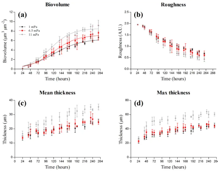
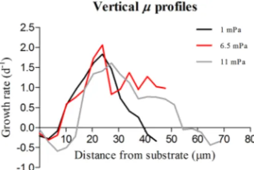
Shear stress affects the architecture and cohesion of Chlorella vulgaris biofilms
12
0
0
Texte intégral
Figure

Documents relatifs