Science and Technology
OPEN ACCESS
Review—Two Different Multiple Photosynthetic Reaction Centers Using
Either Zinc Porphyrinic Oligopeptide-Fulleropyrrolidine or Free-Base
Porphyrinic Polypeptide-Li+@C
60
Supramolecular Complexes
To cite this article: Nathalie Solladié et al 2020 ECS J. Solid State Sci. Technol. 9 061026
View the article online for updates and enhancements.
Review—Two Different Multiple Photosynthetic Reaction Centers
Using Either Zinc Porphyrinic Oligopeptide-Fulleropyrrolidine or
Free-Base Porphyrinic Polypeptide-Li+@C
60Supramolecular
Complexes
Nathalie Solladié,
1,zShunichi Fukuzumi,
2,3,4Kei Ohkubo,
2,3Francis D
’Souza,
5,6Régis Rein,
1Kenji Saito,
2Vincent Troiani,
1Hongjin Qiu,
1Suresh Gadde,
5and
Tetsuya Hasegawa
21
CNRS, LCC (Laboratoire de Chimie de Coordination), 205 route de Narbonne, 31077 Toulouse (France) and Université de Toulouse, UPS, INPT, 31077 Toulouse, France
2
Department of Material and Life Science, Graduate School of Engineering, Osaka University, ALCA and SENTAN, Japan Science and Technology Agency (JST), 2-1 Yamada-oka, Suita, Osaka 565-0871, Japan
3
Department of Bioinspired Science, Ewha Womans University, Seoul, 120-750, Republic of Korea
4Faculty of Science and Technology, Meijo University, ALCA and SENTAN, Japan Science and Technology Agency (JST),
Nagoya, Aichi 468-8502, Japan
5Department of Chemistry, Wichita State University, 1845 Fairmount, Wichita, Kansas 67260-0051, United States of
America
6Department of Chemistry, University of North Texas, 1155, Union Circle, #305070, Denton, Texas 76203-5017, United
States of America
An overview of two successful examples of photosynthetic reaction center models combined with light-capturing antenna chromophores is presented. In the first example, supramolecular complexes are formed between flexible zinc porphyrinic oligopeptides and fulleropyrrolidine bearing either a pyridine or imidazole functionalized C60via a coordination bond plusπ−π
interactions. The excited energy migration occurs between porphyrin units followed by charge separation. The charge separation (CS) lifetimes of the supramolecular oligopeptide complexes have been elongated by increasing the generation of the porphyrins, enabling us to attain the longest lifetime (0.84 ms) for the P(ZnP)8-ImC60supramolecular system in PhCN solution at 298 K, ever
reported for supramolecular complexes. In the second example, free-base porphyrin polypeptides (P(H2P)n; n= 4 and 8) form
supramolecular complexes with Li+@C60in PhCN, in which the binding is much stronger than C60. Efficient energy migration
occurs between porphyrins in P(H2P)n. The triplet CS states derived from3Li+@C60had long lifetimes due to spin-forbidden back
electron transfer. The triplet CS lifetime becomes longer upon increasing the number of H2P due to the charge migration among
porphyrins. The present study provides valuable insight into the energy and electron transfer processes leading to long-lived charge separated states in artificial photosynthetic antenna-reaction center models.
© 2020 The Author(s). Published on behalf of The Electrochemical Society by IOP Publishing Limited. This is an open access article distributed under the terms of the Creative Commons Attribution 4.0 License (CC BY,http://creativecommons.org/licenses/ by/4.0/), which permits unrestricted reuse of the work in any medium, provided the original work is properly cited. [DOI:10.1149/ 2162-8777/abaaf5]
Manuscript submitted June 10, 2020; revised manuscript received July 22, 2020. Published August 10, 2020.This paper is part of the JSS Focus Issue on Porphyrins, Phthalocyanines, and Supramolecular Assemblies in Honor of Karl M. Kadish.
In natural photosynthetic systems, the solar energy is collected by pigment molecules attached to the light harvesting complexes. In these units, the chlorophylls are held in a favored spacing and orientation by fairly shortα-helical polypeptides.1–3When a photon hits one of the chlorophylls, the absorbed energy spreads extremely rapidly to the others until the reaction center is reached within the cell membrane where the solar energy is converted into chemical energy used by the cell to grow. In this way, the energy contained in a single photon is conducted in a very short time and with minimal loss of energy from the point where it is absorbed to where it is needed.
Extensive effort has so far been devoted to mimicking light harvesting and charge separation processes in natural photosynthesis.4–20Multiporphyrin arrays have been employed as light-harvesting units.21–36Light-harvesting and charge-separation units have been combined by coordination bonds between metal-loporphyrins and electron acceptor moieties bearing Lewis base ligands.36–44Thus, metal centres were required for the construction of supramolecular complexes between porphyrins acting as light harvesting units and electron acceptors containing Lewis base ligands for coordination to the metal centres.36–44
To mimic the natural light harvesting antennae, we synthesized 15 years ago a family of porphyrin-functionalized α-polypeptide such as the octamer (Fig. 1).45 A peptidic backbone was chosen owing to the remarkable ability of peptides to establish secondary
structures such as α-helices, that renders reasonable to expect, beyond a certain degree of oligomerisation, a helical spatial arrangement of the chromophores. Such a conformation should induce an overlap of the porphyrins, which may then undergo sufficient electronic coupling to promote a good exciton migration along the set of porphyrins. It has been thefirst results presented at the ECS meeting in 2003. . Since then, these polypetides have been extensively studied and many properties have been highlighted.46–51 In the present mini-review, we focuss on the preparation and study of multiple reaction centers.
Mimicking the natural photosynthetic process requires both synthesis of the light-harvesting and charge-separation units, which should be linked by covalent or noncovalent bonding.8–12,52–54 Non-covalent bonding is certainly more favorable as compared to covalent bonding from the viewpoint of mimicking the biological photosystems, in which non-covalent interaction plays a pivotal role to regulate the energy- and electron-transfer processes, and easy access toward construction of a variety of donor-acceptor systems.16–19,54–60 A variety of multiporphyrin arrays have so far been utilized as the light-harvesting units to mimic efficient energy transfer in the photosynthesis.50,51,61–64 Among multi-porphyrin arrays, porphyrin oligomers with oligopeptidic backbones in which porphyrins are held in a favored spacing and orientation by fairly short helical oligopeptides have merited special attention in relevance to the natural light-harvesting complex in which chlor-ophyll and oligopeptides are well-organized in the protein.65,66We highlight in this mini-review the elaboration of two different multiple photosynthetic reaction centres using either zinc zE-mail:nathalie.solladie@lcc-toulouse.fr
porphyrinic oligopeptide–fulleropyrrolidine or free-base por-phyrinic polypeptide–Li+@C60 supramolecular complexes.
Results and Discussion
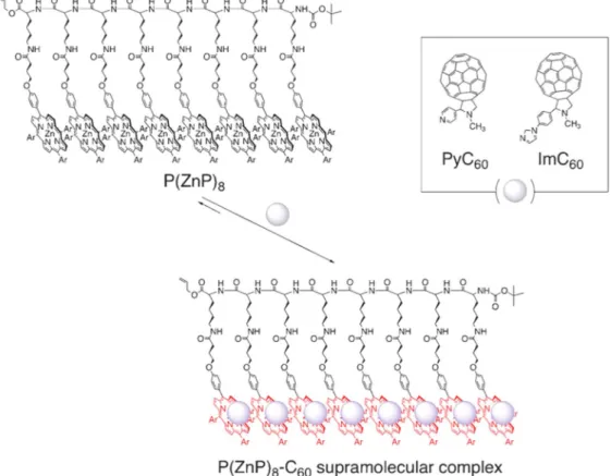
Multiple photosynthetic reaction centers using zinc porphyrinic oligopeptide–fulleropyrrolidine supramolecular complexes.—We first synthesized multiple photosynthetic reaction centers composed of light-harvesting multiporphyrin units and charge-separation units, which have been combined by using non-covalent binding including coordination bond andπ−π interaction. Zinc porphyrinic oligopep-tides with various numbers of porphyrin units [P(ZnP)n; n= 2, 4,
8]48,49,65,66are used as light harvesting multiporphyrin units, which are bound to electron acceptors of fulleropyrrolidine bearing a pyridine (PyC60)67 or imidazole coordinating ligand (ImC60)48,49
as shown in Fig.2.
The laser flash photolysis of the supramolecular complexes of P(ZnP)n with PyC60 and ImC60 has revealed the formation of the
long-lived charge-separated (CS) state. The details of synthesis of porphyrin–peptide oligomers have been reported previously.65,66 The UV–visible spectra of P(ZnP)8in benzonitrile (PhCN) at 298 K
changed upon addition of PyC60, where the Soret band is red-shifted
with an isosbestic point at 436 nm. The absorbance change exhibits a saturation behavior with increasing PyC60 concentration. This
indicates that PyC60forms a 1: 1 supramolecular complex with the
ZnP moiety of P(ZnP)8.39 It has well been established that such
nitrogenous bases readily bind to zinc porphyrins with a 1: 1 stoichiometry.68
As P(ZnP)8forms the supramolecular complex with PyC60, the
fluorescence of P(ZnP)8is strongly quenched by the
intra-supramo-lecular electron transfer from the singlet excited state of the ZnP moiety (1ZnP*) in P(ZnP)8 to bound PyC60(i.e., static quenching)
and not by an intermolecular electron transfer from 1ZnP* to unbound PyC60(i.e., dynamic quenching).39In fact, if the
fluores-cence quenching is analyzed by the Stern-Volmer equation of bimolecular electron transfer [Eq.1], the rate constant of bimole-cular photo-induced electron transfer is estimated as 6 × 10¹³M−1s−1, which is significantly larger than the diffusion rate constant in PhCN (5.6× 10⁹M−1s−1).
I
I0 1 ket PyC60 1 ( / )= + t[ ] [ ] The values of the formation constants (K) of zinc porphyrinic oligopeptides and C60derivatives (PyC60vs ImC60) determined by
the absorption spectral change are listed in TableI. In the case of PyC60as an electron acceptor, the K value increases with increasing
number of zinc porphyrins in an oligopeptide unit. No supramole-cular complex formation was observed in the case of zinc tetraphenylporphyrin with C60 derivatives67 or the porphyrinic
oligopeptides with pristine C60in PhCN. 48,49
The strong association between P(ZnP)8 and C60Py may originate from the strong π−π
interaction between two zinc porphyrins and C60Py in addition to the
axial coordination of C60Py to the zinc porphyrin. In the case of
ImC60, however, the highest K value was obtained for the
P(ZnP)4-ImC60 complex. This indicates that ImC60 is much more
strongly bound by the oligopeptide, P(ZnP)4, than PyC60.
It should be noted that the apparent formation constants determined from thefluorescence quenching are significantly larger than those determined from the UV–vis spectral change, and the difference in such association constants increases with increasing generation of porphyrinic oligopeptides. This indicates that the excited energy migration between the porphyrin units occurs efficiently prior to the electron transfer to the bound C60 moiety.
Such difference in the formation constants is more enhanced in the P(ZnP)n-ImC60 supramolecular complexes as compared to that of
PyC60(TableI). For example, an extremely efficient energy transfer
may occur in the P(ZnP)8-ImC60 system judging from the large
difference in the K values determined by the absorption change and by thefluorescence quenching (1.5 × 10⁴vs 3.3× 10⁵M−1). We have also checked the structural change of the oligopeptidic back-bone when the porphyrinic oligopeptide forms the supramolecular complex with C60 derivatives by the circular dichroism
spectro-scopy, which exhibited the Cotton effect originating from the porphyrin Soret band at 431 nm.
The occurrence of photoinduced electron transfer in the supra-molecular complex in PhCN was confirmed by the transient absorption spectra of the supramolecular complex of P(ZnP)8with
PyC60 using nanosecond laser flash photolysis. The laser
photo-excitation at 561 nm of the supramolecular complex of P(ZnP)8with
PyC60 results in the formation of the CS state as indicated by the
transient absorption spectra in Fig.3a. The absorption band due to PyC60
.− is clearly observed at 1000 nm together with that due to
ZnP.+at 630 nm after laser excitation where only the ZnP moiety is excited (Fig.3a). The CS state detected decays obeyingfirst-order kinetics (Fig. 3b): the first order plots at different initial CS concentrations afford linear correlations with the same slope (inset of Fig. 3b). If there is any contribution of intermolecular back electron transfer from unbound PyC60
.−to ZnP.+, the second-order
kinetics would be involved for the decay time profile. In fact, the corresponding second-order plots (Fig.3c) are clearly non-linear and the initial slope varies depending on the CS concentration. Thus, the decay process is ascribed to back electron transfer in the supramo-lecular complex rather than intermosupramo-lecular back electron transfer between ZnP.+and PyC60
.−.
The CS lifetimes of the supramolecular complexes of other porphyrins [P(ZnP)n: n= 2, 4] and fullerene derivatives (PyC60and
ImC60) were also determined and listed in TableI. The CS lifetime
becomes longer with increasing generation of porphyrinic oligopep-tides. This may result from efficient hole migration between the porphyrin units following photo-induced electron transfer in the supramolecular complex.
Thus, we have succeeded in attaining the longer CS lifetime by increasing the generation of porphyrinic oligopeptides. Such a long-lived CS state was also confirmed by the ESR measurements under photo-irradiation of the supramolecular complex of P(ZnP)8 with
PyC60 or ImC60 in frozen PhCN at 173 K. 48,49
Under photo-irradiation, the isotropic ESR signals corresponding to the zinc porphyrin radical cation and fullerene radical anion are clearly observed at g= 2.002 and 2.000, respectively.16,55
In conclusion, multiple photosynthetic reaction centers combined with light-capturing antenna chromophores have successfully been constructed using supramolecular complexes formed between flex-ible porphyrinic oligopeptides and fulleropyrrolidine bearing a pyridine (PyC60) or imidazole (ImC60) ligand via a coordination
bond plus π−π interaction. The excited energy migration occurs between porphyrin units followed by charge separation. The CS lifetimes of the supramolecular oligopeptide complexes have been
elongated by increasing the generation of the porphyrins, enabling us to attain the longest lifetime (0.84 ms) for the P(ZnP)8-ImC60
supramolecular system in PhCN solution at 298 K ever reported for supramolecular complexes.
Multiple photosynthetic reaction centers of porphyrinic polypeptide–Li+@c60 supramolecular complexes.—In the latter
example, metal centers were required for the construction of supramolecular complexes between porphyrins acting as light harvesting units and electron acceptors containing Lewis base ligands for coordination to the metal centers.35–44 As such there have been only a few reports on supramolecular complexes of free baseporphyrin arrays and electron acceptors without Lewis base ligands, in which the binding is rather weak.56,57We thus focused on the construction of supramolecular complexes of free base porphyrin polypeptides (P(H2P)n: n= 4 and 8) with lithium ion-encapsulated
C60(Li+@C60) (Fig.4),69–71in which binding is much stronger than
the case of C60in benzonitrile (PhCN). The photo-dynamics were
studied by femtosecond and nanosecond laser-induced transient absorption andfluorescence lifetime measurements.
Free base porphyrin polypeptide (P(H2P)n) were synthesized and
characterized as reported previously.65,66 Upon mixing a PhCN solution of Li+@C60with that of P(H2P)8, the intensity of the Soret
band decreased with an increasing concentration of Li+@C60. A
Soret band was slightly red-shifted from 425 to 427 nm by the addition of Li+@C60 to the solution. From the net absorption
change at 425 nm in which the absorption due to Li+@C60 was
subtracted, a linear Benesi–Hildebrand plot72 was obtained, indi-cating that each porphyrin unit of P(H2P)8 forms a 1: 1
supramo-lecular charge-transfer complex with Li+@C60independently with
approximately the same binding constant of the unit of M−1. The binding constant at 298 K was determined from the intercept and the slope to be 2.1× 10⁴M−1, which is significantly larger than that of C60 (5.3 × 10³ M−1).48 The stronger binding of Li+@C60 as
compared with C60may result from the stronger electron acceptor
ability of Li+@C60, which facilitates the charge-transfer interaction
Figure 2. Illustration of a supramolecular complex composed of the porphyrin–peptide octamer P(ZnP)8and PyC60or ImC60. Ar=3,5-(t-Bu)2C6H3.
Table I. Formation constants (K), lifetimes and quantum yield of the CS state of the supramolecular complexes in PhCN at 298 K.
Ka/M−1
Porphyrin Acceptor UV–vis Fluorescence
τCSa) ms−1 P(ZnP)8 PyC60 2.3× 10⁴ 1.5× 10⁵ 0.70 P(ZnP)4 PyC60 5.2× 10³ 1.2× 10⁴ 0.40 P(ZnP)2 PyC60 4.0× 10³ 9.0× 10³ 0.34 P(ZnP)8 ImC60 1.5× 10⁴ 3.3× 10⁵ 0.84 P(ZnP)4 ImC60 5.9× 10⁴ 8.5× 10⁴ 0.38
a) Experimental error within 5%.
as reported for the stronger charge-transfer binding of Li+@C60with
corannulene.73Similarly the binding constant of the supramolecular complex of P(H2P)4was determined from the spectral titration to be
6.2× 10³M−1. In the case of P(H2P)2and P(H2P)1, however, the
spectral change was too small to be able to determine the binding constants accurately. The binding constants are summarized in Table II. Thus, multiporphyrins may facilitate charge transfer interactions through the encapsulation of Li+@C60 by multiple
porphyrins.
Fluorescence of P(H2P)8 was quenched by the addition of a
PhCN solution of Li+@C60. A Benesi-Hildebrand plot for
fluores-cence quenching afforded the apparent binding constant of the supramolecular complex of P(H2P)8 with Li+@C60 being 8.5 ×
104M−1, which is significantly larger than the value determined from the absorption spectral titration (2.1× 10⁴M−1). Similarly the apparent binding constant of P(H2P)4with Li+@C60was determined
from thefluorescence quenching to be 1.6 × 10⁴M−1, which is also larger than the value determined from the absorption spectral titration (Table II). Such larger apparent binding constants deter-mined from thefluorescence quenching than those determined from the absorption spectral titration indicate that energy migration occurs between the singlet excited state of H2P (1H2P*) and adjacent H2P in
P(H2P)n(n= 4 and 8).
The energy migration rate constant was determined by the fluorescence lifetime measurements of P(H2P)8 with Li+@C60.
Thefluorescence of1P(H2P)8* exhibited a single exponential decay
with the lifetime of 11 ns. In the presence of Li+@C60, the decay of 1
P(H2P)8* is well analyzed by three exponentials with lifetimes of
100 ps, 3.0 ns and 11 ns. The fast component corresponds to the lifetime of 1H2P* in the supramolecular complex with Li+@C60.
95% of the fast component agrees with that predicted by the binding constant determined from the absorption spectral titration. The third component has the same lifetime as that without Li+@C60,
corresponding to thefluorescence lifetime of1(H2P)8*. The second
component corresponds to energy migration between1H2P* and the
adjacent H2P. 5% of the sum of the first and second components
agrees with that predicted from the binding constant determined fromfluorescence quenching.
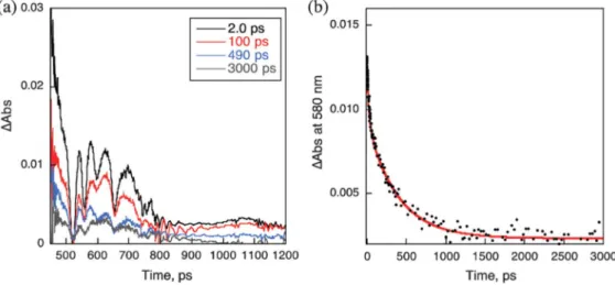
Femtosecond laser-induced transient absorption spectra of P(H2P)8 were recorded in the absence and presence of Li+@C60
(Fig. 5). In each case, only the singlet-singlet absorption of
1
P(H2P)8* was observed, because H2P was exclusively excited at
the excitation wavelength (425 nm) in Fig. 5. The decay of
1
P(H2P)8* exhibits a fast component with a lifetime of 6 ps. A
similar fast component was observed for1P(H2P)4* and1P(H2P)2*.
In the case of P(H2P)1, however, no fast component was observed.
The ratio of the fast component increased upon increasing the number of H2P in P(H2P)n. 96% of the fast component of P(H2P)8
decreased upon decreasing the laser intensity (Fig.5b). These results indicate that the fast component results from the singlet–singlet annihilation of two1H2P* moieties in P(H2P)n(n= 4 and 8).
In the presence of Li+@C60(20 mM), the decay of 1P(H2P)8*
exhibited three exponentials with lifetimes of 6 ps, 90 ps and 2 ns. The first fast component resulted from the singlet-singlet
Figure 3.(a) Transient absorption spectra of P(ZnP)8(2.9× 10⁻⁶M) in the presence of PyC60(4.9× 10⁻³M) in deaerated PhCN at 298 K taken at 70 (solid
line with black circles) and 350 ms (solid line with white circles) after laser excitation at 561 nm (4 mJ pulse−1), respectively. (b) Time profiles of the absorption at 1000 nm due to PyC60.– with different laser powers (4 and 1 mJ pulse−1) at 298 K. Inset:first-order plots. (c) Second-order plots.
annihilation as observed without Li+@C60. The second component
(90 ps) may result from electron transfer from1H2P* to Li+@C60in
the supramolecular complex. Because the transient absorption due to the charge-separated state (H2P
.+and Li+@C 60
.−) was not observed
in Fig.5a, where only the singlet-singlet absorption of1H2P* is seen,
back electron transfer may be much faster than the forward electron transfer. The third component may correspond to the energy migration between1H2P* and the adjacent H2P, because its lifetime
agrees with the lifetime of energy migration in the fluorescence lifetime measurements. In the presence of high concentration of Li+@C60 (120 mM), the component of electron transfer from1H2P*
to Li+@C60 in the supramolecular complex increased. The three
component decay was also observed for P(H2P)4and P(H2P)2with
Li+@C60. In the case of P(H2P)1, only single exponential decay
with a lifetime of 11 ns was observed in the absence and presence of Li+@C60 because the binding of Li+@C60 to P(H2P)1 was
negligible.
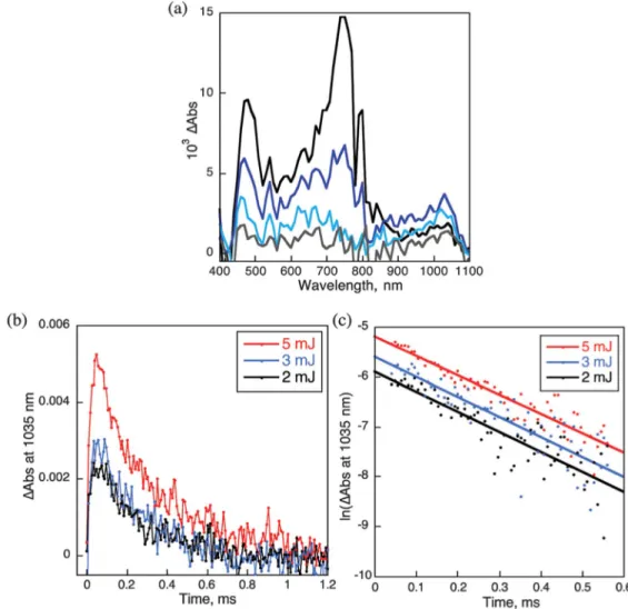
Nanosecond laser-induced transient absorption spectra of P(H2P)8 with Li+@C60 (40 mM) at the excitation wavelength of
532 nm are shown in Fig.6, where the transient absorption band at 730 nm due to the triplet excited state of Li+@C60(3Li+@C60*) is
observed together with the absorption band at 1035 nm due to
Figure 4. Supramolecular formation of peptide porphyrin oligomers with Li+@C60. Ar= 3,5-di-tert-butylphenyl.
Table II. Association constant (K, M−1) of P(H2P)nwith Li+@C60
and C60in PhCN determined from UV–vis absorption and
fluores-cence spectral titration.
Li+@C60 C60b)
Porphyrin UV–vis Fluorescence UV–vis Fluorescence P(H2P)1 __a) 1.1× 10⁴ __a) __a) P(H2P)2 __a) 1.1× 10⁴ __a) __a) P(H2P)4 6.2× 10 3 1.6× 10⁴ 2.7× 10³b) 9.5× 10³b) P(H2P)8 2.1× 10 4 8.5× 10⁴ 5.3× 10³b) 2.2× 10⁴b) a) Too small to determine accurately. b) Taken from Ref.48.
Figure 5. (a) Transient absorption spectra of P(H2P)8(5.2× 10⁻⁶M) with Li+@C60(2.0× 10⁻⁵M) in deaerated PhCN observed after femtosecond laserflash
excitation. Excitation wavelength: 425 nm. (b) Decay time profile at 580 nm with a three-exponential decay curve fitting.
Li+@C60 .−.46
The decay of the absorption at 730 nm coincides with the appearance of Li+@C60
.−. Thus, electron transfer from P(H 2P)8
to 3Li+@C60* occurs to produce the triplet charge-separate (CS)
state of P(H2P)8
.+and Li+@C 60
.−. The transient absorption due to
P(H2P)8
.+is overlapped with that of3
Li+@C60* in the 600–700 nm
region. The photoexcitation of 532 nm resulted in the excitation of Li+@C60 leading to the formation of
3
Li+@C60* via intersystem
crossing. The decay of the absorbance at 1035 nm due to Li+@C60 .−
obeyed first-order kinetics with the lifetime of 210 ms (Fig. 6b). Thus, back electron transfer from Li+@C60
.−to P(H 2P)8
.+occurs in
the supramolecular complex. The lifetime is long because of the spin-forbidden back electron transfer in the triplet CS state.
Similarly the triplet CS states derived from 3Li+@C60* were
observed for P(H2P)n (n = 4, 2, and 1) with Li+@C60. The rate
constants of electron transfer from 1H2P* to Li+@C60 and the
energy migration in the supramolecular complexes of P(H2P)nwith
Li+@C60are summarized in TableIIItogether with the lifetimes of
the triplet CS states. The CS lifetime increases upon increasing the number of H2P in P(H2P)n. Such an increase in the CS lifetime may
result from the charge migration between H2P
.+and the adjacent
H2P in P(H2P)n
.+. The energy diagram of photoexcitation of P(H 2P)n
with Li+@C60 is given in Scheme1. The energies of the excited
state of porphyrin and Li+@C60 were determined by UV–vis,
fluorescence and phosphorescence spectral measurements. The energy of the CS state was determined to be 0.82 eV in PhCN from the one-electron oxidation potential of porphyrin (+0.96 V vs SCE) and reduction potential of Li+@C60(+0.14 V vs SCE) by the
electrochemical measurements. Femtosecond laser excitation of the
supramolecular complex of P(H2P)8 with Li+@C60 at 425 nm
resulted in electron transfer from H2P to the bound Li+@C60 to
produce the singlet CS state and energy transfer from1H2P* to the
adjacent H2P, followed by electron transfer to Li+@C60, when it is
bound H2P. The singlet CS state decays rapidly to the ground state.
Nanosecond laser excitation at 532 nm resulted in the formation of the singlet excited state of excess Li+@C60, followed by intersystem
crossing to produce3Li+@C60*, to which intermolecular electron
transfer occurs from H2P to produce the triplet CS state with a long
lifetime because of the spin-forbidden back electron transfer to the ground state.
We can thus confirm that free base porphyrin polypeptides (P(H2P)n; n = 4 and 8) form supramolecular complexes with
Li+@C60 in PhCN, in which the binding is much stronger than
C60. Efficient energy migration occurs between porphyrins in
P(H2P)n. Although the lifetimes of the singlet CS states were too
short to be detected, the triplet CS states derived from3Li+@C60had
long lifetimes because of the spin-forbidden back electron transfer. The triplet CS lifetime becomes longer upon increasing the number of H2P due to the charge migration among porphyrins. The present
study provides valuable insight into the energy and electron transfer in multiple reaction centers.
Conclusions
In conclusion, multiple photosynthetic reaction centers combined with light-capturing antenna chromophores have successfully been constructed using supramolecular complexes formed between
Figure 6. (a) Transient absorption spectra of P(H2P)8(2.6× 10⁻⁶M) with Li+@C60(4.0× 10⁻⁵M) in deaerated PhCN observed after nanosecond laserflash
excitation taken at 8 (black), 32 (blue), 96 (sky blue) and 200 ms (gray). Excitation wavelength: 532 nm. (b) Decay time profile at 1035 nm with the different laser pulse intensities (2–5 mJ pulse−1). (c) First-order plots.
flexible porphyrinic oligopeptides and fulleropyrrolidine bearing a pyridine (PyC60) or imidazole (ImC60) ligand via a coordination
bond plus π−π interaction. The excited energy migration occurs between porphyrin units followed by charge separation. The CS lifetimes of the supramolecular oligopeptide complexes have been elongated by increasing the generation of the porphyrins, enabling us to attain the longest lifetime (0.84 ms) for the P(ZnP)8-ImC60
supramolecular system in PhCN solution at 298 K ever reported for supramolecular complexes.
In thefirst exemple, metal centers were required for the construction of supramolecular complexes between porphyrins acting as light harvesting units and electron acceptors containing Lewis base ligands for coordination to the metal centers. As such there have been only a few reports on supramolecular complexes of free base porphyrin arrays and electron acceptors without Lewis base ligands, in which the binding is rather weak. Our second study confirms that free base porphyrin polypeptides (P(H2P)n; n= 4 and 8) form supramolecular complexes
with Li+@C60in PhCN, in which the binding is much stronger than
C60. Efficient energy migration occurs between porphyrins in P(H2P)n.
Although the lifetimes of the singlet CS states were too short to be detected, the triplet CS states derived from 3Li+@C60 had long
lifetimes because of the spin-forbidden back electron transfer. The triplet CS lifetime becomes longer upon increasing the number of H2P
due to the charge migration among porphyrins.
Acknowledgments
This work was partially supported by a Grant-in-Aid (No. 20108010 and 23750014 to SF and no. 26620154 and 26288037 to K.O.) and a Global COE program from the Ministry of Education, Culture, Sports,
Science and Technology, Japan, KOSEF/MEST through WCU project (R31-2008-000-10010-0), Korea; ALCA, SENTAN projects from JST, Japan (to S.F.), and US-National Science Foundation (2000988 to FD). The synthesis of the familiy of peptides was supported by the CNRS and the French Ministry of Research.
ORCID
Nathalie Solladié https://orcid.org/0000-0001-6530-6391
Shunichi Fukuzumi https://orcid.org/0000-0002-3559-4107
Kei Ohkubo https://orcid.org/0000-0001-8328-9249
Francis D’Souza https://orcid.org/0000-0003-3815-8949
References
1. W. Kuehlbrandt,Nature (London), 374, 497 (1995).
2. G. McDermott, S. M. Prince, A. A. Freer, A. M. Hawthornthwaite-Lawless, M. Z. Papiz, R. J. Cogdell, and N. W. Isaacs,Nature (London, U. K.), 374, 517 (1995).
3. T. Pullerits and V. Sundstroem,Acc. Chem. Res., 29, 381 (1996).
4. R. E. Blankenship, M. T. Madigan, and C. E. Bauer (ed.),Anoxygenic Photosynthetic Bacteria(Springer , Dordrecht) 2 (1995).
5. K. D. Karlin (ed.), Molecular Level Artificial Photosynthetic Materials(Wiley, New York) 44 (1997).
6. R. A. Wheeler, Introduction to the Molecular Bioenergetics of Electron, Proton, and Energy Transfer, ACS Symposium Series, 883, 1 (2004).
7. W. Leibl and P. Mathis, Electron Transfer in Photosynthesis. Series on photo-conversion of solar energy, 2, 117 (2004).
8. D. Gust, T. A. Moore, and A. L. Moore,Acc. Chem. Res., 42, 1890 (2009). 9. G. Bottari, G. de la Torre, D. M. Guldi, and T. Torres,Chem. Rev., 110, 6768
(2010).
10. V. Sgobba and D. M. Guldi,Chem. Soc. Rev., 38, 165 (2009). 11. M. R. Wasielewski,Acc. Chem. Res., 42, 1910 (2009). 12. S. Fukuzumi,Phys. Chem. Chem. Phys., 10, 2283 (2008). 13. F. D’Souza and O. Ito, Chem. Commun., 4913 (2009). 14. F. D’Souza and O. Ito,Coord. Chem. Rev., 249, 1410 (2005). 15. C. B. Kc and F. D’Souza,Coord. Chem. Rev., 322, 104 (2016). 16. S. Fukuzumi et al.,Chem. Commun., 47, 7980 (2011).
17. T. Kojima, T. Honda, K. Ohkubo, M. Shiro, T. Kusukawa, T. Fukuda, N. Kobayashi, and S. Fukuzumi,Angew. Chem. Int. Ed., 47, 6712 (2008). 18. S. Fukuzumi, T. Honda, K. Ohkubo, and T. Kojima, Dalton Trans., 3880 (2009). 19. T. Kojima, K. Hanabusa, K. Ohkubo, M. Shiro, and S. Fukuzumi,Chem.—Eur. J.,
16, 3646 (2010).
20. S. Fukuzumi, Functional Organic Materials, ed. Dr. Thomas, J. J. Muller, and U. H. F. Bunz (Wiley-VCH, Verlag GmbH) Chap. 13, p. 465 (2007).
21. T. Tanaka and A. Osuka,Chem. Soc. Rev., 44, 943 (2015).
22. H.-W. Jiang, T. Tanaka, H. Mori, K. H. Park, D. Kim, and A. Osuka,J. Am. Chem. Soc., 137, 2219 (2015).
23. N. Aratani, D. Kim, and A. Osuka,Acc. Chem. Res., 42, 1922 (2009). 24. J. Yang, M.-C. Yoon, H. Yoo, P. Kim, and D. Kim,Chem. Soc. Rev., 41, 4808
(2012).
25. T. Miyatake and H. Tamiaki,Coord. Chem. Rev., 254, 2593 (2010).
Table III. Rate constants of intra- and inter-molecular photo-induced electron transfer (kETand ket) and lifetimes (τ(CS)) and
quantum yields (Φ(CS)) of the charge-separated state. Porphyrin kET, M−1s−1 ket, M−1s−1 τCS,μs ΦCS P(H2P)1 7.7× 10⁵ 1.7× 10⁹ __a) 0.70 P(H2P)2 6.7× 10⁴ 1.8× 10⁹ __a) 0.34 P(H2P)4 9.5× 10⁴ 8.0× 10⁸ 160 0.16 P(H2P)8 1.5× 10⁵ 1.5× 10⁸ 210 0.13
a) Bimolecular decay in the radical ion pair.
Scheme 1. Reaction course of photo-induced charge separation and charge recombination in the supramolecular complex between P(H2P)8and Li+@C60with
the values of energy levels.
26. S. Sengupta and F. Würthner,Acc. Chem. Res., 46, 2498 (2013). 27. T. S. Balaban,Acc. Chem. Res., 38, 612 (2005).
28. H.-J. Son et al.,J. Am. Chem. Soc., 135, 862 (2013).
29. I. Cohen-Ofri, M. van Gastel, J. Grzyb, A. Brandis, I. Pinkas, W. Lubitz, and D. Noy,J. Am. Chem. Soc., 133, 9526 (2011).
30. J. L. R. Anderson et al.,Chem. Sci., 5, 507 (2014).
31. F. A. Tezcan, B. R. Crane, J. R. Winkler, and H. B. Gray,Proc. Natl. Acad. Sci. U. S. A., 98, 5002 (2001).
32. T. Koshiyama, M. Shirai, T. Hikage, H. Tabe, K. Tanaka, S. Kitagawa, and T. Ueno,Angew. Chem. Int. Ed., 50, 4849 (2011).
33. R. A. Miller, N. Stephanopoulos, J. M. McFarland, A. S. Rosko, P. L. Geissler, and M. B. Francis,J. Am. Chem. Soc., 132, 6068 (2010).
34. L. S. Witus and M. B. Francis,Acc. Chem. Res., 44, 774 (2011).
35. Y. S. Nam, T. Shin, H. Park, A. P. Magyar, K. Choi, G. Fantner, K. A. Nelson, and A. M. Belcher,J. Am. Chem. Soc., 132, 1462 (2010).
36. M. Endo, M. Fujitsuka, and T. Majima,Chem.—Eur. J., 13, 8660 (2007). 37. S. Fukuzumi and K. Ohkubo,J. Mater. Chem., 22, 4575 (2012).
38. S. Fukuzumi, K. Ohkubo, and T. Suenobu,Acc. Chem. Res., 47, 1455 (2014). 39. S. Fukuzumi, K. Saito, K. Ohkubo, V. Troiani, H. Qiu, S. Gadde, F. D’Souza, and
N. Solladié,Phys. Chem. Chem. Phys., 13, 17019 (2011).
40. A. Takai, C. P. Gros, R. Guilard, and S. Fukuzumi,Chem.—Eur. J., 15, 3110 (2009). 41. A. Takai, C. P. Gros, J.-M. Barbe, and S. Fukuzumi,Phys. Chem. Chem. Phys., 12,
12160 (2010).
42. A. Takai, C. P. Gros, J.-M. Barbe, and S. Fukuzumi,Chem.—Eur. J., 17, 3420 (2011).
43. Y. Pareek and M. Ravikanth,RSC Adv., 4, 7851 (2014).
44. V. S. Nair, Y. Pareek, V. Karunakaran, M. Ravikanth, and A. Ajayaghosh,Phys. Chem. Chem. Phys., 16, 10149 (2014).
45. N. Solladié et al.,J. Porphyrins Phthalocyanines, 7, 270 (2003).
46. P. Ceroni, G. Bergamini, N. Aubert, V. Troiani, and N. Solladié,ChemPhysChem, 6, 2120 (2005).
47. T. Hasobe, P. V. Kamat, V. Troiani, N. Solladié, T. K. Ahn, S. K. Kim, D. Kim, A. Kongkanand, S. Kuwabata, and S. Fukuzumi,J. Phys. Chem. B, 109, 19 (2005). 48. T. Hasobe et al.,J. Mater. Chem., 17, 4160 (2007).
49. M. Fujitsuka, M. Hara, S. Tojo, A. Okada, V. Troiani, N. Solladié, and T. Majima,
J. Phys. Chem. B, 109, 33 (2005).
50. M. Fujitsuka, D. W. Cho, N. Solladié, V. Troiani, H. Qiu, and T. Majima,
J. Photochem. Photobiol., A, 188, 346 (2007).
51. K. Saito, V. Troiani, H. Qiu, N. Solladié, T. Sakata, H. Mori, M. Ohama, and S. Fukuzumi,J. Phys. Chem. C, 111, 1194 (2007).
52. D. Gust, T. A. Moore, and A. L. Moore,Acc. Chem. Res., 34, 40 (2001). 53. D. M. Guldi, G. M. A. Rahman, F. Zerbetto, and M. Prato,Acc. Chem. Res., 38, 871
(2005).
54. S. Fukuzumi and T. Kojima,J. Mater. Chem., 18, 1427 (2008).
55. M.-S. Choi, T. Yamazaki, I. Yamazaki, and T. Aida,Angew. Chem. Int. Ed., 43, 150 (2004).
56. M. Tanaka, K. Ohkubo, C. P. Gross, R. Guilard, and S. Fukuzumi,J. Am. Chem. Soc., 128, 14625 (2006).
57. A. Takai, M. Chkounda, A. Eggenspiller, C. P. Gros, M. Lachkar, J.-M. Barbe, and S. Fukuzumi,J. Am. Chem. Soc., 132, 4477 (2010).
58. J. L. Sessler, B. Wang, and A. Harriman,J. Am. Chem. Soc., 115, 10418 (1993). 59. J. L. Sessler, E. Karnas, S. K. Kim, Z. Ou, M. Zhang, K. M. Kadish, K. Ohkubo,
and S. Fukuzumi,J. Am. Chem. Soc., 130, 15256 (2008).
60. F. D’Souza, E. Maligaspe, K. Ohkubo, M. E. Zandler, N. K. Subbaiyan, and S. Fukuzumi,J. Am. Chem. Soc., 131, 8787 (2009).
61. A. Tsuda and A. Osuka,Science, 293, 79 (2001).
62. Y. Nakamura, I.-W. Hwang, N. Aratani, T. K. Ahn, D. M. Ko, A. Takagi, T. Kawai, T. Matsumoto, D. Kim, and A. Osuka,J. Am. Chem. Soc., 127, 236 (2005). 63. C. Maeda, P. Kim, S. Cho, J. K. Park, J. M. Lim, D. Kim, J. Vura-Weis,
M. R. Wasielewski, H. Shinokubo, and A. Osuka,Chem.–Eur. J., 16, 5052 (2010). 64. A. Satake and Y. Kobuke,Org. Biomol. Chem., 5, 1679 (2007).
65. N. Solladié, A. Hamel, and M. Gross,Tetrahedron Lett., 41, 6075 (2000). 66. F. Aziat, R. Rein, J. Peon, E. Rivera, and N. Solladié, J. Porphyrins
Phthalocyanines, 12, 1232 (2008).
67. F. D’Souza, P. M. Smith, S. Gadde, A. L. McCarty, M. J. Kullman, M. E. Zandler, M. Itou, Y. Araki, and O. Ito,J. Phys. Chem. B, 108, 11333 (2004).
68. J. K. M. Sanders, N. Banpos, Z. Clude-Watson, S. L. Darling, J. C. Hawaley, H. J. Kim, C. C. Mak, and S. J. Webb, The Porphyrin Handbook (Academic, San Diego) 3, p. 1 (2000).
69. S. Aoyagi et al.,Nat. Chem., 2, 678 (2010).
70. Y. Kawashima, K. Ohkubo, and S. Fukuzumi,Chem.—Asian J., 10, 44 (2015). 71. S. Fukuzumi, K. Ohkubo, Y. Kawashima, D. S. Kim, J. S. Park, A. Jana,
V. M. Lynch, D. Kim, and J. L. Sessler,J. Am. Chem. Soc., 133, 15938 (2011). 72. H. Benesi and J. Hildebrand,J. Am. Chem. Soc., 71, 2703 (1949).
73. M. Yamada, K. Ohkubo, M. Shionoya, and S. Fukuzumi,J. Am. Chem. Soc., 135, 13240 (2014).