HAL Id: hal-01730990
https://hal.archives-ouvertes.fr/hal-01730990
Submitted on 13 Mar 2018
HAL is a multi-disciplinary open access
archive for the deposit and dissemination of
sci-entific research documents, whether they are
pub-lished or not. The documents may come from
teaching and research institutions in France or
abroad, or from public or private research centers.
L’archive ouverte pluridisciplinaire HAL, est
destinée au dépôt et à la diffusion de documents
scientifiques de niveau recherche, publiés ou non,
émanant des établissements d’enseignement et de
recherche français ou étrangers, des laboratoires
publics ou privés.
Distributed under a Creative Commons Attribution| 4.0 International License
A modified multilocus sequence typing protocol to
genotype Kingella kingae from oropharyngeal swabs
without bacterial isolation
Nawal El Houmami, Janek Bzdrenga, Jean-Christophe Pons, Philippe
Minodier, Guillaume Andre Durand, Anis Oubraham, Dimitri Ceroni, Pablo
Yagupsky, Didier Raoult, Philippe Bidet, et al.
To cite this version:
Nawal El Houmami, Janek Bzdrenga, Jean-Christophe Pons, Philippe Minodier, Guillaume Andre
Durand, et al.. A modified multilocus sequence typing protocol to genotype Kingella kingae from
oropharyngeal swabs without bacterial isolation. BMC Microbiology, BioMed Central, 2017, 17,
pp.200. �10.1186/s12866-017-1104-5�. �hal-01730990�
M E T H O D O L O G Y A R T I C L E
Open Access
A modified multilocus sequence typing
protocol to genotype Kingella kingae from
oropharyngeal swabs without bacterial
isolation
Nawal El Houmami
1*, Janek Bzdrenga
2, Jean-Christophe Pons
1, Philippe Minodier
3, Guillaume André Durand
1,
Anis Oubraham
1, Dimitri Ceroni
4, Pablo Yagupsky
5, Didier Raoult
1, Philippe Bidet
6and Pierre-Edouard Fournier
1Abstract
Background: Outbreaks of Kingella kingae infection are an emerging public health concern among daycare attendees carrying epidemic clones in the oropharynx. However, genotyping of such epidemic clones from affected cases is limited by the low performance of current methods to detect K. kingae from blood samples and lack of specimens available from infected sites. We aimed at developing a modified multilocus sequence typing (MLST) method to genotype K. kingae strains from oropharyngeal samples without prior culture. We designed in silico MLST primers specific for K. kingae by aligning whole nucleotide sequences of abcZ, adk, aroE, cpn60, recA, and gdh/zwf genes from closely related species belonging to the Kingella and Neisseria genera. We tested our modified MLST protocol on all Kingella species and N. meningitidis, as well as 11 oropharyngeal samples from young children with sporadic (n = 10) or epidemic (n = 1) K. kingae infection.
Results: We detected K. kingae-specific amplicons in the 11 oropharyngeal samples, corresponding to sequence-type 6 (ST-6) in 6 children including the epidemic cases, ST-25 in 2 children, and 3 possible novel STs (ST-67, ST-68, and ST-69). No amplicon was obtained from other Kingella species and N. meningitidis.
Conclusions: We herein developed a specific MLST protocol that enables genotyping of K. kingae by MLST directly from oropharyngeal samples. This discriminatory tool, with which we identified the first K. kingae outbreak caused by ST-6 in Europe, may be used in further epidemiological investigations.
Keywords: Kingella kingae, MLST, Pediatrics, Outbreaks, Bone and joint infections Background
Outbreaks of Kingella kingae infections are emerging as a public health issue in daycare facilities [1–3]. Defined as the occurrence of at least two epidemio-logically connected cases of K. kingae infections within a 1 month-period, they are characterized by a high attack rate and spread of a virulent clone among children aged from 6 to 36 months sharing the same classroom, and causing a variety of osteoarticular and
soft tissue infections, and occasionally endocarditis [1–3]. Epidemiological investigation of these events implies isolation and genotypic characterization of the strain causing the outbreak. At the same time, asymp-tomatic daycare center attendees and staff may be colonized by this virulent strain and, thus, deemed to be at risk to develop an invasive infection and/or to serve as reservoirs and sources of further dissemin-ation of the disease [1, 2]. However, not all colonizing strains are capable of penetrating the epithelial layer and invading the bloodstream, and it is currently rec-ognized that worldwide outbreaks are caused by a limited number of particularly invasive clones [2–4]. Epidemiological investigations revealed that only K.
* Correspondence:[email protected]; [email protected]
1Aix-Marseille Univ, UM63, CNRS 7278, IRD 198, Inserm 1095, Assistance
Publique– Hôpitaux de Marseille, URMITE, Institut Hospitalo-Universitaire Méditerranée Infection, 19-21 Boulevard Jean Moulin, 13385 Marseille, France Full list of author information is available at the end of the article
© The Author(s). 2017 Open Access This article is distributed under the terms of the Creative Commons Attribution 4.0 International License (http://creativecommons.org/licenses/by/4.0/), which permits unrestricted use, distribution, and reproduction in any medium, provided you give appropriate credit to the original author(s) and the source, provide a link to the Creative Commons license, and indicate if changes were made. The Creative Commons Public Domain Dedication waiver (http://creativecommons.org/publicdomain/zero/1.0/) applies to the data made available in this article, unless otherwise stated.
kingae clones belonging to the hypervirulent sequence types 6 (ST-6), ST-14, ST-23, ST-25, and ST-66 have caused in the past few years outbreaks in the USA, Israel and France [2–4].
Since K. kingae is notoriously difficult to recover in culture, real-time polymerase chain reaction (PCR) assays have been developed during the last 10 years and gained increasing acceptance for the diagnosis of K. kingae infections [1, 3, 5]. These culture-independent methods exhibit higher sensitivity compared to conven-tional cultures, shorten the time of detection from days to a few hours, enable the diagnosis in patients being administered antibiotics, as well as identification of asymptomatic K. kingae carriers [2, 5].When no surgical specimen is available and blood cultures are negative, al-ternative strategies have been developed [1, 5, 6]. Notably, the presence of an oropharyngeal K. kingae car-riage in children under the age of four with sporadic osteoarticular infection was demonstrated to have a 90.5% positive predictive value for K. kingae infection [6]. On this point, it was demonstrated that K. kingae clones carried in the oropharynx of children with K. kingae infection are genotypically identical to those detected within infected sites [7].
Although the apparent increase in reported cases of K. kingae infections can be partly explained by im-proved isolation methods and better recognition of this emerging pathogen, the drawback of molecular detection tests is that, until now, they did not enable typing of the colonizing organisms and, thus, did not distinguish between individuals carrying non-invasive K. kingae strains and those colonized by the strain which caused the outbreak. We herein report the de-velopment of a modified multilocus sequence typing protocol (MLST) which enables to genotype K. kingae in oropharyngeal samples with no prior culture. This method was applied in clinics and successfully used to investigate an outbreak of invasive K. kingae infec-tion that occurred in a daycare facility in 2016 in the Marseille area (France).
Methods
Development of a novel specific MLST typing tool for K. Kingae
We started with the analysis of MLST primers previ-ously described in the Institut Pasteur MLST K. kingae database [8], and we observed a lack of in silico primer specificity between the Kingella and Neisseria genera. Therefore, we designed specific MLST primers for K. kingaeby using the following criteria:
1) maximizing mismatches against other Kingella and Neisseriaspecies, especially at the 3′ end;
2) maximizing consensus between distinct K. kingae sequence types;
3) selecting hybridization temperatures close to 58 °C (Additional file1: Figure S1). We designed thereafter a modified MLST method for K. kingae, consisting in PCR amplification and sequencing of 6
housekeeping genes, namely abcZ, adk, aroE, cpn60, recA, and gdh/zwf (Table1). We first aligned the whole nucleotide sequences of the 6
above-mentioned housekeeping genes from fourty K. kingae strains, as well as those from closely-related species including, K. negevensis Sch538T[9], K. denitrificans ATCC 33394T, K. oralisATCC 51147T, N. meningitidis Z2491, N. lactamica 020–06, and N. elongata subs. N. elongatasubs. glycolytica ATCC 29315T.
Finally, we tested this novel K. kingae MLST protocol on 11 oropharyngeal samples that had previously been tested positive for K. kingae by specific real-time PCR targeting the cpn60 gene [10], and on DNA from K. denitrificansCIP 103803, K. oralis CIP 103473, K. potus CIP 108935, K. negevensis Sch538T, and N. meningitidis CSUR P782.
Results
K. kingae-specific amplicons were detected by Sanger se-quencing in all tested oropharyngeal specimens corre-sponding to ST-6 in 6, ST-25 in 2, and possible novel STs in 3, but in none of the strains from others Kingella species and N. meningitidis (Table 2). A few single nu-cleotide polymorphisms (SNPs) were detected in some alleles for 7 specimens. In these cases, the highest peak of the chromatogram was selected to determinate the dominant sequence (Fig. 1). Given that only one copy of each reference housekeeping gene was found in the K.
kingae KWG1 genome, the only strain for which the
whole genome was sequenced using the highly reliable Pacific Biosciences SMRT technology [11], we postulate that K. kingae clones belonging to different STs may co-exist in the oropharynx of these individuals where one clone dominated.
This method was then applied in clinics in 2016. The study was approved by the Ethics committee of the IHU Mediterranee-Infection under reference num-ber 2016–024. From June to July 2016, an outbreak of K. kingae osteoarticular infection involving two infants (aged 17 and 19 months) who shared the same classroom was identified in a daycare facility in southern France. The first patient sustained a left ankle arthritis and the second a first metatarsophalan-geal joint’s arthritis. Both had presented with herpan-gina, fever, and peri-oral rash in the 2 previous weeks. Blood cultures were negative and no joint fluid was surgically collected in either case. Both children
recovered with no sequelae after receiving intravenous cefamandole followed by oral amoxicillin. An oropha-ryngeal sample from the second case was collected prior to antibiotic therapy. Detection of K. kingae using specific real-time PCR was positive in this spe-cimen. By using our modified MLST typing tool, we unambiguously identified K. kingae belonging to ST-6 composed of abcZ-5, adk-2, aroE-4, cpn60–5, gdh/ zwf-5, and recA-1 alleles.
Discussion
We herein developed a specific MLST method enab-ling to genotype K. kingae in oropharyngeal samples without requiring prior strain isolation. Given the fas-tidious nature of the species and the increasing use of molecular techniques for investigating epidemics or
sporadic infections, such an improved genotyping tool is relevant. Indeed, it was previoulsy demonstrated that the presence of an oropharyngeal invasive K. kingae carriage in children under four with sporadic osteoarticular infections had a 90.5% positive predict-ive value for K. kingae infection [6]. Regarding this matter, it is important to note that, of the eleven K.
kingae outbreaks in daycare centers that have been
reported to date [2, 3], only 30% of children (10/33) underwent surgical procedures to obtain synovial fluid or tissue samples. This may be explained by the fact that most infected sites during K. kingae outbreaks were located within small joints located in hands, wrists and feet, ankles, as well as bony sites rich in growth cartilage such as epiphysis of long bones and spine [2]. Since these are regions where joint fluids
Table 1 PCR protocol for specific Kingella kingae multilocus sequence typing Primer design
Gene Primers name Primers Primer length (bp) Amplicon length (bp) abcZ abcZ_Kki_Fwd CGCAAGAAAGCGTGTTTGAC 20 532
abcZ_Kki_Rev CAATTCCTGCGCCTTTTTCTC 21
adk adk_Kki_Fwd CACACAAGCGCAATTTATTACG 22 491 adk_Kki_Rev AAACTTCGGTTTGTTCGTGATAT 23
aroE aroE_Kki_Fwd CAAATCCCCACAAATTCATCAATG 24 621 aroE_Kki_Rev AACGCGGTGGGCTGGTTC 18
cpn60 cpn60_Kki_Fwd CATGGGCGCACAAATGGTT 19 467 cpn60_Kki_Rev CAAACAACAACACAAATGGGC 21
recA recA_Kki_Fwd GACGGCAGCCACCAAGAC 21 456
recA_Kki_Rev TCCTGCCAGTTTACGCAAG 19
gdh/zwf gdh/zwf_Kki_Fwd GAGCGCGGCGAGTTTTAT 18 671 gdh/ zwf_Kki_Rev CAGTTGTCCAAAATTGGCATG 21
10× PCR Buffer 1×
25 mM MgCl2 2.0 mM
dNTP mix (10 mM of each) 200μM of each dNTP
Forward primer 0,1μM
Reverse primer 0,1μM
HotStarTaq DNA Polymerase 2.5 units/ reaction Distilled water variable Template DNA < 0,5μg
Total volume 50μl
PCR protocol
Cycle step 3 step-protocol Cycles
Temperature Time
Initial denaturation 95 °C 15 min 1 Denaturation 95 °C 1 min
35
Annealing 58 °C 30 s
Elongation 72 °C 1 min 30 s
are uncommonly sampled and epiphyseal bone, verte-brae, or intervertebral disks specimens are rarely obtained, many cases remain unconfirmed [1–3] but are still treated since K. kingae clones carried in the oropharynx of children with K. kingae infection are
genotypically identical to those detected within
infected sites [7]. In two K. kingae outbreaks involv-ing four children in Israel, no suspected cases could be formally confirmed [1, 2, 12]. In this peculiar
context, the genotype of epidemic clones was
obtained from K. kingae oropharyngeal isolates culti-vated from either presumed cases or from healthy classmates sharing the same classroom [1, 12].
Blood cultures and even PCR on blood specimens are really disappointing; although skeletal system infections re-sult from the blood-borne dissemination of the bacterium, the prerequisite bacteremic episode is short and most of the time, when a localized infection has been established, the pathogen has usually been cleared from the blood.
Moreover, given that 60% of epidemic cases are not microbiologically confirmed during K. kingae outbreaks, and that K. kingae may be difficult to isolate from polymi-crobial samples even on appropriate culture media [2], this specific K. kingae MLST tool may be helpful when oropharyngeal swabs are the only biological samples avail-able for genotyping, as was the case in this report. Clones belonging to ST-6 are among the most invasive and disseminated worldwide and the main cause of K. kingae outbreaks in Israel [2, 4]. To the best of our knowledge, we here report the first K. kingae outbreak caused by ST-6 in Europe. Therefore, this genotype appears to be respon-sible for 50% of outbreaks worldwide.
Conclusions
This modified, specific K. kingae MLST tool demon-strated a high discriminatory power and may be used in further epidemiological investigations for sporadic and epidemic K. kingae infections.
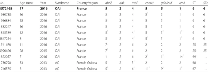
Table 2 Specific multilocus sequence typing (MLST) for Kingella kingae performed by Sanger sequencing method on DNA directly extracted from 11 oropharyngeal specimens (=11 children) with no prior bacterial isolation allowed to detect K. kingae clones belonging to ST-6 in 6 children, ST-25 in 2, and possible new STs in 3, namely ST-67, ST-68, and ST-69
No. Age (mo) Year Syndrome Country/region abcZ adk aroE cpn60 gdh/zwf recA ST STc
1572468 17 2016 OAI France 5 2 4 5 5 1 6 6 1980738 16 2016 OAI France 5 2 4 5* 5 1 6 6 1956884 18 2016 OAI France 5 2 4 5 5 1 6 6 1882247 16 2016 OAI France 5 2 4 5 5 1 6 6 1815589 12 2016 OAI France 5* 2 4* 5 5* 1* 6 6 6847254 8 2016 OAI France 5 2 4* 5* 5 1* 6 6 1541670 11 2016 OAI France 7 2 6 2 2 2 25 25 0990626 28 2015 OAI France 7* 2 6 2 2 2 25 25 1822057 7 2016 OAI France 1 2 6 2* 1* 14* 69 … 1730798 33 2013 AC French Guiana 5 2 3* 2 2 2 68 … 1746575 8 2013 AC French Guiana 5* 2 6* 11* 9* 1* 67 … *
: detection of SNPs OAI osteoarticular infections, AC asymptomatical carriage, mo: months…: not defined Data referring to the second epidemic case of the Châteauneuf-Grasse K. kingae outbreak 2016 are indicated in bold
Fig. 1 Chromatograms illustrating single nucleotide polymorphisms that were detected in nucleotide position 150 of the allele abcZ-5 and those identified in nucleotide 336 of the allele gdh/zwf-5 from oropharyngeal sample No. 1815589. In these cases, the highest peak was selected to determinate the dominant clone. The nucleotide positions refer to the corresponding allele reference numbers provided in the Institut Pasteur database (http://bigsdb.pasteur.fr/perl/bigsdb/bigsdb.pl?db=pubmlst_kingella_seqdef_public&page=downloadAlleles)
Additional file
Additional file 1: Figure S1. MAFFT alignment of MLST genomic regions of the abcZ, adk, aroE, cpn60, gdh/zwf, recA genes from the 40 Kingella kingae strains that were used in this study, and those from 6 closely related Kingella and Neisseria species. Only each distinct variant of K. kingae sequence types is represented. MAFFT alignment and figures were performed by using Geneious 10.2.3 (Biomatters). (PPTX 24674 kb) Abbreviations
CIP:Collection de l’institut pasteur; CSUR: Collection de souches de l’unité des rickettsies; DNA: Deoxyribonucleic acid; IHU: Institut hospitalo-universitaire; MLST: Multilocus sequence typing; OAI: Osteoarticular infection; PCR: Polymerase chain reaction; SMRT: Single molecule real time; SNP: Single nucleotide polymorphism; ST: Sequence type; STc: Sequence type complex Acknowledgements
Not applicable. Funding
This work was supported by the Méditerranée Infection foundation. Availability of data and materials
The datasets supporting the conclusions of this article are indicated within the article and its additional files.
Authors’ contributions
NEH, PM, and PEF conceptualized the study. NEH and JB designed the modified MLST protocol. NEH and PM collected clinical samples. NEH, JCP, AO, GD, and JB collected data and carried out the initial analyses. NEH drafted the initial manuscript that was critically revised by PEF, PY, PM, DR, and DC. All authors approved the final manuscript as submitted.
Ethics approval and consent to participate
The study was approved by the Ethics committee of the IHU Mediterranee-Infection under reference number 2016–024.
Consent for publication Not applicable. Competing interests
The authors declare that they have no competing interests. Author details
1
Aix-Marseille Univ, UM63, CNRS 7278, IRD 198, Inserm 1095, Assistance Publique– Hôpitaux de Marseille, URMITE, Institut Hospitalo-Universitaire Méditerranée Infection, 19-21 Boulevard Jean Moulin, 13385 Marseille, France.
2University of Grenoble Alpes, CEA, CNRS, IBS, F-38000, Grenoble, France. 3
Department of Pediatric Emergency Medicine, North Hospital, Aix-Marseille University, Marseille, France.4Département de l’enfant et de l’adolescent,
HUG-Hôpital des Enfants, Geneva, Switzerland.5Clinical Microbiology Laboratory, Soroka University Medical Center, Beer-Sheva, Israel.6Laboratoire
de Microbiologie, Hôpital Robert Debré, Assistance Publique– Hôpitaux de Paris, Université Paris Diderot, Sorbonne Paris Cité, Inserm, IAME, UMR 1137, Paris, France.
Received: 26 May 2017 Accepted: 5 September 2017
References
1. Yagupsky P, El Houmami N, Fournier PE. Outbreaks of invasive Kingella kingae infections in daycare facilities: approach to investigation and management. J Pediatr. 2017;182:14–20.
2. El Houmami N, Minodier P, Dubourg G, Mirand A, Jouve JL, Basmaci R, et al. Patterns of Kingella kingae disease outbreaks. Pediatr Infect Dis J. 2016;35:340–6.
3. El Houmami N, Cointat V, Mirand A, Fouilloux V, Bakour S, Minodier P, et al. An outbreak of Kingella kingae infections complicating a severe hand, foot, and mouth disease outbreak in Nice, France, 2016. Pediatr Infect Dis J. 2017;36:530–2.
4. Basmaci R, Bidet P, Yagupsky P, Muñoz-Almagro C, Balashova NV, Doit C, et al. Major intercontinentally distributed sequence types of Kingella kingae and development of a rapid molecular typing tool. J Clin Microbiol. 2014;52:3890–7.
5. El Houmami N, Minodier P, Dubourg G, Martin-Laval E, Lafont E, Jouve JL, et al. An outbreak of Kingella kingae infections associated with hand, foot and mouth disease/herpangina virus outbreak in Marseille, France, 2013. Pediatr Infect Dis J. 2015;34:246–50.
6. Ceroni D, Dubois-Ferriere V, Cherkaoui A, Gesuele R, Combescure C, Lamah L, et al. Detection of Kingella kingae osteoarticular infections in children by oropharyngeal swab PCR. Pediatrics. 2013;131:e230–5.
7. Basmaci R, Ilharreborde B, Bidet P, Doit C, Lorrot M, Mazda K, et al. Isolation of Kingella kingae in the oropharynx during K. kingae arthritis in children. Clin Microbiol Infect. 2012;18:E134–6.
8. Basmaci R, Yagupsky P, Ilharreborde B, Guyot K, Porat N, Chomton M, et al. Multilocus sequence typing and rtxA toxin gene sequencing analysis of Kingella kingae isolates demonstrates genetic diversity and international clones. PLoS One. 2012;7:e38078.
9. El Houmami N, Bakour S, Bzdrenga J, Rathored J, Seligmann H, Robert C, et al. Isolation and characterization of Kingella negevensis sp. nov., a novel Kingella species detected in a healthy paediatric population. Int J Syst Evol Microbiol. 2017;67:2370–6.
10. Levy PY, Fournier PE, Fenollar F, Raoult D. Systematic PCR detection in culture-negative osteoarticular infections. Am J Med. 2013;126:1143.e25–33. 11. Bidet P, Basmaci R, Guglielmini J, Doit C, Jost C, Birgy A, et al. Genome
analysis of Kingella kingae strain KWG1 reveals how aβ-Lactamase gene inserted in the chromosome of this species. Antimicrob Agents Chemother. 2015;60:703–8.
12. Yagupsky P, Ben-Ami Y, Trefler R, Porat N. Outbreaks of invasive Kingella kingae infections in closed communities. J Pediatr. 2016;169:135–9.
• We accept pre-submission inquiries
• Our selector tool helps you to find the most relevant journal
• We provide round the clock customer support
• Convenient online submission
• Thorough peer review
• Inclusion in PubMed and all major indexing services
• Maximum visibility for your research Submit your manuscript at
www.biomedcentral.com/submit