Nephrol Dial Transplant (2015) 30: 330–335 doi: 10.1093/ndt/gfu389
Advance Access publication 23 December 2014
The Bench-to-Bedside Transition
Paradoxical response to furosemide in uromodulin-associated
kidney disease
Laura Labriola
1,*, Eric Olinger
2,*, Hendrica Belge
2, Yves Pirson
1, Karin Dahan
3and Olivier Devuyst
1,2 1Cliniques Universitaires Saint-Luc, Université Catholique de Louvain, Brussels, Belgium,2Institute of Physiology, University of Zurich, Zurich, Switzerland and3Institute of Pathology and Genetics, Gosselies, BelgiumCorrespondence and offprint requests to: Olivier Devuyst ; E-mail: [email protected] *
These authors contributed equally to this study.
A B S T R AC T
Mutations in the UMOD gene coding for uromodulin cause autosomal dominant tubulointerstitial kidney disease. Uromo-dulin is known to regulate transport processes in the thick as-cending limb, but it remains unknown whether UMOD mutations are associated with functional tubular alterations in the early phase of the disease. The responses to furosemide and to a water deprivation test were compared in a 32-year-old female patient carrying the pathogenic UMOD mutation p.C217G and her unaffected 31-year-old sister. A single dose of furosemide induced an intense headache with exaggerated decrease in blood pressure (Δsyst: 30 versus 20 mmHg; Δdiast: 18 versus 5 mmHg) and body weight (Δ2.6 kg versus Δ0.9 kg over 3 h) in the proband versus unaffected sib. The diuretic re-sponse and the fall in urine osmolality were also more import-ant and detected earlier in the affected sib. Water deprivation led to increased plasma osmolality and urine concentration in both siblings; however, the response to desmopressin was attenuated in the affected sib. These data reveal that mutations of uromodulin cause specific transport alterations, including exaggerated response to furosemide and a failure to maximally concentrate urine, in the early phase of the disease.
Keywords: autosomal dominant tubulointerstitial kidney disease, furosemide, NKCC2, TAL, Tamm–Horsfall protein
INTRODUCTIO N
Uromodulin (Tamm–Horsfall protein) is exclusively pro-duced in the epithelial cells lining the thick ascending limb (TAL) of Henle’s loop and is the most abundant protein in
the normal urine. Evidence obtained from in vitro studies and knock-out mouse studies have shown that uromodulin protects against urinary tract infection and kidney stones, and regulates transport systems operating in the TAL [1]. The latter include the sodium–potassium–chloride cotran-sporter NKCC2 and the potassium channel ROMK, which operate in parallel and are essential to reabsorb 25% of the filtered NaCl and to mediate the urinary concentrating ability [2].
Mutations in the UMOD gene encoding uromodulin have been shown to cause‘medullary cystic kidney disease type 2’ (MCKD2; MIM 603 860), ‘familial juvenile hyperuricaemic nephropathy’ (FJHN; MIM 162 000) and ‘glomerulocystic kidney disease’ (GCKD; MIM 609 886) [3–5] which are col-lectively referred to as uromodulin-associated kidney diseases (UAKD). UAKD are autosomal dominant disorders character-ized by hyperuricaemia and gout early in life, alteration of urinary concentrating ability and tubulointerstitial fibrosis with occasional cysts at the cortico-medullary junction [1]. UAKD invariably lead to chronic renal failure during the third to seventh decade of life [6]. There is no specific therapy to slow renal disease progression, and most patients end up with end-stage renal disease.
The fact that hyperuricaemia is the most consistent feature observed in patients harbouring UMOD mutations has led to suggestions that the disease causes a dysfunction of the TAL, with NaCl loss and secondary reabsorption of uric acid in the proximal tubule [6, 7]. This hypothesis has been sup-ported by functional defects evidenced in thefirst transgenic mouse model of disease [8]. However, the existence of a specif-ic dysfunction of the TAL as an early consequence of defective uromodulin processing has not been evaluated in patients har-bouring a pathogenic UMOD mutation.
To investigate whether a pathogenic UMOD mutation is as-sociated with functional alterations in the TAL in the early stage of the disease, we compared the response to a furosemide test and to a water deprivation test in a proband carrying a patho-genic mutation of UMOD and her unaffected sister.
M AT E R I A L S A N D M E T H O D S
The proband, a 32-year-old female harbouring a missense ( p.C217G) mutation in UMOD (III,2), and her 31-year-old sister tested negative for the mutation (III,4) belong to a four-generation family. The pathogenic nature of the p.C217G mu-tation of UMOD was evidenced by the typical course of UAKD observed in the other sister of the proband (III,3) who reached end-stage renal disease at age 37 years (Figure 1A). The proband and her unaffected sister had an estimated glomerular filtration rate (eGFR) according to the CKD-EPI equation from the Chronic Kidney Disease - Epidemiology Collaboration ≥60 mL/min/1.73 m2 at time of testing, and none of them was diabetic. They were not taking any drugs, in-cluding diuretics, allopurinol or antihypertensive medications. The protocol was approved by the Ethical Committee of the UCL Medical School (Brussels), and informed consent was provided.
A 24-h urine collection was obtained in both sisters imme-diately before the furosemide test. On the day of the furo-semide test, a baseline clinical and biological evaluation was performed at 9:30AM, 4 h before giving a single oral dose of
80 mg furosemide. Baseline values for the furosemide test (Figure1B) correspond to a 3-h urine collection preceding im-mediately the administration of furosemide. After furosemide administration, urine samples were collected hourly during 3 h and analysed for Na+and K+, creatinine, urea and uric acid concentrations, osmolality and pH. Clinical and biological evaluations were repeated at the end of the test (time +3 h; Table 1). The following day (i.e. after 18 h of normal hydra-tion), both individuals underwent an 8-h water deprivation test, including a 30-min intravenous infusion of desmopressin acetate (Minirin®, 0.3μg/kg; Ferring AG, Baar, Switzerland) after 6 h of water deprivation. Desmopressin (or deamino-8D-arginine vasopressin, dDAVP) is a peptidic analogue of en-dogenous antidiuretic hormone (ADH) with a higher affinity for V2 receptors and negligible vasopressive potential. A clin-ical evaluation was performed hourly and plasma and urine samples were taken every 2 h during the water deprivation and 1-h and 2-h after initiation of desmopressin perfusion. Dietary intake was identical in the two sisters before the furosemide test and throughout the water deprivation test.
R E S U LT S
The baseline osmolar balance in the two sisters immediately before the functional tests was similar, thus allowing direct inter-individual comparison of the responses to furosemide and to water restriction (values for affected sib versus control sib: 24-h Na+ excretion: 228 versus 307 mmol; 24-h urea excretion:
29.1 versus 23.1 g; osmolar excretion rate: 779.2 versus 787.5 µosm/min; plasma renin activity: 1.1 versus 0.8 ng/mL/h; plasma aldosterone: 0.31 versus 0.30 nM, respectively. Plasma ADH: <0.2 pg/mL in both sibs).
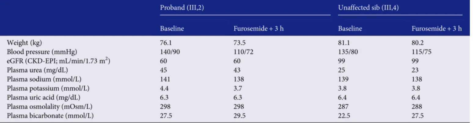
The furosemide test was poorly tolerated by the proband, who reported intense headache, a severe drop in blood pressure (Δ30/Δ18 mmHg versus Δ20/Δ5 mmHg in the unaffected sister) and a weight loss of 2.6 kg (versus weight loss of 0.9 kg in the un-affected sister) in 3 h (Table1). After furosemide administration, both siblings showed the expected increase in diuresis with con-comitant fall in urine osmolality. However, the changes were more important and were detected earlier in the proband, who also showed a more pronounced and earlier increase of fractional excretion of Na+than the control sibling (Figure1B).
The water deprivation test was well tolerated by both parti-cipants. Blood pressure and weight remained constant during all the procedures. As expected, water deprivation resulted in a progressive increase in plasma osmolality with an increased urine osmolality and a reduced urine flow in both subjects (Figure1B). The response to desmopressin was normal in the control sibling, with an increase in urine osmolality (from 983 to 1124 mOsm/kg H2O, 2 h after infusion), a decrease in urine flow (from 30 to 18 mL/h) and a decrease in plasma osmolal-ity (from 298 to 295 mOsm/kg H2O). In contrast, the affected sib showed a blunted increase in urine osmolality (from 834 to 869 mOsm/kg H2O), an increase in urine flow (from 17.5 to 20 mL/h) and a paradoxical increase in plasma osmolality (from 298 to 305 mOsm/kg H2O) (Figure1B).
Pathology examination of the end-stage kidney biopsy of patient III,3 (Figure 1C) revealed marked tubulointerstitial lesions, with thickening of the tubular basement membrane and abnormal processing and accumulation of uromodulin in the tubular cells. Of note, there was no detectable immunos-taining for NKCC2 in the biopsy.
D I S C U S S I O N
In this study, we performed a sib-pair functional testing to evaluate whether pathogenic mutations of UMOD are asso-ciated with dysfunction of the TAL in the early phase of UAKD. Our data reveal that the proband carrying an UMOD mutation shows a clinically and biologically exaggerated re-sponse to furosemide and a failure to maximally concentrate urine after desmopressin administration. To the best of our knowledge, this is thefirst report of abnormal tubular func-tional testing in a subject harbouring a pathogenic UMOD mutation.
Several lines of evidence suggest that uromodulin plays an important role in regulating NKCC2 and ROMK in the TAL [9–11]. These two transport processes mediate the furosem-ide-sensitive NaCl reabsorption and the generation of the osmotic gradient that drives the urine concentrating ability. Patients with UMOD mutations are characterized by an early defect in urine concentration [3–5], which could be paralleled with a discrete NaCl-losing phenotype explaining hyperuri-caemia [6,7]. Studies of mice harbouring a pathogenic muta-tion of uromodulin revealed that the urinary concentrating
THE B ENCH-T O-BEDSIDE T RANSITION
F I G U R E 1 :Pedigree of the family, UMOD mutation, response to furosemide and water deprivation and end-stage kidney biopsy. A, Pedigree of the family and UMOD mutation. Individuals with a history of renal disease are depicted by black symbols. Circles denote females, squares males. The proband carrying the UMOD mutation (III,2) and her unaffected sister (III,4) are marked by an arrow and an arrowhead, respective-ly. Individuals tested for mutation are tagged by an asterisk (*). Patient III,3 showed a typical course of uromodulin-associated kidney disease, with hyperuricaemia at age 21 years and end-stage renal disease at age 37 years. Sequence analysis of the UMOD gene revealed a thymine to guanine transition at nucleotide 794 resulting in the substitution of a well-conserved cysteine by a glycine at position 217 ( p.C217G). Encoded amino acid sequence is indicated above the DNA sequence. Mutated nucleotide is boxed. The mutation has been previously reported by Dahan et al. [4]. B, Biological parameters at baseline and evaluation after furosemide administration and water deprivation with desmopressin perfu-sion. The patient harbouring the UMOD mutation is indicated by red dots and her unaffected sister by blue squares. Baseline values in the fur-osemide test have been obtained from a 3-h urine collection immediately preceding furfur-osemide administration. This same baseline diuresis has been plotted in the water deprivation test curve. Urine and plasma osmolality values at time 0 of the water deprivation test were obtained from urine and blood samples collected before the test. C, End-stage kidney biopsy from patient III,3 harbouring the p.C217G UMOD mutation. Haematoxylin–eosin staining (panel a) reveals interstitial fibrosis and tubular atrophy with thickening of the tubular basement membrane (inset, arrows). Immunostaining ( panel b) reveals diffuse intracellular accumulation of uromodulin in a subset of cells lining enlarged tubules (arrow) as well as cells displaying the normal apical staining for uromodulin (arrowhead). There is no detectable immunostaining for NKCC2 ( panel c) in the kidney of patient III,3 compared with normal staining pattern (inset). Original magnifications: a, ×350; b, ×200, c, ×700.
THE B ENCH-T O-BEDSIDE T RANSITION
defect precedes renal failure and is related to a specific defect in the TAL [8]. The latter includes defective expression of NKCC2 and other markers, secondary to the accumulation of mutant uromodulin in the endoplasmic reticulum [8]. We confirm such lesions in the end-stage kidney biopsy of patient III,3, showing extensive tubulointerstitial damage with abnor-mal processing of uromodulin and loss of NKCC2 immunor-eactivity.
The demonstration of an exaggerated response to furosem-ide in the proband, despite the lower eGFR than her control sister, is of particular interest. Since uromodulin expression levels regulate NKCC2 activity [10, 11], it is conceivable that the reduced trafficking of native uromodulin in UAKD might lead to a decreased amount of active NKCC2 on the apical cell membrane. This effect would be amplified by the fact that disease-causing UMOD mutations also lead to decreased surface expression of ROMK [9]. The paradoxically increased response to furosemide, as observed here, might therefore in-dicate that there is still sufficient NaCl uptake capacity (e.g. due to hyperactivated residual NKCC2) to maintain a steady-state in the early stage of disease. Administration of furosem-ide may disrupt this equilibrium, by inhibiting active NKCC2 transporters with no immediately available compensation in other tubule segments. The normal serum uric acid levels
(Table1) and the slightly higher uric acid fractional excretion in the proband (5.9 versus 5.1% in the healthy sib calculated from the baseline 3-h urine collection) may indeed suggest a maintained sodium balance by residual TAL function without proximal compensatory adaptation.
Alternatively, the exaggerated response to furosemide in the proband with early UAKD could unmask a shift of steady-state NaCl reabsorption inside the TAL. Based on functional and anatomical differences, the TAL segment could indeed be subdivided into a cortical (cTAL) and a medullary (mTAL) portion. The cTAL, which lies in an environment isosmotic to the plasma, is responsible for the majority of NaCl reabsorp-tion and is the true‘diluting segment’ of the nephron [12,13]. In the rat kidney, two different cell types coexist in the TAL: cells with a smooth surface and a dense subapical NKCC2-containing vesicle pool, which predominate in the mTAL, and cells with a rough surface and a much less abundant vesicle system, which largely dominate in the cTAL [14]. Although human UMOD transcript levels have been found to be quite similar in mTAL and cTAL [15], human uromodulin protein levels were 4-fold greater in the TAL-enriched outer medulla than in the cortex by immunoblotting [16]. In healthy indivi-duals, steady-state NaCl reabsorption is probably achieved by the early part of cTAL, leaving a‘physiological reserve’ in the
F I G U R E 1 : C o n t i n u e d THE B ENCH-T O-BEDSIDE T RANSITION
late part of the cTAL [12]. In the proband with early UAKD, a reduced function of NKCC2 in mTAL could be compensated by a larger implication of the cTAL, possibly spared from the transport alterations associated with UAKD. Thus, a shift of NaCl reabsorption to downstream/late part of the cTAL may have used up the ‘physiological reserve’ in the affected sib, which could then contribute to the differential response to fur-osemide. The fact that, in contrast to the control sib, the proband was unable to lower its urinary sodium concentration below plasma sodium (data not shown) might further indicate that the diluting function of the TAL was completely abolished by furosemide. Taken together, these data indicate that the re-sponse to furosemide in UAKD patients is diphasic, with an exaggerated response in early disease and an anticipated loss of response as predicted by the loss of NKCC2 in end-stage kidney. Loop diuretics in these patients should thus be managed with great caution.
At baseline, the proband showed a trend for increased plasma osmolality contrasting with lower urine osmolality, compatible with a slight alteration of water homeostasis. The observation of a preserved response to water deprivation followed by a blunted response to desmopressin may again indicate a reduced‘physiological reserve’ of the urinary con-centrating ability in early UAKD. The normal response of the proband to water deprivation is distinct from the severe nephrogenic diabetes insipidus observed in the transgenic mutant mouse model [8]. The difference is probably due to the fact that the tubular and interstitial lesions observed in the patient are less severe than in the transgenic mice analysed at a late stage of the disease. Additionally, the water restriction test challenges mostly the inner medulla with its ADH-sensitive urea transporters in the terminal collecting duct, where uro-modulin expression is absent [13]. Possible explanations for the blunted response to desmopressin include (i) a failure to increase or maintain interstitial medullary hyperosmolality and/or (ii) a decreased response to desmopressin. A less active osmolar gradient generator, namely NKCC2 transporter, or a leaky TAL allowing water influx into the interstitium or NaCl backleak into the lumen could underlie the former hypothesis. Considering the interactions of uromodulin with NKCC2 and the physical properties (including water impermeability) of uromodulin polymers formed under specific ionic conditions [1], an altered interstitial osmolality could be very well
explained by a reduced excretion of uromodulin. In fact, the Umod KO mice showed no increase in pNKCC2 after stimula-tion with dDAVP, suggesting that uromodulin is important for the TAL sensitivity to vasopressin [10].
The main limitations of this study include the small num-ber of siblings involved and the lack of histopathology data in the early stage of disease. However, we feel that these observa-tions give insights into the biology of uromodulin and the pathophysiology of UAKD. We investigated a patient with pre-clinical disease, with presumably limited interstitial in flamma-tion/fibrosis (normal urinary sediment), and an unaffected sibling of the same gender and similar age. The deleterious effect of the mutation was evidenced by the end-stage kidney biopsy of a third sibling.
In conclusion, our study suggests that, in the early phase of UAKD, the lack of functional uromodulin leads to a discrete dysfunction of the TAL, with maintenance of a precarious clinical equilibrium that can be disturbed by specific testing. As the disease progresses, the aggravation of the tubulointersti-tial lesions leads to overt tubular dysfunction and compensa-tory mechanisms, culminating with chronic kidney disease.
AC KN OW L E DG E ME NTS
We acknowledge Mrs Y. Cnops, Prof. J-P. Cosyns and Mrs N. Van Oost for their help and the reviewers for their helpful suggestions and comments.
F U NDI NG
These studies were supported by the European Community’s 7th Framework Programme (FP7/2007–2013) under grant agreement no 246539 and 608847 (IKPP Marie Curie) and grant no 305608 (EURenOmics); Action de Recherche Con-certée (ARC10/15-029, Communauté Française de Belgique); the FNRS and FRSM; Inter-University Attraction Pole (IUAP, Belgium Federal Government); supported by the Fonds Na-tional de la Recherche, Luxembourg (6903109); the NCCR Kidney.CH program (Swiss National Science Foundation); the Gebert Rüf Stiftung (Project GRS-038/12); and the Swiss Na-tional Science Foundation 310030–146490.
Table 1. Clinical and biological response to furosemide in the proband and the unaffected sibling
Proband (III,2) Unaffected sib (III,4)
Baseline Furosemide + 3 h Baseline Furosemide + 3 h
Weight (kg) 76.1 73.5 81.1 80.2
Blood pressure (mmHg) 140/90 110/72 135/80 115/75
eGFR (CKD-EPI; mL/min/1.73 m2) 60 60 99 99
Plasma urea (mg/dL) 45 43 25 23
Plasma sodium (mmol/L) 141 138 139 138
Plasma potassium (mmol/L) 4.4 3.7 3.8 3.8
Plasma uric acid (mg/dL) 6.3 6.3 6.4 6.4
Plasma osmolality (mOsm/L) 298 298 287 288
Plasma bicarbonate (mmol/L) 27.5 29.5 22.5 27.5
The parameters were similarly recorded 4 h before (Baseline) and 3 h after furosemide administration (Furosemide + 3 h).
THE B ENCH-T O-BEDSIDE T RANSITION
CON F L I C T O F I N T E R E S T S TATE M E N T
None declared.
(See related article by Zacchia and Capasso. The importance of uromodulin as regulator of salt reabsorption along the thick ascending limb. Nephrol Dial Transplant 2015; 30: 158–160.)
R E F E R E N C E S
1. Rampoldi L, Scolari F, Amoroso A et al. The rediscovery of uromodulin (Tamm-Horsfall protein): from tubulointerstitial nephropathy to chronic kidney disease. Kidney Int 2011; 80: 338–347
2. Devuyst O. Salt wasting and blood pressure. Nat Genet 2008; 40: 495–496 3. Hart TC, Gorry MC, Hart PS et al. Mutations of the UMOD gene are
re-sponsible for medullary cystic kidney disease 2. J Med Genet 2002; 39: 882–892
4. Dahan K, Devuyst O, Smaers M et al. A cluster of mutations in the UMOD gene causes Familial Juvenile Hyperuricemic Nephropathy with abnormal expression of uromodulin. J Am Soc Nephrol 2003; 14: 2883–2893
5. Rampoldi L, Caridi G, Santon D et al. Allelism of MCKD, FJHN and GCKD caused by impairment of uromodulin export dynamics. Hum Mol Genet 2003; 12: 3369–3384
6. Bollée G, Dahan K, Flamant M et al. Phenotype and outcome in heredi-tary tubulointerstitial nephritis secondary to UMOD mutations. Clin J Am Soc Nephrol 2011; 6: 2429–2438
7. Scolari F, Caridi G, Rampoldi L et al. Uromodulin storage diseases: clinical aspects and mechanisms. Am J Kidney Dis 2004; 44: 987–999
8. Bernascone I, Janas S, Ikehata M et al. A transgenic mouse model for uro-modulin-associated kidney diseases shows specific tubulo-interstitial damage, urinary concentrating defect and renal failure. Hum Mol Genet 2010; 19: 2998–3010
9. Renigunta A, Renigunta V, Saritas T et al. Tamm-Horsfall glycoprotein in-teracts with renal outer medullary potassium channel ROMK2 and regu-lates its function. J Biol Chem 2010; 286: 2224–2235
10. Mutig K, Kahl T, Saritas T et al. Activation of the bumetanide-sensitive Na+, K+,2Cl-cotransporter (NKCC2) is facilitated by Tamm-Horsfall protein in a chloride-sensitive manner. J Biol Chem 2011; 286: 30200–30210
11. Trudu M, Janas S, Lanzani C et al. Common noncoding UMOD gene var-iants induce salt-sensitive hypertension and kidney damage by increasing uromodulin expression. Nat Med 2013; 19: 1655–1660
12. Burg M. Thick ascending limb of Henle’s loop. Kidney Int 1982; 22: 454–464
13. Bankir L, De Rouffignac C. Urinary concentrating ability: insights from comparative anatomy. Am J Physiol 1985; 249: 643–666
14. Nielsen S, Maunsbach AB, Ecelbarger CA et al. Ultrastructural localization of Na-K-2Cl cotransporter in thick ascending limb and macula densa of rat kidney. Am J Physiol 1998; 275: 885–893
15. Chabardès-Garonne D, Mejéan A, Aude JC et al. A panoramic view of gene expression in the human kidney. Proc Natl Acad Sci USA 2003; 100: 13710–13715
16. Serafini-Cessi F, Malagolini N, Cavallone D. Tamm-Horsfall glycoprotein: biology and clinical relevance. Am J Kidney Dis 2003; 42: 658–676
Received for publication: 4.9.2014; Accepted in revised form: 22.11.2014
THE B ENCH-T O-BEDSIDE T RANSITION