Host-Bacteria Interactions in
Foreign Body Infections
Patrice François, PhD; Pierre Vaudaux, PhD; Timothy J. Foster, PhD; Daniel P. Lew, MD
Persistent staphylococcal infections are a major medical problem, especially when they occur on implanted materials or intravascular catheters. This review describes some of the recent-ly discovered molecular mechanisms of Staphylococcus aureus attachment to host proteins coating biomedical implants. These interactions involve specific surface proteins, called bacterial adhesins, that recognize specific domains of host proteins
deposit-ed on indwelling devices, such as fibronectin, fibrinogen, or fibrin. Elucidation of molecular mechanisms of S aureus adhesion to the different host proteins may lead to the development of specific inhibitors blocking attachment of S aureus, which may decrease the risk of bacterial colonization of indwelling devices (Infect Control Hosp Epidemiol1996;17:514-520).
INTRODUCTION
Despite the continuing development of potent antimi-crobial agents, acute septic and chronic persistent infec-tions due to Staphylococcus aureus1 and coagulase-negative staphylococci (CNS) have increased in recent years.2-4 Major infections due to S aureus are either non–device-related, such as surgical wound infections, or device-related. The clinical significance of CNS in human infections was rec-ognized only in the past 2 decades.2-4 This recognition was delayed by the fact that these organisms were found to be less virulent than S aureus in animal models of experimental infection and frequently were considered to be contami-nants of blood cultures. Coagulase-negative staphylococci now are considered as the leading pathogens of indwelling catheter and prosthetic device infections, thus contributing to the majority of hospital-acquired bacteremias.2-4
Irrespective of their different virulence and persis-tence characteristics, over the last decades, both S aureus and CNS have accumulated multiple, unrelated resistance determinants. Methicillin-resistant strains of S aureus or
Staphylococcus epidermidis, which frequently harbor sever-al additionsever-al resistance determinants, represent a high risk for severely ill patients. Control of such infections requires expensive surveillance programs.
ROLE OF IMPLANTED FOREIGN BODIES ON SUSCEPTIBILITY TO PERSISTENT STAPHYLOCOCCAL INFECTION
The growing number of staphylococcal nosocomial infections is due to the constant increase in transient or permanent medical devices.5,6 Independently of their physical and chemical composition, and whatever their anatomical location, all ar tificial devices (eg, cere-brospinal fluid shunts, intraocular lenses, pacemaker wires and electrodes, prosthetic cardiac valves, vascular grafts, prosthetic joints, intravascular catheters) exhibit high susceptibility to microorganism infection. The pre-dominant pathogens are S aureus1 and S epidermidis or other CNS species.2,3 Both the presence of implanted materials and the growing proportion of multiply antibiotic-resistant strains of staphylococci complicate the therapy of such infections, which are persistent and difficult to cure without implant removal and replacement.7
DEVELOPMENT OF EXPERIMENTAL MODELS OF FOREIGN BODY
INFECTIONS
The development of an animal model that could reproduce some major features of clinically encountered
From the Division of Infectious Diseases, University Hospital (Drs. François, Vaudaux, and Lew), Geneva, Switzerland; the Group of Applied Physics, University of Geneva (Dr. François), Geneva, Switzerland; and the Microbiology Department, Moyne Institute, Trinity College (Dr. Foster), Dublin, Ireland.
This work was supported by the Swiss National Foundation (grant 3200-045810.95/1), by the Health Research Board of Ireland, and by the Wellcome Trust (project grant 033403). The services of Patrice François were funded in part by Ohmeda, Swindon, England, by the ITI Foundation, Switzerland, and by grant 20-40579.94 from the Swiss National Research Foundation.
Address reprint requests to Pierre Vaudaux, PhD, Division of Infectious Diseases, University Hospital, CH-1211 Geneva 14, Switzerland. 96-RVC-096. François P, Vaudaux P, Foster TJ, Lew DP. Host-bacteria interactions in foreign body infections. Infect Control Hosp Epidemiol 1996;17:514-520.
ABSTRACT
From the Fourth International Conference
on the Prevention of Infection
foreign body infections was essential for studying various aspects of bacterial colonization and the blunted host response to a low microbial challenge. Another useful appli-cation of animal models was to evaluate the most effective antibiotic regimens for prophylaxis or therapy of implant-associated infections. The tissue cage model that we have used in the past 15 years is composed of polymeric multi-perforated cylinders (called tissue cages) that are implant-ed subcutaneously in guinea pigs8,9 or rats10,11 for sever-al weeks. The perforations in tissue cages sever-allow repeated sampling of fluids for analysis of proteins, host cells, and microbial organisms during infection. In addition, tissue cages may be implanted with inserted polymethyl-methacrylate (PMMA) coverslips, which are useful tools for studying the reactive extracellular matrix deposited dur-ing implantation and for evaluatdur-ing its impact on microbial adhesion and colonization.
The tissue cage model has been used in two different animals for two separate applications: in guinea pigs, implanted tissue cages are very sensitive to a low bacterial challenge and are most appropriate for virulence8,9 and prophylactic studies12,13; in rats, implanted tissue cages are infected in a more chronic mode (by S aureus only). These animals are more tolerant to prolonged antibiotic courses, thus allowing therapeutic trials to be extended for periods of 1 to 3 weeks.10,11,13
PHAGOCYTIC DEFECTS
Polymorphonuclear neutrophils collected from tis-sue cage fluid of guinea pigs were found to be markedly defective in their content of granule-associated bactericidal enzymes and in their oxidative burst-dependent bacterici-dal activity against S aureus,9,14 compared to those of blood9,14 or peritoneal15 neutrophils. These defects were reproduced partly in vitro by exposure to artificial surfaces in suspension and suggest mechanisms of frustrated phagocytosis.14 Additional studies were performed of poly-morphonuclear neutrophil functions in relationship with phagocytosis of S aureus colonizing implants in order to analyze surface phagocytosis of bacteria colonizing artifi-cial surfaces. In vitro coating of polymer surfaces by native plasma or matrix proteins markedly improved the phago-cytic killing of surface-attached S aureus.16 These con-trasting in vitro versus in vivo observations suggest that chronically implanted materials may contain regions where plasma and extracellular matrix proteins have been degrad-ed locally by releasdegrad-ed proteolytic enzymes, which may impair phagocytic recognition and killing of surface-attached bacteria and lead to their improved survival.
Partial restoration of locally deficient neutrophils in tissue cage fluid and increased resistance to a bacterial chal-lenge can be achieved not only by local transfusion of fresh neutrophils14 but also by local injection of particulate immunomodulators (ie, cell-wall components of S aureus) that can increase the concentrations of local cytokines,8 in particular, of tumor necrosis factor. A causal relationship was found between the locally increased tissue cage fluid levels of tumor necrosis factor, which is known to promote neutrophil functions, and the prevention of foreign body infections.8
ROLE OF PLASMA AND
EXTRACELLULAR MATRIX PROTEINS FOR PROMOTING STAPHYLOCOCCAL ADHESION TO FOREIGN IMPLANT SURFACES
Microbial adhesion is a key step for the colonization of indwelling devices. On their surface, staphylococci, in particular S aureus, express several specific receptors or adhesins17-20 for interacting with a number of host pro-teins such as fibrinogen,21-24 fibronectin,22,25-28
colla-gen,29-32 vitronectin,33,34 laminin,22,35
thrombospondin,36 bone sialoprotein,37,38 elastin,39 and a recently described extracellular matrix-binding protein with broad specificity.40 Several in vitro studies have demonstrated that these adhesins promoted S aureus attachment to each of the mentioned plasma or extracellu-lar matrix proteins individually adsorbed onto polymeric or metallic surfaces. In contrast to S aureus, interaction of S
epidermidisand other CNS species with plasma and extra-cellular matrix proteins has been characterized less well.17 Most in vitro studies of CNS attachment and colonization of artificial surfaces were carried out in the absence of any pro-tein coating and have focused on either slime production or describing a capsular surface polysaccharide that promotes adhesion to uncoated plastic.41,42
During the past 15 years, our group studied the role of plasma or extracellular matrix proteins, in particular, fibronectin, fibrinogen, or fibrin, in promoting S aureus (and to some extent S epidermidis) adhesion to implanted or inserted foreign materials.25 For in vivo studies, our group used PMMA coverslips that were inserted in subcu-taneously implanted tissue cages as described above. Coverslips explanted 4 weeks after surgery are coated with a complex network of matrix proteins and cellular ele-ments: among them, fibronectin plays an important role in promoting S aureus adhesion to those explanted cover-slips.25 Adhesion of a protein A-defective strain of S aureus to fibronectin deposited in vivo was inhibited by antibodies to fibronectin.27
In a simplified in vitro assay, fibronectin22 strongly promoted adhesion of all bacteremic isolates of S aureus and S epidermidis, as opposed to fibrinogen and laminin, which promoted S aureus, but rarely S epidermidis adhe-sion.22 Thrombospondin,36 a glycoprotein present in alpha-granules of platelets, also showed more binding activity and promotion of bacterial attachment with clini-cal isolates of S aureus than of S epidermidis. Activated platelets bound to plastic surfaces also may promote attachment to S aureus cells by interacting with fibrinogen or fibrin.23
Further bacterial adhesion studies, relevant to clini-cal situations of orthopedic infections of implanted metallic devices and performed in collaboration with the Clinique d’Orthopédie (University Hospital, Geneva, Switzerland) also demonstrated that fibronectin was an important deter-minant of S aureus and S epidermidis adhesion to either stainless steel, pure titanium, or titanium-aluminum-niobium–alloy coverslips.43 Bacterial adhesion to each cat-egory of metallic surfaces coated in vitro with fibronectin and to coverslips explanted from the subcutaneous space of
guinea pigs was promoted strongly over albumin-coated controls and was sensitive to inhibition by antifibronectin antibodies.43
The clinical relevance of our experimental observa-tions also was evaluated on a large number of peripheral or central intravenous cannulas removed from hospital-ized patients.27 Compared to uninserted catheters, which allowed only minimal adhesion, previously inser ted catheters promoted significant adhesion of staphylococcal isolates.27 To define the respective contribution of fib-rinogen or fibrin and fibronectin in promoting S aureus adhesion to central venous catheters, the amount, chemi-cal integrity, and biologic activity of these proteins adsorbed on intravenous lines inserted in hospitalized patients were studied prospectively.44 Polyurethane catheters promoted a significantly lower adhesion of S
aureusthan polyvinyl chloride or Hickman cannulas and contained the lowest amount of immunologically assayed fibronectin, but not of fibrinogen or fibrin.44 Fibrinogen showed an extensive loss of adhesion-promoting activity on inserted cannulas, which was related to its proteolytic breakdown, as detected by SDS-PAGE and immunoblots with antifibrinogen antibodies25 and confirmed by in vitro studies with purified protein fragments.44 In contrast, either intact or fragmented fibronectin, although present in much lower amounts than fibrinogen or fibrin, could actively promote S aureus adhesion onto intravenous catheters.44
MOLECULAR AND FUNCTIONAL CHARACTERIZATION OF S AUREUS ADHESINS
Significant progress recently was made in the molec-ular identification, cloning, and sequencing of bacterial genes coding for bacterial adhesins.17-20 These molecular studies allowed structural and functional characterization of the genes for a collagen adhesin29,30,45 and two distinct but related fibronectin-binding proteins (FBP).46,47 Putative adhesins of fibrinogen, whose molecular structure was related closely to that of staphylococcal coagulase, also were described.48,49
To either identify novel bacterial adhesins, such as the fibrinogen-binding protein (also called clumping factor, see below), or to confirm the functional significance of already described adhesins, a combined molecular and functional approach was necessary. These studies were promoted by the production of site-specific mutants of S
aureus specifically defective in adhesion to a single host protein such as fibrinogen24 or fibronectin.50,51 Some of these mutants were instrumental in allowing the cloning and sequencing of a structural gene coding for a major fib-rinogen adhesin.24 Other mutants defective for the pro-duction of fibronectin adhesins demonstrated their role in bacterial attachment to the host protein.
In addition, the function of adhesin(s)-defective mutants could be restored by complementation with func-tional genes located either on multicopy plasmids or inte-grated into the bacterial chromosome.24,51 Some of these restored mutants were useful for identification of host pro-teins specifically contributing to bacterial attachment in vivo.52
Fibrinogen-Binding Protein (Clumping Factor)
This recently discovered 92 kDa surface protein24 now is considered the major fibrinogen adhesin and medi-ator of S aureus clumping.17,20,24,53 This discovery was prompted by the isolation of four transposon-generated mutants of S aureus strain Newman, which were defective in the fibrinogen receptor (clumping factor) and mapped in the same locus (clfA). All the mutants failed to form clumps in soluble fibrinogen and lost attachment to PMMA cover-slips coated in vitro with fibrinogen.24 The transposon-generated mutants defective in fibrinogen binding were very helpful for the cloning of the wild-type clumping factor locus (clfA).24 The clfA gene is predicted to encode a cell surface-associated fibrinogen-binding protein of 896 residues with a predicted molecular mass of 92 kDa (Figure 1). The clfA protein has in its C-terminal region features found in many cell surface-associated proteins in gram-positive bacteria, namely an LPXTG motif, a hydrophobic putative transmembrane domain and posi-tively charged residues at the extreme C-terminus (Figure 1).17,20
A single copy of the clfA gene, when introduced into the chromosome of the mutant strains, fully complemented the clumping deficiency of these strains and restored the ability of these mutants to adhere to fibrinogen-coated PMMA. In addition, the cloned clfA gene introduced on a shuttle plasmid into a weakly fibrinogen-adherent strain of
S aureustransformed it into a strain of strong affinity for surface-bound fibrinogen.24
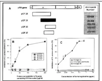
The 520-residue N-terminal region A contains the fib-rinogen-binding domain.54 Studies with recombinant trun-cated derivatives of region A localized the fibrinogen-bind-ing domain to a 218-residue segment (residues 332-550). One truncate (220-550) retained the ability to bind fibrino-gen, to block the attachment of bacteria to solid phase fib-rinogen (Figure 2), and to prevent bacterial clumping in a solution of fibrinogen.54 Antibodies raised against truncate 220-550 strongly inhibited both bacterial adhesion to fib-rinogen-coated surfaces (Figure 2) and cell clumping. All the smaller truncates lost the ability to interact with fib-rinogen. One truncate (332-550) neutralized the ability of polyclonal antibodies raised against truncate 220-550 to block bacterial binding to fibrinogen. Antibodies raised against the smaller protein also blocked bacterial interac-tions with fibrinogen. This suggests that the ligand-binding domain is located between residues 330-550 and that residues 220-330 are required to promote correct folding of the fibrinogen-binding domain.
ClfA contains an unusual 308-residue region com-prising 154 mainly Ser-Asp dipeptide repeats. A recombi-nant protein comprising region R did not bind fibrinogen and failed to block bacterial attachment to fibrinogen-coated surfaces,54 and antiserum raised against region R did not block bacterial attachment to fibrinogen-coated surfaces (Figure 2). A deletion mutant lacking region R failed to bind fibrinogen, suggesting that the function of this region is to display the ligand-binding domain region A correctly on the cell surface (O.M. Hartford, PhD, unpublished data, August 1995).
To evaluate the in vivo contribution of fibrinogen or fibrin to S aureus attachment during brief exposure of
intravascular devices to flowing blood, we tested the clfA-defective mutants or their complemented derivatives in an ex vivo canine arteriovenous shunt model.52 After expo-sure of polymer tubings to circulating canine blood for 5 to 60 minutes, segments of blood-exposed tubings were tested for promotion of bacterial adhesion. S aureus mutants lacking the fibrinogen adhesin were more defec-tive in attachment to the blood-exposed tubings than those lacking the fibronectin adhesin(s), compared to their respective parental strains.52 In contrast, a highly adhesive derivative strain of S aureus 8325-4, produced by complementation with the plasmid pCF4 expressing mul-tiple copies of the clfA gene, exhibited a strong increase over the parental strain in bacterial adhesion to blood-exposed tubing segments.52 These findings indicate that fibrinogen and fibrin are the most active components for initially promoting in vivo adhesion of S aureus, whereas fibronectin and fibronectin proteolytic fragment are more active in central venous catheters inser ted for >24 hours.44
In a collaborative work with Philippe Moreillon, MD (Infectious Diseases, University Hospital, Lausanne, Switzerland), the contribution of the clfA protein to the initial attachment and virulence of S aureus recently was evaluated in a rat model of endocarditis.55 In this animal model of endocarditis with catheter-induced aortic vegetations, the virulence of various single or combined clfA- and coagulase-defective mutants was tested. A significant reduction in the ability of the clfA-defective mutant of S aureus to colonize rat valves was observed. In contrast, the coagulase-defec-tive mutant showed no significant difference with the parental strain. Thus, the clumping factor also may con-tribute to S aureus attachment to damaged heart valves leading to bacterial endocarditis.55
Fibronectin-binding proteins. Molecular analysis of the genetic locus encoding fibronectin-binding surface component(s) of the laboratory strain 8325-4 of S aureus revealed two related genes, called fnbA and fnbB, located 682 base pairs apart.19,46,47,56 Both fnb genes cloned in
Escherichia coli could express functional FBPs, called FnBPA and FnBPB.18,19,46,47
Closely related ligand-binding domains have been identified in both FnBPs using truncated protein fragments and synthetic peptides in binding and inhibition studies. This common binding domain is composed of a 38-amino-acid unit that is repeated three times and partially a fourth, which is called the D-repeat unit or D1-D417,19,56 (Figure
3). The functional role of the D-repeat region has been con-firmed by blocking studies with recombinant and synthetic peptides, which produce a very strong dose-dependent inhibition of S aureus binding to either fluid-phase or solid-phase fibronectin.19,56-59 These peptides exhibit a stronger blocking activity than that of any polyclonal anti-bodies thus far developed against the fibronectin adhesins.60
To evaluate the role of each of the fibronectin adhesins in vitro and in vivo, mutants of strain 8325-4 defec-tive in the production of FnBPA, FnBPB, or both proteins were produced by inserting DNA fragments encoding antibiotic resistance into the fnb genes (a tetracycline resis-tance marker into fnbA and an erythromycin resisresis-tance marker into fnbB).51 Adhesion properties of either fnbA or
fnbBsingle mutants were not altered markedly compared to those of the parental strain.51 In contrast, the double
fnbAfnbBmutant was completely defective for attachment to either PMMA coverslips coated in vitro with fibronectin or to coverslips explanted from the subcutaneous space of guinea pigs.51 The parallelism between in vitro and in vivo observations indicate an important role for FBPs in staphy-lococcal attachment, colonization, and infection of biomate-rial implants.
Another category of bacterial mutants defective in FnBP production was generated by Kuypers and Proctor in strain 879R4S of S aureus by transposon insertion.50 One fibronectin-adhesin-defective mutant showed a markedly reduced ability to adhere to traumatized heart valves and to induce bacterial endocarditis in rats.50 A recently discov-ered characteristic of the parental and mutant strains of S
aureus879R4S is that it contains a single fnb gene closely related to the fnbA of strain 8325-4.61 Recent molecular studies of the fibronectin-adhesin-defective mutant strain 879R4S/1536 and its spectinomycin-derivative R4SSp/1536
FIGURE 2. (A) Schematic diagram showing the location of the clfA fragments and the amino-acid numbers represented. (B) Inhibition of Staphylococcus aureus adhesion to fibrinogen-coated poly-methylmethacrylate (PMMA) coverslips by lysates containing clfA truncates. (C) Inhibition of S aureus adhesion to fibrinogen-coated PMMA coverslips by immunoglobulins purified from anti-clfA sera and preimmune sera. Modified from reference 54 with permission from Molecular Microbiology (1995;16:895-906).
FIGURE 1. Schematic drawing showing the domain organization of Staphylococcus aureus fibrinogen-binding clfA protein. S, signal sequence; A, nonrepeat domain; R, repeat domain; W, wall region; M, membrane-spanning domain; +, positively charged residues. The black box indicates the fibrinogen-binding region.
demonstrated that the Tn918 transposon was inserted between the promoter and coding sequence of the single
fnbgene,61 thus leading to its decreased expression.61 The mutant strain 879R4S/1536 showed interesting adhesion defects in a novel animal model of orthopedic col-onization developed in the orthopedic clinic of our hospi-tal.62 In this model, which mimics conditions of internal fix-ation devices, titanium miniplates were fixed onto the iliac bones of guinea pigs or implanted into their subcutaneous space as controls for a period of 5 to 6 weeks. A significant reduction in adhesion of the fibronectin adhesin-defective mutant compared to its isogenic parent occurred on the metallic plates explanted from either the subcutaneous space or the iliac bone. These data suggest that fibronectin also may be present on bone-implanted metallic devices and promote attachment of S aureus to their surface.62 STRATEGIES FOR DECREASING HOST BACTERIA INTERACTIONS ON
FOREIGN BODIES
The combined development of molecular techniques and experimental models of bacterial adhesion should help to elucidate some of the most important clinically and epi-demiologically relevant mechanisms of S aureus attach-ment to major categories of biomaterial implants or indwelling devices.
The ligand-binding domains of both fibronectin and fibrinogen adhesins have been identified using truncated protein fragments and synthetic peptides in binding and inhibition studies. The binding domain of both fibronectin adhesins is the D-repeat unit or D1-D4 (Figure 3).17,19,56 Synthetic peptides based on this fibronectin-binding domain exhibit a stronger blocking activity on S aureus binding to fibronectin19,56-59 than any polyclonal antibod-ies thus far developed against the fibronectin adhesins.60 In contrast, polyclonal antibodies raised against the fibrino-gen-binding domain of the clfA protein, which is located within a 218-residue segment of its amino-terminal region A (Figure 2), have shown a more potent activity against
bacterial clumping and adhesion to fibrinogen-coated poly-meric surfaces than truncated peptides encompassing the ligand-binding domain of clfA.54
The important advances in the molecular characteri-zation of the fibrinogen-binding protein clfA24,54 and of both FBPs A and B46,47,56 in S aureus, and the functional demonstration24,51,52,54 that they contribute to attach-ment to the purified host proteins, offer interesting per-spectives for the development of a novel category of specif-ic antiadhesive agents.17,56 Such agents would offer a use-ful alternative to antibiotic prophylaxis in circumventing the emergence of multiple resistance determinants in noso-comial strains of staphylococci.
In addition to the fluid-phase specific antiadhesive agents mentioned above, surface-bound molecules chang-ing the chemical or physical properties of indwellchang-ing devices also should be evaluated carefully. We recently have described the impact of a surface coating procedure on the physical and biological properties of a polyurethane catheter.63 Coating of this polyurethane catheter with a hydrogel based on cross-linked polyvinyl pyrrolidone led to a dramatic increase in its surface smoothness and hydrophilic properties.63 The hydrophilic polyurethane catheter showed a strong reduction in the in vitro adsorp-tion of either fibrinogen or fibronectin, leading to a propor-tional reduction in protein-mediated adhesion of either S
aureusor S epidermidis.63
In conclusion, the combined development of specific fluid-phase antiadhesive agents and improved surface treat-ments of indwelling devices may lead to a significant reduc-tion in bacterial colonizareduc-tion of the foreign bodies. Further studies are needed to evaluate the clinical significance of these in vitro observations.
REFERENCES
1. Waldvogel FA. Staphylococcus aureus (including toxic shock syndrome). In: Mandell GL, Bennet JE, Dolin R, eds. Principles and Practice of Infectious Diseases. New York, NY: Churchill Livingstone; 1995:1754-1784.
2. Kloos WE, Bannerman TL. Update on clinical significance of coagulase-negative staphylococci. Clin Microbiol Rev 1994;7:117-140.
3. Rupp ME, Archer GL. Coagulase-negative staphylococci: pathogens associated with medical progress. Clin Infect Dis 1994;19:231-243.
4. Christensen GD, Baldassarri L, Simpson WA. Colonization of medical devices by coagulase-negative staphylococci. In: Bisno AL, Waldvogel FA, eds. Infections Associated With Indwelling Medical Devices. Washington, DC: American Society for Microbiology Press; 1994:45-78.
5. Pittet D, Wenzel RP. Nosocomial bloodstream infections: secu-lar trends in rates, mortality, and contribution to total hospital deaths. Arch Intern Med 1995;155:1177-1184.
6. Pittet D, Tarara D, Wenzel RP. Nosocomial bloodstream infec-tion in critically ill patients: excess length of stay, extra costs, and attributable mortality. JAMA 1994;271:1598-1601. 7. Bisno AL, Waldvogel FA. Infections Associated With Indwelling
Medical Devices. Washington, DC: American Society for Microbiology Press; 1994:398.
8. Vaudaux P, Grau GE, Huggler E, et al. Contribution of tumor necrosis factor to host defense against staphylococci in a FIGURE 3. Schematic drawing comparing the domain organization
of the two fibronectin-binding proteins of Staphylococcus aureus 8324-5. The percentage residue identity between different regions of FnBPA and FnBPB is indicated. S, signal peptide; A and C, non-repeated regions of unknown function; B and D, repeat regions out-side the cell-wall domain; Wr, repetitive region in the cell-wall bind-ing domain; Wc, nonrepeat region in the cell-wall bindbind-ing domain; M, membrane-spanning domain; +, positively charged residues. The black boxes indicate the fibronectin-binding domain.
guinea pig model of foreign body infections. J Infect Dis 1992;166:58-64.
9. Zimmerli W, Waldvogel FA, Vaudaux P, Nydegger UE. Pathogenesis of foreign body infection: description and charac-teristics of an animal model. J Infect Dis 1982;146:487-497. 10. Lucet JC, Herrmann M, Rohner P, Auckenthaler R, Waldvogel
FA, Lew DP. Treatment of experimental foreign body infection caused by methicillin-resistant Staphylococcus aureus. Antimicrob Agents Chemother 1990;34:2312-2317.
11. Chuard C, Herrmann M, Vaudaux P, Waldvogel FA, Lew DP. Successful therapy of experimental chronic foreign-body infec-tion due to methicillin-resistant Staphylococcus aureus by antimicrobial combinations. Antimicrob Agents Chemother 1991;35:2611-2616.
12. Bouchenaki N, Vaudaux P, Huggler E, Waldvogel FA, Lew DP. Successful single-dose prophylaxis of Staphylococcus aureus foreign body infection in guinea pigs by fleroxacin. Antimicrob Agents Chemother 1990;34:21-24.
13. Schaad HJ, Chuard C, Vaudaux P, Waldvogel FA, Lew DP. Teicoplanin alone or combined with rifampin compared with vancomycin for prophylaxis and treatment of experimental for-eign body infection by methicillin-resistant Staphylococcus aureus. Antimicrob Agents Chemother 1994;38:1703-1710. 14. Zimmerli W, Lew DP, Waldvogel FA. Pathogenesis of foreign
body infection. Evidence for a local granulocyte defect. J Clin Invest 1984;73:1191-1200.
15. Zimmerli W, Lew DP, Cohen HJ, Waldvogel FA. Comparative superoxide-generating system of granulocytes from blood and peritoneal exudates. Infect Immun 1984;46:625-630.
16. Herrmann M, Jaconi MEE, Dahlgren C, Waldvogel FA, Stendahl O, Lew DP. Neutrophil bactericidal activity against Staphylococcus aureus adherent on biological surfaces. J Clin Invest 1990;86:942-951.
17. Foster TJ, McDevitt D. Molecular basis of adherence of staphy-lococci to biomaterials. In: Bisno AL, Waldvogel FA, eds. Infections Associated With Indwelling Medical Devices. Washington, DC: American Society for Microbiology Press; 1994:31-44.
18. Höök M, Switalski LM, Wadström T, Lindberg M. Interactions of pathogenic microorganisms with fibronectin. In: Mosher DF, ed. Fibronectin. San Diego, CA: Academic Press; 1989:295-308. 19. Lindberg M, Jönsson K, Müller HP, et al. Fibronectin-binding proteins in Staphylococcus aureus. In: Novick RP, ed. Molecular Biology of the Staphylococci. New York, NY: VCH; 1990:343-356. 20. Foster TJ, McDevitt D. Surface-associated proteins of Staphylococcus aureus: their possible roles in virulence. FEMS Microbiol Lett 1994;118:199-206.
21. Cheung AL, Fischetti VA. The role of fibrinogen in staphylo-coccal adherence to catheters in vitro. J Infect Dis 1990;161:1177-1186.
22. Herrmann M, Vaudaux P, Pittet D, et al. Fibronectin, fibrinogen and laminin act as mediators of adherence of clinical staphylo-coccal isolates to foreign material. J Infect Dis 1988;158:693-701. 23. Herrmann M, Lai QJ, Albrecht RM, Mosher DF, Proctor RA.
Adhesion of Staphylococcus aureus to surface-bound platelets: role of fibrinogen/fibrin and platelet integrins. J Infect Dis 1993;167:312-322.
24. McDevitt D, François P, Vaudaux P, Foster TJ. Molecular char-acterization of the clumping factor (fibrinogen receptor) of Staphylococcus aureus. Mol Microbiol 1994;11:237-248. 25. Vaudaux P, Lew DP, Waldvogel FA. Host factors predisposing to
and influencing therapy of foreign body infections. In: Bisno AL, Waldvogel FA, eds. Infections Associated With Indwelling Medical Devices. Washington, DC: American Society for Microbiology Press; 1994:1-29.
26. Vaudaux P, Pittet D, Haeberli A, et al. Host factors selectively increase staphylococcal adherence on inserted catheters: a role for fibronectin and fibrinogen/fibrin. J Infect Dis 1989;160:865-875.
27. Vaudaux P, Suzuki R, Waldvogel FA, Morgenthaler JJ, Nydegger UE. Foreign body infection: role of fibronectin as a ligand for the adherence of Staphylococcus aureus. J Infect Dis
1984;150:546-553.
28. Vaudaux P, Waldvogel FA, Morgenthaler JJ, Nydegger UE. Adsorption of fibronectin onto polymethylmethacrylate and promotion of Staphylococcus aureus adherence. Infect Immun 1984;45:768-774.
29. Patti JM, Jonsson H, Guss B, et al. Molecular characterization and expression of a gene encoding Staphylococcus aureus colla-gen adhesin. J Biol Chem 1992;267:4766-4772.
30. Patti JM, Bremell T, Krajewska-Pietrasik D, et al. The Staphylococcus aureus collagen adhesin is a virulence determi-nant in experimental septic arthritis. Infect Immun 1994;62:152-161.
31. Patti JM, Boles JO, Höök M. Identification and biochemical characterization of the ligand binding domain of the collagen adhesin from Staphylococcus aureus. Biochemistry 1993;32:11428-11435.
32. Patti JM, House-Pompeo K, Boles JO, Garza N, Gurusiddappa S, Höök M. Critical residues in the ligand-binding site of the Staphylococcus aureuscollagen-binding adhesin (MSCRAMM). J Biol Chem 1995;270:12005-12011.
33. Chhatwal GS, Preissner G, Müller-Berghaus, Blobel H. Specific binding of the human S protein (vitronectin) to streptococci, Staphylococcus aureus, and Escherichia coli. Infect Immun 1987;55:1878-1883.
34. Liang OD, Maccarana M, Flock J-I, Paulsson M, Preissner KT, Wadström T. Multiple interactions between human vitronectin and Staphylococcus aureus. Biochim Biophys Acta 1993;1225:57-63.
35. Lopez JD, Dos Reis M, Bretani RR. Presence of laminin recep-tors in Staphylococcus aureus. Science 1985;229:275-277. 36. Herrmann M, Suchard SJ, Boxer LA, Waldvogel FA, Lew DP.
Thrombospondin binds to Staphylococcus aureus and promotes staphylococcal adherence to surfaces. Infect Immun 1991;59:279-288.
37. Ryden C, Yacoub AI, Maxe I, et al. Specific binding of bone sialoprotein to Staphylococcus aureus isolated from patients with osteomyelitis. Eur J Biochem 1989;184:331-336.
38. Yacoub A, Lindahl P, Rubin K, Wendel M, Heinegård D, Rydén C. Purification of a bone sialoprotein-binding protein from Staphylococcus aureus. Eur J Biochem 1994;222:919-925. 39. Woo Park P, Roberts DD, Grosso LE, et al. Binding of elastin to
Staphylococcus aureus. J Biol Chem 1991;266:23399-23406. 40. Jönsson K, McDevitt D, McGavin MH, Patti JM, Höök M.
Staphylococcus aureus expresses a major histocompatibility complex class II analog. J Biol Chem 1995;270:21457-21460. 41. Muller E, Takeda S, Shiro H, Goldmann D, Pier GB.
Occurrence of capsular polysaccharide/adhesin among clinical isolates of coagulase-negative staphylococci. J Infect Dis 1993;168:1211-1218.
42. Shiro H, Muller E, Gutierrez N, et al. Transposon mutants of Staphylococcus epidermidis deficient in elaboration of capsular polysaccharide/adhesin and slime are avirulent in a rabbit model of endocarditis. J Infect Dis 1994;169:1042-1049. 43. Delmi M, Vaudaux P, Lew DP, Vasey H. Role of fibronectin on
staphylococcal adhesion to metallic surfaces used as models of orthopaedic devices. J Orthop Res 1994;12:432-438.
44. Vaudaux P, Pittet D, Haeberli A, et al. Fibronectin is more active than fibrin or fibrinogen in promoting Staphylococcus aureus adherence to inserted intravascular catheters. J Infect Dis 1993;167:633-641.
45. Switalski LM, Patti JM, Butcher W, Gristina AG, Speziale P, Höök M. A collagen receptor on Staphylococcus aureus strains isolated from patients with septic arthritis mediates adhesion to cartilage. Mol Microbiol 1993;7:99-107.
46. Flock JI, Froman G, Jonsson K, et al. Cloning and expression of the gene for a fibronectin-binding protein from Staphylococcus aureus. EMBO J1987;6:2351-2357.
47. Jonsson K, Signäs C, Muller HP, Lindberg M. Two different genes encode fibronectin binding proteins in Staphylococcus aureus. The complete nucleotide sequence and characteriza-tion of the second gene. Eur J Biochem 1991;202:1041-1048. 48. Cheung AI, Projan SJ, Edelstein RE, Fischetti VA. Cloning,
expression, and nucleotide sequence of a Staphylococcus aureus gene (fbpA) encoding a fibrinogen-binding protein. Infect Immun 1995;63:1914-1920.
49. Bodén MK, Flock J-I. Cloning and characterization of a gene for a 19 kDa fibrinogen-binding protein from Staphylococcus aureus. Mol Microbiol 1994;12:599-606.
50. Kuypers JM, Proctor RA. Reduced adherence to traumatized heart valves by a fibronectin low-binding mutant of Staphylococcus aureus. Infect Immun 1989;57:2306-2312. 51. Greene C, McDevitt D, François P, Vaudaux P, Lew DP, Foster
TJ. Adhesion properties of mutants of Staphylococcus aureus defective in fibronectin binding proteins and studies on the expression of fnb genes. Mol Microbiol 1995;17:1143-1152. 52. Vaudaux PE, François P, Proctor RA, et al. Use of
adhesion-defective mutants of Staphylococcus aureus to define the role of specific plasma proteins in promoting bacterial adhesion to canine arteriovenous shunts. Infect Immun 1995;63:585-590. 53. Dickinson RB, Nagel JA, McDevitt D, Foster TJ, Proctor RA,
Cooper SL. Quantitative comparison of clumping factor- and coagulase-mediated Staphylococcus aureus adhesion to surface-bound fibrinogen under flow. Infect Immun 1995;63:3143-3150. 54. McDevitt D, François P, Vaudaux P, Foster TJ. Identification of the ligand-binding domain of the surface-located fibrinogen receptor (clumping factor) of Staphylococcus aureus. Mol Microbiol 1995;16:895-907.
55. Moreillon P, Entenza JM, Francioli P, et al. Role of Staphylococcus aureus coagulase and clumping factor in patho-genesis of experimental endocarditis. Infect Immun 1995;63:4738-4743.
56. Signäs C, Raucci K, Jönsson K, et al. Nucleotide sequence of the gene for a fibronectin-binding protein from Staphylococcus aureus: use of this peptide sequence in the synthesis of biolog-ically active peptides. Proc Natl Acad Sci U S A 1989;86:699-703. 57. Raja RH, Raucci G, Höök M. Peptide analogs to a fibronectin
receptor inhibit attachment of Staphylococcus aureus to fibronectin-containing substrates. Infect Immun 1990;58:2593-2598.
58. McGavin MJ, Raucci G, Gurusiddappa S, Höök M. Fibronectin binding determinants of the Staphylococcus aureus fibronectin receptor. J Biol Chem 1991;266:8343-8347.
59. McGavin MJ, Gurusiddappa S, Lindgren P-E, Lindberg M, Raucci G, Höök M. Fibronectin receptors from Streptococcus dysgalactiae and Staphylococcus aureus. Involvement of con-served residues in ligand binding. J Biol Chem 1993;268:23946-23953.
60. Rozalska B, Sakata N, Wadström T. Staphylococcus aureus fibronectin-binding proteins (FnBPs). Identification of antigenic epitopes using polyclonal antibodies. APMIS 1994;102:1-7. 61. Greene C, McDevitt D, Vaudaux PE, et al. A low-fibronectin
binding mutant of Staphylococcus aureus 879R4S has Tn918 inserted into its single fnb gene. Microbiology. In Press. 62. Fischer B, Vaudaux P, Magnin M, et al. A novel animal model
for studying the molecular mechanisms of bacterial adhesion to bone-implanted metallic devices: role of fibronectin in Staphylococcus aureusadhesion. J Orthop Res. In press. 63. François P, Vaudaux P, Nurdin N, Mathieu HJ, Descouts P, Lew
DP. Physical and biological effects of a surface coating proce-dure on polyurethane catheters. Biomaterials 1996;17:667-678.
Gina Pugliese, RN, MS; Martin S. Favero, PhD
Medical News Editors
Bartonella quintana first was identified as an important human pathogen during World War I, when it caused epidemics of louse-borne trench fever that affected an estimat-ed 1 million troops in Europe. B
quin-tana infections rarely were recog-nized from the end of the war until the 1980s, when the organism re-emerged as an opportunistic pathogen among HIV-infected per-sons.
B quintanahas emerged among homeless persons in North America and Europe. In 1993, the organism first was isolated from the blood spec-imens of 10 patients at a single hospi-tal in Seattle, Washington, within a 6-month period. These patients had
ill-nesses characterized by fever and persistent bacteremia. Endocarditis developed in two patients, one of whom required a heart valve replace-ment. All 10 patients had chronic alco-holism, eight were homeless, and the six who were tested for HIV were HIV negative. These six were the first cases of invasive B quintana infection among HIV-negative persons report-ed in the Unitreport-ed States.
As a follow up to this Seattle out-break, Dr. Lisa Jackson and Dr. David Spach, from the University of Washington in Seattle, conducted a seroprevalence study of
anti-Bar tonella antibodies among 192 patients at a community clinic in the “skid row” section of Seattle, which serves a primarily homeless and indi-gent population. B quintana IgG titers >64 were detected among 39 (20%) of the 192 clinic patients.
The researchers note that multi-ple factors likely have contributed to the emergence of B quintana among the homeless, including those related to disease transmission, host suscep-tibility, and ability to detect the organ-isms. For example, transmission of B
quintana from human to human by the body louse has been documented experimentally.
As with other emerging infec-tious diseases, further efforts to iden-tify, evaluate, and control these infec-tions will require coordinated effort of clinicians, microbiologists, and public health officials.
FROM: Jackson LA, Spach DH. Emergence of Bartonella quintana infection among homeless persons.
Emerg Infect1996;2(2):141-143.