In Vitro CeIL Dev. Biol.IAnima134:,168-477, June 1998 © 1998 Society for In Vitro Biology
1071-2690/98 $05.00 + 0.00
ISOLATION OF EpH4 MAMMARY EPITHELIAL CELL SUBPOPULATIONS WHICH DIFFER
IN THEIR MORPHOGENETIC PROPERTIES
R. MONTESANO, ~ J. K SORIANO, I. FIALKA, AND L. ORCI
Department of Morphology, University of Geneva Medical Center, Geneva, Switzerland (R. M., J. V. S., L. 0.); and Research Institute of Molecular Patholog); Vienna, Austria (1. F.)
(Received 30 June 1997; accepted 2 October 1997)
SUMMARY
EpH4 is a nontumorigenic cell line derived from spontaneously immortalized mouse mammary gland epithelial cells (Fialka et al., 1996). When grown in collagen gels, EpH4 cells give rise to different types of structures, e.g., solid cords or branching tubes. By removing and subsequently dissociating single three-dimensional colonies of defined morphology, we have isolated six clonal subpopulations of EpH4 cells which display distinct morphogenetic properties in collagen gel cultures. Thus, cells from the H~B clone form branching cords devoid of a central lumen, K3A3 cells form cords enclosing small multifocal lumina, and J3B1 ceils form large cavitary structures containing a wide lumen. I3G 2 cells form either cords or tubes, depending on the type of serum added to the culture medium. Finally, when grown in serum-free medium, Bela cells form spherical cysts, whereas Be4a cells form long, extensively branched tubes. In additional assays of morphogenesis, i.e., cell sandwiching between two collagen gels or culture on a thick layer of Matrigel (a laminin-rich extracellular matrix), all clones form epithelial-cell-lined cavitary structures, except H~B cells which are unable to generate lumina under these conditions. The EpH4 sublines we have isolated provide an in vitro system for studying the mechanisms responsible for lumen formation and branching morphogenesis, as well as for identifying the factors which subvert these developmental processes during mammary carcinogenesis.
Key words: cell cloning; extracellular matrix; collagen gel; Matrigel. INTRODUCTION
The postnatal development of the mammary, gland involves a tightly scheduled sequence of morphogenetic processes, which in- clude the elongation and branching of lactiferons ducts and the sub- sequent budding of alveoli from the growing ducts (reviewed by Dan- icl and Silberstein, 1987). These events can be recapitulated in vitro by growing mammary epithelial cells (either in primary culture or as established cell lines) within reconstituted three-dimensional matri- ces (see for example Yang et al., 1980; Barcellos-Hoff et al., 1989; Darcy et al., 1991; Kanazawa and Hosik, 1992; Petersen et al., 1993; Soriano et al., 1995). Thus, when embedded in collagen gels, a num- ber of immortalized mammary epithelial cell lines have been reported to form histotypic structures resembling branching ducts or alveoli (Ormerod and Rudland, 1982; Danielson et al., 1984; Reichmann et al., 1989; Soule et al., 1990; Berdichevsky et al., 1994; Keely et al., 1995; Soriano et al., 1995).
Because the orderly architecture of mammary" gland tissue is dis- rupted during the development of breast cancer, it is important to develop experimental models in which to study the mechanisms that subvert the normal morphogenetic program of mammary epithelial cells. The nontumorigenic Epl (Reichmann et al., 1992) and EpH4 (Fialka et al., 1996; Oft et al., 1996) cell lines, derived from spon-
~To whom correspondence should be addressed at Dapartement de Mor- phologie, Centre M4dical Universitaire, 1, rue Michel-Server, 1211 GENEVE 4, SUISSE.
taneously immortalized mouse mammary cells (Reichmann et al., 1989), provide a powerful tool for the identification of factors that modulate epithelial architecture. Epl and EpH4 cells exhibit a fully polarized epithelial phenotype (as revealed by their high transepi- thelial electrical resistance and appropriate localization of apical and basolateral marker proteins), and form branching tubular structures when grown in collagen gels. These differentiated properties are how- ever lost following the experimental induction of activated proto- oncoproteins. Thus, in epithelial tubes formed by EpH4 cells in col- lagen gels, activation of c-Jun protein results in redistribution of polarity markers, disappearance of the central lumen, and formation of solid cord-like structures (Fialka et al., 1996). Activation of c-Fos oncoprotein induces more pronounced changes, including the ac- quisition of invasive properties and conversion to a fibroblastoid phe- notype (Reichmann et al., 1992). Similar alterations are observed upon incubation of nontransformed EpH4 cells with thyroid hormone (L6pez-Barahona et al., 1995), or upon incubation of Ha-Ras-EpH4 cells with transforming growth factor beta-1 (Oft et al., 1996).
A complementary approach for identifying molecular determinants of normal and aberrant morphogenesis could involve a comparative analysis of subpopulations derived from the same mammary epithe- lial cell line, yet differing in their organizational behavior in three- dimensional substrata. As an initial step towards this goal, we have isolated six subclones of EpH4 cells which exhibit distinct morpho- genetic properties. Here we describe the characterization of these clones with respect to their ability to form Iumina and to undergo branching morphogenesis in reconstituted extracellular matrices.
MORPHOGENIC BEHAVIOR OF Eptt4 CLONES 469
MATERIALS AND METHODS
Culture of EpH4 cells. Parental EpH4 cells (Fialka et al., 1996) were grown in tissue culture flasks (Falcon, Becton-Dickinson, San Josd, CA) in l)ul- becco's modified Eagle's medium (DMEM, GIBCO, Basel, Switzerland) sup- plemented with 10% fetal calf serum (FCS, GIBCO) and 2 nvl'/L-glutamine (this medium will heretofore be referred to as complete medium). For collagen gel cultures, EpH4 ceils were harvested with trypsin-EDTA, centrifuged, and suspended in three-dimensional collagen gels as described (Montesano et al., 1983; Montesano et al., 1991). In brief, 8 volumes of rat tail tendon collagen solution (approximately 1.5 mg/ml) were mixed with 1 volmne of 10 × con- centrated minimal essential medium (GIBCO) and 1 volume of sodium bi- carbonate (11.76 mg/ml) in a sterile flask kept on ice to prevent premature collagen gelation. Cells were resuspended in the cold mixture, and either 0.4 ml or 2 ml aliquots of cell suspension were dispensed into 16-ram wells (Nunc, Kamstrup, Roskilde, Denmark) or 35-ram dishes (Nunc), respectively. After 10 min incubation at 37 ° C to allow collagen gelation, complete medium was added and changed every 2-3 d.
Establishment ofEpH4 subclones. Subclones of parental EpH4 cells were obtained, as described below, by a two-step procedure involving: a) removal of an epithelial colony of defined morphology from a collagen gel culture, followed by its enzymatic dissociation into single' cells, and b) cloning of the resulting cell population by limiting dilution.
To facilitate the removal of individual colonies frmn collagen gel cultures, parental EpH4 cells were suspended at very low density (100-200 cells/roll in a collagen solution cast into 35-ram dishes, and subsequently incubated in complete medium for 10-14 d. Colonies of different morphology (e.g., solid branching cords, cords containing small multifocal lmnina, or tubulocystic structures with widely patent luminal were identified by phase-contrast optics with a Nikon Diaphot TMD inverted photomicroscope and delimited by a circle drawn on the underside of the dish. Collagen gel fragments containing a single colony were manually removed under sterile conditions with the aid of fine forceps and subsequently digested by a 10-rain incubation with 4 mg Clostridium histolyticum collagenase per ml (Worthington Biochemical Cor- poration, Freehold, NJ) as previously described (Soriano et al., 1995). The released epithelial colonies were dissociated into single cells with trypsin- EDTA, and the resulting cell suspension was plated into 16-ram wells in complete medium. At confluence, the cells were trypsinized, expanded suc- cessively in tissue culture dishes and flasks, and frozen. In some cases, cells obtained by this procedure were suspended once again in collagen gels and subjected to a second cycle of colony isolation and dissociation.
In additional experiments, colonies were isolated from collagen gel cul- tures performed under serum-free conditions, as follows: EpH4 cells were suspended in collagen gels at 300-500 eellstml and incubated with a serum- free medium composed of mammary epithelial cell basal medium (MEBM, Cloneties Corporation, San Diego, CA), 0.4% bovine pituitary extract (Col- laborative Biomedical Products, Bedford, MA), 2 lag insulirdml (Sigma Chem- ical Co., St. Louis, MO), 1 gM isoproterenol (Sigma), 25 ng hydroeortisone per ml (Sigma) and 2.5 ng transforming growth factor alpha per m] (Bachem, Bubendorf, Switzerland) (Fialka et al., 1996). The colonies developed under serum-free conditions were then isolated and dissociated as described above. To ensure the monoclonality of cell populations obtained through colony isolation, we devised a novel limiting dilution procedure, which will be re- ferred to as "droplet cloning." Cells were harvested from confluent monolayer cultures with trypsin-EDTA, centrifuged, resuspended in complete medium, and diluted to a density of 50 cells/ml. A single 20-gl droplet of cell sus- pension (theoretically containing one cell) was placed in the center of each of several 35-ram dishes, within a 5-ram circle drawn on the dish underside. Following a 20-rain incubation at 37 ° C to allow cell attachment, the droplets were carefully inspected by phase-contrast microscopy, and dishes with a droplet containing either no cells or more than one cell were discarded. The remaining dishes, which contained a single cell, were incubated at 37 ° C for a further 90-120 min to allow cell spreading, at which time 2 ml complete medium was added. The circular area in which the droplet was initially de- posited was reexamined to confirm the presence of one cell, and the dishes were incubated at 37 ° C for 1-2 wk to allow formation of colonies approxi- mately 1 mm in diameter. These were subsequently trypsinized within glass rings and the resulting clonal populations expanded and frozen.
Morphogenetic assays. To assess the morphogenetic properties of EpH4 subpopulations, cells from each clone were suspended in three-dimensional collagen gels at a concentration of 5 × 10³ to 2 x l04 cells/ml and grown for 1 to 3 wk. The morphology of the resulting colonies was monitored by
phase contrast optics and further evaluated by light and electron microscopy after fixation and embedding (see below).
The potential ability of the various clones to form lumina was more spe- cifically assessed by "sandwiching" the cells between two collagen layers. Cells were seeded at 1-2 × 10⁵ cells per 35-ram dish onto the surface of a collagen gel (1 ml) and grown to near confluence (2-3 d). The monolayer was subsequently overlaid with a second 1 ml collagen gel as described by Mon- tesano et at. (1983) and incubated at 37 ° C for a further 6--7 d.
An additional assay of morphogenetic behavior consisted in seeding the cells onto a thick layer of Matrigel, a laminiu-rich matrix derived frmn the Engelbreth-Hohn-Swarm (EHS) tumor (Kleinman et al., 1986). First, 150-p,l aliquots of Matrigel (Collaborative Biomedical Products) were dispensed onto a preformed 250-gl collagen gel (base layer) and allowed to gel at 37 ° C for 30 rain. Cells from the various clones were then plated onto the Matrigel surface at concentrations ranging from 2 × 10⁴ to 1 X 10 s cells/well. Al- ternatively, the cells were seeded onto a 125-gl layer of Matrigel, allowed to attach for 30 min, and subsequently covered with a second layer of Matrigel (150 ~tl). Complete medimn was then added on top of the gelified Matt/gel.
Processing for light and electron microscopy. Collagen or Matrigel cultures were fixed in situ overnight with 2.5% glutaraldehyde in 0,1 M sodium cac- odylate buffer (pH 7.4). After extensive rinsing in the same buffer, the gels were gently removed from the dishes or wells and cut into 3- X 3-mm frag- ments. These were postfixed in 1% osmium tetroxide in Veronal acetate buffer for 45 rain, stained en bloc with 2.5% uranyl acetate in 50% ethanol, de- hydrated in graded ethanols, and embedded in Epon 812 as described (Mon- tesano et al., 1991). Semithin (1-gin-thick) sections were cut with an LKB Uhramicrotome, stained with 1% methylene blue, and photographed under transmitted light with a Zeiss photomicroscope. Thin sections were stained with uranyl acetate and lead citrate and examined with a Philips CM 10 electron microscope.
RESUUFS
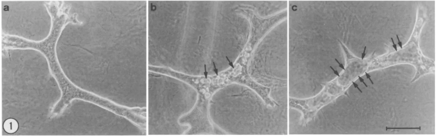
This study was prompted by the finding that E p H 4 mammary ep- ithelial cells (Fialka et al., 1996) form different types of three-di- mensional structures when grown in collagen gels in serum-supple- mented medium (Fig. 1). In cultures examined by phase-contrast microscopy, the majority of the epithelial colonies consisted of solid cords apparently devoid of lumen (Fig. 1 a). A number of colonies, however, contained either muhifocal lumen-like spaces (Fig. 1 b) or widely patent cylindrical lumina virtually occupying the entire cord width (Fig. 1 c). Conceivably, these morphologically different stRic- tures could develop from a homogeneous cell population, either be- cause of local variations in culture microenvironment or as sequential phases in colony evolution. Ahernatively, they could result from the clonal growth of phenotypically distinct cells.
To discriminate between these two possibilities, we set out to as- sess the morphogenetic properties of clonal cell populations derived from individual colonies. Single three-dimensional structures exhib- iting a well-defined morphotype (e.g., solid cords, cords with multi- focal lumina, and tubes with a wide lumen; see Fig. 1) were removed from low density collagen gel cultures (see Materials and Methods) and enzymatically dissociated. The resuhing cell populations were then expanded and assayed for their organizational behavior in col- lagen gels. The finding that each cell subpopulation formed three- dimensional structures similar to the colony from which it was ini- tially isolated supported the view that the E p H 4 cell line is composed of phenotypically distinct epithelial cells. However, because the pop- ulations obtained by colony isolation were not demonstrated to have a monoclonal origin, they were recloned with an improved limiting dilution procedure ("droplet cloning"; see Materials and Methods). Four clones (designated HIB, IzG2, J:~BI and KzA3), each of which gave rise to a different type of colony in collagen gels (see below), were selected for further characterization. In light of our previous finding that epithelial cells s u s p e n d e d in collagen gels can form col-
470 MONTESANO ET AL.
FiG. 1. Parental EpH4 cells form different types of three-dimensional structures in collagen gels. EpH4 cells were suspended in a collagen gel at a concentration of 5 × l03 cells/ml and incubated in FCS-supplemented DMEM for 7 d, at which time colonies of different morphology were photographed with an inverted phase-contrast microscope. (a) Predominant type of colony consisting of smooth-surfaced branching cords devoid of a visible lumen; (b) flattened cords containing muhiple small lumina (arrows); (c) thin-walled tube-like structure containing a wide lumen (arrows).
Bar
= 100 pro.onies of different morphology depending on whether they are grown in the presence of fetal calf serum (FCS) or donor calf serum (DCS) (unpublished observations), the behavior of EpH4 clones was as- sessed in both culture conditions.
Cell suspension in three-dimensional collagen gels.
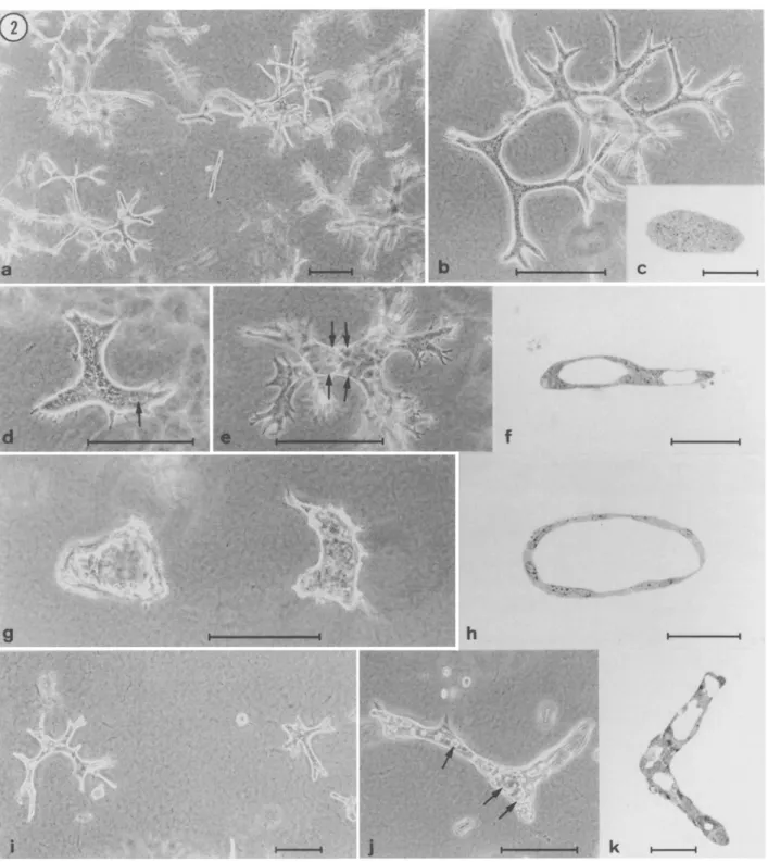
When embed- ded in collagen gels, HIB cells gave rise to long branching cords devoid of central lumen, irrespective of whether the culture medium (DMEM) was supplemented with FCS or DCS (Fig. 2a-c).
In con- trast, the behavior of clone IzG 2 w a s highly dependent on the type of serum added to the cultures: in the presence of FCS, 13G 2 cells formed solid, flattened cords which occasionally contained tiny lu- men-like spaces (Fig. 2 d), whereas in the presence of DCS, they formed branching tubes containing a wide lumen (Fig. 2 e~. JaB1 cells formed cavitary structures, whose morphology ranged from short ectatic tubes (in FCS; not shown) to irregularly-shaped cysts (in DCS; Fig. 2g,h).
Finally, K3A 3 cells gave rise to branching cords contain- ing numerous heterogeneously-sized muhifocal lumina, which were usually larger in DCS than in FCS (Fig. 2i-k).
Thus, suspension in collagen gels in serum-supplemented medium demonstrated first that the four EpH4 clones described above differ in their morphogenetic properties, and second that DCS is superior to FCS in promoting lumen formation (except for HIB cells, which are apparently inca- pable of forming lumina in collagen gels).Because parental EpH4 ceils have been reported to form branch- ing tubes when grown in collagen gels in serum-free medium (Fialka et al., 1996; Oft et al., 1996), we also assessed the behavior of EpH4 clones in serum-free culture. Surprisingly, all four clones examined proliferated slowly under these conditions, and in no instances were tubular structures formed (results not shown). This finding led us to hypothesize that the procedure of isolating colonies formed in serum- containing cultures had selected for EpH4 subpopulations other than those which have the ability to generate branching tubes in serum- free medium. To explore this possibility, parental EpH4 cells were allowed to form three-dimensional structures in serum-free collagen gel cultures, at which time individual colonies were removed and enzymatically dissociated. The cell populations thus obtained were subsequently cloned by the "droplet" method, and the resulting
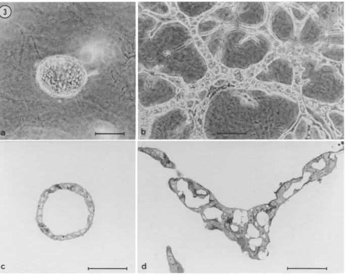
clones were assayed for their morphogenetic properties in collagen gels. Two clones capable of forming cavitary structures (clones Bela and Be4a) were further characterized. When ~'own in collagen gels in serum-free medium, Bela cells gave rise to spherical cysts (Fig.
3 a,c),
whereas Be4a cells formed extensively branched cords con- taining muhifocal or continuous lumina (Fig. 3 b,d). Bela and Be4a cells also exhibited a different organizational behavior in serum- supplemented cultures. Thus, in the presence of DCS, Bela cells formed slowly growing cystic structures, whereas Be4a ceils formed branching tubes (not shown).In summary, the four clones (H1B, I:~G2, JIB~, and KzA3) estab- lished from EpH4 colonies formed in serum-supplemented cultures, as well as the two clones (Bela and Be4a) established from EpH4 colonies formed under serum-free conditions, were found to differ considerably in their ability to undergo branching morphogenesis and to form lumina when suspended in three-dimensional collagen gels (Table 1).
Response to collagen gel overla).
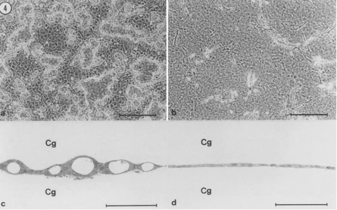
Because the clones described above differed markedly in their capacity to form lumina when sus- pended in collagen gels, we investigated this property in more detail using the collagen sandwich technique. When polarized epithelial cells are grown on a collagen gel and subsequently overlaid with a second layer of collagen, the initial monolayer undergoes a profound reorganization which culminates in the creation of newly formed lu- mina (Chambard et al., 1981; Hall et al., 1982; Schwimmer and Ojakian, 1995; Zuk and Marlin, 1996). To study the response of EpH4 clones to collagen overlay-, cells were seeded onto the surface of a collagen gel and grown to near confluence, at which time the monolayer was covered with an additional collagen gel. Irrespective of whether the culture medium (DMEM) was supplemented with FCS or with DCS, collagen overlay induced lumen formation by all clones except HtB cells (Fig. 4, Table 1). Lumen formation was first de- tectable approximately 2 d following collagen overlay as small re- fractile spaces between adjacent cells (not shown). Over the next few days of culture, these spaces increased in number, enlarged, and coalesced to form wider cavities (shown for JzB1 cells in Fig. 4 a). Sections perpendicular to the culture plane showed that the cavitiesMORPHOGENIC BEHAVIOR OF EpH4 CLONES 471
i ¸ ~
I
I- !
k : ~..
FIG. 2. Clones established from different types of EpH4 colonies exhibit distinct morphogenetic properties. Four clones isolated from EpH4 colonies formed in FCS-supplemented DMEM were suspended in collagen gels and grown either in DMEM + FCS or in DMEM + DCS. (a) Cells from the H1B clone grown in the presence of DCS form solid branching cords; (b) higher magnification of a colony of HxB cells; (c) semithin section of a cord of HxB cells showing the lack of a central lumen. (d) IzG 2 cells grown in FCS-supplemented medium form flattened solid cords which occasionally contain small lumen-like translucent spaces (arrow); (e) 13G2 cells grown in DCS- supplemented medium form branched structures containing a widely patent lumen; (f) semithin section of the culture shown in (e) confirming the tubular nature of IsG 2 colonies. (g,h) large cyst-like structures formed by J:~B~ cells grown in the presence of DCS. K3A 3 cells grown in the presence of FCS (i,]) or DCS (k) form branching cords containing multifocal ]nmina of various sizes. Bars correspond to 200 ~m for phase-contrast micrographs (a,b,d,e,g,i, and j) and to 50 ~m for semithin sections (cf,,h, and k).
472 MONTESANO ET AL.
C I I d I I
FIG. 3. Morphogenetic properties of clones Bela and Be4a. Unlike the four clones illustrated in Figure 2, clones Bela and Be4a were established from colonies of EpH4 cells formed under serum-free culture conditions (see Materials and Methods). (a,c) When suspended in collagen gels in sermn-free medium, Bela cells form spherical cysts; (b,d) Under the same experimental conditions, Be4a cells form long branching cords containing clearly defined lumina. (a,b) Bars = 100 gin; (c,d) Bars = 50 gin.
TABLE 1
SYNOPTIC VIEW OF THE MORPHOGENETIC PROPERTIES OF EPH4 CLONES
Clones
Lunlen
formation Dome Lumen Behavior in collagen formation formation in collagen gels • sandwich on plastic in Matrigel
H~B Solid branching cords - - + - -
I3G 2 In FCS: cords with rare small + - - + lumina. In DCS: tubes
JsBI Tubulocystic structures b + - - + KsA3 Cords with multifocal lumina b + + + Bela Spheroidal cysts (in SFM ~) + + + Be4a Long, highly branched tubes + +a +
(in SFM ¢)
aFor simplicity, only the most salient properties of each clone are indicated. bin DCS, lumina are larger than in FCS.
~SFM = serum-free medium.
aDomes are more flattened than in the other clones.
seen by phase-contrast microscopy were, in fact, lumina enclosed by two epithelial monolayers, one attached to the upper gel and another to the lower (Fig. 4 c). Ultrastructural analysis revealed that the ep- ithelial cells lining the lumen were correctly polarized and provided with apical mierovilli (not shown). In sharp contrast, collagen overlay did not elicit lumen formation by H~B ceils (Fig. 4 b,d). Instead, ceils migrated from the sandwiched monolayer into the upper collagen gel (Fig. 4 b). The inability of H1B cells to form lumina in response to collagen overlay therefore correlates with the finding that these ceils form solid cords when suspended in collagen gels (Fig. 2 a--c).
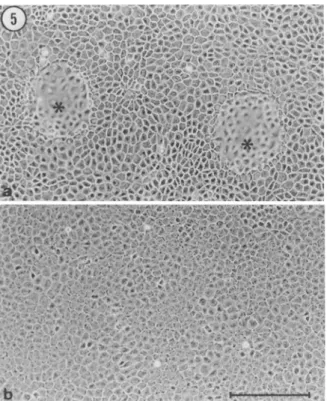
Dome formation in conventional monolayer culture. Like other po- larized epithelial cell lines (see, for example, Wbhlwend et al., 1986), parental E p H 4 cells generate fluid-filled blisters, or domes, when grown on an impermeable substratum. We wished to determine whether the differential ability of E p H 4 clones to form lumina in collagen gels would correlate with the ability to form domes on tissue culture plastic. In postconfluent cultures, H1B, KzA3, B e l a , and Be4a cells formed numerous domes (shown in Fig. 5 a for B e l a ceils). In contrast, domes were not observed in monolayers of either IzG 2 (not
MORPHOGENIC BEHAVIOR OF EpH4 CLONES 473
i
i • ~ ~ ~ • !$°,, )
Cg
Cg
Cg
Cg
~-P I ! d I IFIG. 4. Differential response of EpH4 subclones to collagen overlay. Cells were grown on a coltagen gel until a nearly confluent monolayer was obtained, and were subsequently covered with a second collagen gel, as described in Materials and Methods• (a) Four days following collagen overlay, JaB1 cells have formed numerous irregularly-shaped cavities delimited by refringent borders; (b) six days following collagen overlay, HIB cells have not formed cavities (notice that a number of elongated ceils have migrated from the sandwiched monolayer into the upper collagen gels); (c) cross-section of a culture similar to that shown in (a) demonstrating that
JzBI
cells have reorganized between the two collagen layers to form multiple lumina; (d) cross-section of the culture in (b) demonstrating the lack of lumen formation by H1B cells. (a,b): Bars = 200 ~m; (c,d): Bars = 100 p.m.shown) or JzB1 cells (Fig. 5 b), even when the cultures were main- tained for as long as 3 wk after confluence. These findings indicate that lumen formation in collagen gets and dome formation in con- ventional monolayer culture are unrelated phenomena.
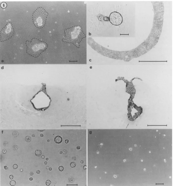
Behavior o f EpH4 clones on Matrigel. Culture of mammary epithe- lial ceils on Matrigel, a reconstituted laminin-rich matrix extracted from the EHS tumor (Kleinman et al., 1986), has been shown to result in the formation of well organized, alveolar-like "mammospheres" (Barcellos-Hoff et al., 1989; Petersen et al., 1993; Hurley et al., 1994). To establish whether EpH4 subpopulations would differ in their ability to form three-dimensional structures on this substrate, ceils from each clone were seeded onto a thick layer of Matrigel and incubated for 10-15 d in complete medium. Cells from all clones attached rapidly to the gel and initially had a homogeneous distri- bution on the substrate. During the next few hours of incubation, however, the cells progressively associated with each other, so that by 24 h they had clustered into compact muhicellular aggregates. The nature of the structures formed on Matrigel was dependent on plating density. When seeded onto the gel at a concentration of 1 X l0 S ceils per 16-ram well, the ceils organized into a network of anas- tomosing cords (not shown). In contrast, at plating densities of 2 X 10⁴ cells/well, cells from all clones clustered into discrete cell ag- gregates (Fig. 6 a). Similar density-dependent differences in asso- ciative behavior have previously been reported for other mamma~
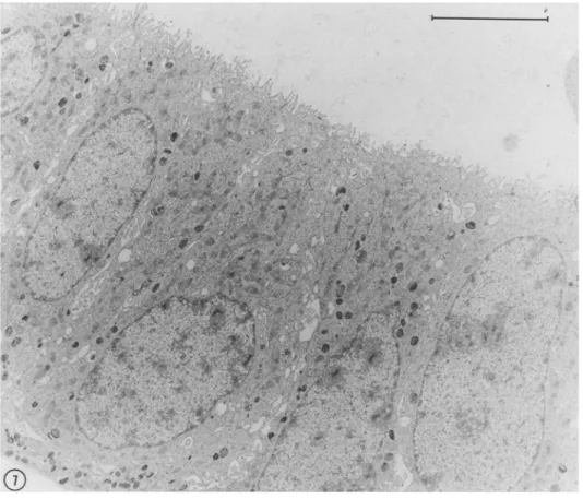
epithelial ceils grown on Matrigel (Stahl et al., 1997). Although the network of cords formed in high density cultures showed little mod- ification following extended incubation, the structures formed after low-density seeding underwent clearly identifiable changes. Cell ag- gregates induced a rearrangement of the surrounding matrix, which was recognizable as a dense array of radially disposed striations (Fig. 6 a) surrounding each aggregate. Matrigel remodeling has been shown in other systems to result from a process of cell traction on the substrate (Vernon et ah, 1992). Daily examination of the cultures after seeding showed that the aggregates gradually sank into the mal- leable matrix and became ensheathed by a thick layer of reorganized Matrigel. Concomitantly with the invasion of the underlying sub- strate, the aggregates underwent a process of cavitation which within 1-2 wk resulted in the formation of cystic structures (Fig. 6 b). The latter were often connected by a stalk to a cluster of cells remaining on the surface of the gel (Fig. 6 d,e). As previously described for primary mammary epithelial cells (Barcellos-Hoff et al., 1989), the lumen of the cysts was delimited by a layer of cubic or columnar epithelial cells (Fig. 6 c-e), which were provided with junctional complexes and numerous apical microvilli (Fig. 7). Cyst formation in Matrigel was observed with all EpH4 clones except H~B cells, which gave rise to solid ball-like structures that only occasionally contained small lumen-like spaces (not shown). Likewise, when
4 7 4 MONTESANO ET AL.
FIG. 5. EpH4 subclones differ in their ability to form domes. Cells were seeded into 35-ram dishes and grown to postcmdluence. (a) Bela cells main- tained in culture for 11 d have generated characteristic blisters or domes
(asterisks); (b) J3B1 cells maintained in culture for 13 d have not formed domes (the same culture was reexamined 10 d later and found to be totally devoid of domes). Bar = 200 p,m.
sandwiched between two Matrigel layers, all clones formed spherical cysts (shown in Fig. 6ffor IzG 2 cells), except H~B cells, which formed small lumen-less aggregates (Fig. 6 g). Similar results were obtained when ceils from the various clones were suspended into Matrigel (data not shown).
DISCUSSION
By cloning the EpH4 mammary epithelial cell line, we have iso- lated six cell subpopulations which display markedly different mor- phogenetic properties. The most distinctive features of each subclone are summarized below (see also Table 1).
When suspended in collagen gels, H~B cells form smooth-con- toured branched cords which superficially resemble mammary gland ducts. The cords, however, lack a central lumen, and the inability of HaB cells to form cavitary structures was confirmed in both the col- lagen gel sandwich and the Matrigel assay. Interestingly, H~B cells form domes when grown to postconfluence in tissue culture dishes, which indicates that despite their inability to form lumina in three- dimensional cultures, these cells have retained some characteristics of the polarized epithelial phenotype.
Unlike
HIB
cells, I3G ~ cells form lumen-containing cords in col- lagen gels. The extent of lumen development, however, is critically dependent on the type of serum added to the culture medium. Whereas in the presence of FCS the cords are mostly solid and only occasionally contain small focal lumina, in the presence of DCS they contain wide lumina and therefore have a tubular structure. Thesefindings indicate that when grown under appropriate conditions, I3G 2 cells have the capacity to form duct-like structures. The factors in DCS which stimulate lumen formation by I3G~ cells are not known, but their lack of effect on H~B cells suggests that they can only act on cells which are intrinsically competent to form lumina.
The most characteristic feature of J3B~ cells is their ability to form large cavitary structures when suspended in collagen gels. Unlike all other EpH4 clones, J~B~ cells form thin-walled branching tubes con- taining a widely patent continuous lumen when grown in FCS-sup- plemented medium. Lumen formation is even more prominent in the presence of DCS, under which condition J3BI cells generate large, irregularly-shaped cyst-like structures.
KzA 3 cells suspended in collagen gels in either FCS or DCS form branching cords containing numerous muhifocal spaces, which sometimes coalesce into larger lmnina.
Bela cells have the unique ability to form spheroidal cysts when grown in collagen gels in serum-free medium. In sharp contrast, un- der the same serum-free culture conditions, Be4a cells generate ex- tensively branched duct-like structures enclosing multifocal or con- tinuous lumina.
When the different clones are evaluated for their ability to mimic morphogenetic events occurring during mammary gland develop- ment, Be4a cells, which generate highly arborized tubules in collagen gels, appear to more accurately recapitulate the process of ductal morphogenesis. Other clones mimic this process only incompletely, due to their limited capacity to form lumina (e.g., K3Az cells) or to branch (e.g., I3G 2 and J3B1 cells). Finally, a selective deficiency in either lumen formation or branching, which are two essential com- ponents of ductal morphogenesis, is observed in clones H,B and Bela, respectively. Thus, while capai31e of undergoing repetitive branching, H1B cells are unable to form lumina. Conversely, when embedded in collagen gels in serum free-medium, Bela cells form hollow cysts, but lack the ability to branch and generate tube-like structures, in contrast to what is observed with Be4a cells grown under the same conditions.
The spectrum of cell subpopulations present in the parental EpH4 cell line is unlikely to be restricted to the six phenotypically distinct clones we have isolated. Indeed, besides the prototypic structures illustrated in Figure 1, collagen gel cultures of parental EpH4 ceils also contained branching structures consisting of short stubby cords with bulbous extremities, which were reminiscent of mammary gland "end buds." Despite repeated attempts, we have not succeeded in establishing EpH4 subclones capable of forming this type of colony in collagen gels. An additional variety of colony occasionally ob- served in cultures of parental EpH4 cells consisted of loose associ- ations of spindle-shaped cells. Since these aggregates lacked an ep- ithelial-like organization, we did not attempt to isolate them. Cloning this putative cell subpopulation remains nonetheless an interesting prospect, because spindle-shaped cells may have arisen through a process of epithelial-mesenchymal conversion.
It may be at first glance surprising that as many as six distinct sublines have been isolated from the EpH4 cell line, which was clonally derived (Fialka et al., 1996). It is conceivable, however, that variant subpopulations have arisen during propagation of the original cell line. Notably, in serum-free collagen gel cultures, onty Be4a cells exhibit morphogenetic properties similar to those of parental EpH4 cells, which suggests that this clone represents the direct descen- dants of the originally established EpH4 ceils. The other clones we have characterized are instead likely to have been generated by in-
MORPHOGENIC BEHAVIOR OF EpH4 CLONES 475
d
QFIG. 6. Morphogenetic behavior of EpH4 clones grown on Matrigel. (a) Three-dimensional aggregates formed by JaB1 ceils 48 h after having been plated on Matrigel at 2 X 10⁴ cells/16-mm well. The hatched lines delimit a zone of substrate remodeling which is recognizable as an array of radially disposed striations surrounding each cell aggregate. (b) Detail of the culture shown in (a) 11 d after cell seeding. An initially solid cell aggregate has invaded the underlying basement membrane gel and has concomitantly undergone a process of cavitation resulting in the formation of a cystic structure. The aggregate is surrounded by a layer of reorganized Matrigel. (c) Semithin section through a cyst-like structure formed by J3B~ cells grown on Matrigel for 10 d. The wall of the cyst is composed by an ordered palisade of cylindrical epithelial cells.
(d,e)
I3G 2 cells grown for 14 d on Matrigel have invaded the underlying matrix, into which they have formed eavitary structures. The cystic portion of the colony is connected through a solid stalk to a cluster of cells remaining on the surface of the gel. Notice that the colonies are ensheathed by a thick layer of reorganized Matrigel. ~ IaG2 cells were first allowed to attach on Matrigel and subsequently overlaid with a second Matrigel layer. Thirteen d later, the cells have formed numerous cystic structures between the two gel layers. (g) HIB cells sandwiched between two Matrigel layers as in (D have formed small aggregates devoid of a central lumen. Bars = 100gm, except for (e) where bar = 50pro.476
MONTESANO ET AL.FIG. 7. Thin section across the wall of a cyst formed by J3B~ cells grown on Matrigel. The cells have a cylindrical shape and project numerous microvilli towards the lumen of the cyst. Bar = 5 p.m.
~ili,
!
d e p e n d e n t mutations within an initially homogenous population. It is equally possible that parental E p H 4 cells are not fully differen- tiated and b e h a v e as precursor-like cells (stem cells?) capable of giving rise to phenotypically different subpopulations.
Lumen formation and ductal b r a n c h i n g are fundamental events in the morphogenesis of mammary gland and other epithelial tissues (Gumbiner, 1992). However, very little is known about the mecha- nisms which control these biological processes (Soriano et al., 1995; Yang et al., 1995; Soriano et al., 1996). The E p H 4 sublines we have isolated should provide a useful in vitro system for investigating the molecular basis of b r a n c h i n g morphogenesis and lumen formation, as well as the factors (e.g., oncoproteins) that disrupt these processes. Thus, a comparative analysis of the expression of potentially relevant proteins (e.g., integrins, extraeellular matrix components, proteases/ protease inhibitors) in different E p H 4 subelones may help to eluci- date the mechanisms responsible for aberrant morphogenesis, i.e., failure of b r a n c h i n g or lack of lumen formation. In addition, the likely common origin of the E p H 4 subpopulations described in this study may offer an opportunity for the identification of genes which are differentially expressed by clones endowed with distinct morphoge- netic properties.
ACKNOWLEDGMENTS
We are grateful to J. Rial-Robert for setting up the "droplet cloning" pro- cedure. We also thank M. Eissler for skillful technical assistance, J. P. Gerber for photographic work, F. Hellal for secretarial assistance, and Drs. H. Beug and M. Pepper for their comments on the manuscript. This study was sup- ported by a grant from the Swiss National Science Foundation (number 31- 43364.95).
REFERENCES
Barcellos-Hoff, M, H.; Aggeler, J.; Ram, T. G., et al. Functional differentiation and alveolar morphogenesis of primary mammary cultures on recon- stituted basement membrane. Development 105:223-235; 1989. Berdichevsky, E; Alford, D.; D'Souza, B., et al. Branching morphogenesis of
human mammary epithelial cells in collagen gels. J. Cell Sci. 107:3557-3568; 1994.
Chambard, M.; Gabrion, J.; Mauchamp, J. Influence of collagen gel on the orientation of epithelial cell polarity: follicle formation from isolated thyroid cells and from preformed monolayers. J. Cell Biol. 91:157-
166; 1981.
Daniel, C. ~\; Silberstein, G. B. Postnatal development of the rodent mam- mary gland. In: Neville, M. C.; Daniel, C. ~(, ed. The mammary gland. Development, regulation and function. New York: Plenum Press; 1987:3-36.
Danielson, K. G.; Oborn, C. J.; Durban, E. M., et al. Epithelial mouse mare- mar), cell line exhibiting normal morphogenesis in vivo and functional differentiation in vitro. Proc. Natl. Acad. Sci. USA 81:3756-3760; 1984.
Darcy, K. M.; Black, J. D.; Hahm, H. A., et al. Mammary organoids from immature virgin rats undergo ductal and alveolar morpbogenesis when grown within a reconstituted basement membrane. Exp. Cell Res. 196:49~5; 1991.
Fialka, I.; Schwartz, H.; Reichmann, E., et al. The estrogen-dependent c- JunER protein causes a reversible loss of mammary epithelial cell polarity involving a destabilization of adherens junctions. J. Cell Biol. 132:1115-1132; 1996.
Gumbiner, B. M. Epithelial morphogenesis. Cell 69:385-387; 1992. Hall, H. G.; Farson, D. A.; Bissell, M. J. Lumen formation by epithelial cell
lines in response to collagen overlay: a mo~phogenetic model in cul- ture. Proc. Natl. Acad. Sci. USA 79:4672M676; 1982.
Hurley, 9- L.; Blatchford, D. R.; Hendry, K. A. K., et al. Extracellular matrix and mouse mammary cell function: comparison of substrata in culture. In Vitro Cell. Dev. Biol. 30A:529-538; 1994.
MORPHOGENIC BEHAVIOR OF EpH4 CLONES 4 7 7 Kanazawa, T.; Hosik, H. L. Transformed growth phenotype of mouse mam-
mary epithelium in primal 3, euhure induced by specific fetal mes- enchymes. J. Cell. Physiol. 153:381-391; 1992.
Keely, E J.; Fong, A. M.; Zutter, M. M., et al. Alteration of collagen-dependent adhesion,
motility,
andmorphogenesis
by the expression of antisense % integrin mRNA in mammary cells. J. Cell Sci. 1 0 8 : 5 9 ~ 0 7 : 1995. Kleinman, H. K.; McGarvey, M. L.; Flassel, J. R., et aI. Basement membrane complexes with biological activity. Biochemistry 25:312-318; 1986. Ldpez-Barahona, M.; Fialka, I.; Gonzalez-Sancho, J. M., et al. Thyroid hor-mone regulates stromelysin expression, protease secretion and the morphogenetie potential of normal polarized mammary epithelial cells. EMBO J. 14:1145-1155; 1995.
Montesano, R.; Orci, L.; Vassalli, R In vitro rapid organization of endothelial cells into capillary-like networks is promoted by collagen matrices. J. Cell Biol. 97:1648-I652; 1983.
Montesano, R.; Schallel; G.; Orci, L. Induction of epithelial tubular morpho- genesis in vitro by fibroblast-derived soluble factors. Cell 66:697- 711; 1991.
Oft, M.; Peli, J.; Rudaz, C., et al. TGF-~I and Ha-Ras collaborate in mod- ulating the phenotype plasticity and mvasiveness of epithelial tumor cells. Genes Devel. 10:2462-2477; 1996.
Ormerod, E. J.; Rudland, R S. Mammary gland morphogenesis in vitro: for- mation of branched tubules in collagen gels by a cloned rat mammary cell line. Dev. Biol. 91:360375; 1982.
Petersen, O. W.; ROnnov-Jensen, L.; Howlett, A. R., et al. Interaction with basement membrane serves to rapidly distinguish growth and differ= entiation pattern of normal and malignant human breast epithelial cells. Proc. Natl. Acad. Sei. USA 89:9064-9068; 1993.
Reichmann, E.; Ball, R.; Groner, B., et ah New mamma~ epithelial and fibrobiastic cell clones in coeuhure form structures competent to dif- ferentiate functionally. J. Cell Biol. 108:1127-1138; 1989. Reichmann, E.; Schwartz, H.; Deiner, E. M., et al. Activation of an inducible
c-FosER fusion protein causes loss of epithelial polarity and triggers epithelial-fibroblastoid cell conversion. Cell 7 I:1103-1116; 1992.
Schwimmer, R.: Ojakian, G. K. The a213L integrin regulates collagen-mediated MDCK epithelial membrane remodeling and tubule formation. J. Cell Sci. 108:2487-2498; 1995.
Soriano, J. V.: Orci. L.; Montesano. R. TGF-[31 induces morphogenesis of branching cords by cloned mammary epithelial cells at subpieomolar concentrations. Biochem. Biophys. Res. Comnmn. 220:879485; 1996.
Soriano, J. K; Peppm, M. S.; Nakamura, T., et al. Hepatocyte growth factor stimulates extensive development of branching duct-like structures by cloned mammary, gland epithelial cells. J. Cell Sci. 108:413M,30; 1995.
Soule. H. D.; Malonev. T. M.; ~blman, S. R., et al. Isolation and character- ization of a spontaneously, immortalized human breast epithelial cell line, MCF-10. Cancer Res. 50:607.5~i086" 1990.
Stahl, S.: ~%itzman, S.; Jones. J. C. R. The role of laminin-5 and its receptor in mammary epithelial cell branching morphogenesis. J. Cell Sci. 110:52>63; 1997.
Vernon. R. B.: Angello, J.-C.: Iruela-Arispe, M. L., et al. Reorganization of basenmnt membrane matrices by cellular traction promotes the for- mation of cellular networks in vitro. Lab. Invest. 66:536-547; 1992. ~bhlwend, A.: Vassalli, J.-D.; Belin, D.. et ah LLC-PK~ cells: cloning of
phenotypically stable subpopalations. Am. J. Physiol. 250:C682- C687; 1986.
Yang, J.: Guzman, R.: Richards, J., et al. Primm?, culture of human mammary epithelial cells embedded in collagen gels. J. Natl. Cancer Inst. 65:337-343; 1980.
Yang, Y.: Spitzer. E.; Meyer, D., et al. Sequential requirement of bepatocyte growth factor and neuregulin in the morphogenesis and differentiation of the mammary gland. J. Cell Biol. 131:215-226; 1995.
Zuk, A.: Matlin. K. S. Apical 13~ integrin in polarized MDCK cells mediates tubulocyst formation in response to type I collagen overlay. J. Cell Sci. 109:1875-1889: 1996.