Key words: bacteria, copper, Enterococcus, homeostasis, metallochaperone, proteolysis
Abstract
The cop operon is a key element of copper homeostasis in Enterococcus hirae. It encodes two copper ATPases, CopA and CopB, the CopY repressor, and the CopZ metallochaperone. The cop operon is induced by copper, which allows uncompromised growth in up to 5 mM ambient copper. Copper uptake appears to be accomplished by the CopA ATPase, a member of the heavy metal CPx-type ATPases and closely related to the human Menkes and Wilson ATPases. The related CopB ATPase extrudes copper when it reaches toxic levels. Intracellular copper routing is accomplished by the CopZ copper chaperone. Using surface plasmon resonance analysis, it was demon-strated that CopZ interacts with the CopA ATPase where it probably becomes copper loaded. CopZ in turn can donate copper to the copper responsive repressor CopY, thereby releasing it from DNA. In high copper, CopZ is proteolyzed. Cell extracts were found to contain a copper activated proteolytic activity that degrades CopZ in vitro. This post-translational control of CopZ expression presumably serves to avoid the accumulation of detrimental Cu-CopZ levels.
Copper circulation in Enterococcus hirae
Copper is a cofactor in many redox reactions through its ability to cycle between oxidized Cu(II) and re-duced Cu(I). Among the over 30 enzymes using copper as a cofactor are Cu/Zn superoxide dismu-tase, cytochrome c oxidase, lysyl oxidase, tyrosinase, coagulation factors Va and VIII, and dopamine β-hydroxylase, to name just a few. Yet, copper can be very toxic to cells through its ability to form free radicals and cells must protect themselves from such copper-induced damage by tightly controlling the form, complex type, and concentration of cytoplasmic copper. This is accomplished on one hand by keeping intracellular copper complexed at all times. In yeast, it has been estimated that there is less than one free cop-per ion cop-per cell in the cytoplasm (Rae et al. 1999). On the other hand, cellular copper levels are tightly con-trolled by copper homeostatic machinery that presents itself in ever increasing complexity. Many proteins
involved in copper homeostasis have thus far been identified in prokaryotes and eukaryotes (for review see Camakaris et al. 1999; Harrison et al. 2000; Horn & Tümer 1999; Mercer 2001; Pena et al. 1999).
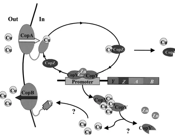
In the Gram-positive bacterium Enterococcus hi-rae, copper homeostasis is understood in some detail and the current model of copper circulation is summa-rized in Figure 1. The organism possesses two copper ATPases, CopA and CopB, that are localized in the cy-toplasmic membrane. Previous studies had indicated that CopA is responsible for copper uptake under cop-per limiting conditions and CopB for copcop-per export if copper reaches toxic levels (Odermatt et al. 1992, 1993). While the function of CopB in copper excretion
had been shown by direct demonstration of64Cu+as
well as m110Ag+ transport , the evidence for CopA
being involved in copper uptake is still indirect. It rests on the following three properties of a copA knock-out strain: (i) it grows like wild-type under normal or elevated copper conditions, (ii) it cannot grow in
Fig. 1. Copper circulation in E. hirae. Model of copper circulation in E. hirae. The import and export ATPase pumps, CopA and CopB, regulate
the intracellular concentration of copper by pumping metal across the membrane into and out of the cytoplasm. CopZ transfers copper from CopA to the repressor CopY. In CopY, one zinc is replaces by two copper(I) ions, which releases it from the DNA and induces expression of the cop operon. Cu-CopY may be degraded, as is excess Cu-CopZ under high copper conditions.
copper-depleted media, and (iii) it is more silver resis-tant than the wild-type, presumably because CopA can be a route for silver entry into the cell (Odermatt et al. 1993).
Intracellular copper routing: the copper chaperones
The fate of copper that has entered the cell remains unclear in several regards. However, a major func-tion in intracellular copper routing is taken by the copper chaperones, the specialized proteins which de-liver copper intracellularly to copper utilizing enzymes (Harrison et al. 2000; O’Halloran & Culotta 2000). In Enterococcus hirae, the 69 amino acid protein CopZ has been shown to function as a chaperone and to specifically deliver copper to the CopY repressor. In its zinc form, the CopY repressor binds to the cop promoter and represses transcription of the four cop
genes, copY, copZ, copA, and copB (cf. Figure 1). When CopZ donates copper to CopY, copper displaces its bound zinc and the repressor dissociates from the promoter, allowing expression of the downstream genes (Cobine et al. 1999).
The solution structure of CopZ has been solved by NMR. It exhibits a βαββαβ global structure: two α-helices laying on a 4-stranded, antiparallel β-sheet, a structure colloquially called an ‘open face sandwich’. A key element of the structure is a cysteine-x-x-cysteine motif, located between the firs β-sheet and the first α-helix. It binds copper(I) in a novel, sol-vent exposed binding site. The same fold identified for CopZ has also been determined for the related eukary-otic copper chaperones, Atx1 from yeast, and Atox1 (formerly HAH1) from humans (Rosenzweig et al. 1999; Rosenzweig 2001). CopZ-like structural ele-ments are a widespread feature of proteins involved in metal ion homeostasis and essentially identical
struc-Chaperone-target interactions
The function initially demonstrated for CopZ is the transfer of copper from Cu-CopZ to the CopY repres-sor for regulation (Cobine et al. 1999). In contrast, the eukaryotic chaperones Atx1 from yeast and Atox1 from humans have been demonstrated to deliver cop-per to copcop-per ATPases located in the trans-Golgi network (Ccc2 in yeast, Menkes/Wilson ATPase in humans; Larin et al. 1999; Pufahl et al. 1997)). These trans-Golgi copper ATPases in turn pump cop-per into the Golgi network where it is required for the biosynthesis of cuproenzymes, such as the Fet3p iron reductase in yeast or ceruloplasmin in mammalian cells (Dancis et al. 1994; Yuan et al. 1995).
How chaperones recognize their targets and how they interact with them at the molecular level has been studied by site directed mutagenesis and struc-tural investigations (Huffman & O’Halloran 2001). The surface residues presented by each partner provide complementarity of the interfaces. Multiple lysine residues clustered on the surfaces of Atox1, Atx1 and CopZ appear to play key roles in proteprotein in-teractions, albeit the location of these lysine patches is different in CopZ and Atx1/Atox1 (Figure 2; Wim-mer et al. 1999). In CopZ, the lysine residues 30, 31, 37, and 38 appear to be important for the interaction with the CopY repressor. MNKr2, the second CopZ-like copper binding domain of the human Menkes copper ATPase, cannot donate copper to CopY in spite of its similar predicted structure. However, if four extra lysine residues are introduced into MNKr2 at positions corresponding to the lysine residues in CopZ (cf. Figure 2), the resultant mutant molecule MNKr2K4 becomes competent in donating copper to CopY. This ‘gain-of-function’ mutation of MNKr2 de-lineates these four lysine residues as key features in the interaction of the CopZ chaperone with the CopY
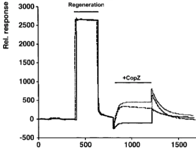
when analyte binds to the immobilized ligand causes a change in refractive index at the sensor chip sur-face and can be measured as a change in the intensity of light reflected from the chip surface. We found that CopZ specifically binds to CopA and that copper modulates this binding. The association of CopZ with CopA and the dissociation of CopZ from CopA in the absence and presence of copper(I) are shown in Fig-ure 3. Maximal binding of CopZ to CopA is observed in the presence of sub-stoichiometric amounts of cop-per(I) (10 µM copcop-per(I) corresponding to a molar sto-ichiometry of 0.9 copper(I):1CopZ; copper was added as the stable copper(I)acetonitrile complex; Hem-merich & Sigwart 1963). In the presence of 100 µM copper(I), the steady-state association of CopZ with CopA was reduced to approximately 25%. In the ab-sence of supplemented copper(I), a transient binding maximum is observed due to incomplete copper de-pletion through contaminating copper in the running
buffer. The association rate ka is 2.4× 10³ M−1s−1
without added copper and is only slightly affected
cop-per. The dissociation rate kd is 14× 10−3s−1 and is
strongly influenced by added copper with a decrease of up to 15-fold. This results in a 7 and 16-fold
in-crease of the affinity constant KD in the presence of
100 and 10 µM copper(I), respectively. It was also shown that mutating the cysteine-x-x-cysteine copper binding motif in the N-terminus of CopA to serine-x-x-serine abolishes the copper induced decrease in the dissociation rate, without significantly affecting the association rate (Multhaup et al. 2001). Thus, the CopZ chaperone can interact with the CopA ATPase in a structure and copper dependent manner. Which residues of CopZ and CopA are involved in this in-teraction remains to be shown. However, the CxxC motif is not the guiding element for this interaction. Figure 4 shows a model of a possible mechanism for CopZ-CopA interaction and metal transfer.
Fi g . 2 . Critical lys ine res idues o f C opZ , A tx1, and M NKr2K4. T h e lys ine res idues critical for C opZ function are box ed and are on the upper side o f the CopZ fold sho wn. T hes e res idues are not pres ent in A tx1 o r M NKr2. T h e res idues critical for A tx1 chaperone function are enlar g ed and underlined, as are the corres ponding lys ine res idues o f u nm odified M NKr2. In the repres entations of the A tx1 and MNKr2K4 folds , thes e lys ines are located in the lo w er half. T he four lys ine res idues that h av e b een introduced into MNKr2 to obtain the ga in-of-function m utant m olecule MNKr2K4 are sh o w n b elo w the sequence alignm ent. T h e lys ine m utations introduced into MNKr2K4 are Q30K, R 31K, D 37K, and N38K. T he st ructure o f M NKr2K4 w ith the four ex tr a lys ine res idues w as m odeled.
Fig. 3. Association-dissociation of CopZ and wild-type CopA.
CopA-CopZ interaction was measured by surface plasmon reso-nance analysis with the Biacore apparatus. Following regeneration of a CopA loaded chip with high salt and a reducing agent, CopZ at a concentration of 100 µg/ml was interacted in the presence of 0 (thick trace), 10 (top trace), and 100 µM (bottom trace) cop-per(I)acetonitrile, respectively. CopZ injection was started at 800 s and continued until 1200 s, followed by injection of protein-free buffer.
Fig. 4. Model of CopA-CopZ interaction and copper transfer. The
CopZ chaperone docks on the CopA ATPase by protein-protein interaction involving positively charged surface lysine residues on CopZ and negatively charged amino acids on CopA. Copper is then transferred from CopA to CopZ by sequential ligand transfer. Fi-nally, CopZ dissociates from CopA. This step could be facilitated by conformational changes of CopA associated with the pumping cycle.
Proteolytic degradation of CopZ
Proteolysis provides cells with an additional means to modulate protein availability in response to alterations in cellular physiology or external stress. However, these processes have received considerably less atten-tion than transcripatten-tional or translaatten-tional regulaatten-tion and our understanding of them is only fragmentary (see Gottesman & Maurizi 1992; Gottesman 1996 for re-view). In Saccharomyces cerevisiae copper induced
per conditions was observed. CopZ is encoded by a polycistronic message also encoding CopA, CopB, and CopY and these four genes are thus co-induced by copper. Levels of mRNA increase with increasing am-bient copper levels and reached a 1000-fold induction at 0.25 mM copper, as assessed by real-time quantita-tive PCR. However, CopZ expression increases only up to 0.5 mM copper and declines at higher cop-per concentrations, to become nearly undetectable at 3 mM copper (Lu & Solioz 2001). It was concluded that CopZ overexpression is toxic to cells, based on the following observations: (i) growth of a strain overex-pressing CopZ from a plasmid is inhibited by ambient copper in excess of 0.1 mM and ceases to grow in 1.5 mM copper, while growth of wild-type E. hirae is not markedly affected by this copper concentra-tion, and (ii) CopZ overexpression makes E. hirae more sensitive to oxidative stress, evident by increased
sensitivity to H2O2and paraquat.
Proteolysis of CopZ could also be demonstrated in vitro. When cytosolic extracts are mixed with purified CopZ, it is rapidly degraded (Lu & Solioz 2001). In-terestingly, apo-CopZ is significantly more resistant to degradation than Cu-CopZ. The more rapid degrada-tion of Cu-CopZ is in line with its proposed toxicity. Copper bound to CopZ is solvent exposed (Cobine et al. 1999) and this copper can most likely participate in Fenton-type reactions, leading to the generation of reactive hydroxyl radicals and cell damage.
The protease responsible for CopZ degradation is not induced by exposure of cells to copper and is also present in cells in which protein synthe-sis has been inhibited for one hour with chlo-ramphenicol. Rather, the proteolytic activity ap-pears to be constitutive, but stimulated by cop-per(I) or Ag(I), a potential Cu(I) mimetic. The serine protease inhibitors p-phenylmethylsulfonyl fluoride (PMSF) and p-aminobenzamidine inhibit
the degradation of CopZ, while N-α-p-tosyl-L-lysine chloromethyl ketone (TLCK) and N-tosyl-L-phenylalanine chloromethyl ketone (TPCK), which are also serine protease inhibitors, are without effect. The metallo-proteinase inhibitor o-phenanthroline does also not inhibit CopZ degradation. It was thus concluded that the protease degrading CopZ is a serine type protease (Lu & Solioz 2001). On zymograms, the CopZ degrading activity was tentatively identified as a protein of 58 kDa. This protein displays the expected properties, namely activation by copper and inhibition by p-aminobenzamidine.
Taken together, these results show the specific degradation of the CopZ copper chaperone under high copper conditions. This step adds another level of con-trol to the copper homeostatic system of E. hirae (cf. Figure 1).
Conclusions
The cop operon of E. hirae, encoding the four genes CopY, CopZ, CopA and CopB, is regulated at the tran-scriptional level by the copper responsive repressor, CopY. It is released from the promoter by increased copper levels, thereby allowing transcription to pro-ceed. For this process, Cu-CopZ donates copper to the CopY repressor, which leads to the loss of the zinc(II) from CopY and its ability to bind to DNA. In addition to this transcriptional control, the intra-cellular concentration of CopZ is also regulated at the post-translational level by proteolytic degradation of CopZ under high copper conditions. The latter implies that CopZ is dispensable under high copper condi-tions. Conceivably, excess Cu-CopZ becomes toxic to cells as such or interferes with another detoxification process that sets in under high copper conditions. A corollary to CopZ degradation is that CopZ is dispens-able for the secretion of excess copper, which proceeds via the CopB copper ATPase. Conceivably, copper complexed to glutathione or other biomolecules is the substrate for CopB. The proteolytic degradation of CopZ is a novel mechanism in copper homeosta-sis. Given the high evolutionary conservation of some components of copper homeostatic systems, similar mechanism are likely to operate in other cell types as well. The interaction of CopZ with the putative copper import ATPase CopA, revealed by surface plas-mon resonance analysis suggests that CopZ picks up copper from CopA under copper limiting conditions. Surface plasmon resonance analysis is a novel,
power-ful tool for the analysis of protein-protein interactions in copper homeostasis.
Acknowledgements
Part of the work described here was supported by grant 32-56716.99 from the Swiss National Foundation, and grants from the International Copper Association and the Australian Research Council.
References
Camakaris J, Voskoboinik I, Mercer JF. 1999 Molecular mecha-nisms of copper homeostasis. Biochem Biophys Res Commun 261, 225–232.
Cobine P, Wickramasinghe WA, Harrison MD et al. 1999 The
Ente-rococcus hirae copper chaperone CopZ delivers copper(I) to the
CopY repressor. FEBS Lett 445, 27–30.
Dancis A, Yuan DS, Haile D et al. 1994 Molecular characterization of a copper transport protein in S. cerevisiae – an unexpected role for copper in iron transport. Cell 76, 393–402.
Gottesman S. 1996 Proteases and their targets in Escherichia coli.
Annu Rev Genet 30, 465–506.
Gottesman S, Maurizi MR. 1992 Regulation by proteolysis: energy-dependent proteases and their targets. Microbiol Rev 56, 592– 621.
Harrison MD, Jones CE, Solioz M, Dameron CT. 2000 Intracellular copper routing: The role of copper chaperones. Trends Biochem
Sci 25, 29–32.
Hemmerich P, Sigwart C. 1963 Cu(CH3CN)+2, ein Mittel zum
Studium homogener Reaktionen des einwertigen Kupfers in wässriger Lösung. Experientia 19, 488–489.
Horn N, Tümer Z. 1999 Molecular genetics of intracellular copper transport. J Trace Elem Exp Med 12, 297–313.
Huffman DL, O’Halloran TV. 2001 Function, structure, and mech-anism of intracellular copper trafficking proteins. Annu Rev
Biochem 70, 677–701; 677–701.
Lamb AL, Torres AS, O’Halloran TV, Rosenzweig AC. 2001 Het-erodimeric structure of superoxide dismutase in complex with its metallochaperone. Nat Struct Biol 8, 751–755.
Larin D, Mekios C, Das K et al. 1999 Characterization of the in-teraction between the Wilson and Menkes disease proteins and the cytoplasmic copper chaperone, HAH1p. J Biol Chem 274, 28497–28504.
Li HH, Merchant S. 1995 Degradation of plastocyanin in copper-deficient Chlamydomonas reinhardtii. Evidence for a protease-susceptible conformation of the apoprotein and regulated prote-olysis. J Biol Chem 270, 23504–23510.
Lu ZH, Solioz M. 2001 Copper Induced Proteolysis of the CopZ Copper Chaperone of Enterococcus hirae. J Biol Chem (in press). Mercer JF. 2001 The molecular basis of copper-transport diseases.
Trends Mol Med 7, 64–69.
Multhaup G, Strausak D, Bissig K-D, Solioz M. 2001 Interaction of the CopZ copper chaperone with the CopA copper ATPase of
Enterococcus hirae. Biochem Biophys Res Commun 288, 172–
177.
Odermatt A, Suter H, Krapf R, Solioz M. 1992 An ATPase operon involved in copper resistance by Enterococcus hirae. Ann NY
1999 Undetectable Intracellular Free Copper: the Requirement of a Copper Chaperone for Superoxide Dismutase. Science 284, 805–808.