COMPUTED TOMOGRAPHY
Computed tomography of the chest with model-based
iterative reconstruction using a radiation exposure similar
to chest X-ray examination: preliminary observations
Angeliki Neroladaki&Diomidis Botsikas&
Sana Boudabbous&Christoph D. Becker&Xavier Montet
Received: 5 June 2012 / Revised: 18 July 2012 / Accepted: 18 July 2012 / Published online: 15 August 2012 # European Society of Radiology 2012
Abstract
Objectives The purpose of this study was to assess the diagnostic image quality of ultra-low-dose chest computed tomography (ULD-CT) obtained with a radiation dose com-parable to chest radiography and reconstructed with filtered back projection (FBP), adaptive statistical iterative recon-struction (ASIR) and model-based iterative reconrecon-struction (MBIR) in comparison with standard dose diagnostic CT (SDD-CT) or low-dose diagnostic CT (LDD-CT) recon-structed with FBP alone.
Methods Unenhanced chest CT images of 42 patients ac-quired with ULD-CT were compared with images obtained with SDD-CT or LDD-CT in the same examination. Noise measurements and image quality, based on conspicuity of chest lesions on all CT data sets were assessed on a five-point scale.
Results The radiation dose of ULD-CT was 0.16 ± 0.006 mSv compared with 11.2±2.7 mSv for SDD-CT (P <0.0001) and 2.7±0.9 mSv for LDD-CT. Image quality of ULD-CT increased significantly when using MBIR com-pared with FBP or ASIR (P<0.001). ULD-CT reconstructed with MBIR enabled to detect as many non-calcified pulmo-nary nodules as seen on SDD-CT or LDD-CT. However, image quality of ULD-CT was clearly inferior for character-isation of ground glass opacities or emphysema.
Conclusion Model-based iterative reconstruction allows de-tection of pulmonary nodules with ULD-CT with radiation exposure in the range of a posterior to anterior (PA) and lateral chest X-ray.
Key Points
• Radiation dose is a key concern with the increased use of thoracic CT
• Ultra-low-dose chest CT approximates the radiation dose of conventional chest radiography
• Ultra-low-dose chest CT can be of diagnostic quality • Solid pulmonary nodules are clearly depicted on ultra-low-dose chest CT
Keywords Computed tomography . Chest . Low dose . Pulmonary nodules . Model-based iterative reconstruction (MBIR)
Abbreviations
ULD-CT Ultra-low-dose chest CT SDD-CT Standard-dose diagnostic CT LDD-CT Low-dose diagnostic CT FBP Filtered back projection
ASIR Adaptive statistical iterative reconstruction MBIR Model-based iterative reconstruction DLP Dose-length product
CXR Chest X-ray
Introduction
Computed tomography (CT) is one of the key investigative techniques in diagnostic imaging, and is particularly useful in chest disease. The diagnostic benefit of CT is, however, associated with the inherent risk of ionizing radiation, and its widespread use places CT among the main sources of ionizing radiation in the general population of industrialised countries [1]. On a statistical level, there are concerns that the widespread use of CT might increase the risk of radiation-induced cancer [2].
A. Neroladaki
:
D. Botsikas:
S. Boudabbous:
C. D. Becker:
X. Montet (*)Department of Radiology, Geneva University Hospital, Rue Gabrielle-Perret-Gentil 4,
1211 Geneva 4, Switzerland e-mail: [email protected]
Major efforts are therefore being made by vendors of CT equipment, radiologists and technicians to minimise the radiation dose of CT examination protocols while maintain-ing adequate diagnostic image quality. Among the different strategies for radiation-dose reduction, the optimisation of CT hardware like X-ray tube and detectors, the use of X-ray dose modulation [3] and the choice of the most adequate CT examination protocol obviously play a key role. Major progress has also recently been made with regard to the algorithms used for reconstruction of CT images. The tradi-tional method, called filtered back projection (FBP) may now be completed with iterative reconstruction algorithms that intend to decrease the radiation dose while maintaining image quality. The first generation of iterative reconstruc-tion tools, such as adaptive statistical image reconstrucreconstruc-tion (ASIR) or iterative reconstruction in image space (IRIS) achieve this mainly by decreasing the noise in the recon-structed CT images [4–9]. More recent developments, such as model based iterative reconstruction (MBIR), include an algorithm that accurately models the entire optical chain (real size of focal spot and detectors) and takes into account the noise of the system (photons statistics and electronic noise). As CT data sets reconstructed with an MBIR algo-rithm have a very low level of noise [10], MBIR carries the potential for even more drastic reduction in dose for obtain-ing images of diagnostic quality.
The purpose of this study was to investigate whether the use of the MBIR algorithm combined with the latest CT hardware technology allows CT images of the chest of diagnostic quality to be obtained while lowering the radia-tion dose close to the level of a posterior to anterior (PA) and lateral chest X-ray (CXR).
Materials and methods
Patients
The study was approved by the ethics committee of our hospital. The study included 42 consecutive patients who underwent non-enhanced chest CT between June and Au-gust 2011. They were 24 men, aged between 19 and 80 years old, with an average body mass index (BMI) of 25.8 ± 3.9, and 18 women aged between 23 and 70 years old, with an average BMI of 22.8 ± 4.5. CT indications were: follow-up or suspected nodules (n032), COPD evaluation (n06), lung transplantation control (n02) and suspicion of pneumotho-rax (n02).
After a first diagnostic CT (either standard-dose diagnos-tic [SDD] CT or low-dose diagnosdiagnos-tic [LDD] CT), a subse-quent acquisition was realised at ultra-low-dose (ULD) CT during the same examination. Twenty patients underwent SDD-CT, whereas 22 patients underwent LDD-CT. As
LDD-CT equals SDD-CT for lesion detection, they were handled together under the name of diagnostic CT [11,12].
CT protocols and image reconstruction
All chest CT examinations were acquired using 64-slice multi-detector CT (GE Discovery 750 HD; GE Healthcare, Milwaukee, USA). For SDD-CT acquisition, the usual clin-ical chest CT protocol was used in helclin-ical mode: 0.6-s gantry rotation time, 120 kVp, 0.984:1 beam pitch, 40-mm table feed per gantry rotation, a z-axis tube current modula-tion was used, with a noise index of 28 (min/max mA, 100/ 500) and a 64×0.625-mm detector configuration.
For LDD-CT acquisition, the parameters were maintained except that a noise index of 26 (min/max mA, 80/100) was used. Z-axis tube current modulation was still used.
For ultra-low-dose acquisition, all parameters remained unchanged, except that the kVp was lowered to 100 kVp and the tube current was lowered to 10 mA without dose modulation.
SDD-CT and LDD-CT images were reconstructed at 0.625-mm thickness with filtered back projection, whereas ultra-low-dose images were reconstructed at 0.625-mm thickness with filtered back projection, with 40% and 80% adaptive statistical iterative reconstruction (ASIR-40 and ASIR-80, respectively) and with model based iterative re-construction (MBIR, Veo™ GE Healthcare, Milwaukee, USA).
Assessment of image quality
Objective image noise was calculated using a lung window setting. Regions of interest were drawn inside the trachea immediately over the carina, while carefully avoiding the trachea wall. The mean HU as well as its standard deviation, considered to be noise, were recorded.
Subjective assessment of image quality of all data sets was realised by three radiologists (X.M. with 9 years of experience, D.B. with 7 years of experience, S.B. with 7 years of experience). Images were displayed with a lung window setting (window level, -500; window width, 1,400) and a soft tissue window setting (window level, 40; window width, 350). The images were anonymised, aggregated in folders in a random way and then evaluated by the three readers for their quality.
The radiologists evaluated the normal lung structures (major fissures and small vessels) and bronchi (< 2 mm diameter) based on a five-point scale defined as follows: 1 point: Anatomical structures visible at 100% (excellent) 2 points: Anatomical structures visible at > 75% (good) 3 points: Anatomical structures visible between 25% and
4 points: Anatomical structures visible < 25% (poor) 5 points: Landmarks were not visible at all (unacceptable)
Diagnostic findings
Radiologists were also asked to record any abnormal struc-tures seen in each pulmonary lobe. Abnormal strucstruc-tures were defined following the glossary of terms for thoracic imaging from the Fleischner Society [13]. The number of micro-nodules (≤ 3 mm in size) was recorded. If the number of micro-nodules was fewer than 5, their number was noted, if the number of micro-nodules was more than 5, > 5 (with-out exact number) was noted. The size of any nodules (> 3 mm) was noted. The presence of ground glass opacity, consolidation, emphysema or bronchiectasis was also noted. Finally, the presence of pleural and pericardial effusion, as well as the presence of mediastinal adenopathy, was noted. Abnormal structures were described on a five-point scale: 1 point: Abnormal structures clearly visible with good
demarcation
2 points: Structures visible with blurring but without re-striction for diagnosis
3 points: Abnormal structures visible, with blurring and uncertainties about the evaluation
4 points: Abnormal structures barely visible with unreli-able interpretation
5 points: Abnormal structures not seen
Radiation dose assessment
To assess the radiation dose associated with the chest CT, the total dose-length product (DLP) was recorded. The effective dose was retrospectively calculated by multiplying the DLP by a factor of 0.020 [14].
Statistical analysis
Statistical analysis was performed with Prism (Prism, ver-sion 5d, 2010; GraphPad Software, San Diego, CA, USA) and Statistica (version 8; Statsoft, Tulsa, OK, USA).
Inter-observer agreement was performed with R using the kappa fleiss function (R package version 0.83, using the irr li-brary). For ordinal values, the results are presented as me-dian. For continuous values, the results are presented as mean ± standard error.
Non-normally distributed data sets (established from Kolmogorov-Smirnov tests) were compared using Friedman test with Dunn post-hoc test. Normally distributed data sets were compared using ANOVA test with Bonferroni post-hoc test. Two-sided testing was used. Differences were considered significant at P<0.05.
Results
Technical parameters
All ULD CTs were reconstructed with FBP, ASIR and MBIR without technical problems. Whereas the FBP and ASIR reconstructions only took a few seconds, about 1-h reconstruction time was needed for t1-he MBIR algorit1-hm.
The dose-length product (DLP) associated with SDD-CT and LDD-CT (diagnostic CT) and ULD CT was 560 ± 138, 133 ± 43 and 8 ± 0.3 mGy·cm, respectively (P<0.0001). Estimated effective doses were 11.2 ± 2.7, 2.7 ± 0.9 and 0.16 ± 0.006 mSv for the SDD-CT, LDD-CT and ULD-CT, respectively.
Image quality
The noise of the SDD-CT and LDD-CT were 24 ± 9 and 28 ± 9 HU, respectively. The noise of the ULD-CT was 107 ± 7, 94 ± 5, 77 ± 4 and 23 ± 4 for FBP, ASIR-40, ASIR-80 and MBIR, respectively. The noise of the ULD-CT was statisti-cally higher than the noise on the diagnostic CT using the FBP, and ASIR algorithms (P<0.0001), whereas it was not different when using the MBIR algorithm (P00.76). The BMI did not influence the image quality/noise of the image. The subjective image quality assessment for the three readers is summarised in Tables1 and2. The normal lung structures on diagnostic CT (either SDD-CT or LDD-CT)
Table 1 Conspicuity of all the fissures
ULD ultra-low dose, FBP fil-tered back projection, ASIR adaptive statistical iterative re-construction, MBIR model-based iterative reconstruction, n.s. not significant Median Fissure R1 R2 R3 P Diagnostic CT (SDD/LDD) 1 1 1 n.s. ULD-CT FBP 3* 3* 3* n.s. ASIR-40 3* 3* 3* n.s. ASIR-80 3* 3* 2* n.s. MBIR 2* 2* 2* n.s. Diagnostic CT vs ULD-CT *P < 0.001 *P < 0.001 *P < 0.001
were of high quality with a median value of 1. When acquired with the ultra-low-dose techniques, the quality of the chest CT increased from FBP to ASIR to MBIR. This increased quality was statistically significant for the three readers on each reconstruction algorithms (P<0.001). In-creased image quality is demonstrated in Fig.1.
Diagnostic findings
The number of solid nodules (> 3 mm) seen on each data set is summarised in Table3. There was no statistical difference between the numbers of nodules seen by the readers, nor between the different reconstruction algorithms. Based on the diagnostic CT, 28 nodules of 5.8 ± 3.2 mm were
depicted by reader one, 29 nodules of 5.9 ± 2.6 mm were depicted by reader 2 and 29 nodules of 5.7 ± 3.1 mm by reader 3. The inter-observer agreement was almost perfect for the diagnostic CT and for the MBIR algorithm (kappa value of 0.815 for both), whereas it was substantial for FBP and ASIR-80 and moderate for ASIR-40. An example of nodules seen on diagnostic CT and on ULD-CT is presented in Fig.2. The sensitivity of nodule detection on ULD-CT compared with SDD-CT was 100% for all readers.
The number of micro-nodules (≤ 3 mm) seen on each reconstruction algorithm is summarised in Table 4. The number of micro-nodules seen by each reader on each reconstruction algorithm was not statistically different. When acquired with the ULD techniques, the number of
Table 2 Conspicuity of the small peripheral artery and bronchi Median Anatomy R1 R2 R3 P Diagnostic CT (SDD/LDD) 1 1 1 ns ULD-CT FBP 4* 3* 3* <0.01 ASIR-40 3* 3* 3* n.s. ASIR-80 2* 2* 2* n.s. MBIR 2* 2* 2* n.s. Diagnostic CT vs ULD-CT *P < 0.001 *P < 0.001 *P < 0.001
Fig. 1 Image quality. Coronal reformatted CT demonstrating the im-age quality of a diagnostic CT (a). The same patient is presented after an ultra-low-dose acquisition, reconstructed with a MBIR (b), FBP (c), ASIR-40 (d) and ASIR-80 (e) algorithms. There was a clear reduction
in noise in the image when iterative reconstructions were used, as well as a clear increase in image quality. The MBIR algorithm achieved the highest subjective image quality among the algorithms used to recon-struct ULD CT (see Tables1and2)
micro-nodules seen increased from FBP to ASIR to MBIR, without reaching a statistical difference. The only statistical difference was noted for the FBP techniques for reader 1, which depicted fewer micro-nodules than all other recon-struction techniques. Nevertheless, the trend to depict more micro-nodules when adding some iterative reconstruction was clear. The sensitivity of ULD-CT for the detection of micro-nodules (compared with SDD-CT) was 107%, 111% and 88% for reader 1,2 and 3, respectively.
Bronchiectasis and architectural distortion were de-scribed with a perfect agreement between the readers on the diagnostic CT as well as the ULD-CT, when recon-structed with MBIR.
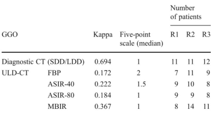
The number of patients having ground-glass opacities is summarised in Table 5. GGO was described on 11 (readers 1 and 2) and 12 (reader3) patients on the diagnostic CT corresponding to a substantial agreement. The inter-observer agreement was slight for the FBP and ASIR-40 data set and was fair for the ASIR-80 and MBIR data set.
The number of patients having emphysema seen on each data set is summarised in Table 6. Emphysema was diag-nosed in nine patients by all readers on the diagnostic CT (perfect agreement). There was almost no agreement be-tween the readers on all the ULD data sets (kappa values between -0.317 and 0.06).
Table 3 Number of nodules
Number of nodules Nodules R1 R2 R3 Kappa Diagnostic CT (SDD/LDD) 28 29 29 0.815 ULD-CT FBP 25 24 19 0.684 ASIR-40 27 28 18 0.578 ASIR-80 28 29 20 0.654 MBIR 28 29 29 0.815 Diagnostic CT vs ULD-CT n.s. n.s. n.s.
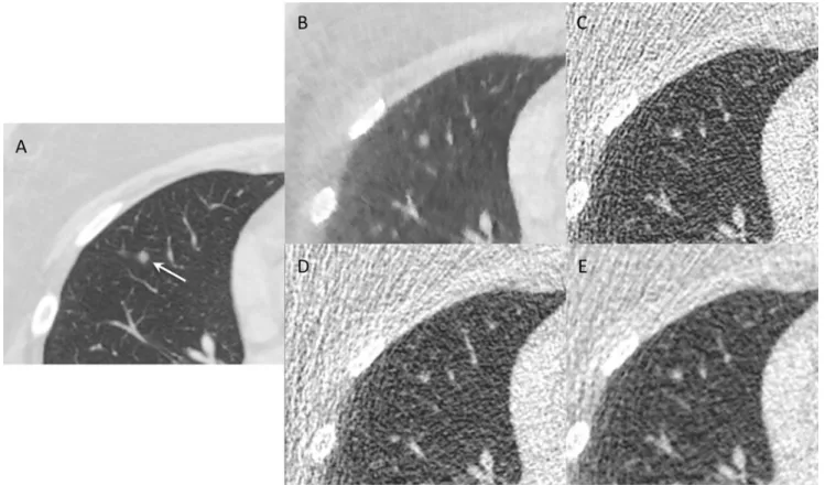
Fig. 2 Nodules. Diagnostic CT showing a 5-mm nodule (arrow) in the right middle lobe (a). The same nodule seen on ULD-CT, reconstructed with MBIR (b), FBP (c), ASIR-40 (d) and ASIR-80 (e) algorithms
Table 4 Number of micro-nodules
Number of micro-nodules Micro-nodules R1 R2 R3 Kappa Diagnostic CT (SDD/LDD) 137 117 126 0.298 ULD-CT FBP 71* 71 75 0.476 ASIR-40 85 86 82 0.386 ASIR-80 98 94 89 0.855 MBIR 147 130 111 0.551 Diagnostic CT vs ULD-CT *P < 0.05 ns ns
Pleural, pericardial effusions and lymph nodes were de-scribed by all readers on SDD-CT and LDD-CT and MBIR data sets. Some subtle pericardial effusions were missed on the ULD-CT reconstructed with FBP or ASIR algorithms.
Discussion
Despite all the technological developments in CT, the re-construction algorithm, namely FBP, has remained un-changed since the 1970s. Although FBP allows very rapid image reconstruction, this algorithm suffers from major drawbacks due to approximation of the real focal spot and detector sizes, and the data are corrupted by quantum and electronic noise during the acquisition, thus propagating image noise [15].
The first generation of iterative reconstruction, including ASIR or IRIS, takes into account how noise propagates during the reconstruction steps and feeds this information back into the loop to iteratively reduce noise in the recon-structed image. Recent clinical data have shown that itera-tive reconstructions with IRIS using three iterations provide similar image quality to chest CT with 35% less dose com-pared with FBP reconstruction [6]. When using ASIR, hy-brid reconstruction algorithms combining FBP and ASIR at varying percentages allow iterative reconstructions to be adapted to different diagnostic settings; ASIR-40 and
ASIR-80 were used arbitrarily in our study protocol, and the data were listed separately.
The second generation of iterative reconstruction algo-rithms, including MBIR, accurately models the production of X-rays in the tube and the attenuation of these X-rays within the patient and the CT system. MBIR also models the measurements inside the detectors and the transformation into the digital signal. All these models are fed into a mathematical model to describe the reconstructed image in a manner representative of the projection data. The resulting function is optimised to find the best possible match be-tween the reconstructed image and the acquired projection data. Hence, this system minimises discrepancies between the projection data and the theoretical model. Our results re-garding image quality demonstrate that the use of MBIR offers even further reduction of noise compared with ASIR alone. Noise reduction of 12%, 28% and 79% was achieved for ASIR-40, ASIR-80 and MBIR, respectively, which is in agreement with recently reported results of other investigators [16].
Clinical evaluation of the potential of dose reduction in chest CT may be done by using variations of different technical parameters that may influence image quality; namely, kilovoltage, tube current and reconstruction algo-rithms. The use of MBIR holds promise to allow even more drastic dose reduction in chest CT compared with ASIR. We designed to investigate the diagnostic value of chest CT reconstructed with ASIR-40, ASIR-80 and MBIR with a radiation dose close to a PA and lateral CXR, in comparison with SDD-CT or LDD- CT.
Our ULD- CT protocol resulted in an exposure of 0.16 ± 0.006 mSv, close to the dose of a PA and lateral CXR, which is reported between 0.05 and 0.24 mSv in the literature [17–21]. This corresponds to a dose reduction of 98.6% compared with SDD-CT and 94% compared with LDD-CT. An increase in subjective image quality with the use of ASIR and even more with the use of MBIR, seems to translate into a higher inter-observer agreement regarding dense lesions encountered in the lung parenchyma. Never-theless, a unique image feature of MBIR data sets (a pixel-lated blotchy appearance, as already described [16]) was clearly seen in this study, but had little effect on diagnostic quality. Despite anonymisation, MBIR data sets were rec-ognised by the readers.
Results of our study suggest that ULD-CT may enable the detection of pulmonary nodules with a similar sensitivity as SDD-CT or LDD-CT. This could perhaps be taken into con-sideration in the context of screening protocols focusing spe-cifically on the detection of pulmonary nodules, since radiation exposure plays a key role in image-based screening protocols [22].
Since most of our patients had normal BMI (25 for male and 22 for female patients), additional data are needed to assess image quality of patients with high BMI (BMI>25).
Table 5 Number of patients having ground glass opacities Number of patients
GGO Kappa Five-point
scale (median) R1 R2 R3 Diagnostic CT (SDD/LDD) 0.694 1 11 11 12 ULD-CT FBP 0.172 2 7 11 9 ASIR-40 0.222 1.5 9 10 8 ASIR-80 0.184 1 9 9 8 MBIR 0.367 1 8 14 11
Table 6 Number of patients having emphysema
Number of patients
Emphysema Kappa Five-point
scale (median) R1 R2 R3 Diagnostic CT (SDD/LDD) 1 1 9 9 9 ULD-CT FBP 0.06 2 5 6 5 ASIR-40 −0.125 1.5 4 5 3 ASIR-80 −0.317 1 8 5 6 MBIR −0.144 2.2 11 16 6
Some drawbacks of the ULD-CT protocol used in our study must, however, be considered. We found that ULD-CT was less adequate for the detection of diagnostic ele-ments of chronic lung disease. Although bronchiectasis was equally well detected as with SDD-CT or LDD-CT, this was not the case for ground-glass opacities and emphysema. Additional studies therefore appear necessary to optimise the image quality in order to detect these diagnostic ele-ments reliably. Implementation of MBIR results in much longer image reconstruction time, requiring an hour or more. In clinical practice, this certainly appears as a drawback in emergency settings, although it may be acceptable in routine indications, such as chronic lung disease, oncological follow-up or screening for lung disease. Nevertheless, due to the constant increase of computing power, one may speculate that in the next few years, the MBIR reconstruc-tion may only take a few seconds.
Finally, we are unable to explain the fact that more micro-nodules were identified on the MBIR data set than on SDD-CT with FBP. Since the spatial resolution increases with the use of MBIR [23], this could possibly lead to better conspi-cuity of micro-nodules on the MBIR data set compared with SDD-CT with back projection, regardless of the reduction in X-ray dose. Since histopathological correlation with the radiological findings was not available it is impossible to exclude false-positive results, i.e. that ULD-CT recon-structed with MBIR algorithm could have shown micro-nodules that did not exist.
In summary, our preliminary data suggest that ULD-CT of the chest acquired with an X-ray dose of a conventional radiography and the MBIR reconstruction algorithm enables detection of non-calcified solid nodules and micro-nodules with similar sensitivity compared with SDD-CT or LDD-CT using FBP. This could perhaps be considered when designing future screening protocols focusing on the detection of solid pulmonary nodules. The detection of subsolid nodules will require additional study, as none were present in this study.
References
1. Brenner DJ, Hall EJ (2007) Computed tomography—an increasing source of radiation exposure. N Engl J Med 357:2277–2284 2. Brenner DJ (2004) Radiation risks potentially associated with
low-dose CT screening of adult smokers for lung cancer. Radiology 231:440–445
3. Lee S, Yoon SW, Yoo SM et al (2011) Comparison of image quality and radiation dose between combined automatic tube cur-rent modulation and fixed tube curcur-rent technique in CT of abdo-men and pelvis. Acta Radiol 52:1101–1106
4. Leipsic J, Nguyen G, Brown J, Sin D, Mayo JR (2010) A pro-spective evaluation of dose reduction and image quality in chest CT using adaptive statistical iterative reconstruction. AJR Am J Roentgenol 195:1095–1099
5. Pontana F, Duhamel A, Pagniez J et al (2011) Chest computed tomography using iterative reconstruction vs filtered back projec-tion (Part 2): image quality of low-dose CT examinaprojec-tions in 80 patients. Eur Radiol 21:636–643
6. Pontana F, Pagniez J, Flohr T et al (2011) Chest computed tomog-raphy using iterative reconstruction vs filtered back projection (Part 1): evaluation of image noise reduction in 32 patients. Eur Radiol 21:627–635
7. Prakash P, Kalra MK, Digumarthy SR et al (2010) Radiation dose reduction with chest computed tomography using adaptive statis-tical iterative reconstruction technique: initial experience. J Comput Assist Tomogr 34:40–45
8. Sato J, Akahane M, Inano S, et al. (2011) Effect of radiation dose and adaptive statistical iterative reconstruction on image quality of pulmonary computed tomography. Jpn J Radiol
9. Singh S, Kalra MK, Gilman MD et al (2011) Adaptive statistical iterative reconstruction technique for radiation dose reduction in chest CT: a pilot study. Radiology 259:565–573
10. Scheffel H, Stolzmann P, Schlett CL, et al. (2011) Coronary artery plaques: Cardiac CT with model-based and adaptive-statistical iterative reconstruction technique. Eur J Radiol
11. Bankier AA, Schaefer-Prokop C, De Maertelaer V et al (2007) Air trapping: comparison of standard-dose and simulated low-dose thin-section CT techniques. Radiology 242:898–906
12. Muangman N, Maitreesorrasan N, Totanarungroj K (2011) Comparison of low dose and standard dose MDCT in detection of metastatic pulmonary nodules. J Med Assoc Thai 94:215–223 13. Hansell DM, Bankier AA, MacMahon H, McLoud TC, Muller NL,
Remy J (2008) Fleischner Society: glossary of terms for thoracic imaging. Radiology 246:697–722
14. Huda W, Magill D, He W (2011) CT effective dose per dose length product using ICRP 103 weighting factors. Med Phys 38:1261– 1265
15. Pan X, Sidky EY, Vannier M (2009) Why do commercial CT scanners still employ traditional, filtered back-projection for image reconstruction? Inverse Probl 25:1230009
16. Katsura M, Matsuda I, Akahane M et al (2012) Model-based iterative reconstruction technique for radiation dose reduction in chest CT: comparison with the adaptive statistical iterative recon-struction technique. Eur Radiol 22:1613-1623
17. Bacher K, Smeets P, Bonnarens K, De Hauwere A, Verstraete K, Thierens H (2003) Dose reduction in patients undergoing chest imaging: digital amorphous silicon flat-panel detector radiography versus conventional film-screen radiography and phosphor-based computed radiography. AJR Am J Roentgenol 181:923–929 18. Mettler FA Jr, Huda W, Yoshizumi TT, Mahesh M (2008) Effective
doses in radiology and diagnostic nuclear medicine: a catalog. Radiology 248:254–263
19. Quaia E, Baratella E, Cernic S et al (2012) Analysis of the impact of digital tomosynthesis on the radiological investigation of patients with suspected pulmonary lesions on chest radiography. Eur Radiol. doi:10.1007/s00330-012-2440-3
20. Samara ET, Aroua A, Bochud FO et al (2012) Exposure of the Swiss population by medical x-rays: 2008 review. Health Phys 102:263–270
21. Schuncke A, Neitzel U (2005) Retrospective patient dose analysis of a digital radiography system in routine clinical use. Radiat Prot Dosimetry 114:131–134
22. Saghir Z, Dirksen A, Ashraf H et al (2012) CT screening for lung cancer brings forward early disease. The randomised Danish Lung Cancer Screening Trial: status after five annual screening rounds with low-dose CT. Thorax 67:296–301
23. Yu Z, Thibault JB, Bouman CA, Sauer KD, Hsieh J (2011) Fast model-based X-ray CT reconstruction using spatially nonhomoge-neous ICD optimization. IEEE Trans Image Process 20:161–175