© Urban & Vogel 2005
Herz
© Urban & Vogel 2005Herz
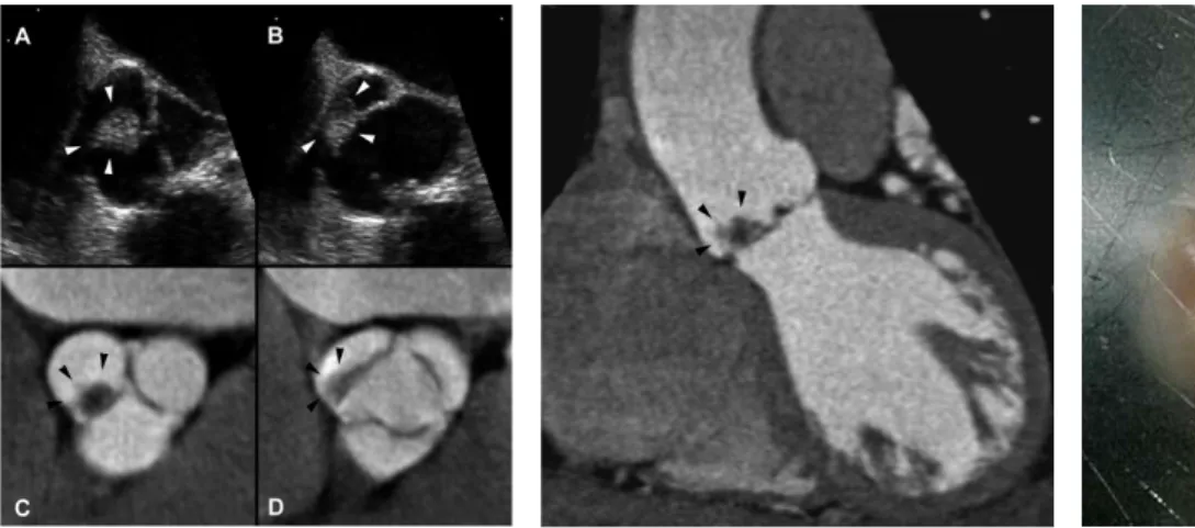
Figures 1A to 1D. Transesophageal echocar-diography (A, B) and corresponding 64-slice CT reconstructions (C, D) demonstrate the spherical tumor with its broad-based attach-ment to the non-coronary cusp of the aortic valve (arrowheads). Images during mid-dias-tole (A, C) and mid-sysmid-dias-tole (B, D) show the change in shape throughout the cardiac cycle which may be explained by the smooth, elas-tic character of the lesion.
Figure 2. Oblique coronal 64-slice CT reforma-tion shows the centrally hypodense lesion with its blurred outer margins (arrowheads) corresponding to the papillary fronds of the tumor.
Figure 3. Macroscopic specimen shows the ap-pearance of the tumor immersed in saline solu-tion resembling a sea anemone which is consid-ered diagnostic of papillary fibroelastoma.
Image of the Month
1 Institute for Diagnostic
Radiology,
2 HerzKreislauf-Zentrum,
Cardiology,
3 Department of Cardiac
and Vascular Surgery, University Hospital, Zurich, Switzerland. Herz 2005;30:438 DOI 10.1007/ s00059-005-2667-8 Adress for Correspondence PD Simon Wildermuth, MD Institut für Diagnosti-sche Radiologie Universitätsspital Zürich Rämistraße 100 8091 Zürich Switzerland Phone (+41/1) 255-3662, Fax -4443 e-mail: simon. [email protected]
anemone (Figure 3) providing a morphological verification of the tumor being a papillary fibro-elastoma.
Cardiac papillary fibroelastoma is a rare tu-mor originating from the endocardium, which is hypothesized to represent a different stage of Lambl’s excrescence. The recognition and correct diagnosis of papillary fibroelastoma is important since it is a treatable cause of cerebral or cardio-vascular emboli. Although usually diagnosed with echocardiography, it is likely that the growing clinical use of cardiac CT may increasingly un-cover this entity.
Fibroelastoma of the Aortic Valve.
Evaluation with Echocardiography
and 64-Slice CT
Hatem Alkadhi1 , Sebastian Leschka1 , David Hurlimann2 , Rolf Jenni2 , Michele Genoni3 , Simon Wildermuth1A 72-year-old women suffered from an acute left hemispheric stroke. Transesophageal echocar-diography for evaluating a possible source of embolus was performed and showed a spherical, hyperechogenic tumor with a broad-based at-tachment to the non-coronary cusp of the aortic valve (Figures 1A and 1B, arrowheads), suggest-ing the diagnosis of a fibroelastoma. The lesion was further characterized with retrospective ECG-gated 64-slice computed tomography (CT) demonstrating an oval lesion attached to the si-nus surface of the non-coronary aortic cusp (Fig-ures 1C and 1D, arrowheads). It was centrally hypodense and had blurred outer margins (Fig-ure 2, arrowheads). The tumor was removed sur-gically and the aortic valve was reconstructed. The gross pathologic specimen showed a lesion with a frond-like, villous surface. After being im-mersed in saline solution, it resembled a sea