Received October 31, 2002; Revised November 15, 2002; Accepted November 15, 2002.
Author to whom all correspondence and reprint requests should be addressed: Pedro L. Herrera, Department of Morphology, University of Geneva Med-ical School, 1 Rue Michel-Servet, CH-1211 Geneva 4, Switzerland. E-mail: [email protected]
Pancreatic Cell Lineage Analyses in Mice
Pedro L. Herrera, Virginie Nepote, and Alexandra Delacour
Department of Morphology, University of Geneva Medical School, 1 Rue Michel-Servet, CH-1211 Geneva 4, Switzerland
267 Considerable knowledge of the ontogeny of the endo-crine pancreas has been gained in recent years, mainly through the use of two complementary genetic ap-proaches in transgenic mice: gene inactivation or over-expression (to assess gene function) and genetic labeling of precursor cells (to determine cell lineages). In recent years, in vivo Cre/loxP-based direct cell tracing expe-riments show that (i) all pancreatic cells differentiate from pdx1-expressing precursors, (ii) p48 is involved in the exocrine and endocrine pancreatic lineages, (iii) islet endocrine cells derive from ngn3-expressing pro-genitor cells, and (iv) insulin cells do not derive from glu-cagon-expressing progenitors. Lineage analyses allow the identification of progenitor cells from which mature cell types differentiate. Once identified, such progeni-tors can be labeled and isolated, and their differentiation and gene expression profiles studied in vitro. Under-standing pancreatic cell lineages is highly relevant for future cell replacement therapies in diabetic patients, helping to define the identity of putative (endodermal) pancreatic stem cells.
Key Words: Pancreas; transgenic mouse; lineage; Cre/ loxP; stem cells.
Pancreas Differentiation and Morphogenesis
The pancreas contains endocrine and exocrine cells. The exocrine pancreas represents more than 95% of the gland; it is composed of the acinar secretory cells, producing dige-stive enzymes such as amylase, and the excretory ducts. The endocrine cells of the pancreas form clusters embedded in the exocrine tissue: the islets of Langerhans. Islets are com-posed of four types of endocrine cells: the centrally located b cells produce insulin, while the peripheral a, d, and PP cells produce glucagon, somatostatin, and pancreatic poly-peptide, respectively.
Pancreas Organogenesis
The pancreas initially develops from distinct dorsal and ventral primordia that later fuse to form the mature organ. This involves a sequential cascade of inductive events in association with the activation of specific transcription fac-tors. Pancreatic buds evaginate from the early foregut endo-derm caudally to the stomach primordium, near the pro-spective liver: the molecular determinants that define the position of the pancreas along the anterior–posterior axis remain unknown (1,2). The first hormone-containing cells found in early primordia are epithelial and appear located exclusively within the walls of the embryonic ducts or cords. Contrary to what is observed in adult islets, these cells fre-quently contain simultaneously more than one hormone; this has suggested the existence of a common precursor cell for all four islet cell types. For instance, because glucagon-con-taining cells are the first to differentiate in early buds, and given the presence of cells co-expressing glucagon and insu-lin, it was proposed that mature insulin cells derive from glucagon progenitors (3).
Commitment to a pancreatic fate occurs as early as embry-onic d 8–8.5 (E8–E8.5) in mice. Permissive signals released from adjacent mesodermal structures, including the noto-chord, aorta, and cardiac mesoderm, are important for induc-tion of the pancreatic program. The notochord induces dorsal pancreas development through the inhibition of SHH (Sonic hedgehog) in pre-pancreatic dorsal endoderm. Shh is expressed in the early gut endoderm and has been shown to pattern it (1,4–6). This Shh repression, which appears to be by activin bB and FGF2 (7), is permissive for the expres-sion of Pdx1. Shh is also repressed, and Pdx1 expressed, in the ventral pre-pancreatic endoderm.
It is intriguing that the same mesenchymal morphogens have opposite inductive effects on the dorsal and ventral pan-creas: in the absence of dorsal mesoderm, the dorsal endo-derm expresses liver markers, whereas, without ventral meso-derm (i.e., cardiac mesomeso-derm and septum transversum), the ventral endoderm “defaults” to pancreas instead of liver. Indeed, signals from ventral mesoderm (FGFs) promote hep-atic development (8).
The first indication of morphogenesis occurs at E9.5, when the dorsal mesenchyme condenses and the underly-ing endoderm evaginates to form a recognizable dorsal pan-creatic bud; the ventral bud appears one day later (E10.5). Both buds subsequently proliferate, stimulated by mesen-chymal signals, to form multiple branches and fuse together
by d E17 (the ventral bud translocates to the dorsal side during gut rotation) to make a functional organ.
Timing of Appearance
of Differentiated Endocrine Phenotypes
In mice, the first differentiated cells, as determined by immunofluorescence, are glucagon-producing cells; they are already detectable at E9.5 in the dorsal pancreatic bud and one day later in the ventral, whereas the first insulin-producing cells are present the following day (E10.5–E11) in both primordia. After branching, at E13.5, somatostatin-positive cells are detectable in both buds, and the exocrine pancreas containing well-formed acini begins to differenti-ate at E14.5. Three days prior to birth, at E16.5, PP-secret-ing cells are also detected, and the first islets of Langerhans are formed at E18.5 (9).
The timing of hormone gene expression was also deter-mined by RT-PCR of total RNAs extracted from pancrea-tic buds and surrounding tissues at different developmental stages (9,10). Glucagon and somatostatin mRNAs are found very early (E8.5–E9), probably from extrapancreatic sources; insulin I transcript was detected in E10.5 buds, whereas insulin II was found only 1 d later. Surprisingly, PP mRNA is detected from E10.5 onward, thus indicating PP gene expression early in the developing mouse pancreas.
Islet Organogenesis
Analysis of endocrine cells in situ suggests three major patterns of endocrine cell localization in embryonic pancreas. The earliest hormone-containing cells are individual cells located within the epithelium of pancreatic ducts or cords. In both dorsal and ventral buds, the majority of these endo-crine cells contain more than one hormone, and this co-localization occurs at the secretory granule level: different hormones are co-stored within the same granules (9). Subse-quently, these cells migrate into the surrounding connective tissue, forming clusters of endocrine cells in the pancreatic interstitia. This process involves interactions between cells through adhesion molecules and active proteolysis of the extracellular matrix (11) [reviewed by Johansson and Grapin-Botton (1)]. For instance, collagen IV, which is a compo-nent of basement membranes, is absent at the places where these first endocrine cells appear (Fig. 1). Finally, starting on E18.5, as indicated above, typical islets are formed with centrally located b cells and with the adult “one cell–one hormone” phenotype (9).
Analysis of Cell Lineage Analyses: A Brief Historical Overview
The origin of multicellularity during evolution required, and allowed, that all (or almost all), cells forming a particu-lar organism become highly specialized in order to fulfill particular tasks. Yet all these different cell types, more than two hundred in a typical mammalian body, derive and
dif-ferentiate progressively from one single and unique totipo-tent cell: the zygote. Development of metazoan organisms from one-celled embryos is either “mosaic,” meaning that early cell divisions are restrictive—the young blastomeres quickly losing their developmental potential—or “regula-tive,” as in mammals, for each blastomere remains pluripo-tent until later stages, when interactions between neighbor cells determine their respective destiny. Understanding these developmental processes implies knowledge of the precise lineages of cells; the observer therefore requires a method to reliably identify particular cells and all of their progeny, to reveal their fate.
Tracer Marking
The study of cell lineages was begun in the early years of the twentieth century, shortly after the establishment of Schwann and Schleiden’s cell theory. Lineage tracing by directly observing living transparent invertebrate embryos, composed of few cells, was first performed by E.G. Conklin and others (12) [reviewed by Stern and Fraser (13)]. In most embryos, however, direct observation is not possible. W. Vogt then devised a method to follow cells using vital dyes (14,15). A new improvement was introduced with the autoradiography on slides of embryos treated with radio-active nucleotide; Rosenquist was able to draw fate maps of chick embryos in this manner (16). However, the remain-ing limitations were that groups of cells, rather than sin-Fig. 1. Immunohistochemical analysis of pancreatic buds
cul-tured in collagen gels. Two consecutive semithin sections of an E12.5 pancreatic bud cultured for 7 d were stained with two differ-ent antibodies: antiglucagon (A) and anticollagen IV (B). Clusters of endocrine cells stained with antiglucagon antibody (A) migrate from the duct-like cords of epithelial cells into the surrounding mesenchyme. This process involves interactions between endo-crine cells and with the extracellular matrix (ECM), through ad-hesion molecules, and active proteolysis of the ECM, as illustrated with the anticollagen IV staining (B). Collagen IV is a main com-ponent of epithelial basement membranes. Digestion of collagen IV is shown to occur at places were endocrine clusters of cells are budding (asterisks in panel B). Scale bar is 20 µm.
gle cells, were labeled, and, in addition, the soluble dyes or labeled radioactive components could pass between unre-lated adjacent neighbor cells. Several alternatives were then found, such as the use of carbocyanine dyes (DiI and DiO), and intracellular tracers (enzymes, like horseradish peroxi-dase, or fluorochrome-conjugated high-molecular-weight molecules, like dextrans), which can be directly injected into single cells. Carbocyanine dyes are lipid-soluble and inte-grate into the membrane of cells; they were successfully used to trace the growth of axons. More recently, monoclo-nal antibodies coupled to gold particles have been used to mark cells expressing a surface antigen; because the cells internalize the attached labeled antibody, their descendants are also marked (17).
Genetic Marking
Injecting small single cells may prove technically diffi-cult, but the main disadvantage, however, with vital dyes, isotope or antibody labeling, fluorescent markers, carbocy-anine dyes, or enzymes, is that the signal becomes diluted as cells divide. To follow cells over long periods or complex differentiation programs, irreversible tagging is mandatory. Mosaics
The labeling dilution problem can be overcome by genet-ically marking embryos through infection with replication-deficient retroviruses bearing a gene encoding a marker (such as b-galactosidase, alkaline phosphatase, or GFP) (18–21). The approach consists in using very few viral particles so that infection is a rare event; therefore, marked cells in a given infected embryo are probably clonally related. Infected embryos are thus mosaics. The main disadvantage of this approach, apart from the uncertainty regarding the inter-pretation of the results, is that labeled cells are not chosen directly; this lineage analysis is thus “retrospective.” Using this method with pancreatic rudiments cultured in vitro, Fishman and Melton demonstrated that a single infected pancreatic progenitor cell is able to give rise to both exo-crine and endoexo-crine cells (20).
Chimeras
Another way to circumvent the problem of tracer dilu-tion was devised as early as the 1920s, through the genera-tion of interspecies chimeras, by transplantagenera-tion (grafting) or, much later, by aggregating two mouse embryos. Chime-ras are derived from more than one embryo; therefore, their body is formed of cells from different individuals, thus hav-ing different genetic constitutions. In transplantation chi-meras, donor and host cells can be easily distinguished for they are morphologically different (22–26). With this method (grafting quail neural crest cells on chick embryos), Le Doua-rin demonstrated that neural crest cells do not contribute to the formation of pancreatic endocrine cells (27).
In vitro grafting, i.e., tissue co-cultures of rudiments from different embryos, is another variation of the chimera approach. With this system, Martineau and colleagues (28)
demonstrated that there is migration of cells from the meso-nephros into the male gonad during development, but not into the developing ovaries. Percival and Slack (29) showed that the pancreatic mesenchyme does not contribute to pan-creatic cells: when labeled prepanpan-creatic endoderm was cul-tured with unmarked mesenchyme, only the endoderm gave rise to acini, ducts, and islets.
Mouse aggregation chimeras, using, for instance, trans-genic and wild-type morulae, or chimeras generated by injec-tion of pluripotent ES cells into host blastocysts, are power-ful tools to identify different cell populations within a single organ in vivo. With the embryo aggregation approach, Jami and coworkers demonstrated that pancreatic islets of Langer-hans are of polyclonal origin, i.e., that endocrine cells within a particular islet derive from different progenitors (30). Transgenics
In the simplest transgenic experiment, expression of re-porter molecules using cell-type-specific promoters (whether exogenous or endogenous, as in knock-in mice) may be taken as evidence for cell lineage relationships (3). This would be valid if the progeny of labeled cells did not cease express-ing the reporter transgene, but this is not applicable in terms of lineage analysis. When reporter expression stops, this method is dependent on the stability of the transgenic reporter protein and is weakened because cell divisions imply dilu-tion of the marker, as previously discussed. These are impor-tant limitations because many developmentally relevant genes are expressed only transiently. Similarly, endogenous pro-teins, such as peptidic hormones, have been used in the past, with the same serious flaws as lineage tracers. Co-localiza-tion of hormones in single cells during development is not a proof of common ontogeny.
Transgenesis resulting in mosaicism may be used for retro-spective lineage analyses. This is possible using transgenic mice in which a very rare spontaneous mutation activates the expression of a reporter transgene in early embryos (31–33), or also using X-inactivation transgenic mosaic mice, i.e., trans-genic females bearing a X-linked reporter transgene (34–38). Targeted oncogenesis, in transgenic mice expressing powerful oncogenes under the control of cell-specific pro-moters, has also been used as an approach to analyze cell lineages in the pituitary (39,40). This method allows for im-mortalizing rare cell types (41–43) that might recapitulate in culture the characteristics of progenitor cells. The main disadvantage of this conception, however, is that tumor cells maintained in vitro are profoundly altered. Moreover, cells in culture are not in a normal environment, and because many genes act noncell autonomously, the effect of neighboring cells, and vice versa, is critical, and cannot be addressed with this approach.
Two sophisticated methods using transgenic mice have been introduced more recently, and both were first employed to trace pancreatic cell lineages. The first method is the targeted ablations of given cell types using
cell-type-spe-cific promoters to express toxin genes. The rationale is that missing cells are derived from, or are dependent on, the cell type destroyed, which would then be a putative precursor, or adjuvant cell. A refinement of this approach was success-fully used to address the enteroendocrine cell lineages through the conditional ablation of secretin cells, upon administra-tion of ganciclovir, in transgenic mice expressing herpes simplex virus 1 thymidine kinase (HSVTK) under the con-trol of a secretin promoter (44). The viral enzyme HSVTK phosphorylates the thymidine analog ganciclovir, which is mutagenic and cytotoxic.
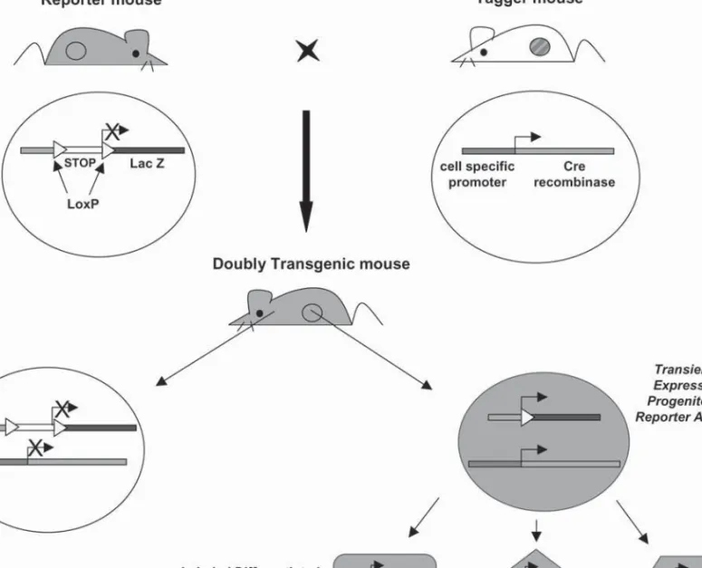
The second approach is the irreversible targeted tagging (labeling) of a given cell type using a promoter to express Cre recombinase (Cre/loxP system) (45,46). This is the most reliable and powerful method to trace cell lineages in Amniota in vivo; it permits detection of descendants that do not express the gene of interest any more (Fig. 2).
Both approaches have pioneered in pancreatic rudiment cell lineage tracing, and will be discussed in more detail below. We now summarize some knockout experiments that have demonstrated a crucial role for a few genes in pancreas development.
Fig. 2. Tracing cell lineages with the Cre/loxP strategy. In order to genetically label pancreatic progenitor cells, two different strains of
transgenic mice are needed. The “reporter mouse” bears a reporter transgene (in the cartoon the b-galactosidase coding region) placed under the control of a promoter (either tissue-specific or ubiquitous) and downstream of a loxP-flanked transcription termination cassette (STOP sequence). The “tagger mouse” (“marker” or “deletor” mouse), has a transgene encoding the Cre recombinase gene under the control of a cell-specific promoter, which should be active in putative pancreatic progenitor cells. In doubly transgenic mice, the expression of the tagger transgene in a precursor cell results in the excision of the reporter’s STOP sequence, thus allowing its expression (b-galactosidase). This tag is then genetically transmitted to all the progeny of this cell and permits the tracing of its lineage during the formation of the pancreas.
Insights from Gene Inactivation Experiments: Examples of Noncell Autonomous vs Cell Autonomous Effects of Gene Expression
Inactivation of genes (gene targeting or ablation) in knock-out (KO) mice is one way to study their function. This may result in loss of some particular cell type(s) and/or activa-tion of reporter gene expression (knockin mice). These results have sometimes been considered as a means to iden-tify precursor cells; this is conceptually wrong. Genes are often expressed transiently: cytodifferentiation during devel-opment is based on differential gene expression. Moreover, many genes act noncell autonomously (i.e., in paracrine ways); as a consequence, cell ablation following gene inac-tivation mutations (knockout mice) cannot be taken as evi-dence of cell lineage relationships among different cell types. Strictly, the gene knockout experiments tell us whether a given gene is necessary for a given cell type to differentiate or fully mature, at the time of inactivation. On the contrary, genetic labeling of cells is aimed at unequivocally identify-ing the progeny of a cell that has expressed a particular gene during differentiation.
Mesenchymal Factors Are Noncell Autonomous
Inducers of Epithelial (Endodermal) Cell Differentiation
Epithelial–mesenchymal interactions are essential for pancreas morphogenesis and cytodifferentiation. Different pancreatic lineages require different mesenchymal factors for their development. For instance, when the pancreatic bud epithelium is cultured in the absence of mesenchyme, or grafted under the kidney renal capsule, only endocrine cells differentiate (47). The identity of most soluble mesenchy-mal factors remains unclear, but the TGFb, FGF, and EGF signaling pathways appear to be involved (1,4,48). The TGFb superfamily member follistatin, which is expressed in pan-creatic mesenchyme, promotes exocrine differentiation (49), but TGFb1 has the opposite effect (50–53).
Targeted disruption of Isl1 (ISLET1, a LIM homeodo-main protein), which is expressed in early endocrine cells, abolishes endocrine differentiation. Isl1 is nevertheless first expressed in the mesenchyme surrounding the dorsal pri-mordium. In Isl1-/- embryos there is no dorsal pancreatic bud, and this is a consequence of the lack of dorsal mesen-chyme (54).
Inactivation of the adhesion molecule N-cadherin also results in agenesis of the dorsal pancreas, which is associated again with a loss of dorsal mesenchyme (55). Interestingly, Isl1 expression persists in the mesenchyme as well as in pro-spective dorsal pancreatic epithelium, whereas Pdx-1 expres-sion is normal by E9, but is then rapidly restricted to the prospective ventral pancreas; because Pdx-1 is similarly dor-sally downregulated in Isl1-/- mice (54), it appears that the dorsal mesenchyme is necessary to maintain Pdx-1 expres-sion. In fact, in vitro recombination experiments demon-strate that wild-type mesenchyme rescues morphogenesis and cytodifferentiation in N-cadherin-/- dorsal endoderm.
Transcription Factors in Pancreas Development (Table 1)
Even though transcription factors are likely to act cell autonomously, the consequences of their activity indirectly affect neighbor cells in a paracrine (noncell autonomous) way. Many transcription factors, mostly homeobox genes, have been mapped to specific regions of the endoderm, as well as to the surrounding visceral mesenchyme, and are likely to modulate the mesoderm–endoderm interactions [reviewed by Schwitzgebel (56)]. Their involvement in pan-creas determination, growth, and differentiation has been assessed by their selective inactivation in mice (summarized in Table 1). The factors that are more relevant for pancreas development are briefly discussed herein.
PDX1 (pancreatic and duodenal homeobox-1) is, together with the homeodomain protein HB9, the earliest marker of pancreatic bud cells, from E8.5. PDX1 is necessary for the development of the pancreas, as Pdx1 deficient mice have pancreatic aplasia (57,58). However, insulin and glucagon cells are present in early embryonic buds of Pdx1-/- mice, and their pancreatic mesenchyme develops normally. Pdx1 is not a “master control gene,” because its ectopic expres-sion in the hindgut endoderm is not sufficient to trigger pancreas formation (59).
The inactivation of two bHLH transcription factors, PTF1-p48 and Ngn3, suggest that they could be implicated in deter-mining whether an early pancreas cell is to become exocrine or endocrine. Mice in which PTF1-p48 is inactivated have no exocrine pancreas (60,61), whereas inactivation of Ngn3 results in agenesis of endocrine pancreas (62). In pancrea-tic buds, NGN3 is detected in proliferating cells expressing
Table 1
Some Factors Involved in Pancreas Development, as Determined Using KO Mice
Targeted Dorsal Ventral
gene pancreas pancreas Reference
Pdx1 Aplasia 57,58,86 Ngn3 No islets 62 PTF1-p48 No exocrine cells 60 Pax4 No b or d cells 66 Pax6 No a cells; 65 endocrine hypoplasia
Isl1 Agenesis; No endocrine 54
no mesenchyme cells
Hlxb9 Agenesis Fewer b cells 87,88 Hes1 Hypoplasia 73
N-cadherin Agenesis; Normal 55
no mesenchyme
Beta2/NeuroD Fewer b cells; 89
distorted islets
Hnf6 Endocrine hypoplasia 90
Pbx1 Hypoplasia 91
PDX1, but not in hormone-containing cells. Overexpres-sion of Ngn3 (under the control of a Pdx1 promoter, in trans-genic mouse embryos, or of an ubiquitous promoter, in electroporated chick embryos) is sufficient to drive differen-tiation of glucagon cells, but not of insulin cells (56,63,64). In the absence of the paired-box domain protein PAX6, known to be downstream of ISL1, and initially expressed in all pancreatic endocrine cells, the development of all endo-crine lineages is affected (65). The PAX4 protein is specifi-cally necessary for the production of mature b and d cells (66). Interestingly, in Pax6-/- and Pax4-/- double mutants islets are totally absent.
In summary, the gene inactivation studies show, among many other interesting findings, that (1) several mesenchy-mal factors, whether soluble or adhesion molecules, or tran-scription factors, are needed for pancreas development by activating genes in the prospective pancreatic endoderm, (2) Pdx1 is necessary for growth of both pancreatic rudi-ments, (3) ptf1-p48 activity is required for exocrine pancreas differentiation, and (4) Ngn3 is necessary for the differen-tiation of islet endocrine cells.
Together, these results suggest some gene products (ISL1, N-Cadherin, PDX1, p48, and NGN3), whose inactivation induces loss of cells, as candidate markers for pancreatic progenitor cells. The KO experiments also point out the principal role of the mesenchyme, whose absence inhibits dorsal pancreas formation, although we know that the mes-enchyme itself does not contribute to pancreatic cells (29). This clearly illustrates that, even though extremely impor-tant, gene inactivation experiments cannot be interpreted as indicative of cell lineages because they result in cell loss. Therefore, cells expressing these candidate genes were sub-sequently genetically marked in transgenic mice, in order to determine whether they give rise to the different adult pancreatic cell types.
Characterizing Pancreatic Progenitor Cells by Irreversibly Deleting or Marking Cell Populations in Transgenic Mice
One approach to cell tracing experiments in vivo is the selective ablation of specific cell types in transgenic mice through the expression of toxin genes (44,67,68). The glu-cagon-promoter-driven expression of a diphtheria toxin (DT) A gene demonstrated that glucagon cells are not precursors of insulin-producing cells, contrary to what colocalization of both hormones in single cells during development would suggest (3,68,69). The analyses were pursued by labeling
progenitor cells through the expression of Cre recombinase in doubly transgenic mice (70). This method established that all pancreatic cells derive from progenitors that do express Pdx1 (71,72). Recent studies also showed that Ngn3-expressing cells are precursors of all islet cell types (72), and that ptf1-p48 is surprisingly implicated not only in exocrine but also in endocrine pancreas lineages (61).
Ablation of Islet Cells by Targeted Expression of Hormone-Promoter-Driven Toxigenes
Direct cell lineage tracing of chosen labeled progenitor cells in mouse embryos was not possible until the Cre/loxP system was developed in the mid 1990s. Therefore, in order to study the ontogenetic, or cell lineage, relationships between the different types of endocrine cells in the islets, targeted cell ablation was the best approach. In a transient transgenic study, we generated embryos in which cells transcribing glu-cagon, insulin, or PP genes were destroyed by the promoter-targeted expression of DT A (68). If, as proposed, insulin cells were the descendants of glucagon-producing precur-sors, then the ablation of all glucagon-expressing cells would preclude their differentiation. On the contrary, we unam-biguously demonstrated that neither glucagon nor insulin gene expressing cells are essential for the differentiation of either b or a cells, respectively. These experiments also indicated a close ontogenetic relationship among insulin, PP, and somatostatin cells. Whether this requirement is through a cell-lineage relationship or because those cells produce paracrine or endocrine factors necessary for their differen-tiation from undefined precursors, remains undetermined.
Islet Cell Lineage Analyses
by Irreversibly Labeling Cell Progenitors (Fig. 2)
Developments in gene recombination technology allowed only recently setting up an approach to “mark” irreversibly cells at one developmental stage using the promoter of a given candidate marker gene, and therefore to identify all their descendants when the promoter is no longer active.
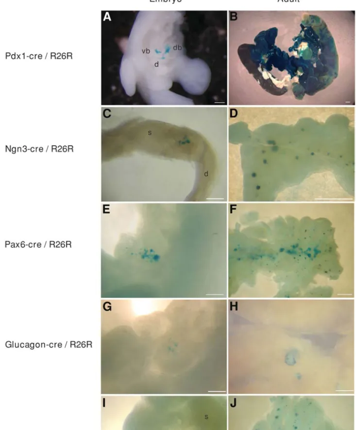
In the context of our work, this involved the genetic labeling of cells that transcribe Pdx1, Ngn3, Pax6, gluca-gon, insulin, or PP genes early during development, and the analysis of islet a and b cells, or the total pancreas, later in development or in the adult, to determine whether they are derived from these early Pdx1, Ngn3, Pax6, glucagon, insu-lin, or PP gene-expressing cells [Fig. 3 and material in prep-aration (70)]. To study the cell lineages of the endocrine pancreas, we used a silent loxP-bearing reporter transgene that is “activated” in a tissue-specific manner through the activity of Cre recombinase made from a second tagger Fig. 3. Pancreatic cell lineage analyses. To study the cell lineages of pancreatic cells, we used, as reporter mice, the R26R strain (74),
and five different transgenic mice as taggers: Pdx1-cre (70,71) (A, B), Ngn3-cre (P.L.H., unpublished results) (C, D), Pax6-cre (85) (E,
F), glucagon-cre (70) (G, H), and insulin-cre (70) (I, J), among others. PDX1 is an early marker of pancreatic bud cells (E8.5) and is
also found in the duodenal wall. In Pdx1-cre/R26R doubly transgenic embryos (A, B), b-galactosidase staining is found from E9.5 in the dorsal and ventral pancreatic buds and in the future duodenum (A, an E11.5 embryo is shown). In adults, the entire pancreas and many duodenal cells are stained (B). These results demonstrate that Pdx1+ cells and their progeny participate in the formation of these
Fig. 3. (Continued). two organs, and are pancreatic progenitor cells. NGN3 and PAX6 are shown to be essential for islet morphogenesis
(Table 1), and are expressed early (from E9.5) in pancreatic development. In Ngn3-cre/R26R (C, D) and Pax6-cre/R26R (E, F) mice, b-galactosidase activity is shown in dorsal pancreatic buds (C, E10.5; E, E11.5), and in all endocrine cells forming the adult islets (D, F), demonstrating that NGN3+ and PAX6+ cells are endocrine progenitor cells. In glucagon-cre/R26R (G, H) and insulin-cre/R26R (I, J) mice, b-galactosidase staining is found in pancreatic buds at E11.5 and earlier (G, I; E11.5 embryos are shown), and in a cells (H) and b cells (J) in mature islets, respectively. (Note in panel H that only peripheral islet a cells are blue.) All these results illustrate the power of the Cre/loxP strategy to study cell lineages. d, duodenum ; db, dorsal bud ; s, stomach ; vb, ventral bud. Scale bars are 0.25 mm in A, C, E, G, H, and I; 1 mm in B, D, F, and J.
transgene (73). The experimental design thus consists in generating mice bearing two transgenes (summarized in Fig. 2). One transgene, “reporter,” placed under the control of either tissue-specific (70) or ubiquitous promoters (61, 71,72,74–76), contains either the human growth hormone (hGH) (70), b-galactosidase (77), human alkaline phospha-tase (71,75), or EGFP (76) coding region placed downstream of a “floxed” (i.e., loxP-flanked) transcription termination site (STOP sequence) (78–80). Expression of the native reporter transgene requires the deletion of this sequence, which is obtained in the presence of Cre recombinase.
The tagger transgene consists of the Cre recombinase gene placed under the control of a promoter (transgenic or endog-enous, as in knockin mice) active in putative progenitor cells; its expression results in the irreversible deletion of the STOP (blocking) sequence of the reporter transgene, thereby allowing their expression, and thus labeling all progeny derived from those progenitors that express the tagger. Others have used this approach to activate the transcription of SV40 T antigen, LacZ, or placental alkaline phosphatase genes in transgenic mice (73,79,80), and to analyze the life span of circulating memory T cells (78), but not to trace cell lineages.
Using this genetic approach, which allows following cell lineages through the expression of Cre recombinase, it was first shown that islet a- and b-cell lineages appear to arise independently during ontogeny from a common Pdx1-expressing precursor [Fig. 3A and B; Fig. 4 (70)]. The cell ablation results (68) were thus confirmed and, in addition, it was discovered that the insulin cell lineage express the PP gene transiently, and that glucagon cells, which do not express Pdx1, actually descend from PDX1+ progenitors. This observation, combined with the fact that glucagon cells are present in early pancreatic buds in Pdx1-/- embryos, is illustrative of the different nature of early (embryonic) and mature (islet) glucagon cells.
A newly available ubiquitous reporter mouse, R26R, made it possible to analyze all pancreatic cell lineages (71,72,74). These studies confirmed that Pdx1 is expressed, at least transiently, in all pancreatic cell types (71), and that all islet endocrine cells derive from progenitors having transiently expressed Ngn3 [P.L.H. unpublished observations; Figs. 3C–D and 4 (72)].
The Cre-loxP system was initially devised to inactivate genes in selected tissues, rather than in the whole organism (45,46). A subsequent refinement of this system allowed
Fig. 4. Schematic representation of endodermal cell lineages as deduced from the Cre/loxP-mediated irreversible labeling of precursor
cells. Liver, pancreas, and intestine derive from the same embryonic layer, the endoderm. Several reports show that Pdx1 and p48 transcription factors are expressed in early presumptive pancreatic endodermal cells that are the progenitors of the entire pancreas
(61,71,72). p48 has been reported to be restricted later to the exocrine progenitor cells. Ngn3, which is expressed from E9.5 in early ducts
or cords, has been demonstrated to be expressed in the progenitors of endocrine cells [(72); present report, Figs. 3C and D]. Pax6, another transcription factor acting downstream of Pdx1, p48, and Ngn3, has also been shown to be expressed in endocrine progenitor cells [(85); present report, Figs. 3E and F]. That glucagon-producing a cells represent an independent lineage was shown in two different reports [(68,70); present report, Fig. 3H, in which a glucagon-cre transgene labels only a cells]. Insulin-containing b cells derive from PP-expressing progenitors, which are also required for the differentiation of somatostatin-containing d cells (68), either directly or noncell autonomously.
the exquisite temporal control of the recombinant activity of Cre in selected cells. One of these approaches consists of a fusion enzyme, Cre-ERT, in which the ligand-binding domain of a mutated estrogen receptor (ERT) that recognizes the antiestrogen tamoxifen (hydroxytamoxifen, 4-OHT), has been fused to Cre. In the absence of 4-OHT, Cre-ERT remains in the cytoplasm. Administration of 4-OHT results in its migration into the nucleus, as expected for a steroid hormone receptor/ligand complex. Accordingly, Cre-medi-ated recombination of genomic DNA is 4-OHT-dependent in mice bearing a Cre-ERT transgene (81,82). Because 4-OHT is active for less than 48 h in mouse embryos, Gu and co-workers were able to pulse labeling Ngn3- or Pdx1-expressing cells during 1–2 d windows of development by using Ngn3-CreERT and Pdx1-CreERT transgenes. The results with the latter transgene were unexpected, for early labeling (at E8.5) marked only acinar and islet cells, but not ducts; indeed, duct cells were labeled only when 4-OHT was administered between E9.5 and E11.5. At E12.5, or later, only acini and islets were marked. This indicates that ducts develop independently of Pdx1 activity after E12.5; in accor-dance with this interpretation, another elegant study in which pancreas development is modulated with doxycycline through activation or inactivation of Pdx1 activity, also points to a Pdx1-independent ductal development after this stage (83). The presence of NGN3+ cells in the adult islet was also demonstrated in Ngn3-CreERT transgenics. Such cell population might contribute to the maintenance of the adult islet mass (72).
Ptf1-p48 inactivation suggested that this gene is required for exocrine pancreas development (60). To trace the lin-eage of Ptf1-p48-expressing cells, Kawaguchi et al. (61) placed the Cre recombinase under the control of the endog-enous Ptf1-p48 promoter in a knockin experiment. Surpris-ingly, nearly all acinar, ductal, and insular cells expressed the ubiquitous reporter transgene in heterozygous Ptf1-p48-cre mice, indicating that Ptf1-p48 expression does not commit precursors to exocrine lineages, but to all pancre-atic cell fates (Fig. 4). In homozygous animals, the pheno-type was similar to that observed in Ptf1-p48-deficient mice (60), and, in addition, suggested that in the absence of PTF1-p48, pancreatic cells may revert to an intestinal epi-thelial fate. In summary, this result indicates that Ptf1-p48, like Pdx1, is expressed in the exocrine (acinar and ductal) and endocrine cell lineages of the pancreas, and is silenced in endocrine and duct cells shortly after they are committed.
Conclusions and Prospective
Understanding pancreatic morphogenesis and cell dif-ferentiation has obvious clinical implications. The poten-tial to form pancreatic endocrine tissue, whether from stem or differentiated cells, located in ducts or elsewhere in the pancreas (or even in extrapancreatic tissues), should not be restricted to the period of embryonic development. After
experimental injury, whether surgical (partial pancreatec-tomy, duct ligation) or chemical (such as streptozotocin or exendin-4 treatment), formation of new islets in adult rodents occurs with some efficiency. Pdx1 is expressed in pancreatic ductal cells during regeneration (84), as is ex-pressed in early pancreatic buds during development (57). However, we still must determine whether differentiation of new islets in the adult recapitulates the events taking place during development. In this context, the power and elegance of the Cre/loxP-based approach to track cell line-ages also reside in the versatility of the transgenic mice encoding Cre recombinase in different subsets of pancre-atic cell types. Thus, in order to address these challenging questions, after these mice have been used to determine cell lineage relationships during development, they are now unique tools to address the lineages during adult regenera-tion (neogenesis) and to inactivate specific genes in these same pancreatic cell lineages (“conditional” KO mice).
In conclusion, the Cre/loxP lineage tracing approach allows a better characterization of progenitor cells not only for the pancreas, but also for any organ. Improvement of this fundamental knowledge, i.e. the understanding of our body’s cell lineages, should be of help to devise, in the future, new sources of replacement tissues to treat degener-ative diseases resulting from massive cell demise.
Acknowledgments
We are grateful to Drs. K.A. Johansson, I. Rodríguez, P. Vassalli, J.-D. Vassalli, J. Huarte and S. Nef, and to Mss. A. Strom and C. Bonal, for carefully reading the manu-script. P.L.H. is supported by grants of the Juvenile Diabe-tes Research Foundation and the Swiss National Science Foundation.
References
1. Johansson, K. A. and Grapin-Botton, A. (2002). Clin. Genet. 62, 14–23.
2. Kim, S. K. and Hebrok, M. (2001). Genes Dev. 15, 111–127. 3. Alpert, S., Hanahan, D., and Teitelman, G. (1988). Cell 53,
295–308.
4. Kim, S. and MacDonald, R. (2002). Curr. Opin. Genet. Dev. 12, 540–547.
5. Hebrok, M., Kim, S. K., St Jacques, B., McMahon, A. P., and Melton, D. A. (2000). Development 127, 4905–4913. 6. Hebrok, M., Kim, S. K., and Melton, D. A. (1998). Genes Dev.
12, 1705–1713.
7. Kim, S. K., Hebrok, M., Li, E., et al. (2000). Genes Dev. 14, 1866–1871.
8. Gualdi, R., Bossard, P., Zheng, M., Hamada, Y., Coleman, J. R., and Zaret, K. S. (1996). Genes Dev. 10, 1670–1682. 9. Herrera, P. L., Huarte, J., Sanvito, F., Meda, P., Orci, L., and
Vassalli, J. D. (1991). Development 113, 1257–1265. 10. Gittes, G. K. and Rutter, W. J. (1992). Proc. Natl. Acad. Sci.
USA 89, 1128–1132.
11. Miralles, F., Battelino, T., Czernichow, P., and Scharfmann, R. (1998). J. Cell. Biol. 143, 827–836.
12. Conklin, E. G. (1905). J. Acad. Nat. Sci. Phila. 13, 1–119. 13. Stern, C. D. and Fraser, S. E. (2001). Nat. Cell. Biol. 3, E216–
14. Vogt, W. (1929). Roux’ Arch. EntwMech. Org. 120, 384–706. 15. Vogt, W. (1924). Wilhelm Roux Arch. Entwicklungsmech. Org.
120, 384–706.
16. Rosenquist, G. C. (1966). Contrib. Embryol. Carnegie Inst. 38, 71–110.
17. Stern, C. D. and Canning, D. R. (1990). Nature 343, 273–275. 18. Bagley, J., Aboody-Guterman, K., Breakefield, X., and Iacomini,
J. (1998). Transplantation 65, 1233–1240.
19. Cepko, C. L., Roberts, B. E., and Mulligan, R. C. (1984). Cell 37, 1053–1062.
20. Fishman, M. P. and Melton, D. A. (2002). Int. J. Dev. Biol. 46, 201–207.
21. Sanes, J. R., Rubenstein, J. L., and Nicolas, J. F. (1986). EMBO J. 5, 3133–3142.
22. Spemann, H. (1921). Roux’ Arch. EntwMech. Org. 48, 533–570. 23. Spemann, H. (1924). Roux’ Arch. EntwMech. Org. 100, 599–638. 24. Waddington, C. H. (1932). Phil. Trans. R. Soc. Lond. B 221,
179–230.
25. Le Douarin, N. (1973). Dev. Biol. 30, 217–222.
26. Oshima, H., Rochat, A., Kedzia, C., Kobayashi, K., and Barran-don, Y. (2001). Cell 104, 233–245.
27. Le Douarin, N. M. (1988). Cell 53, 169–171.
28. Martineau, J., Nordqvist, K., Tilmann, C., Lovell-Badge, R., and Capel, B. (1997). Curr. Biol. 7, 958–968.
29. Percival, A. C. and Slack, J. M. (1999). Exp. Cell Res. 247, 123–132. 30. Deltour, L., Leduque, P., Paldi, A., Ripoche, M. A., Dubois, P.,
and Jami, J. (1991). Development 112, 1115–1121.
31. Nicolas, J. F., Mathis, L., Bonnerot, C., and Saurin, W. (1996). Development 122, 2933–2946.
32. Mathis, L., Bonnerot, C., Puelles, L., and Nicolas, J. F. (1997). Development 124, 4089–4104.
33. Bonnerot, C. and Nicolas, J. F. (1993). C. R. Acad. Sci. III 316, 352–357.
34. Tan, S. S., Williams, E. A., and Tam, P. P. (1993). Nat. Genet. 3, 170–174.
35. Tam, P. P., Williams, E. A., and Tan, S. S. (1994). Dev. Genet. 15, 491–503.
36. Tam, P. P., Zhou, S. X., and Tan, S. S. (1994). Development 120, 2925–2932.
37. Tan, S. S., Faulkner-Jones, B., Breen, S. J., Walsh, M., Bertram, J. F., and Reese, B. E. (1995). Development 121, 1029–1039. 38. Bjerknes, M. and Cheng, H. (2001). Proc. Natl. Acad. Sci. USA
98, 12497–12502.
39. Windle, J. J., Weiner, R. I., and Mellon, P. L. (1990). Mol. Endocrinol. 4, 597–603.
40. Lew, D., Brady, H., Klausing, K., et al. (1993). Genes Dev. 7, 683–693.
41. Stewart, T. A., Pattengale, P. K., and Leder, P. (1984). Cell 38, 627–637.
42. Madsen, O. D., Karlsen, C., Blume, N., Jensen, H. I., Larsson, L. I., and Holst, J. J. (1995). Scand. J. Clin. Lab. Invest. Suppl. 220, 27–35.
43. Hanahan, D. (1985). Nature 315, 115–122.
44. Rindi, G., Ratineau, C., Ronco, A., Candusso, M. E., Tsai, M., and Leiter, A. B. (1999). Development 126, 4149–4156. 45. Rajewsky, K., Gu, H., Kuhn, R., et al. (1996). J. Clin. Invest.
98, 600–603.
46. Sauer, B. (1993). Meth. Enzymol. 225, 890–900.
47. Gittes, G. K., Galante, P. E., Hanahan, D., Rutter, W. J., and Debase, H. T. (1996). Development 122, 439–447.
48. Miettinen, P. J., Huotari, M., Koivisto, T., et al. (2000). Devel-opment 127, 2617–2627.
49. Miralles, F., Czernichow, P., and Scharfmann, R. (1998). Development 125, 1017–1024.
50. Sanvito, F., Herrera, P. L., Huarte, J., et al. (1994). Develop-ment 120, 3451–3462.
51. Sanvito, F., Nichols, A., Herrera, P. L., et al. (1995). Biochem. Biophys. Res. Commun. 217, 1279–1286.
52. Crisera, C. A., Maldonado, T. S., Kadison, A. S., et al. (2000). Differentiation 65, 255–259.
53. Bottinger, E. P., Jakubczak, J. L., Roberts, I. S., et al. (1997). EMBO J. 16, 2621–2633.
54. Ahlgren, U., Pfaff, S. L., Jessell, T. M., Edlund, T., and Edlund, H. (1997). Nature 385, 257–260.
55. Esni, F., Johansson, B. R., Radice, G. L., and Semb, H. (2001). Dev. Biol. 238, 202–212.
56. Schwitzgebel, V. M. (2001). Mol. Cell Endocrinol. 185, 99–108. 57. Jonsson, J., Carlsson, L., Edlund, T., and Edlund, H. (1994).
Nature 371, 606–609.
58. Offield, M. F., Jetton, T. L., Labosky, P. A., et al. (1996). Development 122, 983–995.
59. Heller, R. S., Stoffers, D. A., Hussain, M. A., Miller, C. P., and Habener, J. F. (1998). Gastroenterology 115, 381–387. 60. Krapp, A., Knofler, M., Ledermann, B., et al. (1998). Genes
Dev. 12, 3752–3763.
61. Kawaguchi, Y., Cooper, B., Gannon, M., Ray, M., MacDonald, R. J., and Wright, C. V. (2002). Nat. Genet. 32, 128–134. 62. Gradwohl, G., Dierich, A., LeMeur, M., and Guillemot, F.
(2000). Proc. Natl. Acad. Sci. USA 97, 1607–1611.
63. Apelqvist, A., Li, H., Sommer, L., et al. (1999). Nature 400, 877–881.
64. Grapin-Botton, A., Majithia, A. R., and Melton, D. A. (2001). Genes Dev. 15, 444–454.
65. St-Onge, L., Sosa-Pineda, B., Chowdhury, K., Mansouri, A., and Gruss, P. (1997). Nature 387, 406–409.
66. Sosa-Pineda, B., Chowdhury, K., Torres, M., Oliver, G., and Gruss, P. (1997). Nature 386, 399–402.
67. Breitman, M. L., Rombola, H., Maxwell, I. H., Klintworth, G. K., and Bernstein, A. (1990). Mol. Cell Biol. 10, 474–479. 68. Herrera, P. L., Huarte, J., Zufferey, R., et al. (1994). Proc. Natl.
Acad. Sci. USA 91, 12999–13003.
69. Jensen, J., Heller, R. S., Funder-Nielsen, T., et al. (2000). Diabetes 49, 163–176.
70. Herrera, P. L. (2000). Development 127, 2317–2322. 71. Gannon, M., Herrera, P. L., and Wright, C. V. (2000). Genesis
26, 143–144.
72. Gu, G., Dubauskaite, J., and Melton, D. A. (2002). Develop-ment 129, 2447–2457.
73. Jensen, J., Pedersen, E. E., Galante, P., et al. (2000). Nat. Genet. 24, 36–44.
74. Soriano, P. (1999). Nat. Genet. 21, 70–71.
75. Lobe, C. G., Koop, K. E., Kreppner, W., Lomeli, H., Gertsen-stein, M., and Nagy, A. (1999). Dev. Biol. 208, 281–292. 76. Mao, X., Fujiwara, Y., Chapdelaine, A., Yang, H., and Orkin,
S. H. (2001). Blood 97, 324–326.
77. Mao, X., Fujiwara, Y., and Orkin, S. H. (1999). Proc. Natl. Acad. Sci. USA 96, 5037–5042.
78. Jacob, J. and Baltimore, D. (1999). Nature 399, 593–597. 79. Brocard, J., Warot, X., Wendling, O., et al. (1997). Proc. Natl.
Acad. Sci. USA 94, 14559–14563.
80. Feil, R., Brocard, J., Mascrez, B., LeMeur, M., Metzger, D., and Chambon, P. (1996). Proc. Natl. Acad. Sci. USA 93, 10887–10890.
81. Weber, P., Metzger, D., and Chambon, P. (2001). Eur. J. Neuro-sci. 14, 1777–1783.
82. Hayashi, S. and McMahon, A. P. (2002). Dev. Biol. 244, 305– 318.
83. Holland, A. M., Hale, M. A., Kagami, H., Hammer, R. E., and MacDonald, R. J. (2002). Proc. Natl. Acad. Sci. USA 99, 12236–12241.
84. Sharma, A., Zangen, D. H., Reitz, P., et al. (1999). Diabetes 48, 507–513.
85. Ashery-Padan, R., Marquardt, T., Zhou, X., and Gruss, P. (2000). Genes Dev. 14, 2701–2711.
86. Ahlgren, U., Jonsson, J., and Edlund, H. (1996). Development 122, 1409–1416.
87. Harrison, K. A., Thaler, J., Pfaff, S. L., Gu, H., and Kehrl, J. H. (1999). Nat. Genet. 23, 71–75.
88. Li, H., Arber, S., Jessell, T. M., and Edlund, H. (1999). Nat. Genet. 23, 67–70.
89. Naya, F. J., Huang, H. P., Qiu, Y., et al. (1997). Genes Dev. 11, 2323–2334.
90. Clotman, F., Lannoy, V. J., Reber, M., et al. (2002). Develop-ment 129, 1819–1828.
91. Kim, S. K., Selleri, L., Lee, J. E., et al. (2002). Nat. Genet. 30, 430–435.
92. Sussel, L., Kalamaras, J., Hartigan-O’Connor, D. J., et al. (1998). Development 125, 2213–2221.