2To whom correspondence should be addressed
By using the selectively infective phage (SIP) technology, we selected non-repetitive linkers for a single-chain Fv fragment to have genes more robust against deletions in PCR-based gene assembly and directed evolution experi-ments than is the case for the classical (Gly4Ser)3 linker.
We designed linkers encoding turns at both ends and random positions in the middle where glycines and polar and charged residues were allowed to occur. After only a single round of SIP, all clones obtained were fully functional. Properties such as antigen binding constants, urea dena-turation curves and expression of soluble scFv fragments were identical with those of the parental fragment with the (Gly4Ser)3linker. This demonstrates that SIP is a very fast
and powerful technique to remove rapidly sequences of poor functionality, exclusively yielding sequences of the desired overall property in a single round.
Keywords: filamentous phage/fluorescein/library/scFv
frag-ment/selection
Introduction
Immunoglobulin fragments constitute a very popular subject for protein engineering. This is due to a large body of knowledge that has accumulated owing to extensive analyses of structural (Bork et al., 1994; Padlan, 1994) and physico-chemical properties (Plu¨ckthun, 1992) as well as progress in accessing antibody genes from various libraries (Winter et al., 1994). The enormous potential for diagnostic and therapeutic applications (George et al., 1994, 1996) keeps driving the technology forward.
The binding site of an antibody is made up from the VH
and VL domains, which associate only poorly to form an Fv
fragment, however. Thus, in general, they have to be linked to ensure a stable VH–VL complex in solution (Glockshuber
et al., 1990). This is commonly achieved by introducing a
peptidic linker (Bird et al., 1988; Huston et al., 1988) and the sequence (Gly4Ser)3 connecting the domains has been most
popular (Huston et al., 1991). The repetitive sequence of this linker, however, frequently causes problems during PCR-based assembly of amplified VH and VL domains (Winter et al.,
1994) or during the introduction of random mutations by mutagenic DNA shuffling (Stemmer, 1994). In these PCR-based technologies, the repetitive character of the scFv linker frequently leads to irregular annealing of homologous sequences and hence to undesired length variants.
To make scFv fragments more amenable to PCR-based gene
constructed an scFv library with several positions of the linker region fixed to encode particular structural features and with others randomized, but restricted to particular amino acid types. We used the selectively infective phage (SIP) technology (Spada and Plu¨ckthun, 1997; Spada et al., 1997) for identifying the most desirable sequences. In SIP, the scFv fragment is displayed fused to g3p, in all copies of the protein. In the present experiments, the phage is missing the N-terminal domain N1 and hence completely non-infective. Upon being provided with the soluble N1 domains, coupled to the antigen fluorescein, it becomes infective again (Figure 1). Already after one round of SIP, all of the selected clones bound to fluorescein at least as efficiently as the original FITC-E2. Three of the selected clones were characterized in more detail and turned out to be fully comparable to the original scFv fragment containing the (Gly4Ser)3linker in terms of binding,
folding, expression and solubility.
Materials and methods
Construction of linker library and SIP
The region coding for the randomized scFv linker was assembled by PCR from oligonucleotides LiLib (5 9-CT- GGGATCCMGRAGCAGAACYAGTNTBMGRAGCAGAA-CYACYNTBGCTCGCGCCGTTAGG-39) and LiFill (59-CT-GGTCACCGTCTCGAGTCCTAACGGCGCGAGC-39) (M 5 A or C; R5 A or G; Y 5 C or T; N 5 A, T, C or G; B 5 C, G or T). With the resulting cassette, the (Gly4Ser)3 scFv
linker of the scFlu-‘medium’ phage (Krebber et al., 1997b) was replaced in several cloning steps, resulting in the randomized linker shown in Figure 2A. All constructions were performed using standard methods of DNA manipulation (Sambrook et al., 1989). With the resulting scFlu-‘medium’ phage vectors, Escherichia coli XL1-Blue cells (Stratagene) were electroporated and grown overnight at 37°C on 2YTG plates (Krebber et al., 1997b) containing 25 µg/ml chloram-phenicol (cam), yielding 43106colonies, which were scraped
Fig. 1. Schematic representation of the SIP experiment. The N1 domain of
the phage gene 3 protein was replaced by the scFv FITC-E2 containing the randomized linker region. The resulting scFv-displaying phages are non-infective owing to the lack of the N1 domain. Infectivity is restored by binding of adapter molecules (N1-Flu) consisting of fluorescein (black circle) covalently coupled to the missing N1 domain.
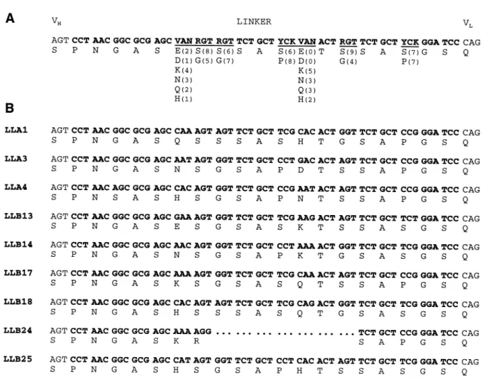
Fig. 2. Sequence of the randomized linker region. (A) The sequence of the partially randomized linker region (bold letters) including the last codon of the VH domain and the first codon of the VLdomain (non-bold letters) is shown. Randomized positions (using the IUPAC nomenclature of mixed bases: V5 A, C or G; N5 A, C, G or T; R 5 A or G; Y 5 C or T; K 5 G or T) are underlined. Below the nucleotide sequence, the amino acid residues which can be encoded are shown. Numbers in parentheses correspond to the frequencies of the respective residue types found in 14 clones of the unselected library. Where the sum of observed residues is 13 instead of 14, a different residue type was found. (B) Nucleotide and deduced amino acid sequences of nine random clones after one round of SIP.
from the plates and resuspended in 50 ml of fresh medium. A 50 ml volume of 2YTG containing 25 µg/ml cam was inoculated with 1 ml of the resulting suspension and incubated overnight at 37°C with shaking. Phage particles were precipitated from the culture supernatant by addition of 0.25 vol. 17% PEG 6000, 3.3 M NaCl, 1 mM EDTA, incubation at 4°C for 30 min and centrifugation at 4°C and 4500 g for 30 min. The pellet was resuspended in 1.5 ml of TBS (25 mM Tris–HCl, 140 mM NaCl, 2.7 mM KCl, pH 7.4), passed through a sterile filter (0.22µm), PEG precipitated again and resuspended in a final volume of 100µl of TBS. For selection of fluorescein binders, 1µl of the resulting phage suspension was incubated with 813µM N1-Flu (prepared as described by Krebber et al., 1997b) in a total volume of 6 µl in TBS. After overnight incubation at 4°C, 1 µl of this mixture was incubated with 100µl of an exponentially growing XL1-Blue culture at 37°C for 1 h and plated on 2YTG agar containing cam (25µg/ml) and tetracycline (15µg/ml).
Phage ELISA
For monovalent display of selected clones, the scFv encoding SfiI fragments were cloned into pAK100 and phage particles were prepared as described (Krebber et al., 1997a). For phage ELISA, a 96-well microtiter plate was coated with
fluorescein isothiocyanate-conjugated bovine serum albumin (FITC–BSA) overnight at 4°C and subsequently blocked for 1 h with 4% skim milk in TBS. Phagemid particles (108
per well) were incubated for 1 h in 100µl TBS 1 2% skim milk in the presence or absence of soluble fluorescein (10–7 M). After incubation with HRP–anti-M13 conjugate
(Pharmacia) for 1 h, relative amounts of bound phages were determined colorimetrically with soluble BM Blue POD sub-strate (Boehringer). For each phagemid clone, three independent measurements were performed in parallel.
Expression and purification of scFv fragments
For soluble expression of the scFv fragments, the correspond-ing SfiI fragments were cloned into pAK400 (Krebber et al., 1997a) and the resulting vectors were transformed into E.coli SB536 (Bass et al., 1996). The cells (1.5 l) were grown at 25°C, induced at an OD550 of 0.5, grown for another
3.5 h, centrifuged and resuspended in 15–20 ml of IMAC buffer (20 mM HEPES, 150 mM NaCl, pH 7.0) on ice and passed through a French press three times. The suspension was centrifuged (48 000 g, 4°C) and the supernatant was passed through a 0.22µm sterile filter. The protein was purified on an Ni-NTA column (Qiagen) and eluted with a step gradient at 100 mM imidazole. The scFv containing fractions were
20 mM HEPES, 150 mM NaCl, pH 6.8, overnight at 10°C. The exact urea concentration in each sample was determined from its refractive index (Pace and Scholtz, 1997). Upon equilibration at 20°C for at least 1 h, unfolding was monitored by fluorescence spectroscopy (λex 5 280 nm,
λem5 320–365 nm). From the wavelength shift of maximum
fluorescence, the fraction of scFv fragment unfolded at each distinct urea concentration was determined according to Pace and Scholtz (1997).
Detection of scFv variants in soluble and insoluble fractions of cell lysate
To compare the soluble expression of the scFv variants, the partitioning into soluble and insoluble fractions of crude E.coli lysate was determined by quantitative Western blotting. E.coli SB536 (Bass et al., 1996), harboring the pAK400-based secretion vectors (Krebber et al., 1997a), were grown at 25°C in 20 ml of 2YT medium (Sambrook et al., 1989) to an OD550
of 0.5, induced by addition of 1 mM IPTG and grown for a further 3.5 h. Upon centrifugation, resuspended cells were disrupted by three passages through a French press and recentrifuged (16 000 g, 15 min, 4°C). Upon removal of the supernatant containing the soluble fraction of total cellular protein, insoluble proteins were solubilized from the pellet in the presence of 2% SDS. Aliquots of soluble and insoluble fractions that corresponded to 25 µl of bacterial suspension with an OD550of 32 were subjected to SDS–PAGE and blotted
onto a nitrocellulose membrane. ScFv fragments were detected with an anti-His tag scFv–alkaline phosphatase fusion protein (Lindner et al., 1997).
Gel filtration
For analytical gel filtration, 50 µg of the purified scFv fragments were run on a Superose 12 PC 3.2/30 column on a SMART System (Pharmacia) in 20 mM HEPES, 150 mM NaCl, pH 6.8, at a flow rate of 60µl/min. The eluted proteins were monitored by measuring the absorbance at 280 nm.
Results
Construction of an scFv library with partially randomized linker region
As a model system for introducing a non-repetitive linker, we used the fluorescein binding antibody scFv fragment FITC-E2 (Vaughan et al., 1996), which was in the orientation VH–(Gly4Ser)3–VL. The linker was replaced by a randomized
oligonucleotide, designed according to the following principles. In modeling the scFv fragment it becomes obvious that the protein chain must make a turn in going from the end of VH
a proline-mediated kink or for the sequence Ser-Gly-Ser-Gln encoding a type II9β-turn (Mattos et al., 1994).
We chose the length of the linker to be 19 residues, which should be sufficient to drive the monomer–diabody equilibrium completely to the side of the monomers. Increasing the length of the linker favors monomers (Desplancq et al., 1994) and in this model system already the (Gly4Ser)3 linker gave
exclus-ively monomers (see Figure 6). In the region between the two turns, we wished to allow charged and polar residues in addition to a number of glycines, such that the conformation could adapt to the requirements and the preferred ionic composition would be selected. For instance, two glycine-rich spacers separate the domains of the minor coat protein g3p (Beck and Zink, 1981) and these contain a number of acidic residues.
We therefore designed the oligonucleotide shown in Figure 2A, where three different codon mixtures were specified, allowing for the amino acids given below the nucleotide sequence to occur with equal probability. To verify random-ization of the respective codons, 20 unselected clones of the library that were correct on the basis of restriction analysis were sequenced. Of these, six had an insertion or deletion in the linker region. Among the remaining 14 clones, the frequen-cies of different residues observed at each randomized position were almost as expected (Figure 2A).
SIP
The constructed scFv library was displayed on the scFv-‘medium’ SIP phage (Krebber et al., 1997b) and selected for fluorescein binding clones by one round of SIP using N1-Flu (Krebber et al., 1997b) as adapter (Figure 1). Fluorescein was chemically coupled to the N1 domain of g3p to give the N1-Flu adapter. This adapter has the advantage over N1-N2-N1-Flu that it does not inhibit infection at high concentrations (Krebber
et al., 1997b) and therefore its concentration is not critical as
long as it is high enough. The use of ‘medium’ phages (containing the N2 domain) results in higher infectivity than that of ‘short’ phages (not containing the N2 domain) (Krebber et al., 1997b). Growth of the phage library at 30°C instead of 37°C led to a larger number of clones obtained after SIP (80 000 vs. 2500), some of which, however, were only functional when regrown at 30°C but not when regrown at 37°C (data not shown).
Phage ELISA
Of the colonies obtained after SIP, 22 were picked randomly, grown in liquid culture and after overnight growth the culture supernatant was screened by ELISA for fluorescein binding
Fig. 3. ELISA of scFv displaying phagemid particles on FITC–BSA-coated
plates in the absence (filled bars) and presence (white bars) of 10–7M fluorescein as competitive inhibitor. Nine clones selected for fluorescein binding by one round of SIP as well as the original clone FITC-E2, containing the (Gly4Ser)3linker, are shown.
phage particles. For all clones, binding to the FITC–BSA coated plate could be detected, whereas before SIP selection, very few (1 out of 23) clones gave rise to an ELISA signal (data not shown). To confirm these results with monovalent display—the SIP phages lead to multivalent display—nine of the selected scFv variants were cloned to the phagemid vector pAK100 (Krebber et al., 1997a) and the antigen-binding ELISA was repeated with the resulting phagemid particles (Figure 3). Clone FITC-E2, containing the original (Gly4Ser)3
linker, also in the pAK100 format, was used as a control. All SIP selected variants investigated gave rise to an ELISA signal at least as high as that of clone FITC-E2 and all were efficiently inhibited in the presence of 10–7 M fluorescein.
Sequences
To determine whether selection for fluorescein binding resulted in selection for distinct sequences, the clones shown in Figure 3 were sequenced (Figure 2B). Except for the second randomized position, where exclusively serine residues were found, no obvious bias towards any particular sequence was observed. Owing to a deletion, clone LLB24 had a severely shortened linker of 12 amino acid residues that sterically should not allow intramolecular association of VHand VL domains. The
most plausible explanation of why this clone gives rise to an antigen-binding ELISA signal is that it contains an scFv diabody on the phage (McGuinness et al., 1996; Schier et al., 1996), with one monomer covalently fused to g3p, the other one being soluble owing to proteolytic cleavage of the scFv– g3p linker.
Determination of dissociation constants
The intensities of phage ELISA signals may be influenced by a number of parameters such as the antigen binding constant and the display frequency on the phage, which in turn is dependent on various properties such as folding efficiency, thermodynamic stability, proteolytic stability and solubility of the displayed scFv fragments. In order to determine which parameters to select for, three of the selected clones (LLA1, LLA4, LLB17) were subjected to a more detailed analysis of physicochemical properties. For this purpose, the respective
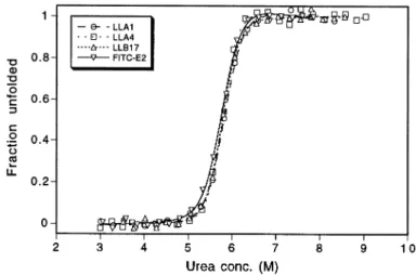
Fig. 4. Urea-induced unfolding of scFv variants.
Fig. 5. Quantitative Western blot analysis of the partitioning of scFv
variants into soluble (s) and insoluble (i) fractions of crude E.coli lysate. Protein samples loaded per lane correspond to equal amounts of cells. The electrophoretic mobility of marker proteins (Mr310–3) is indicated beside the blot.
Table I. Dissociation constants determined by fluorescence quench titration
scFv fragment KD(nM)
LLA1 0.56 (6 0.02)
LLA4 0.53 (6 0.05)
LLB17 0.72 (6 0.02)
FITC-E2 0.73 (6 0.09)
genes were cloned into the expression vector pAK400 (Krebber et al., 1997a) and soluble scFv protein was purified. Determination of affinities to fluorescein in solution by fluor-escence quench titration revealed dissociation constants of ~0.5–0.7 nM for all variants (Table I). The selected linker variants thus exhibited high affinities to fluorescein identical with the original FITC-E2, within experimental error.
Urea unfolding
In order to investigate the influence of the variant linker regions on conformational stability, reversible unfolding of the scFv fragments in the presence of increasing concentrations of urea was monitored (Figure 4). As is evident from the unfolding curves, all variants seem to be fairly stable, the midpoint of transition being ~5.8 M urea in each case. Furthermore, all variants can be closely superimposed on the wild-type with only subtle deviations.
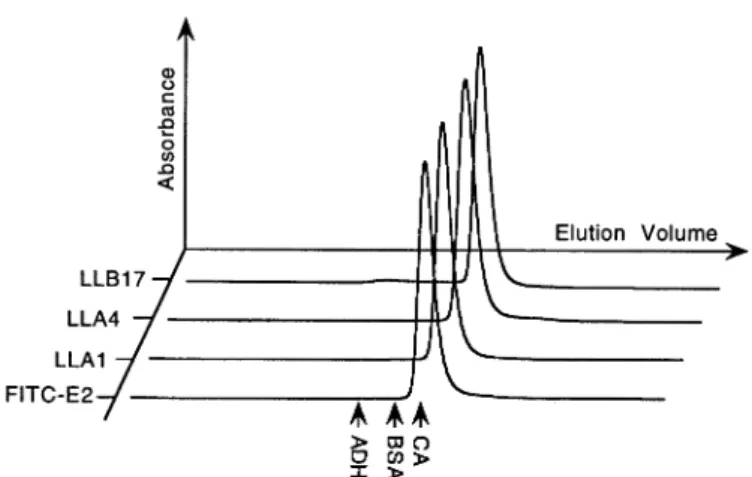
Fig. 6. Analytical gel filtration. The elution profiles of the scFv fragments
LLA1, LLA4, LLB17 and FITC-E2 are shown. The elution volumes of calibration proteins alcohol dehydrogenase (ADH) (Mr5 150 000), bovine serum albumin (BSA) (Mr5 66 000) and carbonic anhydrase (CA) (Mr5 29 000) are indicated by arrows.
Soluble expression of scFv fragments
To compare the soluble expression of the variants, the parti-tioning of the respective scFv proteins into soluble and insoluble fractions of crude E.coli lysates was determined by quantitative Western blot analysis (Figure 5). As is evident from the blot, the total expression level, proteolytic stability and the ratio of soluble and insoluble scFv fragments accumulated inside the cells are about the same for all variants and do not deviate significantly from the wild-type with the (Gly4Ser)3
linker.
Analytical gel filtration
To investigate whether the selected scFv linkers favor intra-molecular VH/VL association over the formation of diabodies,
the purified scFv fragments were subjected to analytical gel filtration (Figure 6). Only a single molecular species could be detected in all cases. From the elution volumes, apparent molecular masses of 28 800 (LLA1), 25 700 (LLA4), 28 300 (LLB17) and 28 100 (FITC-E2) were calculated, which are in good agreement with the theoretical value of 30 000. Hence all variants form exclusively monomers in solution.
Discussion
We have selected, by using SIP technology, a series of scFv linkers from partially designed and partially randomized non-repetitive linker sequences. The selected linkers give rise to scFv fragments which are indistinguishable in properties from that containing the original (Gly4Ser)3 linker, but are highly
advantageous in that they do not contain internal repeats that may lead to recombination and shortening of the linker during PCR. This feature will simplify the assembly of scFv fragments from amplified VL and VHby PCR. In addition,
non-repetit-iveness was previously shown to be an important prerequisite for the generation of scFv fragments by mutagenic DNA shuffling (Stemmer, 1994; Proba et al., 1998). Even though the three Gly4Ser repeats have often been encoded by different
codons to minimize this recombination (Ge et al., 1995; Krebber et al., 1997a), it cannot be completely eliminated (Proba et al., 1998). For example, in phage panning, shortened variants are frequently selected, presumably forming diabodies on the phage (Schier et al., 1996). Furthermore, a sequence stretch homologous to the linker exists at the beginning of
though the trend is very weak. In contrast to Tang et al. (1996) and Turner et al. (1997), we observed no influence of the linker on expression. However, these authors started with poorly expressed scFv fragments with the aim of increasing the yield of functional scFv protein. In contrast, the aim of our study was to generate highly functional scFv fragments containing non-repetitive linkers. Therefore, we used as a model system an scFv fragment with favorable overall proper-ties and compared the selected mutants with it. It seems that, in our study, any poorly expressed mutants which might conceivably have been generated by randomizing the linker sequence have been efficiently counterselected.
The new linkers appear to perform identically with the original (Gly4Ser)3 variant. This shows that, as expected,
the folding and expression properties of the scFv fragments are governed by the antibody domains and not by the linker. The linker thus appears to be a passive connector, consistent with the observation in NMR (Freund et al., 1993) that there are no cross peaks between linker and antibody domains and hence no stable contacts. Furthermore, both NOE and T2values
indicate an extremely high mobility (Freund et al., 1993), which is consistent with the fact that the linker has been invisible in X-ray structures of scFv fragments (Kortt et al., 1994; Zdanov et al., 1994). This is also borne out by molecular dynamics simulations (A.Caflisch, unpublished work), which indicate very high flexibility and almost no contact between the linker and the antibody domains. Nevertheless, we cannot conclude that the set of linkers selected in this study will be generally favorable independent of the context of the scFv sequence. However, here we have presented a reliable pro-cedure for easily adapting the sequences to any desired scFv fragment.
SIP technology was shown to lead to the selection of useful clones in a single round with immediate elimination of all non-functional linker sequences and most of the clones containing insertions or deletions. Although one could conceivably select for the best variants among the selected clones by further rounds of SIP, this would limit the variability to the given library diversity. Instead, we have now constructed a robust system which can be randomized by any number of PCR-based technologies and selected for the best variants including changes all over the protein.
Beyond the mere selection of functional non-repetitive scFv linkers, we have demonstrated with this study the application of SIP technology to the screening of a randomized immuno-globulin library. It stringently selects not only for high binding constants (already present in our starting molecule) but also for satisfactory overall properties of the displayed molecule,
including expression and folding. We believe that this techno-logy is a powerful tool for the molecular optimization of a wide variety of ligand binding proteins.
Acknowledgements
This work was supported by the Schweizerische Nationalfonds, grant 3100-046624.96/1. F.H. is the recipient of an EU fellowship, ERBCHBGCT 940720.
References
Bass,S., Gu,Q. and Christen,A. (1996) J. Bacteriol., 178, 1154–1161. Beck,E. and Zink, B. (1981) Gene, 16, 35–58.
Bird,R.E. et al. (1988) Science, 242, 423–426.
Bork,P., Holm,L. and Sander,C. (1994) J. Mol. Biol., 242, 309–320. Desplancq,D., King,D.J., Lawson,A.D.G. and Mountain,A. (1994) Protein
Engng, 7, 1027–1033.
Freund,C., Ross,A., Guth,B., Plu¨ckthun,A. and Holak,T.A. (1993) FEBS Lett.,
320, 97–100.
Ge,L., Knappik,A., Pack,P., Freund,C. and Plu¨ckthun,A. (1995) In Borrebaeck, C.A.K. (ed.), Antibody Engineering. Oxford University Press, Oxford, pp. 229–266.
George,A.J.T., Spooner,R.A. and Epenetos,A.A. (1994) Immunol. Today, 15, 559–561.
George,A., Huston,J.S. and Haber,E. (1996) Nature Biotechnol., 14, 584. Glockshuber,R., Malia,M., Pfitzinger,I. and Plu¨ckthun,A. (1990) Biochemistry,
29, 1362–1367.
Huston,J.S. et al. (1988) Proc. Natl Acad. Sci. USA, 85, 5879–5883. Huston,J.S., Mudgett-Hunter,M., Tai,M.-S., McCartney,J., Warren,F., Haber,E.
and Oppermann,H. (1991) Methods Enzymol., 203, 46–88. Kortt,A.A. et al. (1994) Eur. J. Biochem., 221, 151–157.
Krebber,A., Bornhauser,S., Burmester,J., Honegger,A., Willuda,J., Bosshard,H.R. and Plu¨ckthun,A. (1997a) J. Immunol. Methods, 201, 35–55. Krebber,C., Spada,S., Desplancq,D., Krebber,A., Ge,L. and Plu¨ckthun,A.
(1997b) J. Mol. Biol., 268, 607–618.
Lindner,P. et al. (1997) BioTechniques, 22, 140–149.
Mattos,C., Petsko,G.A. and Karplus,M. (1994) J. Mol. Biol., 238, 733–747. McGuinness,B.T., Walter,G., FitzGerald,K., Schuler,P., Mahoney,W.,
Duncan,A.R. and Hoogenboom, H.R. (1996) Nature Biotechnol., 14, 1149–1154.
Pace,C.N. and Scholtz,J.M (1997) In Creighton,T.E. (ed.), Protein Structure: a Practical Approach. IRL Press, Oxford, pp. 299–321.
Pace,C.N., Vajdos,F., Fee,L., Grimsley,G. and Gray,T. (1995) Protein Sci., 4, 2411–2423.
Padlan,E.A. (1994) Mol. Immunol., 31, 169–217.
Pedrazzi,G., Schwesinger,F., Honegger,A., Krebber,C. and Plu¨ckthun,A. (1997) FEBS Lett., 415, 289–293.
Perisic,O., Webb,P.A., Holliger,P., Winter,G. and Williams,R.L. (1994) Structure, 2, 1217–1226.
Plu¨ckthun,A. (1992) Immunol. Rev., 130, 151–188.
Proba,K., Wo¨rn,A., Honegger,A. and Plu¨ckthun,A. (1998) J. Mol. Biol., 275, 245–253.
Sambrook,J., Fritsch,E.F. and Maniatis,T. (1989) Molecular Cloning. A Laboratory Manual. Cold Spring Harbor Laboratory Press, Cold Spring Harbor, NY.
Schier,R., Bye,J., Apell,G., McCall,A., Adams,G.P., Malmqvist,M., Weiner,L.M. and Marks,J.D. (1996) J. Mol. Biol., 255, 28–43.
Spada,S. and Plu¨ckthun,A. (1997) Nature Med., 3, 694–696.
Spada,S., Krebber,C. and Plu¨ckthun,A. (1997) Biol. Chem., 378, 445–456. Stemmer,W.P.C. (1994) Proc. Natl Acad. Sci. USA, 91, 10747–10751. Stemmer,W.P.C., Morris,S.K. and Wilson,B.S. (1993) BioTechniques, 14,
256–265.
Tang,Y., Jiang,N., Parakh,C. and Hilvert,D. (1996) J. Biol. Chem., 271, 15682–15686.
Turner,D.J., Ritter,M.A. and George,A.J.T. (1997) J. Immunol. Methods, 205, 43–54.
Vaughan,T.J. et al. (1996) Nature Biotechnol., 14, 309–314. Wilmot,C.M. and Thornton,J.M. (1988) J. Mol. Biol., 203, 221–232. Winter,G., Griffiths,A.D., Hawkins,R.E. and Hoogenboom,H.R. (1994) Annu.
Rev. Immunol., 12, 433–455.
Zdanov,A., Li,Y., Bundle,D.R., Deng,S.-J., MacKenzie,C.R., Narang,S.A., Young,N.M. and Cygler,M. (1994) Proc. Natl Acad. Sci. USA, 91, 6423– 6427.
Received November 12, 1997; revised January 26, 1998; accepted February 4, 1998