ORIGINAL PAPER
Anthelmintic activity of medicinal plants used in Côte
d
’Ivoire for treating parasitic diseases
Witabouna Mamidou Koné&Mireille Vargas&
Jennifer Keiser
Received: 22 October 2011 / Accepted: 6 December 2011 / Published online: 27 December 2011 # Springer-Verlag 2011
Abstract Natural products play an important role in the discovery and development of new pharmaceuticals. In the present study, we assessed the anthelmintic properties of medicinal plants used in Cote d’Ivoire. Ethanolic extracts from 50 medicinal plants were tested in vitro against trem-atodes (Echinostoma caproni, Schistosoma mansoni) and nematodes (Ancylostoma ceylanicum, Heligmosomoides bakeri, Trichuris muris). Active extracts were evaluated for their cytotoxicity and followed up in vivo in mice harbouring adult S. mansoni, E. caproni and T. muris at single oral doses of 400 or 800 mg/kg. All extracts tested were active against at least one helminths species. Ten of the 65 extracts tested (15.4%) in vitro revealed activity against all helminths tested. Of 65 extracts tested in vitro at a concentration of 2 mg/ml, all caused death of schistosomula and 34.4% and 39.1% were lethal against adult S. mansoni and E. caproni 72 h post-incubation, respectively. The
highest activity against A. ceylanicum in vitro was observed with Sclerocarya birrea at 2 mg/ml, which resulted in death of adult worms and inhibition of activity of third-stage larvae (L3). Of the extracts, 41.5% completely inhibited movement of H. bakeri L3 at minimal lethal concentration (MLC) values of 20–200 μg/ml 48 h post-incubation, and 15.4% paralysed adult H. bakeri at 200 μg/ml 72 h after incubation. Of the extracts, 19% resulted in death of adult T. muris at MLC values of 10–100 μg/ml. In vivo, none of the extracts tested revealed activity against E. caproni. Olax subscorpioidea achieved total and female worm burden reductions of 60% and 84%, respectively in S. mansoni-infected mice. Combretum mucronatum was the most active extracts in vivo against T. muris with a worm burden reduc-tion of 85.3%. In conclusion, several of the medicinal plants used in Côte d’Ivoire are active against different helminths, hence might play a role in the treatment of helminthiases. Further studies are necessary to isolate the active compo-nents from these extracts.
Introduction
Soil-transmitted helminth (STH) infections, schistosomiasis and food-borne trematodiases are chronic, disabling parasit-ic diseases affecting millions of people mostly in the devel-oping world. These neglected tropical diseases mainly occur among the rural poor (Hotez and Kamath2009; Feasey et al. 2010). Globally, an estimated 207 million people are infected with schistosomes (Steinmann et al. 2006). A population of more than a billion is affected by soil-transmitted helminthiases (Bethony et al. 2006). Finally, 40 million people are infected with food-borne trematodes. Food-borne trematodiases are emerging in several countries due to a globalization of the food supply, increased
W. M. Koné
UFR des Sciences de la Nature, Université d’Abobo-Adjamé, BP 801,
Abidjan 02, Côte d’Ivoire W. M. Koné
Centre Suisse de Recherches Scientifiques en Côte d’Ivoire, BP 1303,
Abidjan 01, Côte d’Ivoire M. Vargas
:
J. KeiserDepartment of Medical Parasitology and Infection Biology, Swiss Tropical and Public Health Institute,
Socinstrasse 57, 4051 Basel, Switzerland M. Vargas
:
J. Keiser (*) University of Basel, Basel, Switzerland e-mail: jennifer.keiser@unibas.chinternational travel, population growth, pollution, ecological transformations or poverty (Dorny et al.2009; Keiser and Utzinger2009).
The consequences of these parasitic infections are man-ifold. STH, for example exacerbate malnourishment and can lead to anaemia, retarded growth, mental incapacity (Bethony et al.2006). Infections with food-borne tremato-des cause inflammatory lesions and tissue damage, which can result in serious secondary complications such as chol-angiocarcinoma in the case of infections with Clonorchis sinensis and Opisthorchis viverrini (Keiser and Utzinger 2009).
Only a handful of drugs are available for treatment of these diseases and most of them are very old (Keiser et al. 2006, 2010; Utzinger et al.2011). Concerns about the de-velopment of resistance arise from an increasing use of these drugs in mass drug administration programs and experien-ces made in the veterinary medicine, where resistance de-veloped shortly after different of the nematocidal drugs had been introduced (Chartier et al. 2001; Kotze et al. 2002; Almeida et al.2010). Hence new drugs should be discovered and developed.
The use of drugs derived from plants, fungi, bacteria and marine organism has a long tradition in medicine. Plants constitute a rich source of bioactive chemicals (Newman and Cragg 2007; Mondal and Khalequzzaman 2010). By 1990, approximately 80% of drugs were either natural prod-ucts or derivatives inspired by natural precursors (Li and Vederas2009). In addition, herbal medicines are generally more accessible and affordable (Fennell et al.2004) and are an important part of the culture and traditions of many populations. In the past years, many studies have been undertaken to assess the potential of medicinal extracts against parasitic diseases (Elango and Rahuman 2011; Ghosh et al. 2011; González-Coloma et al. 2011). Many patients from resource poor settings have strong beliefs in the use and efficacy of ethnomedicines (Chinsembu and Hedimbi2010). In Côte d’Ivoire, medicinal plants are wide-ly used against various diseases from parasitic and non-parasitic origin such as malaria, bacterial infection, rheumatic, gastro-intestinal parasite infections, diarrhoea, dysentery, res-piratory diseases and cough (Adjanohoun and Aké Assi1970, 1979; Bouquet and Debray1974; Koné et al.2002; Koné and Kamanzi Atindehou2008; Tra Bi et al.2005; N’Guessan et al. 2009).
The aim of the present study was to investigate the effects of selected medicinal plants from Côte d’Ivoire against a broad range of helminths, including the trematodes Schistosoma man-soni, Echinostoma caproni and the nematodes Ancylostoma ceylanicum, Heligmosomoides bakeri and Trichuris muris. It has been demonstrated in previous studies that different plants from West Africa investigated show a good efficacy against Haemonchus contortus, a sheep nematode (Diehl et al.2004;
Koné et al. 2005; Kamaraj et al. 2011), Caenorhabditis elegans, a free-living nematode (McGaw et al.2000,2007; Beloin et al.2005; Smith et al.2009; Waterman et al.2010) and Strongyloides ratti (Olounladé et al.2011).
The anthelmintic activity of the plants was first evaluated in vitro and promising extracts followed up in vivo.
Materials and methods Plant material
The 50 plants studied in the present work were selected based on results of a survey carried out between 1999 and 2005 in Northern Côte d’Ivoire on the use of medicinal plants for treating human parasitic diseases and associated discomforts (stomach ache, dysentery, diarrhoea (Koné 2005; Koné et al. 2002, 2005), animal parasitic diseases (Koné and Kamanzi Atindehou 2008) and on literature reports (Diehl et al.2004; Tra Bi et al.2005; Togola et al. 2008; N’Guessan et al. 2009). The studied plant material was harvested in Northern (Ferkessédougou), Central (Bouaké, Katiola), Western (Guiglo) and Southern (Petit Yapo) Côte d’Ivoire in 2005, 2008 and 2010. A detailed summary of the investigated plant material, including fam-ily, indications and which parts of the plants are used is presented in Table1. All samples collected were identified at the herbarium of the Centre Suisse de Recherches Scien-tifiques en Côte d’Ivoire (CSRS) where voucher specimens are maintained.
Preparation of plant extracts
The air-dried plant material was grounded and extracted under mechanical stirring in a tenfold excess of ethanol 90% at room temperature during 14 h. The filtrates were evaporated on a rotary evaporator (40°C), frozen and lyoph-ilized to yield the crude extracts. Sixty-five ethanolic crude extracts were then prepared for anthelmintic screening. The lyophilized extracts were solubilised in dimethylsulfoxide (DMSO) to prepare stock solutions (200 mg/ml) for in vitro studies. For in vivo assays, extracts were suspended in a solution composed of Tween 80 (70%) and ethanol (30%) and diluted with distilled water to obtain final concentra-tions of 7% Tween 80 and 3% ethanol.
Animals and parasites
The life cycles of the studied parasites are maintained at the Swiss Tropical and Public Health Institute (Swiss TPH) following animal welfare regulations. In vitro and in vivo studies were carried out in accordance with these guidelines. Female mice (NMRI strain, n090, weight ∼20–22 g) and
Table 1 Traditional uses of medicinal plants from Côte d’Ivoire selected for anthelmintic screening
Plant species (Family) Indications and plant parts used
Acacia nilotica (L.) Willd. ex Delile (Mimosaceae) Veterinary medicine: intestinal worms, anaemia Afzelia africana Sm. (Caesalpiniaceae) Malaria, stomach-ache (l)
Allophyllus africanus P. Beauv. (Sapindaceae) Malnutrition, diarrhoea (l) Ampelocissus africana (Lour.) Merr. (Vitaceae) Constipation, stomach-ache (r)
Annona senegalensis Pers. (Annonaceae) Diarrhoea, dysentery, abdominal pain, malaria (r) Anogeissus leiocarpus (DC.) Guill. & Perr. (Combretaceae) Malaria, diarrhoea, dysentery, anemia (l, sb) Anthostema senegalense A. Juss. (Euphorbiaceae) Intestinal worms (sb)
Antidesma venosum E. Mey. ex Tul. (Euphorbiaceae) Intestinal worms (sb)
Carissa edulis (Forssk.) Vahl. (Fabaceae) Diarrhoea in infants and children (r) Combretum mucronatum Schumach. & Thonn. (Combretaceae) Malaria, wound healing (l) Combretum platypterum (Welw.,) Hutch. & Dalziel (Combretaceae) Malaria (l)
Craterispermum caudatum Hutch. (Rubiaceae) Cough, abdominal pain (l)
Crossopteryx febrifuga (Afzel. ex G. Don) Benth. (Rubiaceae) Diarrhoea, malaria, abdominal pain (r, l) Diospyros mespiliformis Hochst. ex A. DC. (Ebenaceae) Diarrhoea, vomiting (sb)
Eriosema griseum Baker (Fabaceae) Infant cure (l)
Erythrina senegalensis DC. (Fabaceae) Diarrhoea, dysentery, abdominal pain, malaria, and infections of urinary tract, intestinal worms, cough, tuberculosis (r, sb) Flabellaria paniculata Cav. (Malpighiaceae) Malaria, wound healing (l)
Flueggea virosa (Roxb. ex Willd.) Voigt (Euphorbiaceae) Malaria, vomiting (l) Hybanthus enneaspermus (Linn.) F.V. Muell. (Violaceae) Malaria, aphrodisiac, cough (w)
Khaya senegalensis (Desr.) A. Juss. (Meliaceae) Malaria, cough, intestinal worms, diarrhoea (sb) Lannea barteri (Oliv.) Engl. (Anacardiaceae) Diarrhoea, sterility (l)
Lophira lanceolata Van Tiegh. ex Keay (Ochnaceae) Anaemia, diarrhoea with blood, cough, abdominal pain, anaemia (dl, sb) Milbraedia paniculata Pax (Euphorbiaceae) Cough (l)
Mimusops kummel Bruce ex A. DC. (Sapotaceae) Intestinal worms (sb)
Monotes kerstingii Gilg (Dipterocarpaceae) Intestinal worms, diarrhoea (sb, l) Morinda longiflora G. Don (Rubiaceae) Malaria, abdominal pain (r) Miytragyna ciliata Aubrév. & Pellegr. (Rubiaceae) Malaria (l)
Miytragyna inermis (Willd.) O. Kuntze. (Rubiaceae) Intestinal worms, malaria, diarrhoea (sb, l) Napoleona vogelii auct., p.p. Hook. & Planch. (Lecythidaceae) Diarrhoea (l)
Olax subscorpioidea Oliv. (Olacaceae) Intestinal worms, malaria (r)
Parinari curatellifolia Planch. ex Benth. (Chrysobalanaceae) Dysentery, diarrhoea, wound healing (sb) Parinari excelsa Sabine (Chrysobalanaceae) Constipation, abdominal pain (sb) Pavetta crassipes K. Schum. (Rubiaceae) Malaria, anorexia (l)
Pericopsis laxiflora (Benth. ex Baker) Meeuwen (Fabaceae) Intestinal worms, dysentery (sb) Piliostigma thonningii (Schumach. & Thonn.) Milne-Redh. (Caesalpiniaceae) Cough, wound healing (sb) Pseudarthria hookeri Wight & Arn. (Fabaceae) Cough (r)
Pseudocedrela kotsstchyi (Scweinf.) Harms (Meliaceae) Intestinal worms, abdominal pain (sb) Rauvolfia vomitoria Afzel. (Apocynaceae) Malaria, fever (sb)
Sacoglottis gabonensis (Baill.) Urban. (Humuriaceae) Abdominal pain (sb)
Sarcocephalus latifoliusa (Smith) Bruce (Rubiaceae) Intestinal worms, abdominal pain, fever, malaria (r) Sclerocarya birrea (A. Rich.) Hochst. (Anacardiaceae) Abdominal pain, cough (sb)
Securidaca longepedunculata Fres. (Polygalaceae) Intestinal worms, anorexia, urinary infections (r, l) Starchytarpheta cayennensis (Rich.) Vahl (Verbenaceae) Diarrhoea, dysentery (w)
Terminalia avicennioides Guill. & Perr. (Combretaceae) Intestinal worms, diarrhoea (sb) Thallia geniculata L. (Maranthaceae) Anaemia (r)
Trema orientalis (L.) Blume (Ulmaceae) Intestinal worms, diarrhoea (r, l) Vitellaria paradoxaum C. F. Gaertn. (Sapotaceae) Dysentery, diarrhoea (r)
Ximenia americana L. (Olacaeae) Diarrhoea, cough, wound healing (r) Xylopia aethiopica (Dunal) A. Rich. (Annonaceae) Intestinal worms, diarrhoea (sb, f) Ziziphus mauritiana Lam. (Rhamnaceae) Intestinal worms, diarrhoea (sb)
male Golden Syrian hamsters (n02, weight ∼100 g) were purchased from Harlan (Horst, The Netherlands), main-tained under environmentally controlled conditions and ac-climatized for 1 week before infection.
In vitro assays
All in vitro tests were carried out in duplicates. The exper-iment was repeated three times for extracts that showed an inhibitory effect.
In vitro assays against S. mansoni schistosomula S. man-soni schistosomula were prepared as described in previous publications (Manneck et al.2011). Briefly, cercariae of S. mansoni were harvested from infected intermediate host snails (Biomphalaria glabrata) after exposure to light for 4 h and subsequently transformed into schistosomula (newly transformed schistosomula, NTS). NTS were kept in Basch medium (TSS 199), supplemented with 5% heat-inactivated foetal calf serum (iFCS) and 100 U/ml penicillin and 100 μg/ml streptomycin (Invitrogen, Carlsbad, USA) at 37°C in an atmosphere of 5% CO2for a minimum of 3–
12 h before use. NTS-suspension (100μl) containing 500– 1,000 NTS was added to each well of 48-well plates (Costar). Stock solutions of the tested extracts were diluted in TSS 199 in order to obtain final concentrations of 2 mg/ml and 200μg/ml. The plates were incubated at 37°C in 5% CO2.
Morphological changes, paralysis or death of NTS were monitored after 24, 48 and 72 h. Extracts, which showed activity at 200μg/ml were further studied and serial dilutions were prepared from 160 to 5 μg/ml. The minimal lethal concentration (MLC), which is the minimum concentration needed to kill all schistosomula was determined after 24, 48 and 72 h. Schistosomula exposed to the highest concentration of solvent (1% DMSO) and worms incubated with praziquan-tel served as controls.
In vitro assays against adult S. mansoni Only the most active extracts (MLC05–40 μg/ml) against NTS were selected for screening against adult S. mansoni. Worms were collected from infected NMRI mice from the hepatic portal system and the mesenteric veins as described previously (Manneck et al. 2010). Cleaned worms (n04; 2 males and 2 females) were placed in each well of 24-well plates (Costar), in presence of extracts at 2 mg/ml or 200 μg/ml in RPMI 1640 culture medium supplemented with 5% iFCS and 1% pencillin–strep-tomycin. Schistosomes incubated with the highest concentra-tion of drug solvent served as controls. Worms were kept in an incubator at 37°C, 5% CO2for up to 72 h. The effect of the
extracts was assessed under a dissecting microscope with an emphasis on changes in worm motility (activity or paralysis) and death of worms. Death was defined as no movement observed for at least 2 min of examination. Active extracts
were further diluted (160–5 μg/ml) and tested as described above.
In vitro assays against adult E. caproni NRMI mice were orally infected with 25 metacercariae of E. caproni freshly collected from infected B. glabrata. Two weeks post-infection, mice were euthanized with CO2and adult worms
were removed from the intestine. Two worms were placed in each well of 24-well plates (Costar) in RPMI medium supplemented with 1% penicillin–streptomycin and 20% α-D-glucose. Adult E. caproni were incubated in the presence of 2 mg/ml or 200 μg/ml of the extracts in 5% CO2 atmosphere at 37°C for 72 h. Worms exposed to
medium supplemented with the highest concentration of DMSO (1%) served as control. Worms were monitored daily using a dissecting microscope and changes in worm motility and morphology and death recorded. Extracts active at 200μg/ml were diluted tenfold and re-tested.
In vitro assays against larvae of A. ceylanicum Third-stage larvae (L3) were cultured from the faeces of A. ceylanicum-infected Syrian Golden hamsters 20 days post-infection as described recently (Tritten et al. 2012). Thirty larvae were incubated in the presence of the individual extracts (2 mg/ml or 200 μg/ml) and 100 μl of Hank’s Basal Salt Solution (HBSS) supplemented with 10% of amphotericin B and 1% of penicillin–streptomycin in 96-well plates (Costar). Con-trol wells contained 1% DMSO only. The plates were stored at ambient temperature for 72 h. L3 were stimulated by adding 100 μl of warm water (∼80°C) to the wells. The number of moving larvae was counted and the percentage of survival calculated.
In vitro assays against adult A. ceylanicum Syrian Golden hamsters were orally infected with 150 A. ceylanicum L3 (Tritten et al. 2012). Twenty days post infection, hamsters were euthanized with CO2and worms collected from
diges-tive tract were placed in HBSS supplemented with 10% amphotericin B, 1% penicillin–streptomycin and 10% FCS. Three rinsed worms each were incubated in the pres-ence of 2 mg/ml or 200μg/ml of extracts in 48 well plates in 1,000 μl supplemented HBSS for 72 h at 37°C, 5% CO2.
After incubation, worms were stimulated with 500 μl of warm water (∼80°C) and death recorded.
In vitro assays against larvae of H. bakeri L3 were cultured from faeces of infected NRMI mice as described in a recent publication (Nwosu et al.2011). L3 (30 per well) were placed in 96 well plates (Costar) containing 100 μl HBSS supple-mented with 10% of amphotericin B and 1% of penicillin– streptomycin and three dilutions (2 mg/ml, 200 and 20μg/ml) of the extracts. Larvae exposed to 1% DMSO served as con-trols. The larvae were incubated for 72 h at room temperature.
After incubation, they were stimulated with 100 μl of hot water (∼80°C) and the percentage of inhibitory motility (IM) determined. Active extracts (IM value≥80%) at 20 μg/ml were further diluted to 2μg/ml and the activity assessed.
In vitro assays against adult H. bakeri Only extracts show-ing IM values of at least 80% at 20μg/ml against H. bakeri L3 were tested against the adult worms. For this test, NRMI mice were orally infected with 80 H. bakeri L3. One week post-infection, mice were euthanized with CO2 and adult
worms collected from the intestines. Worms were rinsed with RPMI medium and incubated in 48 well plates (Costar) in RPMI medium supplemented with 5% penicillin–strepto-mycin and 0.2% amphotericin. Adult H. bakeri were incu-bated in the presence of extracts (200 or 20μg/ml) at 37°C in 5% CO2atmosphere for 72 h. Changes in the motility and
morphology of adult H. bakeri were monitored under a dissecting microscope.
In vitro assays against adults of T. muris Adult T. muris were collected from the intestines of infected NRMI mice. Worms were rinsed in RPMI and four worms were placed in individual wells of 24 well plates (Costar), containing RPMI medium supplemented with 5% amphotericin and 1% pen-icillin–streptomycin and the test extracts (2 mg/ml and 200μg/ml). Plates were incubated in 5% CO2atmosphere
at 37°C. The effect of the plant extracts on the motility and morphology was documented using a dissecting microscope after 24, 48 and 72 h following incubation. Worms were classified as dead when no movement was observed for at least 2 min of examination. Extracts exhibiting activity at 200 μg/ml were serially diluted from 160 to 5 μg/ml and tested for activity against adult T. muris.
Cytotoxicity test on skeletal myoblast cells using Alamar Blue assay
Cytotoxicity tests on L-6 rat skeletal myoblast cells were carried out for all active (MLC≤80 μg/ml) extracts. L-6 rat skeletal myoblast cells were seeded in 96-well microtiter plates at a density of 105 ml−1in MEM supplemented with 10% heat-inactivated foetal bovine serum. Crude extracts at concentrations ranging from 90 to 0.001μg/ml were added. The plates were incubated for 72 h at 37°C in 5% CO2. After
70 h, 10μl of Alamar Blue® was added to each well. After a further incubation of 2 h, the plates were read using a fluorescence reader and the IC50 values calculated.
Podo-phyllotoxin was included as positive control. In vivo assays
In vivo activity against E. caproni The eight most active extracts from the in vitro screening were selected for in vivo
assays against E. caproni. NRMI mice were orally infected with 25 metacercariae of E. caproni and 2 weeks post-infection E. caproni-infected mice (four mice per extract) were treated with single oral doses of 800 mg/kg of the test extracts. In the first step, a high dose was used in order not to miss any activities. For comparison, 100% worm burden reductions were obtained with a single oral 50 mg/kg dose of praziquantel (Keiser et al. 2006). Four mice remained untreated and served as controls. One week post-treatment, mice were euthanized with CO2and dissected. Worms were
removed from the intestine and counted using a binocular microscope.
In vivo activity against S. mansoni Mice harbouring chronic (49 days post-infection) S. mansoni infections were treated at single oral doses of 400 mg/kg with the 11 extracts, which showed the highest activities in vitro. This dose was selected since praziquantel achieved a worm burden reduction of 93% following a dose of 400 mg/kg (Keiser et al. 2011). Groups of three to four mice were used per extract. One group of untreated mice served as controls (n08). Four weeks post-infection, mice were killed by cervical disloca-tion and the mesentery and the liver were collected as described previously by Manneck et al. (2011). Schisto-somes were removed from the mesenteric veins, sexed and counted using a binocular microscope. The liver was flat-tened and examined for the presence of worms. Worm count was compared to untreated controls.
In vivo activity against T. muris NRMI mice were orally infected with 400 eggs of T. muris. Fourty five days post infection, four mice were treated with a single dose of 400 mg/kg of each of the five extracts, which had shown the highest activities in vitro. One week post-treatment, stools were harvested and analysed under binocular microscope for count-ing expelled worms. Mice were euthanized and dissected, and the intestine was examined for the presence of worms. Worm expulsion rate and worm burden reduction were calculated. Statistical analyses
Statsdirect statistical software (version 2.4.5, Statsdirect Ltd, Cheshire, UK) was used to compare the medians of the worm counts with a Kruskal–Wallis test. Differences in medians were considered to be significant at a significance level of 0.05.
Results
All the extracts screened in this study were active against at least one of the helminth species tested, causing reduced viability, paralysis or death of the worms. Overall, ten
(15.4%) of the 65 extracts tested in vitro showed activity against all (n05) helminths tested, hence displayed both trematocidal and nematocidal properties. The extracts with the broadest spectrum of activity were prepared from the roots and stem bark of Anogeissus leiocarpus, roots of Crossopteryx febrifuga, leaves of Eriosema griseum, leaves of Combretum mucronatum, roots of Monotes kerstingii, stem bark of Xylopia aethiopica, roots of Securidaca longe-pedunculata, roots of Olax subscorpioidea, roots of Saco-glottis gabonensis and roots of Erythrina senegalensis. Of the remaining extracts, 6 (9.2%) extracts showed activity against 4 helminths, 17 (26.2%) revealed activity against 3 worms, and 19 (29.2%) and 13 (20%) were active against 2 helminths and 1 worm, respectively.
Activity against S. mansoni NTS and adults in vitro The screening of plant extracts against schistosomes showed that NTS were more affected by these products than the adult worms of S. mansoni. Of the 65 extracts tested at 2 mg/ml, 53 (84.4%) were active against NTS. Schistoso-mula had died in the presence of these extracts after 24 h. This percentage increased to 98.4% and 100% 48 and 72 h post-incubation, respectively. When tested at 200μg/ml, 28 (42.2%) extracts caused death of NTS after 24 h, 45 (70.3%) after 48 h and 54 (84.4%) after 72 h of incubation. Follow-ing further dilutions, seven extracts still showed a high activity against NTS, namely the stem bark of S. gabonensis (MLC05 μg/ml), roots and stem bark of A. leiocarpus, leaves of Craterispermum caudatum, roots of E. senegal-ensis, leaves of Lannea barteri and stem bark of X. aethiop-ica (all MLC010 μg/ml; Table2). Extracts of the leaves of C. mucronatum, leaves of E. griseum, leaves of Flabellaria paniculata, roots of M. kerstingii, and stem bark of Pseu-docedrela kotsstchyi resulted in death of NTS at 20μg/ml.
Of the 30 extracts tested against adult schistosomes (characterized by an MLC of 5–40 μg/ml against NTS) at a concentration of 2 mg/ml, 17 (26.6%), 21 (31.8%) and 22 (34.4%) extracts caused death of adult schistosomes 24, 48 and 72 h post-incubation, respectively. Fifteen extracts and one product displayed an activity at tenfold and 100-fold lower concentrations (200 and 20μg/ml), respectively 48 h post-incubation. The most active extracts were the roots of C. febrifuga with a MLC of 10μg/ml, the leaves of E. griseum (MLC040 μg/ml), the stem bark of S. gabonensis and the roots of S. longepedunculata with MLCs of 80μg/ml (Table2). Activity against E. caproni in vitro
Fourteen (21.9%), 20 (31.3%) and 25 (39.1%) of the extracts tested caused death of E. caproni at a concentration of 2 mg/ml 24, 48 and 72 h post-incubation, respectively. At 200μg/ml worms were killed by 15 (23.1%) extracts 72 h
post-incubation. The highest echinostomicidal activity (MLC020 μg/ml) was observed with extracts prepared from the leaves of Milbraedia paniculata, roots of S. longepe-dunculata and the stem bark of X. aethiopica (Table 2). These extracts caused death of the worms between 48 and 72 h and a change of morphology (colour, granulation) was observed following incubation with S. longepedunculata and X. aethiopica.
Activity against A. ceylanicum in vitro
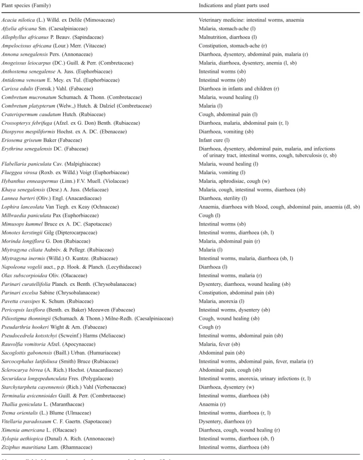
With the exception of nine extracts (roots of Ampelocissus grantii, stem bark of Khaya senegalensis, stem bark of P. kostchyi, stem bark of S. gabonensis, stem bark and roots of Sclerocarya birrea, leaves of E. griseum, stem bark of Termi-nalia avicennioides and stem bark of Ziziphus mauritiana), which exhibited moderate activity (percentage of worms sur-viving ranging between 10% and 40%) 48 h post-incubation, none of the studied extracts showed an activity against A. ceylanicum L3 in vitro at a concentration of 2 mg/ml. At 200μg/ml, only the root extract of S. birrea showed activity (survival of 36.6%) at the 48 h examination point.
Only the root extract of S. birrea killed adult A. ceylani-cum 72 h after incubation, when exposed to a high concen-tration of 2 mg/ml (Fig.1). At the same concentration, six extracts from C. mucronatum (leaves), S. longepedunculata (roots), M. kerstingii (leaves), Z. mauritiana (stem bark), L. barteri (roots) and O. subscorpioidea (roots) reduced the viability of adult worms and paralysed the worms (72 h post incubation), however did not result in killing of worms. Activity against H. bakeri in vitro
Fourteen (23.1%) and 12 (18.5%) of the products (derived from A. leiocarpus, C. febrifuga, E. griseum, Flueggea virosa, L. barteri, Lophira lanceolata, M. kerstingii, Napoleona voge-lii, Pericopsis laxiflora, S. gabonensis and S. longepeduncu-lata) reduced motility of H. bakeri L3 by at least 80% at concentrations of 200 and 20 μg/ml 48 h post-incubation, respectively (Table2). When extracts (IM value≥80%) were tested against adult H. bakeri, only ten (15.4%) extracts exhibited activity. These extracts were prepared from M. kerstingii (leaves), Annona senegalensis (roots), T. avicen-nioides (stem bark), S. longepedunculata (leaves), M. panicu-lata (leaves), Mimusops kummel (roots and stem bark), Miytragyna ciliata (leaves), S. gabonensis (roots) and of C. mucronatum (leaves). All ten extracts caused death of worms at 2 mg/ml 48 h post incubation. Exposure to 200μg/ml of these ten extracts resulted in reduced movements and paralysis of worms 72 h post-incubation. The extracts from the roots of E. senegalensis and the stem bark of X. aethiopica caused twisting and death of the worms at 200μg/ml after 72 h of incubation.
Activity against T. muris
Fourteen (21.5%), 37 (56.9%) and 45 (69.2%) of extracts caused death of adult T. muris at 2 mg/ml 24, 48 and 72 h post-incubation, respectively. Eight extracts displayed a high activity with MLC values ranging between 5 and 20μg/ml 72 h post-incubation. These extracts were prepared from the roots and stem bark of A. leiocarpus, the leaves of C. mucronatum, the roots of C. febrifuga, the stem bark of P. kotsstchyi, the roots
of S. longepedunculata, the stem bark of T. avicennioides and the roots of M. kerstingii (Table2).
Cytotoxicity of the tested extracts
The evaluation of the cytotoxicity of 22 tested extracts on L6 rat skeletal myoblast cells showed either no toxicity (IC50>90μg/ml) or only a very low toxicity of 4 extracts
(roots of A. leiocarpus, leaves of C. mucronatum, roots of
Table 2 Minimum lethal con-centrations (microgrammes per millilitre) of studied medicinal plants against E. caproni, S. mansoni, T. muris and H. bakeri in vitro
nd non determined (MLC against NTS>40μg/ml)
Plant species Plant part tested Helminths tested
E. caproni S. mansoni T. muris H. bakeri Adults NTS Adults Adults L3 larvae Anogeissus leiocarpus Roots 200 10 250 10 20
Stem bark >2,000 10 160 10 >2,000 Anthostema senegalenseis Stem bark >2,000 40 2,000 2,000 200 Combretum mucronatum Leaves >2,000 20 200 10 2,000 Craterispermum caudatum Leaves 2,000 10 200 >2,000 2,000 Crossopteryx febrifuga Roots 160 40 10 20 20 Eriosema griseum Leaves 2,000 20 40 2,000 20 Erythrina senegalensis Roots 80 10 160 2,000 200 Flabellaria paniculata Leaves >2,000 20 200 2,000 200 Khaya senegalensis Stem bark >2,000 40 1,000 2,000 >2,000 Lannea barteri Roots 2,000 40 2,000 2,000 20 Leaves 2,000 10 200 2,000 200 Milbraedia paniculata Leaves 20 40 200 2,000 200 Mimusops kummel Roots 160 80 nd 2,000 2,000 Stem bark 2,000 40 200 2,000 20 Monotes kerstingii Stem bark 2,000 40 160 2,000 200 Leaves >2,000 2,000 nd 2,000 20 Roots 80 20 200 40 2,000 Morinda longiflora Roots 200 200 nd >2,000 2,000 Napoleona vogelii Leaves 2,000 80 nd 2,000 20 Olax subscorpioidea Roots 80 40 160 100 2,000 Parinari curatellifolia Stem bark >2,000 80 nd 2,000 >2,000 Parinari excelsa Stem bark >2,000 160 nd 100 2,000 Leaves >2,000 200 nd >2,000 20 Piliostigma thonningii Stem bark 2,000 40 2,000 >2,000 >2,000 Pseudocedrela kotsstchyi Stem bark >2,000 20 500 20 >2,000 Sacoglottis gabonensis Stem bark 200 5 80 2,000 20 Sarcocephalus latifoliaus Roots 2,000 160 nd >2,000 2,000 Sclerocarya birrea Stem bark >2,000 40 2,000 100 >2,000 Roots >2,000 200 nd 100 2,000 Securidaca longepedunculata Roots 20 40 80 20 20 Leaves 200 40 250 >2,000 200 Starchytarpheta cayennensis Whole plant 160 40 160 2,000 200 Terminalia avicennioides Stem bark 200 40 500 20 2,000 Leaves >2,000 40 500 2,000 2,000 Xylopia aethiopica Stem bark 20 10 160 2,000 200
O. subscorpiodiea and stem bark of S. gabonensis) (IC50
31.8–43.3 μg/ml) when compared to the positive control (Podophyllotoxin IC500.004μg/ml).
In vivo anthelmintic efficacy of plants
In vivo assays were carried out with extracts, which dis-played the highest activity against adult S. mansoni (n011; MLC of 20–160 μg/ml), E. caproni (n08; MLC020– 200 μg/ml) and T. muris (n05; MLC of 10–20 μg/ml) in vitro. We did not carry out in vivo studies in H. bakeri-infected mice because none of the extracts revealed prom-ising activity against adult H. bakeri in vitro. In addition, in vivo studies in A. ceylanicum-infected hamsters were not done, as only the root extract of S. birrea was active at a high concentration of 2 mg/ml against adult worms. Activity against E. caproni
None of the tested extracts showed activity against E. cap-roni in vivo. Worm burden reductions achieved with a single oral dose of 800 mg/kg ranged between 0% and 21% for these extracts (P>0.05; Table3).
Activity against T. muris
At a single oral dose of 400 mg/kg, one of the five extracts tested in vivo against T. muris, prepared from the leaves of C. mucronatum showed a significant effect (P00.038) with a worm burden reduction of 85.3%. Low to moderate worm burden reductions (3.1–40.7 %) were observed with the remaining four extracts (Table3).
Activity against S. mansoni
Low total worm burden (0–16.1%) and female worm burden (0–11.1%) reductions were documented for the extracts of A. leiocarpus (roots), E. senegalensis (roots), M. kerstingii
(roots), S. longepedunculata (roots), Starchytarpheta cayen-nensis (whole plant), S. gabocayen-nensis (stem bark) and X. aethiopica (stem bark). Moderate worm burden reductions (total worm burden reduction of 49.5% and female worm burden reduction of 48.9%) were achieved with the leaves of E. griseum. With the stem bark of A. leiocarpus and roots of C. febrifuga, moderate total worm burden (46.3–57.0%) and high female worm burden (71.1%) were observed. The highest activity in S. mansoni-infected mice (total worm bur-den reduction of 60.2% and female worm burbur-den reduction of 84.5% (which however was not significant since only a small number of mice were used) was observed with the roots of O. subscorpiodea (Table4).
Discussion
In the present study, as many as 50 medicinal plants used in Côte d’Ivoire and elsewhere in West Africa were evaluated against a wide range of helminths, namely the trematodes E. caproni and S. mansoni and the nematodes A. ceylanicum, H. bakeri and T. muris. In addition, with regard to A. ceylanicum, H. bakeri and S. mansoni, the products were not only studied against the adult stages but also against larval stages (A. ceylanicum and H. bakeri) and S. mansoni schistosomula. The rationale for selecting these plants for testing against the helminths arose from their traditional uses against parasitic diseases and the anthelmintic activity of several extracts against L3 of H. contortus (Diehl et al. 2004; Koné et al. 2005) and Caenorhabditis elegans (McGaw et al.2007; Smith et al.2009). To our knowledge, the antischistosomal, trichuricidal, ancylostomicidal and echinostomicidal activities of most of the studied plants are reported here for the first time.
Several plant extracts showed an in vitro effect against all five trematodes and nematodes studied, which might be explained by the synergistic combination among multiple bioactive compounds contained in the crude extracts (Viljoen
Fig. 1 In vitro effect of plant extracts on viability of adult A. ceylanicum. Scl bir,
Sclerocarya birrea; Sec lon, Securidaca longepedunculata Mon ker, Monotes kerstingii; Ziz mau, Ziziphus mauritiana; Ola sub, Olax subscorpioidea; Com muc, Combretum mucronatum; Lan bar, Lannea barteri
et al. 2005; Karmegam et al. 2008). Three compounds revealed high in vivo activities, namely O. subscorpioidea, A. leiocarpus and C. mucronatum. Promising antischistoso-mal properties were observed when S. mansoni-infected mice
were treated with the extract of the root of O. subscorpioidea. The roots of O. subscorpioidea are used in Northern Côte d’Ivoire for treating intestinal worms (Koné et al.2005) and malaria (Gauthier-Beguin1992). It is interesting to note that in
Table 3 Effect on worm burden of 12 active extracts administered at a single dose of in E. caproni-infected (800 mg/kg) and T. muris-infected (400 mg/kg) mice
Plant tested E. caproni T. muris Mean number
of worms (SD)
Worm burden reduction (%)
P values Mean number of worms (SD) Worm expulsion rate (%) Worm burden reduction (%) P values Control 29.6 (10.2) 89 (42.8) 125 (75.7) Anogeissus leiocarpus (sb) ND 57.7 (113.8)a 5.2 6.2 0.436 Anogeissus leiocarpus (r) ND 90.0 (137.8)b 9.4 27.7 0.436 Combretum mucronatum (l) ND 18.3 (18.0)b 44.4 85.3 0.038 Crossopteryx febrifuga (r) 34.3 (5.8) 0 89.7 (171.5)a 3.9 3.1 0.194 Eriosema griseum (l) ND ND Erythrina senegalensis (r) ND ND Monotes kerstingii (sb) 26 (11.3) 12.2 0.46 ND Lannea barteri (l) 26.7 (11.0) 9.9 0.65 ND Olax subscorpioidea (r) 25 (3.0) 15.5 >0.99 ND Sacoglottis gabonensis (sb) 33.3 (9.0) 0 0.55 ND Securidaca longepedunculata (r) 26 (12.7) 12.2 0.44 ND Starchytarpheta 23.3 (5.5) 21.2 0.46 Terminalia avicennioides (sb) ND 52.7 (69.6)a 0 40.7 0.28 Xylopia aethiopica (sb) 32.3 (6.6) 0 0.46 ND Podophyllotoxin ND ND
ND non determined, r roots, l leaves, sb stem bark, w whole plant
a
Versus control 1
bVersus control 2
Table 4 Effect on worm burden of 11 active extracts administered at a single dose of 400 mg/kg to mice harbouring adult S. mansoni Plant species and control Mean number
of worms (SD)
Total worm burden reduction (%)
P values Female worm burden reduction (%) P values Total Females Control 9.3 (7.5) 4.5 (4.2) – – – – Anogeissus leiocarpus (sb) 4.0 (1.0) 1.3 (0.6) 57.0 0.44 71.1 0.49 Anogeissus leiocarpus (r) 7.8 (8.3) 4.3 (3.9) 16.1 0.44 4.5 0.17 Crossopteryx febrifuga (r) 5.0 (2.6) 1.3 (1.5) 46.3 0.49 71.1 0.26 Eriosema griseum (l) 4.7 (2.5) 2.3 (1.8) 49.5 0.55 48.9 0.55 Erythrina senegalensis (r) 14.7 (10.3) 5.7 (4.6) 0 na 0 na Olax subscorpioidea (r) 3.7 (0.6) 0.7 (1.6) 60.2 0.35 84.5 0.12 Sacoglottis gabonensis (sb) 10.8 (11.7) 4.8 (5.4) 0 na 0 na Securidaca longepedunculata (r) 10.7 (1.1) 4.3 (2.1) 0 na 4.5 na Starchytarpheta 13.7 (9.6) 6.0 (5.6) 0 na 0 na Xylopia aethiopica (sb) 9.0 (8.5) 4.0 (4.2) 3.2 >0.99 11.1 >0.99 r roots, l leaves, sb stem bark, w whole plant, na not applicable
particular, female schistosomes were highly affected in vivo (female worm burden reduction of 84.5%). On the other hand, no effect was observed against adult E. caproni in vivo, pointing to a very specific mechanism of action of this drug. The phytochemicals present in O. subscorpioidea are known only for the seeds of this plant, with santalbic acid being the principle component (Cantrell et al.2003). Studies have been launched therefore in our laboratories to isolate the active components of O. subscorpioidea to be able to characterize the interesting antischistosomal properties in further detail. O. subscorpioidea has not only trematocidal but also nematocidal properties. T. muris were highly affected by this plant in vitro. In addition, a previous study found that O. subscorpioidea possessed larvicidal activity against H. contortus L3 (Koné et al.2005).
The extract prepared from the stem bark of A. leiocarpus achieved a worm burden reduction of 71.1% against female S. mansoni. To our knowledge, the antischistosomal activi-ties of A. leiocarpus have been described for the first time. Further studies are necessary to strengthen and confirm these positive findings, including experiments in larger an-imal group sizes and to also study the effect against juvenile schistosomes. Previous studies reported promising anthel-mintic activities of A. leiocarpus against Rhabditis pseudoe-longata (Okpekon et al.2004) and H. contortus (Diehl et al. 2004; Koné et al.2005) and antiparasitic properties against Plasmodium falciparum (Gansané et al. 2010). The stem bark of A. leiocarpus was found to contain gallic and gen-tisic acids (Chaabi et al.2008; Shuaibu et al.2008), which have recently been shown to possess anthelmintic activity (Smith et al.2009). Studies should be undertaken to isolate the components of A. leiocarpus stem bark responsible for the antischistosomal activities.
High trichuricidal activity was documented for C. mucro-natum, a plant used in Côte d’Ivoire against malaria, and in Ghana against guinea worm infestation (Ibrahim1986). We observed a high worm burden reduction of 85.3% in vivo against T. muris. To our knowledge, this is the first report of the anthelmintic activity of C. mucronatum; its phytochemicals have not been studied to date.
Disappointingly, E. senegalensis lacked activity against adult S. mansoni in vivo despite promising activities ob-served against both stages in vitro. This plant is applied in traditional medicine in the treatment of urinary bilharziosis (Togola et al.2008). The efficacy against juvenile S. man-soni in vivo remains to be tested. It is possible that higher worm burden reductions will be achieved against the juve-nile worms in vivo, since this stage was more susceptible than the adult worms in vitro (10 versus 160μg/ml).
In this study, we also noted an activity of E. senegalensis against the mouse hookworm, H. bakeri, however T. muris and A. ceylanicum were not affected. E. senegalensis also lacked activity against H. contortus in a previous study
(Koné et al.2005). One of the phytochemicals of E. senegal-ensis (Taylor et al. 1986; Wandji et al. 1990; Lee et al. 2009), alpinumisoflavone, was also isolated from Milletia thonningii, a medicinal plant used in Ghana as anthelmintic (Abbiw 1990). It has been demonstrated that alpinumiso-flavone present in M. thonningii was highly active against S. mansoni miracidia, cercariae and adult worms in vitro (Perrett et al. 1995; Lyddiard and Whitfield 2001; Lyddiard et al.2002). The presence of alpinumisoflavone in E. senegalensis might contribute to the in vitro activity against juvenile and adult S. mansoni and H. bakeri observed in this study.
In conclusion, the present study screened the in vitro and in vivo anthelmintic activity of 50 extracts of plant species used in Côte d’Ivoire to treat parasitic diseases. Ten of these plants exhibited significant in vitro activity against trematodes (E. caproni and S. mansoni) and nematodes (H. bakeri and T. muris), providing some validation for their traditional uses. In vivo studies showed high antischistosomal activities of O. subscorpioidea and high trichuricidal activity of C. mucro-natum. For O. subscorpioidea, phytochemical investigations are ongoing in order to isolate the active compounds.
Acknowledgements We are grateful to the programme “Echanges Universitaires”", founded by the Swiss Agency for Development and Cooperation. JK and MV are grateful to the Swiss National Science Foundation for financial support (project no. PPOOA-114941 and PPOOP3_135170). We sincerely acknowledge the Centre Suisse de Recherches Scientifiques in Côte d’Ivoire and the students of the Helminth Drug Development Unit for technical assistance.
Conflict of interest The authors declare that they have no conflict of interest.
References
Abbiw (1990) Useful plants of Ghana. West African uses of wild and cultivated plants. Intermediate Technology Publications and the Royal Botanic Gardens, Kew, London, UK, p. 337
Adjanohoun E, Aké Assi L (1970) Plantes pharmaceutiques de Côte d’Ivoire. Rapport au Ministère de la Recherche Scientifique. Abidjan
Adjanohoun E, Aké Assi L (1979) Contribution au recensement des plantes médicinales de la Côte d’Ivoire. Centre National de Floristique. Abidjan
Almeida FA, Garcia KCOD, Torgerson PR, Amarante AFT (2010) Multiple resistance to anthelmintics by Haemonchus contortus and Trichostrongylus colubriformis in sheep in Brazil. Parasitol Int 59:622–625
Beloin N, Gbeassor M, Akpagana K, Hudson J, de Soussa K, Koumaglo K, Arnason J (2005) Ethnomedicinal uses of Momordica charantia (Cucurbitaceae) in Togo and relation to its phytochemistry and biological activity. J Ethnopharmacol 96:49–55
Bethony J, Brooker S, Albonico M, Geiger SM, Loukas A, Diemert D, Hotez PJ (2006) Soil-transmitted helminth infections: ascariasis, trichuriasis, and hookworm. Lancet 367:1521–1532
Bouquet A, Debray M (1974) Plantes médicinales de Côte-d’Ivoire. ORSTOM, Paris
Cantrell CL, Berhow MA, Phillips BS, Duval SM, Weisleder D, Vaughn SF (2003) Bioactive crude plant seed extracts from the NCAUR oilseed repository. Phytomedicine 10:325–333 Chaabi M, Benayache S, Benayache F, N’Gom S, Koné M, Anton R,
Weniger B, Lobstein A (2008) Triterpenes and polyphenols from Anogeissus leiocarpus (Combretaceae). Biochem Syst Eco 36:59–62
Chartier C, Soubirac F, Pors I, Silvestre A, Hubert J, Couquet C, Cabaret J (2001) Prevalence of anthelmintic resistance in gastro-intestinal nematodes of dairy goats under extensive manage-ment conditions in southwestern France. J Helminthol 75:325– 330
Chinsembu KC, Hedimbi M (2010) An ethnobotanical survey of plants used to manage HIV/AIDS opportunistic infections in Katima Mulilo, Caprivi region. Namibia J Ethnobiol Ethnomed 6:25 Diehl MS, Kamanzi Atindehou K, Téré H, Betschart B (2004) Prospect
of anthelmintic plants in the Ivory Coast using ethnobotanical criteria. J Ethnopharmacol 95:277–284
Dorny P, Praet N, Deckers N, Gabriel S (2009) Emerging food-borne parasites. Vet Parasitol 163:196–206
Elango G, Rahuman AA (2011) Evaluation of medicinal plant extracts against ticks and fluke. Parasitol Res 108:513–519
Feasey N, Wansbrough-Jones M, Mabey DCW, Solomon AW (2010) Neglected tropical diseases. Brit Med Bull 93:179–200
Fennell CW, Lindsey KL, McGaw LJ, Sparg SG, Stafford GI, Elgorashi EE, Grace OM, van Staden J (2004) Assessing African medicinal plants for efficacy and safety: pharmacological screening and toxi-cology. J Ethnopharmacol 94:205–217
Gansané A, Sanon S, Ouattara LP, Traoré A, Hutter S, Ollivier E, Azas N, Traore AS, Guissou IP, Sirima SB, Nebié I (2010) Antiplas-modial activity and toxicity of crude extracts from alternatives parts of plants widely used for the treatment of malaria in Burkina Faso: contribution for their preservation. Parasitol Res 106:335– 340
Gauthier-Beguin D (1992) Étude ethnobotanique des plantes de cueil-lettes à utilisation alimentaire dans un village au Sud du V-Baoulé (Côte-d’Ivoire). Université de Genève, Thèse, 318 p
Ghosh S, Debnath S, Hazra S, Hartung A, Thomale K, Schultheis M, Kapkova P, Schurigt U, Moll H, Holzgrabe U, Hazra B (2011) Valeriana wallichii root extracts and fractions with activity against Leishmania spp. Parasitol Res 108:861–871
González-Coloma A, Reina M, Sáenz C, Lacret R, Ruiz-Mesia L, Arán VJ, Sanz J, Martínez-Díaz RA (2011) Antileishmanial, antitrypa-nosomal, and cytotoxic screening of ethnopharmacologically se-lected Peruvian plants. Parasitol Res. doi: 10.1007/s00436-011-2638-3
Hotez PJ, Kamath A (2009) Neglected tropical diseases in Sub-Saharan Africa: review of their prevalence, distribution, and dis-ease burden. PLoS Negl Trop Dis 3:e412
Ibrahim MA (1986) Veterinary traditional practice in Nigeria. In von Kaufmann, R., Chater, S., Blench, R. Livestock system research in Nigeria’s subhumid zone. Proceedings of the second ILCA/ NAPRI Symposium, Kaduna, Nigeria, 29 October–2 November 1984
Kamaraj C, Rahuman AA, Elango G, Bagavan A, Zahir AA (2011) Anthelmintic activity of botanical extracts against sheep gastro-intestinal nematodes, Haemonchus contortus. Parasitol Res 109:37–45
Karmegam N, Karuppusamy S, Prakash M, Jayakumar M, Rajasekar K (2008) Antibacterial potency and synergistic effect of certain plant extracts against food-borne diarrhoeagenic bacteria. Int J Biomed Pharmaceut Sci 2:88–93
Keiser J, Utzinger J (2009) Food-borne trematodiases. Clin Microbiol Rev 22:466–483
Keiser J, Brun R, Fried B, Utzinger J (2006) Trematocidal activity of praziquantel and artemisinin derivatives: in vitro and in vivo
investigations with adult Echinostoma caproni. Antimicrob Agents Chemother 50:803–805
Keiser J, Duthaler U, Utzinger J (2010) Update on the diagnosis and treatment of food-borne trematode infections. Curr Opin Infect Dis 23:513–520
Keiser J, Manneck T, Vargas M (2011) Interactions of mefloquine with praziquantel in the Schistosoma mansoni mouse model and in vitro. J Antimicrob Chemother 66:1791–1797
Koné MW (2005) Potentiel des plantes médicinales de Côte d’Ivoire dans le contrôle des haemonchoses chez les ovins. Thèse Unique, Université de Cocody-Abidjan, Abidjan, Côte d’Ivoire, 220 p Koné WM, Kamanzi Atindehou K (2008) Use of ethnoveterinary
medicinal plants in Northern Côte-d’Ivoire (West Africa). S Afr J Bot 74:76–84
Koné WM, Atindehou Kamanzi K, Traoré D (2002) Plantes et mede-cine traditionnelle dans la region de Ferkessedougou (Cote-d’Ivoire). Annales de Botanique de l’Afrique de l’Ouest 2:13–21 Koné WM, Kamanzi Atindehou K, Traoré D, Betschart B (2005) Anthelmintic activity of medicinal plants used in Northern Côte d’Ivoire against intestinal helminthiasis. Pharm Biol 43:72–78 Kotze CA, Dobson RJ, Tyrrell KL, Stein PA (2002) High-level
iver-mectin resistance in a field isolate of Haemonchus contortus associated with a low of resistance in the larval stage: implications resistance detection. Vet Parasitol 108:255–263
Lee J, Oh WK, Ahn JS, Kim YH, Mbafor JT, Wandji J, Fomum ZT (2009) Prenylisoflavonoids from Erythrina senegalensis as novel HIV-1 protease inhibitors. Planta Med 75:268–270
Li JW, Vederas JC (2009) Drug discovery and natural products: end of an era or an endless frontier? Science 325:161–165
Lyddiard JR, Whitfield PJ (2001) Inhibition of Site I mitochondrial electron transport by an extract of the seeds of Millettia thonnin-gii: a potential mechanism for the plant’s molluscicidal and schis-tosome larvicidal activity. J Helminthol 75:259–265
Lyddiard JRA, Whitfield PJ, Bartlett A (2002) Antischistosomal bio-activity of isoflavonoids from Millettia thonningii (Leguminosae). J Parasitol 88:163–170
Manneck T, Haggenmüller Y, Keiser J (2010) Morphological effects and tegumental alterations induced by mefloquine on schistoso-mula and adult flukes of Schistosoma mansoni. Parasitology 137:85–98
Manneck T, Braissant O, Ellis W, Keiser J (2011) Schistosoma man-soni: antischistosomal activity of the four optical isomers and the two racemates of mefloquine on schistosomula and adult worms in vitro and in vivo. Exp Parasitol 127:260–269
McGaw LJ, Jäger AK, van Staden J (2000) Antibacterial, anthelmintic and anti-amoebic activity in South African medicinal plants. J Ethnopharmacol 72:247–263
McGaw LJ, Van der Merwe D, Eloff JN (2007) In vitro anthelmintic, antibacterial and cytotoxic effects of extracts from plants used in South African ethnoveterinary medicine. Vet J 173:366–372 Mondal M, Khalequzzaman M (2010) Toxicity of naturally occurring
compounds of plants essential oil against Tribolium castaneum (herbst). J Biol Sci 10:10–17
N’Guessan K, Tra Bi FH, Koné MW (2009) Etude ethnopharmacolo-gique de plantes antipaludiques utilisées en médecine traditionnelle chez les Abbey et Krobou’Agboville (Côte d’Ivoire). Ethnophar-macologia 44:42–50
Newman DJ, Cragg GM (2007) Natural products as sources of new drugs over the last 25 Years. J Nat Prod 70:461–477
Nwosu U, Vargas M, Harder A, Keiser J (2011) Efficacy of the cyclo-octadepsipeptide PF1022A against Heligmosomoides bakeri in vitro and in vivo. Parasitology 138:1193–1201
Okpekon T, Yolou S, Gleye C, Roblot F, Loiseau P, Bories C, Grellier P, Frappier F, Laurens A, Hocquemiller R (2004) Antiparasitic activities of medicinal plants used in Ivory Coast. J Ethnophar-macol 90:91–97
Olounladé PA, Azando EV, Hounzangbé-Adoté MS, Ha TB, Leroy E, Moulis C, Fabre N, Magnaval JF, Hoste H, Valentin A (2011) In vitro anthelmintic activity of the essential oils of Zanthoxylum zanthoxyloides and Newbouldia laevis against Strongyloides ratti. Parasitol Res. doi:10.1007/s00436-011-2638-3
Perrett S, Whitfield PJ, Sanderson L, Bartlett A (1995) The plant molluscicide Millettia thonningii (Leguminosae) as a topical anti-schistosomal agent. J Ethnopharmacol 47:49–54
Shuaibu MN, Wuyep PA, Yanagi T, Hirayama K, Tanaka T, Kouno I (2008) The use of microfluorometric method for activity-guided isolation of antiplasmodial compound from plant extracts. Parasitol Res 102:1119–1127
Smith RA, Pontiggia L, Waterman C, Lichtenwalner M, Wasserman J (2009) Comparison of motility, recovery, and methyl-thiazolyl-tetrazolium reduction assays for use in screening plant products for anthelmintic activity. Parasitol Res 105:1339–1343
Steinmann P, Keiser J, Bos R, Tanner M, Utzinger J (2006) Schisto-somiasis and water resources development: systematic review, meta-analysis, and estimates of people at risk. Lancet Infect Dis 6:411–425
Taylor RB, Corley DG, Tempesta MS, Fomum ZT, Ayafor JF, Wandji J, Ifeadike PN (1986) 2,3-Dihydroauriculatin, a new prenylated isoflavone from Erythrina senegalensis. Application of the selective inept technique. J Nat Prod 49:670–673
Togola A, Austarheim I, Theïs A, Diallo D, Paulsen BS (2008) Ethno-pharmacological uses of Erythrina senegalensis: a comparison of three areas in Mali, and a link between traditional knowledge and modern biological science. J Ethnobiol Ethnomed 4:6
Tra Bi FH, Kouamé FN, Traoré D (2005) Utilisation of climbers in two forest reserves in West Côte d’Ivoire. In: Bongers F, Parren MPE, Traoré D (eds) Forest climbing plants of West Africa: diversity, ecology and management. CABI, Cambridge, pp 167–181 Tritten L, Silbereisen A, Keiser J (2012) In vitro and in vivo efficacy of
monepantel (AAD 1566) against laboratory models of human intestinal nematode infections. PLoS NTDs (in press)
Utzinger J, N’Goran EK, Caffrey CR, Keiser J (2011) From innovation to application: social-ecological context, diagnostics, drugs and integrated control of schistosomiasis. Acta Trop 116:185–192 Viljoen AM, Subramoney S, van Vuuren SF, Başer KHC, Demirci B
(2005) The composition, geographical variation and antimicrobial activity of Lippia javanica (Verbenaceae) leaf essential oils. J Ethnopharmacol 96:271–277
Wandji J, Nkengfack AE, Fomum ZT, Ubillas R, Killday KB, Tempesta MS (1990) A new prenylated isoflavone and long chain esters from two Erythrina species. J Nat Prod 53:1425–1429
Waterman C, Smith RA, Pontiggia L, DerMarderosian A (2010) An-thelmintic screening of Sub-Saharan African plants used in tradi-tional medicine. J Ethnopharmacol 127:755–759