Analysis of bacterial and protozoan communities in an aquifer
contaminated with monoaromatic hydrocarbons
Boris Zarda, Geo¡rey Mattison *, Annatina Hess, Dittmar Hahn, Patrick Hoëhener,
Josef Zeyer
Swiss Federal Institute of Technology (ETH Zurich), Institute of Terrestrial Ecology, Soil Biology, Grabenstr. 3, CH-8952 Schlieren, Switzerland
Received 19 March 1998; revised 16 June 1998; accepted 18 June 1998
Abstract
Bacterial and protozoan communities were examined in three cores (A, B and C) from an aquifer located at an abandoned refinery near Huënxe, Germany. Cores were removed along a transect bordering a plume containing various monoaromatic hydrocarbons. Monoaromatic hydrocarbons could not be detected in the unsaturated zone in any core but were present in the saturated zones of core C (between 280 and 42 600 Wmol kg31of core material [dry wt.]) and cores A and B (between 30 and 190
Wmol kg31of core material [dry wt.]). Xylene isomers accounted for 50^70% of monoaromatic hydrocarbons in all cores. The
number of DAPI-stained bacteria was found to increase from the low-contaminated cores A and B (approx. 0.1U108cells and
0.2U108cells g31of core material [dry wt.], respectively) to the high-contaminated core C (2.4U108cells g31of core material
[dry wt.]). The higher bacterial numbers in core C were found to coincide with a higher detection rate obtained by in situ hybridization using probe Eub338 to target the domain Bacteria (13^42% for core C as compared to 3^25% for cores A and B, respectively). Proteobacteria of the N-subdivision (which includes many sulfate-reducing bacteria) were the most predominant of the groups investigated (7^15% of DAPI-stained bacteria) and were followed by Proteobacteria of the Q- and L-subdivisions (4% and 1% of DAPI-stained bacteria, respectively). The total numbers of protozoa and bacteria determined by direct counting occurred in a ratio of approx. 1:103, which was independent of depth or core examined. Most probable number analysis
combined with a subsequent classification of the culturable protozoa revealed nanoflagellates as the major component of the protozoan community. Naked amoebae became increasingly more encysted with depth, except in the high-contaminated core C where vegetative trophozoites were present in the saturated zone. The co-occurrence of bacteria and protozoa in association with high concentrations of monoaromatic hydrocarbons suggests the involvement of trophic interactions in the process of biodegradation. z 1998 Federation of European Microbiological Societies. Published by Elsevier Science B.V. All rights reserved.
Keywords: Bacterial community; Protozoon; In situ hybridization; Monoaromatic hydrocarbon; Bioremediation
1. Introduction
In situ bioremediation is widely considered to be an environmentally friendly and cost e¡ective
tech-* Corresponding author.
Tel.: +41 (1) 633 61 23; Fax: +41 (1) 633 11 22; E-mail: [email protected]
nology for the treatment of hydrocarbon-contami-nated sites [17,26]. The technology relies on the col-lective ability of microorganisms to degrade the contaminants under prevailing environmental condi-tions. Microbial degradation of organic contami-nants such as petroleum-derived hydrocarbons is usually monitored by analyzing changes in chemical parameters including reductions in contaminants and oxidants, and increases in dissolved inorganic carbon and reduced species [20]. In addition, measurements of the13C/12C isotopic ratios in educts and products quite often allow mass balances of the catabolic processes to be established [4,18,23]. Microbiological monitoring of in situ bioremediation has been con-sidered less reliable due to limited information on the requirements and capabilities of the indigenous mi-croorganisms, and a lack of suitable methods for in situ determination of their abundance and activity [6].
Previous studies on bacterial populations in con-taminated aquifers have largely been con¢ned to techniques relying on the culturability of organisms as in the case of plate counts or most probable num-ber techniques [2,11,25,43]. However, these techni-ques only allow a low percentage of the total number of microorganisms to be assessed [2]. Many recent studies have focused on the isolation and character-ization of bacteria capable of degrading speci¢c con-taminants [12,33,48], on determining their catabolic pathways [7,47], or on determining the catabolic potential of natural populations in laboratory aqui-fer columns [21,22]. Consequently, the impact of natural communities of bacteria on the degradation of contaminants in the ¢eld remains poorly under-stood.
Within the last few years, it has been suggested that populations of subsurface protozoa might be a good indicator of in situ biodegradation activity [27,42]. Since many protozoa are bacterivorous, it is thought that high numbers of protozoa are indi-cative of rapidly growing populations of bacteria [30]. Comparatively large populations of sub-surface protozoa have been reported from various organi-cally contaminated sites [27,29,30,41,42]. However, the in£uence of bacterivorous protozoa on the composition and hence degradative ability of the bacterial community at these sites is currently un-known. Since our knowledge of natural populations
of microorganisms is largely derived from culture-dependent studies, it is likely that current informa-tion on the composiinforma-tion of bacterial and protozoan communities is biased by the limitations of cultur-ability.
The present study examined the chemical environ-ment in relation to the microbial community at a ¢eld site contaminated with monoaromatic hydro-carbons. The in situ hybridization technique (for re-view see [1]) was used in association with DAPI epi-£uorescence staining as a culture-independent direct counting method for determining in situ the absolute abundance of bacteria and protozoa. Studies on bac-terial community structure initially focused on the analysis of higher phylogenetic groups and on Azoar-cus sp. which are considered to be involved in the biodegradation of hydrocarbons under oxic and de-nitrifying conditions [22]. A comparison was made between the enumeration of protozoa by in situ hy-bridization and by the culture-dependent most prob-able number (MPN) technique which also provided information on the composition of the protozoan community.
2. Materials and methods
2.1. Field site and sampling procedure
The ¢eld site was an area contaminated with monoaromatic hydrocarbons on the site of an aban-doned re¢nery near Huënxe in the lower Rhine area of Germany [36,37]. The contaminated aquifer con-sisted of a calcite-rich sand predominantly of grain size 0.2^1.0 mm and a fractional organic carbon con-tent of 0.07%. A porosity of 25% was estimated for the saturated zone [37]. The contaminated zone was partially bioremediated as described in [37].
In March 1997, three cores A, B and C (diameter 3.6 cm; length 7.0 m) were taken along a transect bordering the xylene plume and northwest of the area with xylene in free phase, using a pile-driven coring device. Core A was located outside the plume, core B at the edge of the plume and core C inside the plume. In situ temperature was 12³C and no increase was observed due to the sampling procedure. At the time of sampling the water table was 5.4 m below the surface and the saturated thickness was about 15 m.
Cores A, B and C showed a grey coloration below a depth of 6.6 m, 6.4 m and 6.2 m, respectively. Each core was sampled at 50-cm intervals above 4.5 m depth and at 10-cm intervals below this depth. Sam-ples (approx. 10 g) for the analysis of volatile hydro-carbons were collected on site using gas-tight head-space £asks sealed with te£on coated rubber stoppers. For the analysis of microorganisms, core material (5^15 g) was added on site to 15 ml Falcon tubes and either left un¢xed or ¢xed with 5 ml of 4% paraformaldehyde solution. All samples were trans-ferred on ice to the laboratory and then stored at either 4³C or 320³C prior to analysis.
2.2. Chemical analysis
Pore water was obtained by centrifuging approx. 12 g of core material added to 5-ml plastic syringes which were plugged with silane-treated glass wool (Supelco Inc., Bellefonte, PA, USA). The ¢lled sy-ringes were placed in 15-ml Falcon tubes and centri-fuged at 4³C and 2500Ug for 10 min. Concentra-tions of NO3
3, NO32, SO234 , PO334 , and Cl3 were analyzed in 15-Wl samples of pore water by ion chro-matography (Dionex DX-100 ion chromatograph equipped with an IonPac AS4A-SC column; Dionex, Sunnyvale, CA, USA) using an eluent of 1.8 mM Na2CO3 and 1.7 mM NaHCO3 [21]. Data from ion chromatography were analyzed using Chrom-Card for Windows (Fison Instruments, Rodano, Italy) [21].
For the analysis of volatile hydrocarbons, the vol-ume of pore water in each £ask was calculated from the dry weight of the core material, and an equal volume of pentane was injected [16]. After vortex-mixing for 1 min, water and pentane phases were allowed to separate. The pentane phase was trans-ferred to gas-tight glass vials which were then sealed with te£on-lined screw caps. One Wl of each extract was injected into a Carlo Erba GC 8000 gas chro-matograph equipped with a £ame ionization detector (Fison Instruments). A glass column (diameter 3 mm; length 2 m) packed with 5% SP 1200 and 5% Ben-tone 34 on Supelcoport 100/120 mesh (Supelco Inc.) was used to resolve the hydrocarbons. Conditions were 100³C isothermal for 15 min and N2 was used as the carrier gas. Data from gas chromatography were analyzed using Chrom-Card for Windows [16].
Less volatile hydrocarbons were analyzed in ex-tracts of mixed material of each core from a depth of 5^7 m. Extracts were obtained from 6-g samples dried with 20% Na2SO4 by Soxhlet extraction with 35 ml CCl4 at 55³C for 15^20 h [5]. The resulting extract was concentrated to a volume of 2 ml under a gentle stream of N2 at 25³C. Two Wl of extract was injected into a Fisons HRGC Mega II GC (Fison Instruments) equipped with a £ame ionization detec-tor and a BGB-5 fused silica capillary column (length 3 m, inner diameter 0.32 mm) with a 5% phenyl-methyl polysiloxane (0.25 Wm) bonded phase (BGB Analytik AG, Zurich, Switzerland). The con-ditions for analysis were splitless injection with H2as the carrier gas and a column temperature of 40³C for 2 min followed by a temperature increase to 250³C at a rate of 3³C min31. Data were again analyzed using Chrom-Card for Windows with o-terphenyl as a cal-ibration standard [5].
2.3. In situ hybridization
After 36 h of ¢xation in 4% paraformaldehyde, samples for in situ hybridization were washed twice with phosphate-bu¡ered saline (0.13 M NaCl, 7 mM Na2HPO4, 3 mM NaH2PO4; pH 7.2), resuspended in 98% ethanol to a ¢nal density of 0.6 g core ma-terial [dry wt.] per ml and stored at 320³C [15]. These suspensions were then diluted 10-fold in 0.1% pyrophosphate and thoroughly mixed. After 10 s sedimentation, 10 Wl of each dispersed sample was spotted onto gelatin-coated slides, dried at room temperature for at least 4 h, and ¢nally dehydrated in 50%, 80% and 96% ethanol for 3 min in each.
Hybridizations were carried out with Cy3-labeled oligonucleotide probes as described previously [46]. Probes were used to detect the domains Bacteria (probe Eub338) and Eukarya (Euk516), bacteria of the K- (Alf1b), L- (Bet42a), Q- (Gam42a), and N- (SRB385 and SRB385Db) subdivisions of Proteo-bacteria, the high G+C Gram-positive bacteria (HGC69a), the Cytophaga-Flavobacterium cluster (CF319a) [46], and hydrocarbon-degrading Azoarcus sp. (Azo644) [22]. Hybridizations were performed in 9 Wl of hybridization bu¡er (0.9 M NaCl, 20 mM Tris-HCl, 5 mM EDTA, 0.01% SDS; pH 7.2) in the presence of 10^35% formamide, 1 Wl of the probe (25 ng Wl31), and 1 Wl of a solution of the DNA
intercalating dye 4P,6-diamidino-2P-phenylindole (DAPI, 200 ng Wl31, Sigma, Buchs, Switzerland) at 42³C for 2 h [46]. After hybridization, the slides were washed in bu¡er at 48³C for 15 min, rinsed with distilled water and air-dried.
The slides were mounted with Citi£uor solution and the preparations were examined with a Zeiss Axiophot microscope ¢tted for epi£uorescence with a high-pressure mercury bulb (50 W) and ¢lter sets 02 (Zeiss, Oberkochen, Germany; G 365, FT 395, LP 420) and HQ-Cy3 (AHF Analysentechnik, Tuëbingen, Germany; G 535/50, FT 565, BP 610/ 75). Organisms were counted at 1000U magni¢ca-tion in randomly selected ¢elds each covering an area of 0.01 mm2. Twenty ¢elds were examined for probe-conferred signals indicating speci¢c bacterial groups. Hybridization signals obtained with probe Euk516 which was used to detect protozoa were ex-amined in 300 ¢elds. Bacterial and protozoan num-bers are expressed per g core material [dry wt.] for DAPI-stained samples or as a percent of DAPI counts for bacteria detected with speci¢c oligonu-cleotide probes. Numbers were expressed as mean þ standard error.
2.4. MPN enumeration of protozoa
In addition to detection by in situ hybridiza-tion, protozoa were also analyzed by a modi¢cation of a MPN technique [35]. Samples from 1-m depth intervals in each core (approx. 2 g) were weighed, suspended in a protozoan saline [31] and vortex-mixed. A three-fold dilution series was prepared from each suspension using a sterile soil extract diluted 1:4 with deionized water [35]. Dilution levels from 101 to 105 with ¢ve replicates for each were prepared in a 96-multiwell plate and incubated at 21³C for 28 days. Plates were examined micro-scopically and each well scored for the presence or absence of ciliates, of £agellates, and of vegetative and encysted amoebae. An MPN for each sample was calculated from the number of positive end-point dilutions [8] and expressed per g of core material [dry wt.]. The lower limit of detection for the technique was 0.8 protozoa g31 of core material [dry wt.] with an estimated standard error of 50% [40].
Fig. 1. Concentration pro¢les showing total aromatic hydrocarbons (b), total trimethylbenzenes (a) and m- (P), p- (8) and o-xylene (7) in cores A, B, and C at depths between 4.5 and 7.0 m. Note the di¡erent scales used.
3. Results
3.1. Chemical characterization of the aquifer
Monoaromatic hydrocarbons could not be de-tected in any of the cores A, B and C above a depth of 4.8 m. Below this depth, monoaromatic hydrocar-bons were detected in large amounts only in core C (between 280 and 42 600 Wmol kg31 of core material [dry wt.]) and in much lower amounts in cores A and B (up to 190 Wmol kg31 of core material [dry wt.]) (Fig. 1). In core C, they comprised in order of de-creasing concentrations o-xylene, 1,3,5-trimethylben-zene and/or 1,2,4-trimethylben1,3,5-trimethylben-zene (not resolved on the gas chromatograph), 3-ethyltoluene, m-xylene, p-xylene, 4-ethyltoluene, 1,2,3-trimethylbenzene, ethyl-benzene, toluene, and benzene. Xylene isomers ac-counted for 90 þ 1% of monoaromatic hydrocarbons in core A, 77% in core B, and 50 þ 5% in core C (with 6 50% at a depth above 6 m and s 50% fur-ther down this core). Less volatile hydrocarbons were only detected in trace amounts in all three cores after Soxhlet extraction of a mixture of soil samples from below 5 m.
Nitrate concentrations in pore water from the
sa-turated zones (Fig. 2) decreased from core A (be-tween 6 and 135 WM) at the border of the plume towards core C (between 4 and 15 WM), lying inside the plume. Along the depth pro¢le of each core con-centrations of nitrate and sulfate were generally found to decrease with depth. The concentrations of sulfate as well as of nitrite were highest in core C and lowest in core B (Fig. 2). Concentrations of sulfate ranged between 70 and 1090 WM in core A, between 210 and 490 WM in core B, and between 600 and 1800 WM in core C. Concentrations of nitrite ranged between 1 and 22 WM in core A, between 2 and 10 WM in core B, and between 10 and 58 WM in core C.
3.2. Bacterial community structure
After DAPI staining, bacteria were detected in numbers between 4 and 8U108cells g31 of core ma-terial [dry wt.] in the non-contaminated surface layers in all cores (Fig. 3). Numbers decreased within the ¢rst meter of each core to values between 0.1 and 1U108 cells g31 of core material [dry wt.] and did not change signi¢cantly toward the saturated zone. In the saturated zone, a signi¢cant increase in
rial numbers was only detected in contaminated core C with up to 2.4U108cells g31 of core material [dry wt.]. Numbers of bacteria in the low-contaminated cores A and B remained low (approx. 0.1U108 cells and 0.2U108 cells g31 of core material [dry wt.], respectively).
In situ hybridization with probe Eub338 targeting the domain Bacteria detected percentages of DAPI-stained cells of 3^16%, 10^25%, and 13^42% in cores A, B, and C, respectively. Low numbers of bacteria in cores A and B were re£ected in a lower detection rate by in situ hybridization, which rendered the use of oligonucleotide probes di¤cult. Consequently, only spot checks were carried out for higher phylo-genetic groups in these cores. During these checks, only members of the N-subdivision of Proteobacteria were occasionally detected.
In the saturated zone of contaminated core C, 10^ 20% of DAPI-stained bacteria could be assigned to the phylogenetic groups investigated which repre-sented up to 50% of the bacteria detectable by in situ hybridization (Fig. 4). Some 7^15% of DAPI-stained bacteria were detected with a combination
Fig. 4. Prevalence of bacterial groups belonging to the L-subdivi-sion (8; Bet42a), Q-subdiviL-subdivi-sion (O; Gam42a), and N-subdiviL-subdivi-sion of Proteobacteria (b; SRB385 and SRB385Db) and the
hydro-carbon-degrading Azoarcus sp. (7; Azo644) in core C at depths between 4.5 and 7.0 m.
Fig. 3. Total number of bacteria determined after DAPI staining (b) and the percentage of cells detectable after hybridization with the Cy3-labeled probe Eub338 (7) in cores A, B, and C at depths between 0.1 m and 7.0 m.
of probes SRB385 and SRB385Db designed to detect members of the N-subdivision of the Proteobacteria. Their number decreased slightly with increasing depth from 15% at a depth of 5 m to 10% at 5.4 m, and remained quite stable at this level (8% at 5.8 m, 10% at 6.2 m, 7% at 6.5 m, and 10% at 6.9 m). The analysis of bacteria belonging to the family Desulfo-bacteriaceae using probe SRB385Db and unlabeled probe SRB385 as a competitor showed a more pro-nounced decrease of cells with increasing depth. Their numbers decreased from 11% at a depth of 5 m to 7% (5.4 m), 6% (5.8 m), 5% (6.2 m), 3% (6.5 m) and ¢nally 4% at a depth of 6.9 m. Popula-tions of other higher phylogenetic groups remained relatively unchanged along this depth pro¢le. Bacte-ria detected with probe Gam42a designed to detect members of the Q-subdivision of Proteobacteria ac-counted for 2^4% of DAPI-stained bacteria (Fig. 4). Those detected with probe Bet42a designed to detect members of the L-subdivision of Proteobacteria ac-counted for about 1% of DAPI-stained bacteria oc-curring between 5 and 6 m depth and decreased in
prevalence below this depth (Fig. 4). Comparable numbers of bacteria were detected with probe Bet42a and with probe Azo644 which targeted hy-drocarbon-degrading Azoarcus sp. belonging to the L-subdivision of Proteobacteria. In situ hybridization with probes designed to detect other higher phyloge-netic groups such as the Gram-positive bacteria with a high DNA G+C content, the Cytophaga-Flavobac-terium cluster, the K-subdivision of Proteobacteria, and the planctomycetes did not result in signi¢cant detection yields ( 6 1% of DAPI-stained bacteria). 3.3. Protozoan community structure
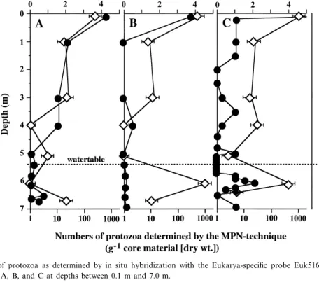
Numbers of protozoa were detected in each core A, B, and C by in situ hybridization with probe Euk516 and by the MPN technique (Fig. 5). Although the actual numbers of protozoa detected by both methods di¡ered considerably (between 2 and 5 orders of magnitude higher using in situ hybridization), a similar trend was found between both sets of data. Numbers of protozoa were highest
Fig. 5. Total number of protozoa as determined by in situ hybridization with the Eukarya-speci¢c probe Euk516 (b) and by the MPN technique (7) in cores A, B, and C at depths between 0.1 m and 7.0 m.
in the surface layer in each core (up to 105 cells g31 of core material [dry wt.] as detected by in situ hybridization) and decreased sharply below about 1 m depth. Elevated numbers of protozoa approach-ing those found in the surface layer were only de-tected in the saturated zone in contaminated core C (Fig. 5). The ratio between total numbers of bacteria determined after DAPI staining and protozoa de-tected with probe Euk516 was quite constant (core A: 0.7 þ 0.4U103; core B: 1.7 þ 0.9U103; core C: 1.2 þ 0.7U103; average of cores A to C: 1.1 þ 0.7U103).
The analysis of the protozoan community using the MPN technique is based on the assignment of culturable protozoa to the morphologically distinct groups of ciliates, £agellates, and amoebae (Table 1). Ciliates accounted for up to 4% of the protozoan population and were con¢ned to the surface layer above 1 m depth in each core. Flagellates and naked amoebae were found to co-occur in variable propor-tions to a depth of about 1.6 m below which the former were the predominant group in each core.
Amoebae became increasingly more encysted with depth, except in contaminated core C, where vegeta-tive trophozoites were also detected in the saturated zone (Table 1).
4. Discussion
4.1. Chemical characterization of the aquifer
The chemical data con¢rmed that cores A, B, and C were sampled in the border zone of the plume since large di¡erences for monoaromatic hydrocar-bons and for nitrate were observed between cores A and C. The highest concentrations of monoaro-matic hydrocarbons (up to 40 mmol kg31 of core material [dry wt.]) were found in core C where they exceeded the individual solubilities of the constituent hydrocarbons. The maximum solubility of the most prominent monoaromatic hydrocarbons found in core C ranged between 0.4 mM (trimethylbenzenes) and 1.7 mM (xylenes) [39]. One kilogram of
water-Table 1
Prevalence of protozoa in cores A, B and C as determined by the MPN technique Core Depth (m) Total protozoa MPN (g31 [dry wt.]) Prevalence (%)
Ciliates Flagellates Amoebae
Total Total Total Trophozoites Cysts
A 0.1^0.3 300 2 56 42 23 19 1.0^1.6 21 0 21 79 38 41 3.0^3.5 26 0 60 40 16 24 4.0^4.5 1 0 50 50 0 50 5.0^5.2 5 0 84 16 0 16 6.0^6.2 1 0 100 0 0 0 6.6^6.8 25 0 100 0 0 0 B 0.1^0.3 640 4 27 69 6 63 1.0^1.6 9 0 8 92 38 54 3.0^3.5 13 0 78 22 22 0 4.0^4.5 1 0 100 0 0 0 5.0^5.2 1 0 0 100 0 100 6.0^6.2 1100 0 99 1 0 1 6.6^6.8 12 0 100 0 0 0 C 0.1^0.3 1200 1 55 44 30 14 1.0^1.6 27 0 6 94 7 87 3.0^3.5 19 0 88 12 5 7 4.0^4.5 35 0 96 4 0 4 5.0^5.2 3 0 77 23 0 23 6.0^6.2 490 0 97 3 1 2 6.4^6.5 6 0 89 11 11 0
saturated core material was found to contain only about 0.14 l of water which corresponded to a water content of approx. 25%. This means that only a very small fraction of the hydrocarbons detected in core C can be solved in the pore water (approx. 200 Wmol kg31of core material [dry wt.] and that a free xylene phase still exists in core C which probably serves as a source for further contamination [36]. Core C con-tained high concentrations of o-xylene, trimethylben-zene and ethyltoluene, in contrast to earlier ¢ndings which reported mainly p- and m-xylene in the plume [36]. The high o-xylene concentrations could re£ect the close proximity of the sampling sites to the for-mer o-xylene production plant.
The concentration of nitrate in each core was found to be inversely related to concentrations of monoaromatic hydrocarbons, suggesting that nitrate reduction and degradation of aromatics are coupled. Furthermore, dissolved oxygen concentrations were previously shown to be high outside the plume but low inside the plume [37]. These observations togeth-er with the accumulation of nitrite in core C suggest that the resident microbial population uses monoar-omatic hydrocarbons as their carbon and energy source under aerobic and nitrate reducing condi-tions. A decrease in sulfate concentrations was ob-served in the lower parts of cores A and C where the sediment also appeared greyish-black. This suggests that sulfate reductive assimilation may be taking place at a depth below about 6.5 m. This suggestion, however, is not supported by the sulfate pro¢le in core B in which a decrease in sulfate concentration was not observed. Because earlier analyses per-formed at the site gave no indication for the occur-rence of high concentrations of sulfate and neither of sulfate reduction [36,37], sul¢de analysis was not considered necessary in our sampling program. Ad-ditional supporting data for sulfate reduction are therefore not available.
4.2. Bacterial community structure
The number of DAPI-stained bacteria increased from core A to core C and corresponded with in-creased levels of contaminants. The higher numbers of bacteria detected after DAPI-staining in the satu-rated zone of contaminated core C were found to coincide with a higher detection rate obtained with
probe Eub338. Detection rates after hybridization with probe Eub338 are used as an indication for the presence of metabolically active cells which con-tain su¤cient amounts of rRNA coupled with a suf-¢cient cell permeability or permeabilization to permit their detection [1], though recent studies have shown that probe Eub338 does not detect all members of the domain Bacteria [46]. The detection rate showed no direct correlation with the growth rate or activity of bacteria and varies according to the species exam-ined and their consumption of nutrients and oxi-dants (for review see [1]). Nevertheless, it is likely that the increase in detectability of bacteria in the saturated zone of contaminated core C (as compared with the low-contaminated cores A and B) is due to the availability of monoaromatic hydrocarbons as growth substrates. The correspondingly low level of oxidants detected in core C provides support for this assumption.
Proteobacteria of the N-subdivision comprising many sulfate-reducing bacteria were predominant in core C and provided support for the assumption of sulfate-reductive conditions. Due to the presence of sulfate-reducing bacteria in core C, the contribu-tion of sulfate-reductive degradacontribu-tion of hydrocar-bons [3,32] might have been underestimated. How-ever, further investigations would be necessary to quantify their contribution. A high proportion of DAPI-stained bacteria (2^4%) was represented by the Q-subdivision of Proteobacteria. Since the Q-sub-division of Proteobacteria is physiologically very het-erogeneous, a higher phylogenetic resolution would be necessary to attribute certain physiological activ-ities to its constituent bacterial groups. Bacteria of the L-subdivision of Proteobacteria only accounted for approx. 1% of DAPI-stained cells. The latter ¢ndings were in contrast to those obtained in a laboratory aquifer column which was set up in order to simulate the aerobic and denitrifying reme-diation processes during degradation of petroleum-derived hydrocarbons. In this column up to 90% of DAPI-stained cells belonged to the L-subdivision of Proteobacteria [22]. Though many of the organisms detected in connection with aerobic [13,14] or deni-trifying [22] degradation of monoaromatic hydrocar-bons belong to the L-subdivision of Proteobacteria, their impact on hydrocarbon degradation at the ¢eld site in Huënxe may be impeded by adverse
environ-mental factors such as low redox potentials. Lower numbers of bacteria belonging to the L-subdivision of Proteobacteria could also occur if these bacteria are less tolerant to high concentrations of aromatic hydrocarbons [14] as, for example, those of the Q-subdivision of Proteobacteria [24].
The proportion of Azoarcus sp. detected in the saturated zone of contaminated core C was similar to that found in a laboratory aquifer column (1^2%) designed to simulate the aerobic and denitrifying re-mediation processes occurring during degradation of petroleum-derived hydrocarbons [22]. The genus Azoarcus has already been implicated in the biode-gradation of monoaromatic hydrocarbons in a num-ber of studies [13,14,22]. The percentage of hydro-carbon-degrading Azoarcus sp. detected accounted for up to 1.4% of the total bacterial community which indicates that they are members of the indig-enous microbiota. Indeed, these Azoarcus sp. ac-counted for more cells in the contaminated aquifer (2U106 g31) than the biogeochemistry model [36] previously estimated for the total number of bacteria (max. 1.3U106 g31) present. Similar to the attribu-tion of signi¢cant denitri¢caattribu-tion activity to Paracoc-cus sp. based on their 3.5% abundance [28], these results suggest that Azoarcus sp. may play an impor-tant role during bioremediation of hydrocarbon-con-taminated aquifers. However, con¢rmation of this assumption depends on the availability of more in-formation on the catabolic activity of the hydrocar-bon-degrading Azoarcus populations both in column studies and in the ¢eld.
4.3. Protozoan community structure
Protozoa are integral members of the microbial community in groundwater aquifers [27,29,40,42]. Numerically, they are second in importance only to bacteria [30]. Evidence from the present study con-¢rmed the discrepancy between culture-dependent and -independent methods with MPN counts be-tween two and ¢ve orders of magnitude less than those using direct counting with eukaryotic probe Euk516. Interestingly though, both the MPN techni-que and probe Euk516 detected elevated numbers of protozoa in the surface layer in each core, which declined sharply with depth but again increased in the saturated zone in contaminated core C. These
¢ndings corresponded with published data from oth-er hydrocarbon-contaminated aquifoth-ers [27,40,42].
The covariance between results from the MPN technique and probe Euk516 suggests some relative con¢dence in each approach. The MPN technique will probably remain an important alternative to di-rect counting methods until the latter have been suf-¢ciently developed to provide information on proto-zoan taxa [35]. In a recent study, oligonucleotide probes were used to enumerate a particular nano£a-gellate species in a mixed population [34]. This dem-onstrates the possibility for developing molecular tools to investigate both the taxonomy and ecology of protozoa as well as of bacteria and their mutual in£uence in a variety of habitats by the same meth-ods. Future studies in our laboratory will focus on the development of such tools.
Protozoan taxa comprising ciliates, £agellates and naked amoebae were identi¢ed during the present study. Ciliates formed a minor proportion of the protozoan community (9 4%) and were con¢ned ex-clusively to the surface layer ( 6 1 m depth) in each core. Similar ¢ndings from other aquifers have been attributed to straining by sediment on large protozoa over 20 Wm in diameter [18,40]. The major compo-nent of the community was comprised of £agellates which increased in prevalence with depth and there-by con¢rmed earlier ¢ndings from other aquifers [29,30,40]. The predominance of £agellates (particu-larly nano£agellates of 2^3 Wm in diameter) has been attributed to their optimal size for transport through the aquifer matrix [19]. Naked amoebae became in-creasingly more encysted with depth except in core C where vegetative trophozoites were present in the saturated zone. Presumably, this was in response to elevated numbers of bacteria associated with con-taminants in the saturated zone.
The role of bacterivorous protozoa in contaminant biodegradation is considered to be an indirect result of their ability to selectively graze on and control the biomass of aquifer bacteria [27,30,42]. This in turn creates a nutritional loop in which protozoa rapidly remineralize nutrients which sustain further bacterial growth [9]. The ratio of protozoa to bacteria has variously been reported as 1:101^105 [41] and 1:103 [42] from other aquifers and corresponds with a ratio of 1:103 found during the present study. Estimates for feeding rates from batch culture studies suggest
that £agellates require 102bacteria whereas amoebae require 103 bacteria per cell division [38,49]. A sim-ilar growth requirement of 102 bacteria per division has been observed for the £agellate Spumella sp. iso-lated from core C (unpublished data).
The growth of bacteria in porous media supplied with nutrients has frequently been observed to cause reductions in hydraulic conductivity due to bioclog-ging [10,44,45]. Perhaps bacterivorous protozoa in contaminated aquifers are capable of limiting bacte-rial bioclogging. The role of bacterivorous £agellates in limiting bacterial bioclogging in model aquifer columns is currently being investigated in our labo-ratory. Further studies should therefore evaluate the merits of incorporating a protozoan dimension into bioremediation.
Acknowledgments
The authors wish to thank Dr. D. Hunkeler (ETH, Zurich) and Mr. C. Fischer (Harness Pickel Consult, Kassel, Germany) for assistance during sampling. We also wish to thank Dr. W. Schaëfer (Ruprecht-Karls-Universitaët, Heidelberg, Germany) for supply-ing detailed information on previous studies at Huënxe. Valuable contributions from discussions held with Dr. W. Schlimme (Novartis, Basel, Swit-zerland) and Dr. G. Novarino (The Natural History Museum, London, UK) are also gratefully acknowl-edged. The study was supported by grants from the Swiss National Science Foundation (Priority Pro-grams Biotechnology and Environment, respec-tively), and the Swiss Federal O¤ce of Environment, Forests and Landscape (BUWAL).
References
[1] Amann, R.I., Ludwig, W. and Schleifer, K.-H. (1995) Phylo-genetic identi¢cation and in situ detection of individual micro-bial cells without cultivation. Microbiol. Rev. 59, 143^169. [2] Atlas, R.M. and Bartha, R. (1992) Hydrocarbon
biodegrada-tion and oil spill bioremediabiodegrada-tion. Adv. Microb. Ecol. 12, 287^ 338.
[3] Beller, H.R., Spormann, A.M., Sharma, P.K., Cole, J.R. and Reinhard, M. (1996) Isolation and characterization of a novel toluene-degrading sulfate-reducing bacterium. Appl. Environ. Microbiol. 62, 1188^1196.
[4] Bolliger, C., Hoëhener, P., Hunkeler, D., Haëberli, K. and Zeyer, J. (1998) Intrinsic bioremediation of a petroleum hy-drocarbon-contaminated aquifer: assessment of mineraliza-tion. Environ. Sci. Technol. (submitted).
[5] Bregnard, T.P.-A., Hoëhener, P., Haëner, A. and Zeyer, J. (1996) Degradation of weathered Diesel fuel by microorgan-isms from a contaminated aquifer in aerobic and anaerobic microcosms. Environ. Toxicol. Chem. 15, 299^307. [6] Brock, T.D. (1987) The study of microorganisms in situ:
progress and problems. Symp. Soc. Gen. Microbiol. 41, 1^17. [7] Chee-Sanford, J.C., Frost, J.W., Fries, M.R., Zhou, J. and Tiedje, J.M. (1996) Evidence for acetyl coenzyme A and cin-namoyl coenzyme A in the anaerobic toluene mineralization pathway in Azoarcus tolulyticus Tol-4. Appl. Environ. Micro-biol. 62, 964^973.
[8] Clesceri, L.S., Greenberg, A.E. and Trussell, R.R. (1989) Standard Methods for the Examination of Water and Waste-water. American Public Health Association, Washington, DC. [9] Coleman, D.C., Cole, C.V., Anderson, R.V., Blaha, M., Cam-pion, M.K., Clarholm, M., Elliot, E.T., Hunt, H.W., Shaefer, B. and Sinclair, J. (1977) An analysis of rhizosphere-sapro-phage interactions in terrestrial ecosystems. Ecol. Bull. 25, 299^309.
[10] DeLeo, P.C. and Baveye, P. (1997) Factors a¡ecting proto-zoan predation of bacteria clogging laboratory aquifer micro-cosms. Geomicrobiol. J. 14, 127^149.
[11] Dobbins, D.C., Aelion, C.M. and Pfaender, F. (1992) Subsur-face, terrestrial microbial ecology and biodegradation of or-ganic chemicals: A review. Crit. Rev. Environ. Control 22, 67^136.
[12] Fries, M.R., Zhou, J., Chee-Sanford, J. and Tiedje, J.M. (1994) Isolation, characterization, and distribution of denitri-fying toluene degraders from a variety of habitats. Appl. En-viron. Microbiol. 60, 2802^2810.
[13] Fries, M.R., Hopkins, G.D., McCarty, P.L, Forney, L.J. and Tiedje, J.M. (1997) Microbial succession during a ¢eld evalu-ation of phenol and toluene as the primary substrates for trichloroethene cometabolism. Appl. Environ. Microbiol. 63, 1515^1522.
[14] Fries, M.R., Forney, L.J. and Tiedje, J.M. (1997) Phenol- and toluene-degrading microbial populations from an aquifer in which successful trichloroethene cometabolism occurred. Appl. Environ. Microbiol. 63, 1523^1530.
[15] Hahn, D., Amann, R.I., Ludwig, W., Akkermans, A.D.L. and Schleifer, K.-H. (1992) Detection of microorganisms in soil after in situ hybridization with rRNA-targeted, £uorescently labelled oligonucleotides. J. Gen. Microbiol. 138, 879^887. [16] Haëner, A., Hoëhener, P. and Zeyer, J. (1997) Degradation of
trimethylbenzene isomers by an enrichment culture under N2O-reducing conditions. Appl. Environ. Microbiol. 63,
1171^1174.
[17] Hart, S. (1996) In situ bioremediation: De¢ning the limits. Environ. Sci. Technol. 30, 398A^401A.
[18] Harvey, R.W. (1991) Parameters involved in modelling move-ment of bacteria in groundwater. In: Modelling the Environ-mental Fate of Microorganisms (Hurst, C.J., Ed.), pp. 89^114. American Society for Microbiology, Washington, DC.
[19] Harvey, R.W., Kinner, N.E., Bunn, A., MacDonald, D. and Metge, D. (1995) Transport behaviour of groundwater proto-zoa and protoproto-zoan-sized microspheres in sandy aquifer sedi-ments. Appl. Environ. Microbiol. 61, 209^217.
[20] Heitzer, A. and Sayler, G.S. (1993) Monitoring the e¤cacy of bioremediation. Trends Biotech. 11, 334^343.
[21] Hess, A., Hoëhener, P., Hunkeler, D. and Zeyer, J. (1996) Bioremediation of a diesel fuel contaminated aquifer: simula-tion studies in laboratory aquifer columns. J. Contam. Hy-drol. 23, 329^345.
[22] Hess, A., Zarda, B., Hahn, D., Haëner, A., Stax, D., Hoëhener, P. and Zeyer, J. (1997) In situ analysis of denitrifying toluene-and m-xylene-degrading bacteria in a Diesel-fuel contaminated laboratory aquifer column. Appl. Environ. Microbiol. 63, 2136^2141.
[23] Hunkeler, D., Hoëhener, P., Haëner, A., Bregnard, T. and Zeyer, J. (1995) Quanti¢cation of hydrocarbon mineralization in a diesel fuel contaminated aquifer treated by in situ bio-restoration. In: Ground Water Quality: Remediation and Protection (Kovar, K. and Krasny, J., Eds.), pp. 421^430. IAHS Press, Wallingford.
[24] Inoue, A. and Horikoshi, K. (1989) A Pseudomonas thrives in high concentrations of toluene. Nature 338, 264^266. [25] Kaëmpfer, P., Steiof, M., Becker, P.M. and Dott, W. (1993)
Characterization of chemoheterotrophic bacteria associated with the in situ bioremediation of a waste-oil contaminated site. Microb. Ecol. 26, 161^188.
[26] MacDonald, J.A. and Rittmann, B.E. (1993) Performance standards for in situ bioremediation. Environ. Sci. Technol. 27, 1974^1979.
[27] Madsen, E.L., Sinclair, J.L. and Ghiorse, W.C. (1991) In situ biodegradation: microbiological patterns in a contaminated aquifer. Science 252, 830^833.
[28] Neef, A., Zaglauer, A., Meier, H., Amann, R., Lemmer, H. and Schleifer, K.-H. (1996) Population analysis in a denitrify-ing sand ¢lter: Conventional and in situ identi¢cation of Paracoccus spp. in methanol-fed bio¢lms. Appl. Environ. Microbiol. 62, 4329^4339.
[29] Novarino, G., Warren, A., Kinner, N.E. and Harvey, R.W. (1994) Protists from a sewage-contaminated aquifer on Cape Cod, Massachusetts. Geomicrobiol. J. 12, 23^36.
[30] Novarino, G., Warren, A., Butler, H., Lambourne, G., Box-shall, A., Bateman, J., Kinner, N.E., Harvey, R.W., Mosse, R.A. and Teltsch, B. (1997) Protistan communities in aqui-fers: a review. FEMS Microbiol. Rev. 20, 261^275. [31] Page, F.C. (1976) An Illustrated Key to Freshwater and Soil
Amoebae. Freshwater Biological Association, Ambleside. [32] Rabus, R., Nordhaus, R., Ludwig, W. and Widdel, F. (1993)
Complete oxidation of toluene under strictly anoxic condi-tions by a new sulfate-reducing bacterium. Appl. Environ. Microbiol. 59, 1444^1451.
[33] Rabus, R. and Widdel, F. (1995) Anaerobic degradation of ethylbenzene and other aromatic hydrocarbons by new deni-trifying bacteria. Arch. Microbiol. 163, 96^103.
[34] Rice, J., O'Connor, C.D., Sleigh, M.A., Burkill, P.H., Giles, I.G. and Zubkov, M.V. (1997) Fluorescent oligonucleotide
rDNA probes that speci¢cally bind to a common nano£agel-late, Paraphysomonas vestita. Microbiology 143, 1717^1727. [35] RÖnn, R., Ekelund, F. and Christensen, S. (1995) Optimizing
soil extract and broth media for MPN-enumeration of naked amoebae and heterotrophic £agellates in soil. Pedobiologia 39, 10^19.
[36] Schaëfer, W., Therrien, R. and Voss, A. (1994) Simulation of a xylene spill in a sandy aquifer with a 3D transport and bio-geochemistry model. In: Transport and Reactive Processes in Aquifers (Dracos, T.H. and Stau¡er, F., Eds.), pp. 463^468. Balkema, Rotterdam.
[37] Schaëfer, W. and Therrien, R. (1995) Simulating transport and removal of xylene during remediation of a sandy aquifer. J. Contam. Hydrol. 19, 205^236.
[38] Schnuërer, J., Clarholm, M., Bostroëm, S. and Rosswall, T. (1986) E¡ects of moisture on soil microorganisms and nema-todes: A ¢eld experiment. Microb. Ecol. 12, 217^230. [39] Schwarzenbach, R.P., Gschwend, P.M. and Imboden, D.M.
(1993) Environmental Organic Chemistry. John Wiley and Sons, New York.
[40] Sinclair, J.L. and Ghiorse, W.C. (1987) Distribution of protozoa in subsurface sediments of a pristine groundwater study site in Oklahoma. Appl. Environ. Microbiol. 53, 1157^1163.
[41] Sinclair, J.L. and Ghiorse, W.C. (1989) Distribution of aero-bic bacteria, protozoa, algae and fungi in deep subsurface sediments. Geomicrobiol. J. 7, 15^31.
[42] Sinclair, J.L., Kampbell, D.H., Cook, M.L. and Wilson, J.T. (1993) Protozoa in subsurface sediments from sites contami-nated with aviation gasoline or jet fuel. Appl. Environ. Micro-biol. 59, 467^472.
[43] Song, H.G. and Bartha, R. (1990) E¡ects of jet fuel spills on the microbial community of soil. Appl. Environ. Microbiol. 56, 646^651.
[44] Taylor, S.W. and Ja¡eè, P.R. (1990) Bio¢lm growth and the related changes in the physical properties of a porous me-dium; 1. Experimental investigation. Water Resources Res. 26, 2153^2159.
[45] Vandevivere, P. and Baveye, P. (1992) Saturated hydraulic conductivity reduction caused by aerobic bacteria in sand col-umns. Soil Sci. Soc. Am. J. 56, 1^13.
[46] Zarda, B., Hahn, D., Chatzinotas, A., Schoënhuber, W., Neef, A., Amann, R.I. and Zeyer, J. (1997) Analysis of bacterial community structure in bulk soil by in situ hybridization. Arch. Microbiol. 168, 185^192.
[47] Zeyer, J. and Kocher, H.-P. (1988) Puri¢cation and character-ization of a bacterial nitrophenol oxygenase which converts ortho-nitrophenol to catechol and nitrite. J. Bacteriol. 170, 1789^1794.
[48] Zhou, J.-Z., Fries, M.R., Chee-Sanford, J.C. and Tiedje, J.M. (1995) Phylogenetic analysis of a new group of denitrifyers capable of anaerobic growth on toluene: description of A. tolulyticus sp. nov. Int. J. Syst. Bacteriol. 45, 500^506. [49] Zwart, K.B. and Darbyshire, J.F. (1992) Growth and
nitro-genous excretion of a common soil £agellate Spumella sp. ^ a laboratory experiment. J. Soil Sci. 43, 145^157.