Failed coronary artery bypass anastomosis detected by
intraoperative coronary flow measurement
Beat H. Walpoth*, Andreas Bosshard, Beat Kipfer,
Pascal A. Berdat, Ueli Althaus, Thierry Carrel
Clinic for Thoracic and Cardiovascular Surgery, University of Berne, Berne, SwitzerlandAbstract
Objectives: To assess intraoperative flow of arterial and venous coronary grafts after myocardial revascularization which may allow early detection of low flow situations, especially during minimally invasive coronary bypass surgery (MIDCAB), and lead to immediate correction of technical problems. Methods: In two patients with severe and diffuse multi-vessel disease the left internal mammary artery (IMA) was connected to the left anterior descending artery (LAD). During reperfusion, the flow was measured in the IMA and vein grafts using a transit time flow meter. Results: In both cases the IMA showed only a systolic pendulating flow curve with a mean flow of 0–1 ml/ min and a high resistance. Manual IMA assessment revealed an adequate pulsation. Both distal IMA anastomoses were re-explored on cardiopulmonary bypass yielding an initial flow of 7 and 14 ml/min, respectively. After treatment with papaverine/adenosine the IMA flow increased from 7 to 26 ml/min (coronary flow reserve (CFR)=3.7) and from 14 to 46 ml/min (CFR=3.3), respectively. Conclusion: Intraoperative flow assessment of IMA and venous bypass grafts can be recommended to monitor flow; especially during MIDCAB procedures.1998 Elsevier Science B.V.
Keywords: Coronary bypass graft; Internal mammary artery; Minimally invasive coronary bypass surgery; Blood flow measurement; Coronary flow reserve; Transit time flow
1. Introduction
With the advent of minimally invasive coronary artery bypass procedures (MIDCAB), assessment of graft flow and coronary vascular resistance are important quality control methods immediately after myocardial revascularization. Intraoperative flow measurement can be recommended to assess graft flow, especially in the left internal mammary artery (IMA) to the left anterior descending artery (LAD) [1]. In MIDCAB procedures, some additional difficulties due to restricted access or cardiac motion may compromise the quality of the anastomosis [2,3,8]. Several reports on the use of volume flow measurements during cardiac surgery with the transit time method have been previously published [4,7–10]
2. Methods and results
2.1. Flow measurement with the transit time method
Intraoperative transit time flow (TTF) and vascular resis-tance were measured with a CardioMed flow meter (CM-4008, Medi-Stim, AS, Oslo, Norway) using 3 and 4 mm probes [6,8–10]. The transit time method is based on the fact that the time required for ultrasound to pass through blood is slightly longer upstream than downstream. As the ultrasound beam is wider than the diameter of the vessel lumen, the ultrasound waves will cover every flow vector in the vessel, thus, making the transit time difference propor-tional to the true blood volume flow in ml/min. The results are therefore not affected by vessel diameter or non-moving objects (i.e. arteriosclerotic plaques) in the acoustic window as the time difference will only be generated by the blood itself. The vascular resistance is calculated by dividing the simultaneously measured arterial pressure by the flow.
0387-7604/98/$19.00 1998 Elsevier Science B.V. All rights reserved P I I S 1 0 1 0 - 7 9 4 0 ( 9 8 ) 0 0 1 1 0 - 9
* Corresponding author. Thoracic and Cardiovascular Surgery, Univer-sity Hospital, Insel, CH-3010 Berne, Switzerland. Tel.: +41 31 6322373/ 6322375; fax:+41 31 6329766/3820279; e-mail: beat.walpoth@insel.ch
2.2. Patients
Insufficient intraoperative graft flow (IMA to LAD) was observed in two patients immediately after weaning from extracorporeal circulation using transit time flow measure-ment. Both patients were operated on a regular basis, using moderate hypothermic cardiopulmonary bypass (CPB) (32°C) and antegrade blood cardioplegia as myocardial pro-tection.
2.2.1. Patient 1 (M.M. 1921)
A 76-year-old woman (66 kg) with three-vessel disease (LAD 70–90% stenosis at two levels including the first diagonal branch; right coronary artery (RCA) occluded) and reduced ejection fraction (45%) after acute myocardial infarction received three grafts (L-IMA to LAD; saphenous vein graft (SVG) to the RCA and to the first diagonal branch). All recipient vessels were severely diseased and of small caliber (1.5 mm). As often encountered in old
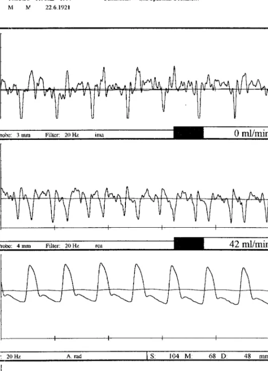
Fig. 1. (Patient 1 M.M.) Transit time flow measurement of the internal mammary artery (IMA) graft with a technical problem at the distal anastomosis showing no-flow and only a systolic pulsatile curve oscillating around the 0 line (upper panel). The saphenous vein graft (SVG) to the right coronary artery (RCA) shows 42 ml/min of mean flow and a pronounced diastolic flow pattern (middle panel). The lower two panels show the radial arterial pressure and heart rate trace.
female patients, the IMA graft was a small and delicate vessel with a tendency to dissection in the distal part. After release of the aortic cross clamp and cessation of CPB, a TTF flow measurement, performed simultaneously on the IMA (3 mm probe; 0 ml/min) and on the SVG to RCA (4 mm probe: 42 ml/min), showed a no flow situation in the IMA despite adequate pulsation of the graft assessed by the surgeon’s fingers. The TTF flow curve showed a systolic flow pattern oscillating around the 0 line (mean arterial pressure (MAP 68 mmHg) and heart rate (91 beats/min (bpm)). In addition vascular resistance was above physiological values. The flow in the SVG to RCA was adequate and showed a normal coronary flow curve with a pronounced diastolic pattern (Fig. 1). CPB was restarted and the IMA to LAD anastomosis was re-explored during a period of electrical ventricular fibrillation. The free flow was 33 ml/min after distal IMA transection. A localized intimal dissecting flap in the IMA was found and the anastomosis was redone using continuous 7.0 polypro-pylene suture in a running fashion. The flow measurement was repeated immediately thereafter and showed a low flow in the IMA to LAD (7 ml/min). This was attributed to severe vasoconstriction. Two sequential doses of diluted papaverine (3 mg) were injected directly intraluminally into the middle section of the IMA graft using a 26 gauge needle. The flow increased rapidly to 17 then 26 ml/min, indicating a coronary flow reserve (CFR) of 3.7 with a drop in resistance (9.6 to 2.7 Q). After administration of an adrenaline bolus (10 mg i.v.) the MAP increased from 70 to 90 mmHg, the heart rate from 91 to 112 bpm and the IMA flow rose further to a maximum of 53 ml/min (Table 1).
Concomitantly the SVG to RCA showed some increase in flow with time from 89 to 115 ml/min. A further increase to 125 ml/min with the pharmacological pressure rise was noted (Table 1). The post operative course of the patient was uneventful; no ECG alterations or enzyme release were found.
2.2.2. Patient 2 (H.W. 1940)
A 57-year-old man (73 kg) with a two- to three-vessel disease (LAD 70–90% stenosis at three levels; first diagonal branch 70–95% stenosis and RCA 50% stenosis) with a good ejection fraction of 82% received four grafts including a L-IMA to proximal LAD. After reperfusion, inadequate contractility of the anterior wall was observed and a loca-lized intramural hematoma was found in the IMA pedicle at the level of the distal anastomosis. TTF measurement showed a no-flow situation (0–1 ml/min) despite change of probes, optimal pulsation and coupling factor. The ana-stomosis was redone on normothermic CPB with cold blood cardioplegic arrest. The segment of the intramural hema-toma was resected. The free flow of the IMA after distal transection was 30 ml/min into a beaker and 34 ml/min by TTF (MAP of 68 mmHg). After cessation of CPB TTF was reassessed showing a flow of 14 ml/min in the IMA, and 48 ml/min in the SVG including a jump anastomosis (Fig. 2a). After adenosine administration (25 mg/kg per min in left ventricle) the flow in the IMA rose to 46 ml/min (CFR=3.3) and in the SVG to 128 ml/min (CFR=2.7) with little change in MAP and heart rate. Simultaneously the resistance decreased in the IMA from 5.1 to 1.5 and in the SVG from 1.5 to 0.5 Q. In addition the shape of the flow curve showed an increase in the diastolic flow component (Table 1 and Fig. 2b). No postoperative problems occurred in this patient.
3. Discussion and conclusions
Our observation is based on two cases of coronary artery bypass grafting among the last 450 cases, in which a tech-nical problem at the IMA anastomosis (dissection and con-secutive adventitial hematoma) was diagnosed with intraoperative transit time flow measurement. In both cases no flow was measurable by TTF despite an adequate free flow of the IMA graft immediately after preparation and
Table 1
Transit time flow measurement before and after redo of distal anastomosis for technical problems (no flow)
Event Q IMA (ml/min) Q SVG (ml/min) MAP (mmHg) HR (BPM) R IMA (Q) R SVG (Q) CFR IMA CFR SVG Patient 1 Occlusion 0 42 68 91 – 1.60 Redo 7 89 67 91 9.60 0.75
Papaverine (IMA i.a. 3 mg) 17 100 66 91 3.90 0.66 2.4 1.1
Papaverine (IMA i.a. 3 mg) 26 115 70 91 2.70 0.61 3.7 1.3
Adrenaline (10 mg i.v.) 53 125 90 112 1.70 0.72 Patient 2 Occlusion 0–1 – 66 70 – – Redo 14 48 71 83 5.20 1.50 Adenosine (25mg/kg per min in LV) 46 128 66 81 1.50 0.51 3.3 2.7
a good pulsatility in the anastomosed IMA graft. Pulsatility is well documented on Fig. 1 showing a real time systolic pulsatile flow curve oscillating around the zero line, yield-ing a no forward flow situation (mean flow=0 ml/min). In addition, coronary vascular resistance (CVR) is elevated above physiological values. The above parameters (all available on the CM 4000), in addition to an insufficient
IMA flow, have led the surgeon to re-explore the anastomo-sis.
In both cases the free flow in the IMA after distal transec-tion before reanastomosis was assessed. The respective free flow values, 33 and 30 ml/min, are somewhat less than the free flow reported in our previous publication of 50.7 ±32.4 ml/min. Similarly the IMA to LAD flow at the end of the
Fig. 2. (Patient 2 H.W.) (a) Baseline after redo IMA: transit time flow of the internal mammary artery (IMA) to the proximal left anterior descending artery (LAD) (upper panel) and a saphenous vein graft (SVG) (second panel) after redoing the distal IMA to LAD anastomosis. Panel 3, radial arterial pressure curve; panel 4, heart rate; panel 5, resistance curve of the IMA. (b) After adenosine: coronary flow reserve (CFR) measurement with adenosine (25 mg/kg per min in left ventricle (LV)) showing a CFR of 3.3 for the IMA and 2.7 for the SVG. Note the marked diastolic flow pattern in the IMA and SVG grafts. Panel description as in (a).
procedure was lower than reported by our group (49.8 ± 32.9 ml/min), especially when no pharmacologic vasodila-tion was used. This may be due to reclamping of the IMA and further preparation without papaverine soaking [10].
The respective IMA flow values (7 ml/min (Patient 1) and 14 ml/min (Patient 2)) after redoing the distal anastomosis and cessation of CPB were rather low and probably indica-tive of severe vasoconstriction and spasm due to prolonged manipulations of the IMA (low flow and high coronary
vascular resistance). Pharmacological vasodilatation (in Patient 1 with papaverine injected in the IMA lumen and in Patient 2 with intraventricular adenosine) showed a three-fold increase in flow with a concomittant marked reduction in coronary vascular resistance. In both cases the coronary flow reserve was around 3.0. Patient 2 (HW) received ade-nosine and showed a marked flow increase in the IMA (14– 46 ml/min) and the SVG (48–128 ml/min). Patient 1 (M.M.), who received papaverine in the IMA graft, showed
a good IMA flow increase but only a gradual flow increase in the SVG to the RCA over time mainly due to a rise of the MAP (67–90 mmHg). This confirms the pressure flow rela-tionship. However, it should be noted that the transit time flow measurement cannot differentiate between a spasm in the IMA, a spasm in the native coronary artery or a technical error at the anastomosis. A low flow situation due to vasos-pasm may be excluded by using pharmacological vasodila-tation. If there is no improvement of flow and change in the curve shape, the surgeon should reevaluate the anastomosis. Further attention should be given to mis-sized or misaligned probes, poor ultrasonic coupling and competitive flow in the native coronary artery.
Intraoperative transit time flow measurement is easy, fast and reliable and may be recommended routinely or for selected conditions (low flow in the free IMA or severe distal coronary disease) after myocardial revascularization to exclude low or no-flow situations. There are many causes of such findings: inflow problems (proximal anastomosis or graft problems such as twist, kink, flap, dissection) vasos-pasms, technical problems at the distal anastomosis and poor run off. Vasospasms and elevated distal coronary vas-cular resistance can be diagnosed and treated pharmacolo-gically with an immediate response on TTF measurement, whereas technical problems usually require surgical correc-tion [5]. However, immediate re-exploracorrec-tion may prevent myocardial infarction and subsequent reoperation with markedly increased morbidity/mortality, hospital stay and cost. Since similar problems might occur during MIDCAB surgery where exposure is sometimes less optimal, we recommend assessing IMA flow immediately after perform-ing the anastomosis. Our initial ex-perience in this field is encouraging – measurement of IMA flow contributes to improved quality control after MIDCAB procedures.
References
[1] Barner HB, Standeven JW, Reese J. Twelve years experience with internal mammary artery for coronary bypass. J Thorac Cardiovasc Surg 1985;90:668–675.
[2] Benetti FJ, Ballester C, Sani G, Doonstra P, Grandjean J. Video assisted coronary bypass surgery. J Cardiac Surg 1995;10:620–625. [3] Calafiore AM, Di Giammarco G, Teodori G. et al. Left anterior descending coronary artery grafting via left anterior small thoracot-omy without cardiopulmonary bypass. Ann Thorac Surg 1996; 61:1658–1665.
[4] Canver, C.H.C., Dame, N.A. Ultrasonic assessment of internal thor-acic artery graft flow in the revascularized heart. Ann Thorac Surg 199; 58: 135–138.
[5] Carrel T, Kujawski T, Zu¨nd G, Schwitzer J, Amann FW, Gallino A, Bertel O, Jenni R, Turina M. The internal mammary artery malperfu-sion syndrome: incidence, treatment and angiographic verification. Eur J Cardio-thorac Surg 1995;9:190–197.
[6] Laustsen J, Pedersen EM, Terp K, Steinbru¨chel D, Kure HH, Paulsen PK, Jorgensen H, Paaske WP. Validation of a new transit time ultra-sound flowmeter in man. Eur J Vasc Endovasc Surg 1996;12:91–96. [7] Louagie YAG, Haxhe JP, Jamart J, Gurne O, Buche M, Schoevaerdts JC. Perioperative hemodynamic study of left internal mammary artery grafts. Thorac Cardiovasc Surg 1995;43:27–34.
[8] Saatvedt K, Dragsund M, Nordstrand K. Mini invasive coronary artery bypass grafting (letter). Ann Thorac Surg 1996;62.
[9] Van der Meulen J, Eijsman IE. Detection of a twisted saphenous vein graft during coronary artery bypass surgery. Medi-Stim Clin Cases 1997;4:1.
[10] Walpoth BH, Mohadjer A, Gersbach P, Rogulenko R, Walpoth BN, Althaus U. Intraoperative internal mammary artery transit-time flow measurements: comparative evaluation of two surgical pedicle pre-paration techniques. Eur J Cardio-thorac Surg 1996;10:1064–1070.