© 1990 European Dialysis and Transplant Association-European
Renal Association
Nephrology
Dialysis
Transplantation
Registry Report
Kidney Transplantation in Primary Oxalosis: Data From the EDTA
Registry
M. Broyer Hopital Necker Enfants-Malades, Paris, France
F. P. Brunner* Departement fur Innere Medizin, Universitat Basel, Switzerland
H. Brynger Department of Surgery 1, Sahlgrenska Sjukhuset, Goteborg, Sweden
S. R. Dykes St Thomas' Hospital, London, United Kingdom
J. H. H. Ehrich Medizinische Hochschule Hannover, Federal Republic of Germany ""Chairman
W. Fassbinder Stadtische Kliniken Fulda, Federal Republic of Germany W. Geerlings Stichting Thuisdialyse Noord
Nederland, Haren, Netherlands G. Rizzoni Ospedale Pediatrico Bambino
Gesii, Istituto di Ricerca Scientifica, Roma, Italy N. H. Selwood St Thomas' Hospital, London,
United Kingdom
G. Tufveson Transplantation Unit, University Hospital, Uppsala, Sweden A. J. Wing St Thomas' Hospital, London,
United Kingdom
Abstract. This paper reports the results of 98 first kidney transplantations in patients with oxalosis as the primary renal disease as recorded by the EDTA Registry. There were 79 patients who received a cadaveric (CAD) graft and 15 patients with a living related donor (LRD) graft; the type of donor was not recorded for four patients. Initial graft survival appeared to be better after LRD as compared to CAD grafts but at 3 years the poor survival was similar with 23% for LRD and 17% for CAD grafts. CAD graft survival did not differ between children and adults and was not affected by the waiting time on dialysis. A slight improvement was observed in grafts performed in the years 1983-1986 as compared to grafts performed in earlier years. The causes of failure reported were mainly rejection (33%) and recurrence of primary renal disease (31 %). In view of the poor results related to recurrence of Correspondence and offprint requests to: Professor F. P. Brunner,
EDTA Registry, St Thomas' Hospital, London SE1 7EH, UK. Tel: 01-633 0636.
oxalosis in the graft, the potential of combined kidney and liver transplantation is discussed.
Key words: Primary oxalosis; Kidney transplantation
Introduction
Primary hyperoxaluria, a synonym for congenital oxalosis, is a rare metabolic disorder caused by accumu-lation of glyoxylate, which leads to increased synthesis and excretion of glycolate and oxalate. The basic biochemical defect described in 1986 by Danpure and Jennings [1], seems to be the absence of peroxisomal alanine glyoxylate aminotransferase, which is located exclusively in the liver. The disease is characterised clini-cally by recurrent calcium oxalate kidney stones and nephrocalcinosis resulting in progressive renal insuffi-ciency. Once end-stage renal failure is reached, further calcium oxalate accumulation cannot be prevented by any
Kidney Transplantation in Primary Oxalosis 333
form of dialysis strategy and leads to extrarenal com-plications with progressive and severely disabling bone disease being the most prominent [2]. Calcification of blood vessels and heart may also occur and cause death [3]. For these reasons, dialysis is quite unsatisfactory and explains why kidney transplantation has been attempted in spite of the almost inevitable recurrence of calcinosis in the graft. Several reports of unsuccessful or successful renal transplantation in patients with primary oxalosis have been published [4,16]. Since successful cases may be reported preferentially, and in order to have a more general view on renal transplantation in primary oxalosis, it was decided to analyse the outcome of all such kidney grafts reported to the Registry of the European Dialysis and Transplant Association—European Renal Association (EDTA Registry) up to 31 December 1966.
Methods
This study is based on information obtained through individual patient questionnaires that have been collected each year since 1965 by the EDTA Registry. The essential structure of the patient questionnaire has remained largely unchanged since 1971 [17]. The coding structure for primary renal diseases was altered in 1973, at which time code 53 was introduced for 'oxalosis'. The definition of code 53 was modified in 1985 to 'primary oxalosis', in order to exclude the rare forms of end-stage renal failure due to secondary hyperoxaluria. Since restricting the study to those cases registered since 1985 would result in too few cases for study, it was felt reasonable to include all cases of oxalosis reported to the Registry. Secondary oxalosis is known to occur more commonly in the older age groups, carries a better prognosis than primary oxalosis, and is also uncommon. For this reason, studying all cases of oxalosis was considered unlikely to affect the results adversely. The method of calculating actuarial survival using life table methods has been previously described [18].
Results
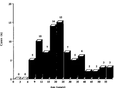
There were 221 patients reported to the Registry up to 31 December 1986 with oxalosis recorded as the primary renal disease: 121 males, 99 females and one with the sex not recorded. The age distribution at start of treatment is given in Fig. 1. Figure 2 shows the age distribution of 79 patients at the time of receiving their first cadaveric graft. Sixty per cent of these graft recipients were younger than 25. The number of new patients accepted for renal replacement therapy per year varied from 9 to 22 in the last 10 years. The number of grafts performed yearly seemed to increase slightly from 1976 to 1986 (Fig. 3).
35 35
12 15 20 25 Age (years)
35 40 45 50 55
Fig. 1. Age distribution at start of renal replacement therapy of 221 patients with oxalosis.
0 3 6 9 12 15 20 25 30 35 40 45 50 55 Age (years)
Fig. 2. Age distribution at first cadaveric graft of patients with oxalosis.
Of a total of 98 patients who received a first transplant (79 cadaveric, 15 living donor and 4 where the source was not recorded), 76% were performed since 1980 and 58% since 1983. The waiting time on dialysis before grafting was variable; five patients were grafted directly without dialysis, 35 were grafted within the first year, 21 within the second year and 36 after a longer interval. In one case, the treatment history prior to transplantation had not been reported to the Registry.
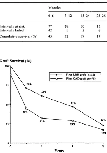
Graft survival, shown in Table 1 and Fig. 4, appeared to be better initially after living related as compared to cadaveric grafting, but this was not sustained. Primary non-function was recorded for 11 % of first cadaveric grafts. Cadaver graft survival was no different in children compared to adults (Fig. 5) and no living donor graft was performed in patients under the age of 15. Graft survival
76 77 78 79 80 81 82 83 84 85 86
Grafts (n) Graft Survival (%)
2 5 100-1 - 20 - 15 - 10 75" 50- 25- n-i \ 4 5 % ^v j v 34% 33% <15 yrs at grafting (n=23) 15 yrs or over at grafting (n=55)
30% 29% ^ ' ^ ^ 24% —• 12% 0 1 2 3 Years
Fig. 3. Number of patients starting renal replacement therapy and ,
number of grafts in patients with oxalosis by year. F i8 -5-F l r s t c a d a v e r 8r a f t s u r v l v a l according to age of patient at grafting.
Table 1. First cadaveric graft survival in patients with oxalosis (n = 79) Graft Survival (%)
100-1 Interval n at risk Interval n failed Cumulative survival (%) Months 0-6 77 42 45 7-12 28 5 32 13-24 20 2 29 25-26 15 6 17 Graft Survival (%) 50-First LRD graft (n=15) First CAD graft (n=79)
1983-1986 (n=36) before 1983 (n=41)
34%
21%
Years
Fig. 6. First cadaver graft survival according to year of grafting.
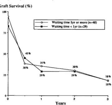
was slightly better for transplants performed more recently, that is between January 1983 and December 1986 (Fig. 6), in keeping with improving graft survival for other primary renal diseases [19,20]. Graft survival was also computed according to waiting time on dialysis: no
17%
difference was found between patients grafted within or after the first year of starting treatment (Fig. 7).
0 , ~"2 " I Of the first grafts that failed, 88% did so within the first
Years year. Among the causes reported for these, only 33% were Fig. 4. First graft actuarial survival according to source of graft. Primary r e l a t e d t o rejection compared to 70%-85% in patients non-function was recorded for 11% of CAD grafts. with other primary renal diseases [21]. Recurrence of
Graft Survival (%)
100 n
Waiting time lyr or more (n=48) Waiting time < lyr (n=29)
38%
Years
Fig. 7. First cadaver graft survival according to time on dialysis prior to
transplantation.
oxalosis in the graft was recorded as the cause of graft failure in 31%. These proportions were similar for the eight second graft failures.
Patient survival after grafting in 1980-1986 and for all patients with oxalosis starting renal replacement therapy during the same period is shown in Table 2. Despite the high graft failure rate, mortality appeared to be similar, or only slightly higher, after grafting. This resulted in a cumulative actuarial survival of 74% at 3 years after receiving a first cadaver or living donor graft. Actuarial survival, irrespective of the mode or renal replacement therapy, was 79% at 3 years. This compares unfavourable to patients with the common primary renal diseases of similar age distribution, who were found to have a 5-year cumulative survival above 80% [19].
Table 2. Per cent actuarial patient survival
After first graft (n = 60)
On RRT (including grafting n = 122) 1980-1986 Year 1 85 91 2 85 87 3 74 79
The distribution of the main causes of death was studied and the results are shown in Table 3 according to whether the patient had ever received a transplant. There were slightly less than twice as many deaths in ungrafted than grafted patients. Cardiovascular causes were the most frequent (51 %) with infection second (14%). Deaths from infection were rarely recorded in ungrafted patients.
Table 3. Per cent distribution of causes of death according to whether or
not the patient had ever received a transplant
Cardiac Vascular Infection Therapy Other ceased Ungrafted (%) (n = 69) Grafted (%) (7i = 39) All (%) (7i=108) 41 36 39 16 5 12 4 31 14 6 3 5 33 25 31
Cardiovascular causes, on the other hand, were more frequent (57%) in ungrafted patients than in those who had received a transplant at some time (41 %).
Discussion
Historically, renal transplantation was considered as contraindicated in primary hyperoxaluria [4,5]. This atti-tude was confirmed by several reports which recorded poor graft survival due to oxalate deposition in the trans-planted kidney [6,10]. Nevertheless, there were some publications reporting a single or a small number of successful kidney transplants [11,15]. These reports stressed the importance of specific measures such as intensive dialysis at time of grafting, and pyridoxine administration, which may decrease oxalate deposition in the graft. The largest series with well-defined prophylactic medical treatment was reported in 1984 by Scheinmann et al [16]. It included ten living related donor transplants, of whom seven had a functioning graft 1-7 years later, and one cadaver graft that lasted for 9 years. Considered together with the poor prognosis of patients with primary oxaluria on any form of dialysis therapy, these limited but rather optimistic reports probably encouraged continued attempts with kidney transplantation in patients with primary oxalosis, since the number of grafts per year recorded in the EDTA Registry increased between 1982 and 1986.
This report confirms the poor prognosis of kidney transplantation in patients recorded by the EDTA Registry with the diagnosis of oxalosis, although it is important to keep in mind that, before 1985, some patients with secondary oxalosis may have been registered under the same code. Since secondary oxalosis is either related to acute but transient oxalate intoxication or to uncontrolled, though in principle controllable, enteric hyperabsorption of oxalate, kidney transplantation for secondary oxalosis is likely to have a much better prog-nosis. Thus, the low graft survival rates reported here might still overestimate the true situation concerning primary hyperoxaluria.
Recipients of living related donor transplants run a lower risk of oliguria due to ischaemic damage at trans-plantation or due to rejection episodes. It is not surpris-ing, therefore, that fewer living related donor grafts were lost during the early post-transplant months. However, their failure rate was higher thereafter and resulted in a graft survival that was as low at 3 years as in cadaveric grafts (Fig. 4).
End-stage renal failure occurring in childhood might point to a more severe form of oxalosis. However, graft survival was no better in adults than in children, indi-cating that once on dialysis the oxalate pool size would probably have the same potentially deleterious effect on the graft. This is also reflected by similarly poor graft survival irrespective of the waiting time on dialysis (Fig. 7). Finally it is also disappointing to note only a slight improvement of graft survival due to fewer early failures but with a similar late failure rate in more recent years. The poor prognosis of kidney transplantation in patients with primary hyperoxaluria may lend support to another approach, namely combined hepatic and renal transplan-tation [22]. Whether or not correction of the underlying metabolic disorder by this approach will improve the outlook for patients with primary oxalosis remains yet to be clearly demonstrated.
Acknowledgements. The work of the EDTA Registry has been supported
by grants from governments or national societies for nephrology. Grants have also been made by the AKZO Group (Fibres and Polymers Division), Asahi Medical Co. Ltd, B. Braun Melsungen AG, Baxter Healthcare Corporation, Bellco SpA, Boehringer Mannheim GmbH, CD Medical International Ltd, Cilag AG Research, Fresenius Ltd, Gambro AB, Hospal Ltd, and Sandoz AG.
We thank all those doctors and their staff who have kindly completed EDTA Registry questionnaires.
References
1. Danpure CJ, Jennings PR. Peroxisomal alanine: glyoxylate amino-transferase deficiency in primary hyperoxaluria type I. FEBS
Utters 1986; 201: 20-24
2. Broyer M, Gagnadoux MF. L'oxalose: aspects cliniques et evolutifs. In Legrain M, Kuss J, eds. Seminaire D'Uro-Nephrologie. Masson Publishers, Paris. 1975: 136-146
3. Blackburn WE, McRoberts JW, Bhathena D et al. Severe vascular complications in oxalosis after bilateral nephrectomy. Ann Intern
Med 1975; 82: 44-46
4. Deoohar DS, Tung KS, Zuhle V et al. Kidney transplantation in primary oxalis. Clin Nephrol 1976; 5: 239-244
5. Klauwers J, Wolf PL, Cohn R. Failure of renal transplantation in primary oxalosis. JAMA 1969; 209: 551
6. Wilson, RE. Renal transplantation in congenital and metabolic diseases.//)AM 1975; 232: 148-153
7. Saxon A, Busch GJ, Merrill JP et al. Renal transplantation in primary hyperoxaluria. Arch Intern Med 1974; 133: 464-467 8. Halverstadt DB, Wenzl JE. Primary hyperoxaluria and renal
transplantation. J Urol 1974; 111: 398^02
9. Jacobs C, Rottenburg J, Reach I. Terminal renal failure due to oxalosis in 14 patients. Proc Eur Dial Transplant Assoc 1974; 11: 359-366
10. Novik AL, Braun WE, Magnusson M, Stowe N. Current status of renal transplantation at the Cleveland clinic. J Urol 1979; 122: 433-437
11. O'Regan P, Constable AR, Joekes AM et al. Successful renal transplantation in primary hyperoxaluria. Postgrad Med J1980; 56: 288-293
12. Bohannon LL, Norman DJ, Barry J, Bennett W. Cadaveric renal transplantation in a patient with primary hyperoxlauria.
Transplan-tation 1983; 36: 114-115
13. David D, Cheigh JS, Stenzel KH, Rubin AL. Successful renal trans-plantation in a patient with primary hyperoxaluria. Transtrans-plantation
Proc 1983; 15:2168-2171
14. Breed A, Chesney R, Friedman A et al. Oxalosis induced bone disease: a complication of transplantation and prolonged survival in primary oxalosis. Report of a case. J Bone Joint Surg 1981; 63A: 310-316
15. Whelchel JD, Alion DV, Luke J et al. Successful renal transplan-tation in hyperoxaluria. Transplantransplan-tation 1983; 35: 161-164 16. Scheinmann JI, Najarian JS, MauerM. Successful strategies in renal
transplantation in primary oxalosis. Kidney Int 1984; 25: 804—811 17. Bryoer M, Brunner FP, Brynger H et al. Demography of dialysis
and transplantation in Europe 1984. Nephrol Dial Transplant 1986; 1:1-8
18. Kramer P, Broyer M, Brunner FP et al. Combined report on regular dialysis and transplantation in Europe, XIV, 1983. Proc
EDTA-ERA 1985; 21: 2-68
19. Brunner FP, Broyer M, Brynger H et al. Survival on renal replace-ment therapy: data from the EDTA Registry. Nephrol Dial
Transplant 1988; 2: 109-122
20. Geerlings W, Tufveson G, Broyer M et al. Combined report on regular dialysis and transplantation in Europe, XVII, 1986. Proc
EDTA-ERA 1980
21. Wing AJ, Broyer M, Brunner FP et al. Combined report on regular dialysis and transplantation in Europe, XIII, 1982. Proc
EDTA-ERA 1983; 20: 2-75
22. Watts RWE, Rolles K, Morgan SH, Williams R, Calne RY et al. Successful treatment of primary hyperoxaluria type I by combined hepatic and renal transplantation. Lancet 1987; 2:474-475
Received for publication 20.10.89 Accepted 7.2.90