HAL Id: hal-00553427
https://hal.archives-ouvertes.fr/hal-00553427
Submitted on 16 Mar 2011HAL is a multi-disciplinary open access archive for the deposit and dissemination of sci-entific research documents, whether they are pub-lished or not. The documents may come from teaching and research institutions in France or abroad, or from public or private research centers.
L’archive ouverte pluridisciplinaire HAL, est destinée au dépôt et à la diffusion de documents scientifiques de niveau recherche, publiés ou non, émanant des établissements d’enseignement et de recherche français ou étrangers, des laboratoires publics ou privés.
A modeling study of the role of tonic vs. phasic
dopamine input to the prefrontal cortex
Denis Sheynikhovich, Satoru Otani, Angelo Arleo
To cite this version:
Denis Sheynikhovich, Satoru Otani, Angelo Arleo. A modeling study of the role of tonic vs. phasic dopamine input to the prefrontal cortex. Cinquième conférence plénière française de Neurosciences Computationnelles, ”Neurocomp’10”, Aug 2010, Lyon, France. �hal-00553427�
A modeling study of the role of tonic vs. phasic dopamine input
to the prefrontal cortex
Denis Sheynikhovich1, Satoru Otani2, Angelo Arleo1
1. CNRS-UPMC UMR7102, Univ. Paris 6, F-75005 Paris, France 2. INSERM-U952, CNRS-UMR7224, Univ. Paris 6, F-75005 Paris, France
ABSTRACT
This work presents a computational model of dopamine (DA) influence on long-term potentiation (LTP) and long-term depression (LTD) in the prefrontal cortex. Distinct properties of the model are a DA-concentration-dependent switch from depression to potentiation during induction of plasticity, and an inverted-U-shaped dependence of protein synthesis threshold on the level of background DA. Protein synthesis is responsible for maintenance and late phase of LTP/LTD in the model. Our simulations suggest that in order to comply with in vitro experimental data, prefrontal synapses must contain a protein that is slowly (on the timescale of minutes) activated in the presence of DA in a concentration-dependent manner. The activation value determines the direction of plasticity during induction, while it also modulates the magnitude of plasticity during maintenance. More generally, our results support the hypothesis that phasic release of endogenous DA is necessary for the induction of long-term changes in synaptic efficacy, while the concentration of tonic DA determines the direction and magnitude of these changes in the PFC.
KEY WORDS
Neuron modeling, synaptic plasticity, dopamine, prefrontal cortex
1. Introduction
The prefrontal cortex (PFC) is thought to mediate executive functions, including strategic organization of behavior [1]. These functions were shown to rely on (short-term) working memory, thought to be represented by persistent activity within the PFC [2], as well as on long-term memory, represented by persistent synaptic changes [1, 3]. Both of these memory representations were shown to be strongly modulated by dopamine (DA) [4, 5]. In particular, for both memory types (i) the role of tonic (background) DA levels is different from that of phasic (or event-related) DA release [6, 4, 5]; and (ii) optimal DA levels are required for best performance. The latter property is expressed by an inverted-U-shaped dependence of task performance (working memory) or magnitude of synaptic changes (long-term memory) on DA levels [7, 4]. Whereas a fair amount of theoretical work has been devoted to DA modulation of persistent activity and
working memory, no theoretical models address the role of DA for long-term plasticity in the PFC.
The present work attempts to fill this gap by proposing a model of DA modulation of long-term plasticity in the PFC. In particular, we focus on the series of studies that investigated DA influence on long-term depression (LTD) and long-term potentiation (LTP) induced by high frequency stimulation (HFS, 50 Hz) in the rat PFC in vitro [8, 9, 10, 5]. In line with general properties of DA in the PFC outlined above, three main conclusions can be made from the results of these studies. First, a phasic DA release during synaptic stimulation is required to trigger plasticity at this frequency [8, 9]. Second, tonic DA levels modulate the direction of plastic changes in the PFC, since the same HFS results in either LTD or LTP depending on DA conditions in prefrontal slices 10-40 min before the stimulation. Namely, insufficient time of DA exposure results in no plasticity or LTD (for concentrations in the range 3-100μM), whereas prolonged bath at a low tonic DA (1-10 μM) results in LTP [(1-10, 5]. Third, tonic DA modulates the amplitude of plastic changes in the PFC. In the LTP regime, different tonic DA concentrations result in LTP of different magnitude such that the dependence of the LTP strength on DA concentration follows an inverted-U-shaped curve [10, 5]. This is in contrast to Hippocampus where DA influence is limited to the maintenance phase of long-term plasticity (via cAMP-dependent pathways) [11, 12, 13] suggesting that direction of plasticity (which is determined during induction) is DA-independent. Moreover, an inverted-U-shaped dependence of memory strength or memory-dependent performance has not been reported for the Hippocampus. The mechanism of this differential influence of DA in the PFC relative to that in the Hippocampus is not clear.
We address these issues by proposing a computational model of long-term plasticity in which DA modulates the direction and magnitude of plastic changes. In this model, the direction of plasticity is determined by DA influence during plasticity induction (i.e. during synaptic tagging phase) while the plasticity amplitude is determined by DA influence during maintenance phase of LTP (i.e. during protein synthesis). Moreover, our simulations suggest that DA actions during both phases can be accounted for by considering DA-dependent activation of a single protein kinase.
2. Model of synaptic plasticity under DA
influence
In our model of long-term plasticity an artificial neuron is first exposed to a ‘bath period’ with different DA conditions and then stimulated by high-frequency synaptic input. The evolution of the average synaptic strength is monitored after the stimulation to see whether long-term synaptic changes were induced. As a result of stimulation, synapses of the neuron can undergo plastic changes that occur in two phases, plasticity induction and plasticity maintenance as described below.
2.1 Neuron model
We used a two-compartment model neuron (i.e. soma and dendrite) with compartment sizes and passive parameters taken from the study of [14]. Ionic conductances, Ca2+ and K+ dynamics were modeled
identically to the same study except that slow potassium current IKslowwith conductance 1.0 mS/cm2 was added
which was modeled as in [15]. The model neuron had 100 AMPA synapses that were plastic and 100 non-plastic NMDA synapses. Parameters of AMPA and NMDA synaptic conductances were taken from [16], except that for a single synapse gAMPA,max = 4 nA
(gNMDA,max = gAMPA,max/50) and only short-term
depression was modeled with utilization parameter USE
= 0.6 and recovery time constant trec= 800 ms [17].
Synaptic conductances were scaled by efficacies wnthat
were initialized at wn(0) = w0 = 1 and remained fixed
for NMDA synapses, while for the AMPA synapses they were updated according to the synaptic plasticity rules described in the following sections. The conductance of the slow potassium current gKslow and
peak synaptic conductances (gAMPA,max, gNMDA,max) were
tuned to reproduce voltage responses of PFC neurons to step currents and HFS recorded in vitro (neuron recording data provided by S. Otani, see also [5]), see Results.
2.2 DA influence during ‘bath period’
A principal feature of the DA-dependent switch from LTD to LTP is that the same high frequency stimulation results in either potentiation or depression, depending on the DA conditions in the slice during 10-40 min time period before the stimulation [10, 5]. This suggests that the direction of synaptic changes, at least in the PFC, depends not only on how the synapse is stimulated but also on a long-term (i.e. on the time scale of tens of minutes) history of the synapse before the stimulation. On the basis of experimental data [5] we propose that the presence of DA in the bath activates an unidentified protein kinase (referred to as DAK in the following), such that the rate of activation depends on the DA concentration in the bath (Fig. 1A). Concentration [DAK] of the DA-activated kinase above threshold θLTP
= 10−4 at the time of stimulation is a necessary
condition for LTP induction (see below). If the activation level is not sufficient, only LTD can be
induced. In addition, we assume that the activation rate is fastest at an optimal DA concentration and decreases at non-optimal concentrations. This leads to an inverted-U-shape dependence of the [DAK] on tonic DA concentration (Fig. 1A).
Figure 1: DA-dependent activation of DAK and its role in plasticity induction and maintenance. A. [DAK] profile at the time of stimulation (t=40 min). Threshold for LTP is not visible at this scale. Inset: zoom-in to the lower part of the plot. Dashed line corresponds to θLTP
B. The concentration of synthesized PRPs starts to grow (p>0) when a phasic DA release is higher than threshold Np (solid line), which depends inversely on
[DAK] (see Eq. 3). The dashed lines: DA concentrations for which experimental data is available [5]. The value of Npcorresponds to t=40 min after the
start of the DA bath.
2.3 DA influence during induction
According to the theory of synaptic tagging and capture [18, 11], synaptic stimulation results in setting of ‘tags’ (thought to be implemented by an activated form of a particular kinase, such as CaMKII for LTP [19]) at some synapses. Tagged synapses are potentiated (if the LTP tag is set) or depressed (if the LTD tag is set) for a period of time <40 min, corresponding to an early phase of LTP or LTD. In order to maintain these changes for a longer period of time, protein synthesis is required (see the next section).
Following [20] we model synaptic tags by binary variables lnand hncorresponding to LTD and LTP tags,
respectively. Only one of the tags can be equal to 1 at any time and non-tagged state corresponds to hn=0,
ln=0. The probability of setting a tag, i.e. plasticity rate
is given by:
ρ = A · gsyn· [V − θV]+ (1)
where A>0 is a constant parameter, gsyn is the total
synaptic conductance, V is membrane voltage and [.]+
denotes rectification, i.e. [x]+=x if x>0 and zero
otherwise. The rise in synaptic conductance gsynserves
as a presynaptic spike detector. According to Eq. 1, a synapse can become tagged only if the neuron is depolarized as a consequence of the synaptic input above the threshold value θV = −50 mV. A tag
represents only a temporary change at the synapse and so a tagged synapse will return to an initial state with stochastic rates kL= 1/(1.5 h) (for LTD tag) and kH=
1/h (for LTP tag), unless it is consolidated.
The plasticity rate ρ (Eq. 1) determines when a tag is set, but it does not determine the type of tag. In the
present model, the tag will be either LTD or LTP depending on the value of [DAK] at the time of the stimulation (see Fig. 1A). Thus, a synapse that has no tags set, can change its state under the following conditions:
• If 0<[DAK]<θLTP, the synapse switches to the state
ln=1, hn=0 (i.e. LTD tag is set) with probability
ρLTD given by Eq. 1 with A=ALTD=1.7 · 10−4
• If [DAK]>θLTP, the synapse switches to the state
ln=0, hn=1 (i.e. LTP tag is set) with probability
ρLTP given by Eq. 1 with A=ALTP=1.0 · 10−4
• otherwise, the synapse remains in the non-tagged state ln=0, hn=0.
2.4 DA influence during maintenance
As a result of stimulation, plastic changes can be induced at some synapses with corresponding transient increase (early-LTP) or decrease (early-LTD) in their strength. These transient changes may become persistent if plasticity related proteins (PRPs) are synthesized and reach the tagged synapse [18]. In the model, the concentration of synthesized PRPs at the synapse is described by variable p, growth of which is under the control of DA.
DA signaling in the PFC occurs in two distinct modes [6, 7]. Tonic, or background, DA signal provides a constant low concentration of DA that is simulated in slice experiments by low-concentration DA bath. This signal is thought to be provided by the background population activity of dopaminergic neurons in the VTA [7, 21]. Phasic, or event-related, release of DA occurs as a result of burst firing of the DA neurons in response to salient environmental stimuli. In slices, high-frequency stimulation results in DA release from dopaminergic axon terminals present in the slice that can be considered as an in vitro model of the event-related DA release [22, 5]. It has been suggested that DA action is determined by the relative values of tonic vs. phasic DA levels [6, 5], but no mechanism has been proposed so far.
In our model, the phasic DA signal is proportional to the total number of synapses that were tagged during stimulation [20]:
DAphasic= ADA·Σ(hn+ ln) (2)
with constant ADA= 0.03 μM, such that a maximal DA
release (when all synapses are tagged) corresponds to 3μM, which is in the range of experimentally observed values [23]. This simple model of phasic DA reflects the experimental observation that stronger stimulation results in larger phasic DA release [22].
The tonic DA signal in our model exerts its influence via the DA-dependent activation of DAK, such that an optimal level of tonic DA results in high concentration of the activated kinase (Fig. 1A). We assume that in addition to determining the direction of weight changes (see the previous section), this concentration influences the maintenance phase of plasticity such that if [DAK]
is low, a strong stimulation is required to trigger PRP synthesis, while only a weak stimulation is sufficient to trigger the synthesis if [DAK] is high. The relation is captured in the model by a threshold Np of PRP synthesis which depends inversely on [DAK] (Fig. 1B): Np([DAK]) = B / ([DAK]+0.01) + C (3)
The protein synthesis is triggered (i.e. p starts to grow) when DA concentration near the synapse due to phasic DA release is higher then Np [20]:
dp/dt = −p/τpd + (1−p)/τpr · H[DAphasic − Np([DAK])] where τpd=60min, τpr=2min and H is the Heaviside step
function, H[x]=1 if x>0 and H[x]=0 otherwise. In Eq. 3, the constants B=0.013 and C=0.3 determine the amount of phasic DA that is necessary to induce plasticity when the tonic DA is absent or present at an optimal concentration. The inverse dependence of protein synthesis threshold Npon [DAK] in Eq. 3 can be
interpreted as the requirement for a sufficient activation of DAK at the synapse before the synthesis of plasticity-related proteins in the nucleus can be started (i.e. suggesting a protein-cascade-like process).
Thus, under a given stimulation strength, the plasticity related proteins will be synthesized faster when [DAK] is high (i.e. Npis low). A faster rate of PRP synthesis
results in a larger number of consolidated synapses (in non-saturating regime), and hence to higher LTP magnitude (measured as an average increase of synaptic strength). Conversely, low values of [DAK], resulting from non-optimal DA conditions, lead to a smaller amount of PRPs to be synthesized leading to smaller LTP magnitude.
Finally, the consolidation process is modeled identically to [20] by introducing a synaptic consolidation variable zngoverned by bistable dynamics:
τz ·dzn/dt= zn(1 − zn)(zn− 0.5) + γ(hn− ln)p (4)
where γ=0.1 and variable zn represents the activated
state of an enzyme responsible for synaptic consolidation (e.g. PKMζ [24]). In the absence of plasticity related proteins (i.e. p = 0) or if no tags are set at the synapses (i.e. hn=0 and ln=0), Eq. 4 describes
bistable dynamics with two stable states z = 0 and z = 1, that represent activated or non-activated enzyme, respectively. When a tag is set at the synapse and enough plasticity related proteins are available, variable zn can pass from one stable state to another, depending on which tag is set. When one of the stable states is reached, it is kept indefinitely long, unless a new stimulation induces a switch between states. Initially, zn=1 for 1/3 of all synapses ensuring the capacity of the
neuron to express late LTD.
The synaptic weight change as a result of stimulation has contributions both from induction and maintenance phases [20]:
Δwn= w0 · (hn− 0.5 · ln+ 2 · zn) (5)
where w0 is the weight value before stimulation. Since
only one of the tags can be set to 1 at a time, Eq. 5 means that the LTD contribution is half of that of LTP, reflecting the experimentally observed difference in the strengths of induced LTD and LTP in minimal stimulation experiments [25].
3. Results
Influence of DA on long-term plasticity was investigated by stimulating the artificial neuron with HFS (3 or 6 trains of 100 pulses at 50Hz) under various DA conditions and monitoring the evolution of the average synaptic strength for several hours after the stimulation. Below we show that the simulated neuron behaves similarly to real PFC neurons in relevant stimulation regimes and that DA-modulation of plasticity in these neurons reproduces neurophysiological data [5].
3.1 Reproduction of voltage response of PFC neurons
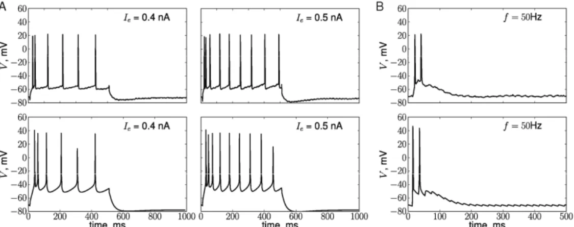
Most of the neurons used in the study [5] were regularly spiking and their response to the HFS exhibited strong adaptation (an example of one such neuron is shown in Fig. 2, top row). Since we were interested in reproducing DA effects on PFC neurons, we first ensured that the neuron model we use is capable of reproducing responses of real PFC neurons to step currents and HFS. Spiking frequency in response to step currents was tuned by adjusting the conductance of the slow potassium current [15] (Fig. 2A, bottom row). Fast adaptation during HFS in the model is due to short term depression [17] (Fig. 2B, lower panel).
3.2 Synaptic plasticity in the absence of tonic DA
We then tested whether long-term synaptic plasticity can be induced by HFS in the absence of tonic DA. Weak stimulation (3 trains of 100 pulses at 50 Hz) did
not result in a significant change of synaptic strength two hours after the stimulation (Fig. 3A, top) whereas a longer stimulus (6 trains at the same frequency) resulted in long-term LTD (Fig. 3B, top). In the absence of tonic DA, DAK is not activated so that only LTD can be induced in our model (see Fig. 1A). That is why HFS results in a depression of synaptic weights after stimulation (Fig. 3, top row). The peak of the depression is larger after the stronger stimulus, since more synapses were tagged, as shown by the higher amplitude of phasic DA release in this case (shown by black lines in Fig. 3, bottom row, see also Eq. 3).
Unless this depression of synaptic strength is consolidated via protein-synthesis dependent process, it will disappear as a result of stochastic decay of synaptic tags (Fig. 3A, top). The concentration p of synthesized PRPs (shown by the dashed line in Fig. 3) starts to increase when the rise in DA concentration due to phasic DA release reaches threshold Np (shown by the Figure 2. Voltage responses of real (top row, data provided by S. Otani) and simulated (bottom row) PFC neurons to injected step currents (A) and HFS at 50 Hz (B). Current amplitudes 0.4 nA (left column) and 0.5 nA (right column), duration 500 ms. B. First 500 ms of the response to the tetanus.
Figure 3. Synaptic plasticity in the absence of tonic DA as a result of stimulation by weak HFS (3 trains at 50 Hz, A) or strong HFS (6 trains at 50 Hz, B) applied at t = 40 min. Top row: Evolution with time of the average weight change of the simulated neuron (in % relative to baseline). Bottom row: Concentration p of synthesized PRPs (dashed line, arbitrary units) grows when the amount of phasic DA (black line) is larger than minimum value Np (dotted line) and decays to zero otherwise.
dotted line in Fig.3). When the DA concentration falls below the threshold (e.g., due to DA re-uptake mechanisms [23]) PRP concentration decays to zero. For the simulated neuron shown in Fig. 3, not enough PRPs were synthesized when the stimulation was weak resulting in the absence of a long-term weight change. In contrast, a stronger stimulation resulted in high PRP synthesis rate and late LTD.
The PRP synthesis threshold Npis determined by the
DAK concentration, which is equal to zero in the present case. The value of Np at zero tonic DA is
constant (see Eq. 3) and can be considered as the minimum amount of phasic DA (which corresponds to the minimal stimulation strength) that is necessary for plasticity to take place.
Since both the magnitude of plasticity and its sign (LTD in this case) are determined by the DAK concentration, the same results will be obtained in our model when the DAK is activated but the activation is not sufficient to pass threshold for LTP (see inset in Fig. 1A). These conditions correspond to either low tonic DA concentrations (i.e., less than 1 μM, corresponding to experimental data from [5]), or short exposure to high DA concentrations (up to 20min at >8μM, corresponding to the data reported in [10]). We also note here that because of stochasticity in the setting and decay of tags, synapses have low but non-zero probability to become consolidated even when stimulation is weak. This will lead to small LTD when results are averaged over many repetitions (see Fig. 5).
3.3 Synaptic plasticity in the presence of tonic DA
Next, we explored how the sign and magnitude of plasticity changes in our model depending on different tonic DA conditions. The only way the tonic DA concentration influences plasticity in our model is through the activation of DAK. Hence, DA bath at different concentrations corresponds in our model to
different values of [DAK] at the time of the stimulation (40 min in the simulations below, see Fig. 1B).
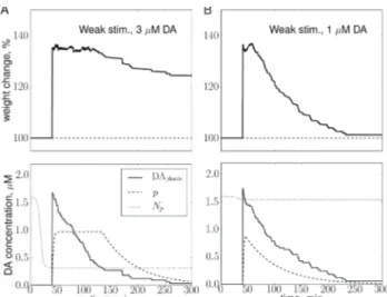
The same HFS (3 trains of 100 pulses at 50 Hz) resulted in strong LTP when the DA concentration was optimal (3 μM, Fig. 4A, top), while the magnitude of LTP decreased at non-optimal concentrations (1 μM, Fig. 4B, top; and 10 μM, not shown) reproducing the data reported in [5]. The mechanism of modulation of the LTP amplitude is similar to that for LTD (see previous section), with two main differences. First, since the DA concentration was equal to or larger than 1 μM, the DAK activation reached the LTP threshold such that the plasticity direction switched from depression to potentiation. Second, the rise in DAK concentration due to the presence of tonic DA lead to a decrease of the threshold Np of PRP synthesis with time during the bath period. At the optimal concentration the decrease is steep so that at the time of the stimulation even a small phasic DA release leads to fast PRP synthesis and high number of consolidated synapses (Fig. 4A). At non optimal concentrations, the decrease in Np is small so that only a weak LTP can be induced by the same HFS (Fig. 4B).
In summary, our model reproduces the main features of DA modulation of long-term synaptic plasticity observed in vitro [5, 10]. First, the model exhibits DA-dependent switch from LTD to LTP under the same HFS (Fig. 5A). Second, in the LTP regime, the magnitude of LTP follows an inverted-U-shaped dependence on the concentration of tonic DA (Fig. 5B). Both of these effects are due to the assumption that extracellular tonic DA activates a putative protein kinase in a DA-concentration dependent manner. The level of activation determines directly whether depression or potentiation can be induced (during plasticity induction phase). In addition, it indirectly determines the magnitude of plasticity by influencing the rate of PRP synthesis. A phasic release of DA is necessary for any plasticity to take place.
3. Conclusion
In this work we proposed a mechanism of DA influence on long-term plasticity within PFC. In this mechanism, the sign and magnitude of plasticity depend on the relative levels of phasic and tonic DA activity. Moreover, the model makes a specific prediction about the existence of a DA-activated protein kinase that influences plasticity during both induction and maintenance phases.
Acknowledgements
This work is granted by French National Research Agency, project EVONEURO ANR-09-EMER-005. Figure 4. Synaptic plasticity after DA bath at
concentrations 3 μM (A) and 1 μM (B). Weak HFS (3 trains at 50 Hz) was applied at t=40 min. See caption of Fig. 3 for details. Protein threshold Np (dotted line) decreases as a function of time due to the DA-concentration dependent increase in [DAK], see Eq. 3.
References
[1] J. M. Fuster. Memory in the cerebral cortex. The MIT Press, Boston: A Bradford Book, 1995.
[2] P. S. Goldman-Rakic. Cellular basis of working memory. Neuron, 14(3):477–485, 1995.
[3] K. Touzani, S. V. Puthanveettil, and E. R. Kandel. Consolidation of learning strategies during spatial working memory task requires protein synthesis in the prefrontal cortex. Proc Natl Acad Sci USA, 104(13):5632–5637, 2007.
[4] J. K. Seamans and C. R. Yang. The principal features and mechanisms of dopamine modulation in the prefrontal cortex. Prog Neurobiol., 74(1):1–58, 2004.
[5] B. Kolomiets, A. Marzo, J. Caboche, P. Vanhoutte, and S. Otani. Background dopamine concentration dependently facilitates long-term potentiation in rat prefrontal cortex through postsynaptic activation of extracellular signal-regulated kinases. Cereb Cortex., 19(11):2708–2718, 2009.
[6] A. A. Grace. Phasic versus tonic dopamine release and the modulation of dopamine system responsivity: A hypothesis for the etiology of schizophrenia. Neuroscience, 41(1):1–24, 1991.
[7] Y. Goto, S. Otani, and A. A. Grace. The yin and yang of dopamine release: a new perspective. Neuropharmacology, 53(5):583 – 587, 2007.
[8] S. Otani, O. Blond, J.M. Desce, and F. Cr´epel. Dopamine facilitates long-term depression of glutamatergic transmission in rat prefrontal cortex. Neuroscience, 85(3):669–676, 1998.
[9] S. Otani, N. Auclair, J. M. Desce, M. P. Roisin, and F. Crépel. Dopamine receptors and groups and mGluRs cooperate for long-term depression induction in rat prefrontal cortex through converging postsynaptic activation of kinases. J Neurosci., 19(22):9788–802., 1999.
[10] Y. Matsuda, A. Marzo, and S. Otani. The presence of background dopamine signal converts long-term synaptic depression to potentiation in rat prefrontal cortex. J Neurosci., 26(18):4803–4810, 2006.
[11] U. Frey and R. G. Morris. Synaptic tagging: implications for late maintenance of hippocampal long-term potentiation. Trends Neurosci., 21(5):181–8, 1998.
[12] S. Navakkode, S. Sajikumar, and J. U. Frey. The type IV-specific phosphodiesterase inhibitor rolipram and its effect on hippocampal long-term potentiation and synaptic tagging. J Neurosci., 24(35):7740–7744, 2004.
[13] S. Navakkode, S. Sajikumar, and J. U. Frey. Synergistic requirements for the induction of dopaminergic D1/D5-receptor-mediated LTP in hippocampal slices of rat CA1 in vitro. Neuropharmacology, 52(7):1547–1554, 2007.
[14] D. Durstewitz and J. K. Seamans. The computational role of dopamine d1 receptors in working memory. Neural Networks., 15(4-6):561–572, 2002.
[15] S. Schreiber, J.-M. Fellous, P. Tiesinga, and T. J. Sejnowski. Influence of ionic conductances on spike timing reliability of cortical neurons for suprathreshold rhythmic inputs. J Neurophysiol., 91(1):194–205, 2004. [16] D. Durstewitz and T. Gabriel. Dynamical basis of irregular spiking in NMDA-Driven prefrontal cortex neurons. Cereb Cortex., 17(4):894–908, 2007.
[17] M. V. Tsodyks and H. Markram. The neural code between neocortical pyramidal neurons depends on neurotransmitter release probability. Proc Natl Acad Sci USA, 94(2):719–23, 1997.
[18] U. Frey and R. G. M. Morris. Synaptic tagging and long-term potentiation. Nature, 385(9):533–536, 1997. [19] K. G. Reymann and J. U. Frey. The late maintenance of hippocampal LTP: Requirements, phases, ‘synaptic tagging’, ‘late-associativity’ and implications. Neuropharmacology, 52(1):24–40, 2007. [20] C. Clopath, L. Ziegler, E. Vasilaki, L. Büsing, and W. Gerstner. Tag-trigger-consolidation: a model of early and late long-term-potentiation and depression. PLoS Comput Biol., 4(12):e1000248, 2008.
[21] S. B. Floresco, A. R. West, B. A., H. Moore, and A. A. Grace. Afferent modulation of dopamine neuron firing differentially regulates tonic and phasic dopamine transmission. Nat Neurosci., 6(9):968–973, 2003. [22] C. E. Young and C. R. Yang. Dopamine d1-like receptor modulates layer- and frequency-specific short-term synaptic plasticity in rat prefrontal cortical neurons. Eur J Neurosci., 21(12):3310–3320, 2005. [23] P. A. Garris and R. M. Wightman. Different kinetics govern dopaminergic transmission in the amygdala, prefrontal cortex, azpqnd striatum: an in vivo voltammetric study. J Neurosci., 14(1):442–450, 1994. [24] S. Sajikumar, S. Navakkode, T. C. Sacktor, and J. U. Frey. Synaptic tagging and cross-tagging: The role of protein kinase mζ in maintaining long-term potentiation but not long-term depression. J Neurosci., 25(24):5750–5756, 2005.
[25] D. H. O’Connor, G. M.Wittenberg, and S. S.Wang. Graded bidirectional synaptic plasticity is composed of switch-like unitary events. Proc Natl Acad Sci USA, 102(27):9679–9684, 2005.
Figure 5. A summary of simulated DA influence on long-term plasticity in the simulated PFC neurons. The bars correspond to the magnitude of plasticity (average weight change over 10 neurons in % relative to baseline ±STD) 260min after the HFS application at different tonic DA concentrations. A. DA-dependent switch from LTD to LTP (DA corresponds to the optimal tonic DA concentration,
DAtonic = 3 μM. Weak stimulus: 3 trains at 50 Hz, Strong
stimulus: 6 trains at 50 Hz. B. The LTP magnitude follows an inverted-U-shaped dependence from tonic DA concentration.