HAL Id: tel-01775072
https://tel.archives-ouvertes.fr/tel-01775072
Submitted on 24 Apr 2018HAL is a multi-disciplinary open access archive for the deposit and dissemination of sci-entific research documents, whether they are pub-lished or not. The documents may come from teaching and research institutions in France or abroad, or from public or private research centers.
L’archive ouverte pluridisciplinaire HAL, est destinée au dépôt et à la diffusion de documents scientifiques de niveau recherche, publiés ou non, émanant des établissements d’enseignement et de recherche français ou étrangers, des laboratoires publics ou privés.
behavior : a potential therapeutic target for psychiatric
disorders
Tanzil Mahmud Arefin
To cite this version:
Tanzil Mahmud Arefin. GPR88 signatures in mouse neuronal connectivity and behavior : a potential therapeutic target for psychiatric disorders. Neurobiology. Université de Strasbourg; Albert-Ludwigs-Universität (Freiburg im Breisgau, Allemagne), 2017. English. �NNT : 2017STRAJ101�. �tel-01775072�
ÉCOLE DOCTORALE SCIENCES DE LA VIE ET DE LA SANTE
Institut de génétique et de biologie moléculaire et cellulaire (IGBMC)
THÈSE
présentée par :Tanzil Mahmud AREFIN
soutenue le : 20 November, 2017
pour obtenir le grade de :
Docteur de l’université de Strasbourg
Discipline/ Spécialité
: Neuroscience
TITRE de la thèse
Signatures du récepteur GPR88 sur la connectivité fonctionnelle et structurelle du cerveau chez la souris : implications pour le développement de la
dépendance à l’alcool
THÈSE dirigée par :
Mme Ipek Yalcin, PhD. Université de Strasbourg M. Ad Aertsen, PhD. University of Freiburg
RAPPORTEURS :
Mme Tracey D. Farr, PhD. University of Nottingham Mme Ilka Diester, PhD. University of Freiburg
AUTRES MEMBRES DU JURY :
M. Yann Herault, PhD. Université de Strasbourg
GPR88 signatures in mouse neuronal
connectivity and behavior:
A potential therapeutic target for
psychiatric disorders
Inaugural-Dissertation
Submitted by
Tanzil Mahmud Arefin
Faculty of Biology
Albert-Ludwigs-Universität Freiburg
Freiburg, Germany
École Doctorale des Sciences de la Vie et de
la Santé Université de Strasbourg
2
Contents
List of manuscripts ... 4
Summary ... 5
Chapter 1: Introduction ... 8
1.1 G-protein coupled receptor (GPCR) – GPR88 ... 10
1.1.1 GPR88 – a novel therapeutic for psychiatric disorders ... 11
1.1.2 Behavioral studies on GPR88 receptor deficient mice ... 13
1.1.3 Generation of Gpr88-/- mice ... 15
1.2 The concept of brain connectivity and brain networks ... 16
1.3 Brain imaging techniques ... 19
1.3.1 Structural connectivity assessment via diffusion tensor imaging (DTI) and fiber tractography .... 19
1.3.2 Functional connectivity assessment via resting state functional MRI (rsfMRI) ... 20
1.3.2.1 Default mode network ... 22
1.3.2.2 Central executive network ... 23
1.3.2.3 Salience network ... 24
1.3.2.4 Reward/aversion network ... 24
1.4 Rodents in neuroscience/neuroimaging research ... 26
1.4.1 rsfMRI and DTI in rodents... 26
1.4.2 Rodents in behavioral neuroscience ... 27
1.4.2.1 IntelliCage ... 27
1.4.2.2 IntelliCage – software overview ... 29
1.5 Implications of GPR88 receptor in alcohol addiction ... 30
1.6 Integration of the introduced techniques into my work ... 31
Chapter 2: Results ... 33
2.1 Characterization of the impact of GPR88 receptor on the mouse brain connectivity ... 35
2.2 Behavioral studies with the Gpr88-/- mice ... 39
2.2.1 Increased alcohol seeking in mice lacking Gpr88 involves dysfunctional addiction networks ... 39
2.2.2 Home cage behavioral phenotyping of Gpr88-/- female mice in group-housed condition ... 42
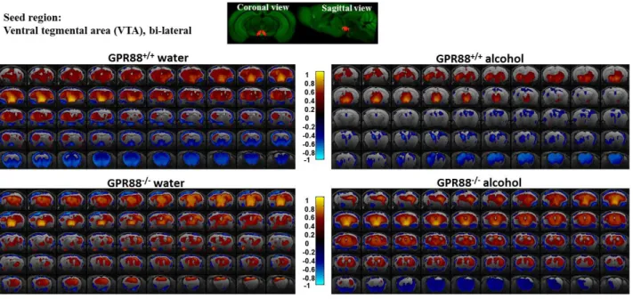
2.3 Gpr88 signatures on the brain reward network connectivity after alcohol exposure in mice ... 46
3
Chapter 3: Discussion ... 50
Conclusion and future perspective ... 59
References... 60 Acknowledgements ... 85 Chapter 4: Annex ... 86 4.1 Abbreviations ... 87 4.2 Tables... 90 4.3 Figures ... 96 4.4 Experimental procedures ... 98 4.4.1 Construction of Gpr88-/- mice ... 98
4.4.2 Animal preparation and MRI data acquisition ... 98
4.4.3 MRI data processing ... 99
4.4.3.1 Data pre-processing ... 99
4.4.3.2 Data post-processing ... 101
4.4.4 Mouse behavioral experiments ... 103
4.4.4.1 Experiments with alcohol ... 103
4.4.4.2 IntelliCage – system overview ... 103
4.4.4.3 Behavioral experiments with IntelliCage ... 109
4.4.4.4 IntelliCage data analysis ... 110
4.5 Principles of MRI ... 110
4.5.1 MR imaging ... 110
4.5.2 Relevant basic pulse sequences ... 112
4.5.3 Resting-state functional magnetic resonance imaging (rsfMRI) ... 113
4.5.4 T2-weighted MRI ... 115
4
List of manuscripts
1. Tanzil M. Arefin,Anna E Mechling, Carole Meirsman, Thomas Bienert, Neele Hübner, Hsu-Lei Lee, Sami Ben Hamida, Aliza Ehrlich, Dan Roquet, Jürgen Hennig, Dominik v. Elverfeldt, Brigitte Lina Kieffer and Laura-Adela Harsan
Remodeling of sensorimotor brain connectivity in Gpr88 deficient mice (Arefin T. et al., 2017, Brain Connectivity, https://doi.org/10.1089/brain.2017.0486)
Own contribution: I designed the study, performed all experiments, analyzed data and wrote the
manuscript in close collaboration with my supervisor.
2. Sami Ben Hamida, Sueli Netto, Tanzil M. Arefin, Md. Taufiq Nasseef, Laura-Joy Boulos, Michael McNicholas, Aliza Toby Ehrlich, Luc Moquin, Alain Gratton, Emanuel Darcq, Laura-Adela Harsan, Rafael Maldonado and Brigitte Kieffer.
Increased alcohol seeking in mice lacking Gpr88 involves dysfunctional mesocorticolimbic
networks (Ben Hamida et al., 2018, Molecular Psychiatry,
https://doi.org/10.1016/j.biopsych.2018.01.026)
Own contribution: I performed MRI experiments, analyzed data and wrote the manuscript parts
related to rsfMRI in close collaboration with my supervisor.
3. Aliza T. Ehrlich, Meriem Semache, Julie Bailly, Stefan Wojcik, Tanzil M. Arefin, Christine Colley, Christian Le Gouill, Florence Gross, Viktoriya Lukasheva, Mireille Hogue, Emmanuel Darcq, Laura-Adela Harsan, Michel Bouvier and Brigitte L. Kieffer
Mapping GPR88-Venus illuminates a novel role for GPR88 in Sensory processing (Ehrlich et al., 2017, Brain structure and function, https://doi.org/10.1007/s00429-017-1547-3)
Own contribution: I performed MRI experiments, analyzed data and wrote the manuscript parts
related to MRI in close collaboration with my supervisor.
4. Gregoire Maroteaux*, Tanzil M. Arefin*, Sami Ben Hamida, Laura-Adela Harsan, Emmanuel Darcq, Brigitte Kieffer. *Co-1st authors
Lack of anticipatory behavior in Gpr88 knockout mice revealed by automatized home cage phenotyping (Maroteaux et al., 2018, Genes, Brain and Behavior,
https://doi.org/10.1111/gbb.12473)
Own contribution: I designed the study, performed all experiments, analyzed data and wrote the
manuscript in close collaboration with my supervisor.
5. Anna E. Mechling, Tanzil Arefin, Hsu-Lei Lee, Thomas Bienert, Marco Reisert, Sami Ben Hamida, Emmanuel Darcq, Aliza Ehrlich, Claire Gaveriaux-Ruff, Maxime J. Parent, Pedro Rosa Neto, Jürgen Hennig, Dominik von Elverfeldt, Brigitte Lina Kieffer and Laura-Adela Harsan. Deletion of the mu opioid receptor gene in mice reshapes the reward-aversion connectome (Mechling et al., 2016, PNAS, doi:10.1073/pnas.1601640113)
5
Summary
Recent studies have demonstrated that the pathological perturbations of the brain and the expression or mutation of single gene influence spatially distinct regions via axonal pathways and result in the modification of overall brain functional and structural network architecture (Cao et al., 2015; Mechling et al., 2016; Richiardi et al., 2015; Thompson et al., 2013, Arefin et al., 2017). Functional and structural connectivity mapping of the brain thus offer a prevailing framework for localizing pathology, identifying the brain regions affected by pathological processes as well as tracking the patterns of psychiatric disorders that disturb higher cognitive functions (Biswal et al., 2010; Craddock et al., 2013; Sporns et al., 2005).
Resting-state functional magnetic resonance imaging (rsfMRI) is a technique that detects low frequency fluctuations (LFFs) of less than 0.1 Hz in the blood oxygen level dependent signal (BOLD) signal and measures functional connectivity (FC) between brain regions as the level of synchrony of spontaneous fMRI time-series during rest (Biswal et al., 1995, 1997; Greicius et al., 2003; Salvador et al., 2005). Diffusion tensor imaging (DTI) on the other hand is a three-dimensional noninvasive imaging modality that measures the diffusion of water molecules as a probe to infer the microstructural features. By combining the directional information and magnitude of anisotropic diffusion of the individual voxels, the course of fiber tracts can be reconstructed, which is known as tractography. Therefore, DTI and fiber tractography provides a unique opportunity to study the fiber architecture in vivo and characterize microstructural changes or differences with neuropathology and treatment.
Both the rsfMRI and DTI have been widely used for functional and structural brain connectivity mapping in human (Fair et al., 2007; Fox and Raichle, 2007; Alexander et al., 2007; Mori et al., 2001), rodents (Jonckers et al., 2011; Mechling et al., 2014, 2016; Harsan et al., 2006, 2010, 2013, Arefin et al., 2017) and primates (Hutchison et al., 2012; Shi et al., 2013; Zhang et al., 2013).
In this study, we combined mouse mutagenesis with functional and structural magnetic resonance imaging (MRI) to determine whether targeted inactivation of a single gene would modify whole-brain connectivity in live animals and how it translates at the behavioral level. The targeted gene encodes GPR88 - an orphan G-protein coupled receptor, robustly expressed in the dorsal and ventral striatum as well as in the amygdala, olfactory tubercle, inferior olive nucleus and neocortex (Ghate et al., 2007; Meirsman et al., 2016; Mizushima et al., 2000) in rodents, monkeys and human being during development and adulthood (Massart et al., 2009). The striatum is a major entry into the basal ganglia (BG) and plays important role in the initiation and patterning of many behaviors. Striatum receives excitatory cortical glutamatergic and thalamic glutamatergic inputs as well as modulatory dopaminergic input from substantia nigra and ventral tegmental area. These glutamatergic inputs together with inhibitory inputs from interneurons are integrated and relayed to other BG components via GABAergic medium spiny neurons (MSNs). MSNs express D1- or D2-dopamine receptors (D1R and D2R), founding the striatonigral (direct) and striatopallidal (indirect) pathways (Gerfen, 1992). GPR88 is abundant in MSNs expressing D1R and D2R (Massart et al., 2009). GPR88 thus plays potential role in psychiatric and
6
neurodegenerative diseases such as schizophrenia, depression, hyperactivity, addiction and bipolar disorder (Del Zompo et al., 2014; Ingallinesi et al., 2015; Logue et al., 2009; Massart et al., 2009; Meirsman et al., 2016; Quintana et al., 2012). However, much remains to be clarified regarding the specific cellular and physiologic roles of GPR88, and its pathophysiologic relevance to brain disorders.
Therefore, the first objective of my PhD project was to investigate the role of GPR88 receptor in living mouse brain structural and functional communication. This comprised, imaging the Gpr88 gene knock-out (Gpr88-/-) mice and their wild-type littermates (CTRL or Gpr88+/+ - mice normally expressing the GPR88 receptor) by means of rsfMRI and DTI with tractography techniques respectively.
Secondly, I investigated the involvement of GPR88 in the development of alcohol seeking and drinking behavior. Gpr88-/- and their littermates Gpr88+/+ mice were exposed to alcohol to examine whether Gpr88 deletion alters alcohol-taking and seeking behaviors. These mice were further imaged to investigate the involvement of GPR88 receptor in neurocircuitries modifications due to alcohol intake. Neuronal connectivity alterations were assessed following similar MR based neuroimaging approaches similar to the first part of my study.
Additionally, Gpr88 deficient mice were characterized by investigating the effects of Gpr88 gene in mouse behaviors using computer-based, fully automated testing apparatus - IntelliCage. It is an automated home cage that monitors group-housed mice implanted with radio frequency identification chips and allows studying multi-dimensional aspects of mice behavior. This longitudinal study was designed to investigate the striatum and hippocampus mediated behaviors with group-housed mice in 4 consecutive phases (free adaptation, nosepoke adaptation, place learning and fixed schedule drinking).
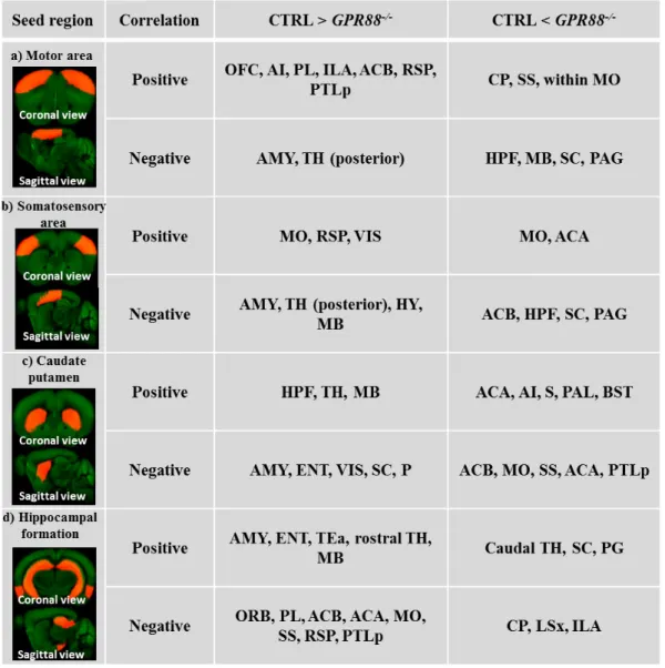
My work provided the first evidence of GPR88 involvement in remodeling the mouse brain functional and structural brain networks, primitive to the repertoire behavior observed in Gpr88-/- mice (Arefin T. et al., 2017). Deletion of Gpr88 in mice resulted extensive remodeling of intra-cortical and cortico-subintra-cortical networks. Most prominent modifications were observed in retrospenial cortex connectivity, a core player of the default mode network (DMN). Indeed, FC modifications in the DMN is considered a hallmark of many psychiatric conditions (Brady et al., 2016; Castellanos et al., 2008; Fair et al., 2010; Garrity et al., 2007). Furthermore, somatosensory and motor cortical networks showed remarkable FC modifications suggesting sensorimotor gating deficiency reported in mutantanimals (Logue et al., 2009; Meirsman et al., 2016a), and also likely underlie their hyperactivity phenotype. Apart from the cortical network, alterations within hippocampal and dorsal striatum FC underscore a specific learning deficit previously reported in Gpr88-/- animals (Meirsman et al., 2016a). Moreover, amygdala connectivity with cortex and striatum was weakened, perhaps underlying the “risk-taking” behavior of these animals (Meirsman et al., 2016b). This study hence implies GPR88 as a core player in brain communication.
7
In addition, we observed that Gpr88 deletion disrupts executive, reward and emotional networks in a configuration that reduces alcohol reward and promotes alcohol seeking and drinking. The FC signature is reminiscent of alterations observed in individuals at-risk for alcohol use disorders (AUDs). The Gpr88 gene, therefore, may represent a vulnerability/resilience factor for AUDs, and a potential drug target for AUDs treatment (Ben Hamida et al., 2018).
Moreover, through the development of IntelliCage protocols, we perceived hyperactivity, non-habituation, repetitive behavior and learning alteration that were previously described using different classical behavioral tests. The novel finding of this study is the lack of anticipatory behavior in mice lacking GPR88 receptor (Maroteaux et al., 2018).
This is the first study demonstrating that GPR88 activity shapes the mouse brain functional as well as structural connectome and how it translates at the behavioral level. Most importantly, the concordance between connectivity alterations and behavior deficits observed in Gpr88-deficient mice suggests Gpr88 as a potential therapeutic target for psychiatric disorders.
8
Chapter 1
Introduction
9
The PhD project presented here relates to the interdisciplinary field of research, merging non-invasive neuroimaging techniques and behavioral investigations in genetically modified mice. This comprised: mapping brain functional and microstructural network by means of resting state functional magnetic resonance imaging (rsfMRI) and high angular resolution diffusion imaging (HARDI) as well as global tractography. Moreover, GPR88 mediated mouse behavioral and cognitive functions were investigated using a computer-based automated system – IntelliCage and mouse behavior in response to alcohol exposure was assessed using conventional type-III cages. Mice exposed to alcohol were further scanned to examine whether deletion of Gpr88 gene remodels the brain functional and structural connectivity. This chapter provides an introductory view of the entire study.
Section 1.1 introduces the novel GPR88 receptor and its expression in the mouse brain, followed by the importance of examining this gene as a therapeutic target for psychiatric disorders (1.1.1). Section 1.1.2 describes behavioral studies characterizing the influence of GPR88 in mouse behavior and thus highlighting the significance of investigating the role of this gene in brain communication.
Section 1.2 briefly describes the concept of brain connectivity and brain networks.
Section 1.3 introduces one of the most widely used non-invasive magnetic resonance imaging (MRI) technique to map the brain connectivity or networks. This section further includes 2 more sub-sections: 1.3.1 illustrates the principles of mapping the brain structural network using diffusion tensor imaging (DTI) and tractography approach and the subsequent section (1.3.2) describes the concept of rsfMRI and some of the methods commonly used for brain functional connectivity mapping, such as: seed correlation analysis, independent component analysis (ICA) and partial correlation analysis. These methods used in my studies to characterize the mouse brain connectivity have been also extensively used in humans, as well as rodents and primates to identify the functional brain networks. Three of the major functional networks: default mode network (DMN), central executive network (CEN) and salience network (SN), have been briefly introduced in the succeeding sub-sections 1.3.2.1, 1.3.2.2 and 1.3.2.3 respectively.
Section 1.4 describes the use of rodents in neuroscience research, particularly in the neuroimaging (rsfMRI and DTI) and behavioral neuroscience (1.4.1 and 1.4.2). Next, sub-sections introduce the computer-based automated system – IntelliCage, used for screening the behavioral and cognitive functions of group-housed mice. This includes the hardware and software of the IntelliCage as well as some of the salient features of this system.
Section 1.5 demonstrates the implication of GPR88 receptor in the development of alcohol addiction.
Section 1.6 finally elucidates how these techniques have been implemented and integrated into my work.
10
1.1 G-protein coupled receptor (GPCR)
– GPR88
G protein-coupled receptors (GPCRs) are the most common targets of the neuro-pharmacological drugs in the central nervous system (CNS). GPCRs are activated by manifold neurotransmitters, and their activation in turn evokes slow synaptic transmission. GPR88 is an orphan G-protein coupled receptor that was first identified in rat brain by Mizushima et al. (Mizushima et al., 2000). Gpr88 gene is initially describes as having almost exclusive expression in dorsal and ventral striatum in rodents and human (Figure 1)
Further studies validate GPR88 expression in the amygdala, olfactory tubercle, inferior olive nucleus, as well as in neocortex (Figure 2, Arefin T. et al., 2017) (Ghate et al., 2007; Aura C. Meirsman et al., 2016; Mizushima et al., 2000) in rodents, monkeys and human being during development and adulthood (Massart et al., 2009).
Figure 1: Northern blot hybridization analysis for strg/GPR88 expression: (A) Distribution of rStrg/rGpr88 mRNA in rat central nervous system. (B) Distribution of mStrg/mGpr88 mRNA in adult mouse tissues. (C) Distribution of human STRG/GPR88 mRNA in human brain tissues. The lower panels show the ethidium bromide-stained gel to confirm the quality and relative amount of the RNA in each lane (A) and control hybridization with the b-actin probe (B, C). [Adapted from (Mizushima et al., 2000)].
11
1.1.1 GPR88 – a novel therapeutic for psychiatric disorders
The striatum is a major entry into the basal ganglia (BG) and plays important role in the initiation and patterning of many behaviors. Dorsal striatum (Caudoputamen – CP) contributes directly to decision making, especially to action selection and initiation, through the integration of sensorimotor, cognitive, and motivational/emotional information within specific cortico-striatal circuits involving discrete regions of striatum (Balleine and Killcross, 2006; Barnes et al., 2005; Cromwell and Schultz, 2003; Hikosaka et al., 1989; Jog et al., 1999; Kawagoe et al., 1998; Killcross and Coutureau, 2003; Shidara et al., 1998). Nucleus accumbens (ACB), the ventral striatal complex on the other hand serves as a critical region where motivations derived from limbic regions interface with motor control circuitry to regulate appropriate goal-directed behavior (Groenewegen et al., 1996; Mogenson et al., 1980; Nicola et al., 2000; Wise, 2004a; Zahm, 2000).
The olfactory tubercle is interconnected with endocrine, sensory, and cognitive related centers in the brain (Luskin and Price, 1983; Reep and Winans, 1982; Santiago and Shammah-Lagnado, Figure 2: Localization of GPR88 receptor via In situ hybridization (ISH): ISH expression of GPR88 in cortical and subcortical regions: i. Cortical regions of GPR88 expression in the layers 4 and 5 of somatosensory cortex (SS), and caudate putamen (CP). ii. The SS layer 4 and 5 enrichment of GPR88. iii. Amygdalar GPR88 expression is predominately localized to the central extended amygdala areas (CEA) and intercalated amygdalar nucleus (IA) compared with the lack of expression in basolateral amygdala (BLA). iv. GPR88 is expressed in the nucleus accumbens (ACB) and olfactory tubercle (OT). Corpus callosum (cc) and anterior commissure (aco) is
12
2004; Scott et al., 1980; Ubeda-Bañon et al., 2008; White, 1965). It is also heavily interconnected with the reward system (Ikemoto, 2007).
The amygdala is particularly important for conditioned forms of learning. It helps to establish associations between environmental cues and whether or not that particular experience is rewarding or aversive. It also interacts with the ventral tegmental area (VTA) – ACB reward pathway to determine the rewarding or aversive value of an environmental stimulus (natural reward, drug of abuse, stress) (Adolphs et al., 1995, 1995; Baxter and Murray, 2002; Berridge and Kringelbach, 2008; Ikemoto, 2007; LeDoux et al., 1990). Some other studies suggest that the projection from amygdala to ACB modulates cue-triggered motivated behaviors and thus facilitates reward seeking (Ambroggi et al., 2008; Cador et al., 1989; Di Ciano and Everitt, 2004; Stuber et al., 2011).
Optimal functioning of somatosensory system is crucial for learning and development of cognitive functions (Yochman et al., 2006). Several studies have documented on abnormal somatosensory processing in children with attention deficit hyperactivity disorder (ADHD)
(Miyazaki et al., 2007; Mostofsky et al., 2006; Parush et al., 1997, 2007; Visser and Geuze, 2000; Yochman et al., 2006).
Robust expression of GPR88 in the striatal MSNs, amygdala, somatosensory area and olfactory tubercle, highlighted this gene as a potential target to treat several neuropsychiatric diseases that are caused due to abnormal function of striatal GABAergic MSNs, as well as malfunctioning of somatosensory system such as Parkinson’s, Huntington’s, bipolar disorder, learning disabilities, ADHD and addiction (Di Chiara and Imperato, 1988; Everitt et al., 2001; Gerfen, 1992; Graybiel et al., 1994; Ingallinesi et al., 2015; Joshi et al., 2009; Surmeier et al., 2009; Wise, 1996). Moreover, in recent years, it has attracted considerable attention because of its modulated expression observed in several anti-depressant therapies and pharmacological interventions (Befort et al., 2008; Böhm et al., 2006; Conti et al., 2006) and induced both by glutamate and dopamine (Logue et al., 2009; Massart et al., 2009). In humans, GPR88 was associated with bipolar disorders and schizophrenia (Del Zompo et al., 2014). Additionally, it has been reported that GPR88 deficiency alters sensory-motor gating in mice (Logue et al., 2009). These findings highlight the involvement of GPR88 in multiple psychiatric/neurodegenerative disorders and promote further investigations to unveil the neurobiological functions and molecular mechanisms of GPR88.
13
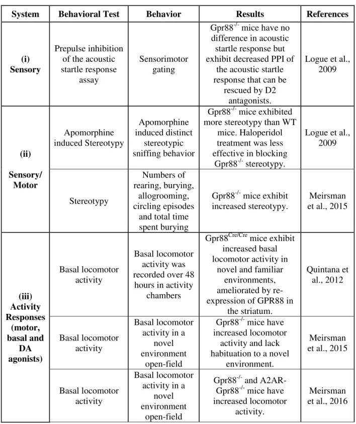
1.1.2 Behavioral studies on GPR88 receptor deficient mice
Up to date, emerging investigations have been carried out to reveal the implication of GPR88 in rodents behavior as listed in Annex: Table 1. Figure 3 summarizes some of the behavioral characteristics of Gpr88 gene knock-out mice reported earlier. It has been shown that in the absence of GPR88, MSNs have increased glutamatergic excitation and reduced GABAergic inhibition that together promote enhanced firing rates in vivo (Quintana et al., 2012). In mice, deletion of the GPR88 has been studied with a primary focus of striatal-mediated behaviors and null mutant mice show hyperactivity, poor motor-coordination, and impaired cue-based learning (Ingallinesi et al., 2015; Logue et al., 2009; Massart et al., 2009; Quintana et al., 2012). For example, mice were placed in activity chambers for 48 hours to elucidate the role of GPR88 in basal locomotor activity. Gpr88Cre/Cre (Gpr88-knockout) mice were more active during the first few hours, reflecting the response to novelty (Figure 3A; adapted from (Quintana et al., 2012)). All animals increased their activity during the nocturnal cycle, however greater in Gpr88Cre/Cre mice and daytime activities were comparable (Figure 3B; adapted from(Quintana et al., 2012)). As a step forward, mice were placed on top of an accelerating rod and the latency to fall was scored to assess the motor coordination and balance. While control group (GPR8+/+) improved their performance with each experimental session, Gpr88Cre/Cre mice fell more quickly and showed no improvement with training (Figure 3C; adapted from (Quintana et al., 2012)), confirming impairments in motor coordination or strength of Gpr88Cre/Cre mice.
Logue et al., showed that Gpr88-/- mice have lower pre-pulse inhibition (PPI) (Figure 3D (i), adapted from (Logue et al., 2009)), which can then be rescued by treating the mice with D2 antagonists (Figure 3D (ii), adapted from (Logue et al., 2009)).
Meirsman et al., demonstrates that Gpr88-/- mice show improved hippocampal-dependent learning and reduced anxiety levels (Aura C. Meirsman et al., 2016). Striatum and hippocampus compete to drive during learning and memory (Poldrack and Packard, 2003). Thus, altered striatal functions in the GPR88 receptor deficient mice may influence hippocampal mediated behaviors as well. Meirsman et al., evaluated the hippocampal dependent behaviors in the Gpr88
-/- mice through several ways. For example, by scoring spontaneous attention in Y-maze revealed
that knock-out mice entered into arms of the maze more often than the control group (Figure 3E; adapted from (Aura C. Meirsman et al., 2016)), consistent with locomotor hyperactivity observed in former study (Quintana et al., 2012). Mutant mice showed a trend toward higher spontaneous alteration and returned significantly less into the same arm, indicative of less preservative errors, while alternate arms returns were unchanged. Hippocampal/striatal balance in learning was specifically assessed by testing mutant mice in a dual-solution cross-maze task. Performance at early stage of the experiment reveals hippocampal facilitated allocentric strategy (place), whereas striatal conditioned egocentric strategy (response) during later stages. Gpr88-/- mice showed longer choice latencies, however, acquired the task more rapidly and reached higher levels of performance than Gpr8+/+ mice. A probe trail after eight training sessions showed that knockout mice shifted toward an egocentric strategy to solve the task, while the control mice still used the
14
allocentric strategy at the same stage, suggesting higher levels of performance in mutant mice compared to the control. Interestingly, probe trial performed after two reversal sessions indicated that mutant mice reshifted to an allocentric strategy (Figure 3F; adapted from (Aura C. Meirsman et al., 2016).
15
These findings suggest GPR88 receptor lacking mice have facilitated hippocampus dependent behaviors. Recent work from the same group reports decreased threat avoidance and exhibit increased risk-taking behavior in both the Gpr88-/- and Gpr88A2A-Cre mice. However, impaired fear conditioning in the Gpr88-/- but not Gpr88A2A-Cre mice (Figure 3G and 3H; adapted from (Aura Carole Meirsman et al., 2016).
This receptor, therefore, controls a much larger behavior repertoire than initially anticipated and, beyond motor activity, also engages spatial learning, emotional processing, sensorimotor gating and fear conditioning as well as in the risk-taking behavior. Nevertheless, the impact of Gpr88 gene in brain connectivity has not been reported yet, promoting further investigations to address how this receptor reshapes the neural architecture at structural and functional level. Implications of GPR88 in brain communication may underscore the molecular mechanisms underlying the behavioral traits observed in Gpr88-/- mice. Thus, one of the main objectives of my study was to assess the brain connectivity modifications in response to the deletion of GPR88 in living mouse brain.
1.1.3 Generation of Gpr88-/- mice
GPR88 floxed mice (Gpr88fl/fl) were generated at the Institut Clinique de la Souris (Strasbourg) using Cre-LoxP technology. Briefly, exon 2 was flanked by loxP sites and a Lox-flippase recognition target neomycin-resistance cassette was inserted downstream exon 2 using homologous recombination (Figure 4, adapted from Aura C. Meirsman et al., 2016). F1 heterozygous Gpr88fl/+ mice were bred with CMV-Flip mice to remove the neomycin cassette and produce a conditional GPR88 floxed line. For this study, constitutive knockout animals were further created by breeding conditional animals with a general CMV-Cre driver
Figure 3: Impact of GPR88 receptor in mice behavior: (A) Gpr88Cre/Cre mice are more active
than the Gpr88+/+ mice. (B) Animals from both group increased their activities during the nocturnal cycle, however greater response was observed in Gpr88Cre/Cre mice. (C) Gpr88Cre/Cre mice show poor motor coordination in rotarod performance test. (D (i)) GPR88 receptor lacking mice show decreased pre-pulse inhibition (PPI) than the wild-type (WT). (D (ii)) D2 antagonists rescue PPI deficiency in the Gpr88-/- mice. (E) When exploring a Y-maze, mutant mice display more arm entries, evoking hyperactivity, and make less perseverative arm reentries. (F) Mutant animals acquire earlier and better a dual solution cross-maze task using distal extra-maze cues, shift sooner to a response strategy to solve the task (probe trial 1), and reacquire more rapidly this task after spatial reversal than Gpr88+/+ animals, by shifting sooner to an allocentric strategy (probe trial 2). AAR, alternate arm return; E, east; N, north; S, south; SAR, same arm return; SPA, spontaneous alternation; W, west. (G) Gpr88-/- and Gpr88A2A-Cre mice show increased risk taking behavior. (H) Gpr88-/- but not A2AR- Gpr88-/- mice impairs fear expression.
Figure 4: Gene targeting strategy to generate
16
line (Gaveriaux-Ruff et al., 2011; Metzger and Chambon, 2001). This led to germline deletion of
GPR88 exon 2 on a hybrid 50% C57BL/6J-50% 129Sv genetic background. Gpr88fl/fl ×
CMV-CreTg/+ and Gpr88+/+ × CMVCre0/+ were used as experimental (Gpr88-/- mice) and control (Gpr88+/+: CTRL) animals, respectively (see Annex 3.4 for details).
1.2 The concept of brain connectivity and brain networks
Brain networks consist of spatially distributed but functionally connected regions that process information through their afferent and efferent connections in an orchestrated manner and thus enabling different sensorimotor and cognitive tasks to be performed.
Structural connectivity (SC) is defined as the formation of networks through synaptic contacts between neighboring neurons or fiber tracks connecting neuron pools in spatially distant brain regions. Functional connectivity (FC) on the other hand, is defined as the temporal dependency of neuronal activation patterns of anatomically separated brain regions (Friston, 1994). It reflects statistical dependencies between distinct and distant regions of information processing neuronal populations. A central paradigm in modern neuroscience is that structural and functional connections between brain regions are organized in a way such that information processing is near optimal.
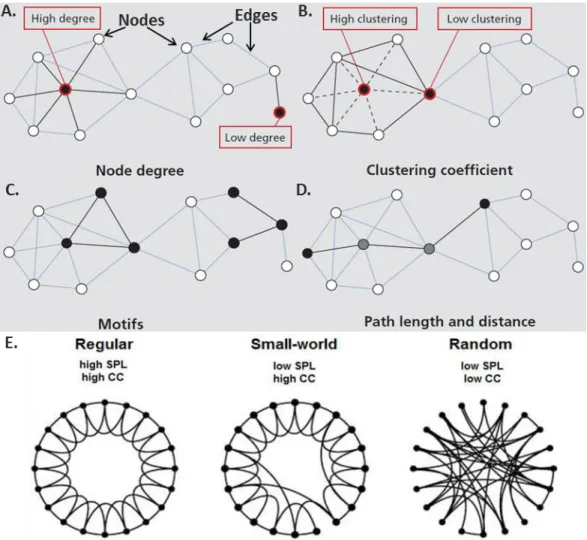
Brain networks can be defined based on the structural connectivity or functional interdependence between brain regions. The structural network organization of the brain is based on the anatomical linkage of its neurons that are connected locally by synapses from short axons, dendrites and gap junctions. Although neuronal populations throughout the brain have a variety of different internal circuitry configurations, they can be represented as network nodes if they have a uniquely identifiable local structural organization. Large-scale functional network on the other hand can be defined as a collection interconnected brain areas that interact to perform circumscribed functions. Large-scale brain networks therefore provide a comprehensive description of the brain's structural and functional connections among brain areas that expedites signaling along preferred pathways in the service of specific cognitive tasks. It is essential to identify the brain areas that constitute structural network nodes and the connecting pathways that serve as structural network edges to know which configurations of interacting areas are possible. Graphical representation of a brain network provides quantitative information on how the network is structured or organized in order to segregate and integrate information among brain regions (Newman, 2006; Rubinov and Sporns, 2010, 2011; Sporns, 2013; Watts and Strogatz, 1998). Several parameters are used to characterize a brain network.
Nodes: Nodes in a network represent brain areas, however, their definition slightly differs while characterizing in structural and functional large-scale network. The nodes of large-scale structural networks are typically considered to be brain areas defined by: (i) cytoarchitectonics; (ii) local circuit connectivity; (iii) output projection target commonality; and (iv) input projection source commonality. A network node in a functional brain network can be a circumscribed brain region
17
displaying elevated metabolism in positron emission tomography (PET) recordings or elevated blood perfusion in functional magnetic resonance imaging (fMRI) or regions of interest (ROIs) based on anatomical knowledge or brain regions identified via independent component analysis (ICA) in resting state fMRI (rsfMRI) (Bressler and Menon, 2010).
Edges: The edges are the long-range axonal-fiber (white matter) pathways that connect brain areas in large-scale structural networks. Network edges are directed because axonal fiber pathways have directionality from the somata to the synapses, and can be bidirectional when fiber pathways run in both directions between brain areas. All blobs in figure 5 (adapted from (Sporns, 2013; Watts and Strogatz, 1998)), represent the nodes, connected via lines that are termed as edges.
Figure 5: Characterization of a brain network: (A) Nodes, edges and the degree of nodes in a network. (B) Clustering coefficient expresses the extent to which a node’s topological neighbors are connected among themselves. (C) Motifs – the subgraph of a network. (D) Path length and distance of a network. (E) Network configuration. Regular, small-world and random network are represented considering identical number of nodes and edges.
18
Node degree: The node degree is the number of edges attached to a specific node. Higher number
of edges from a node denotes the higher degree of that corresponding node and vice-versa (Figure 5A).
Clustering coefficient (CC): It is the measure of density of connections among a node’s topological neighbors (Figure 5B). If the neighbors of a given node are densely interconnected, they are said to form a cluster. The average of clustering coefficients over all nodes defines the clustering coefficients of the network.
Motifs: Motifs constitute the subgraph of a network. Every network can thus be subdivided into a
set of motifs of a given size (Figure 5C).
Path, path length, shortest and characteristics path length: Path is the sequence of edges that connects two nodes with each other. Path length is therefore defined as the number of steps or the sum of the edge lengths in a network (Figure 5D). The length of the shortest path between each pair of nodes resembles their distance and referred to as the shortest path length (SPL). The global average of all distances across the entire network is called the characteristic path length.
Brain network features: Depending on the CC and SPL, the feature of a given brain network can
be determined. Small-world feature of a network is a standard of complexity and efficiency of global network structures (Watts and Strogatz, 1998). Small world networks have a topology with a level of randomness between that of a regular and random network (Figure 5E). A small world topology with high local clustering of links and still short travel distances between any links of the network has been demonstrated for living organisms brain in former studies (Bullmore and Sporns, 2009; Mechling et al., 2014). Furthermore, this small-world feature has been investigated in several human brain disorders and indeed, alterations could be demonstrated in dementia, multiple sclerosis, traumatic brain injury or epilepsy (Stam, 2014). In contrast to the small-world network, regular network exhibits high SPL and CC, while these parameters are low in the random network (Figure 5E).
Modern noninvasive imaging techniques applied to humans and animals brain allow the mapping of such complex structural and functional brain networks. Emerging studies demonstrate disrupted brain communication between distinct regions due to psychiatric illness. Analysis of the neuronal connectivity within a brain network is thus important to understand the organization of brain, as well as to reorganization during disease, learning and aging. Additionally, insight to the brain functional and structural connectivity alterations due to the expression or restriction of specific gene, treatments with drugs and/or through disease modelling may endorse pathologies and potential treatment regimes.
Recent years have seen a dramatic increase in the advances and applications of non-invasive neuroimaging techniques, such as Magnetic Resonance Imaging (MRI), Electro/Magneto- encephalogram (EEG/MEG) as well as other invasive techniques like Computed Tomography (CT), Positron Emission Tomography (PET) or Single-Photon Emission Computed Tomography (SPECT). With the advent of these techniques, brain connectional fingerprints have become accessible, offering the unique possibility to identify altering brain functions and structures
19
persuaded by numerous factors including experience, pathology or genetics (Greenough, 1984; Kolb et al., 1998; Thompson et al., 2013).
1.3 Brain imaging techniques
MRI is one of the most widely used non-invasive imaging techniques in clinical and pre-clinical research now-a-days for mapping the brain structural and functional connectivity. In medicine, MRI is primarily used to produce structural images of organs, including the central nervous system (CNS), but it can also provide information on the physico-chemical state of tissues, their vascularization, and perfusion. Emergence of functional MRI (fMRI) - to measure the hemodynamic changes after enhanced neural activity in response to a task or stimulus, as well as in the absence of task (during rest), namely resting state functional MRI (rsfMRI) and diffusion tensor imaging (DTI) with fiber tracking - to study the brain microstructural modifications, had a real impact on basic cognitive neuroscience research. Since then, these techniques have been remarkably applied in the clinical and pre-clinical research. In my study, rsfMRI as well as DTI and fiber tracking techniques were implemented in the Gpr88 gene knockout living mouse brain, to map the functional and structural fingerprints respectively. The combined analysis of structural and functional connectivity provides insight on how brain structure shapes the brain function, to what degree brain function feeds back to change its structure, and what functional or structural aspects of physiology ultimately drive cognition and behavior (Sui et al., 2014). Following two sections (1.3.1 and 1.3.2) describe the principles of these techniques and how to map the large-scale structural and functional brain network respectively.
1.3.1 Structural connectivity assessment via diffusion tensor imaging (DTI) and fiber tractography
The brain contains more than 100 billion neurons that communicate with each other via axons and forms complex neural networks. The structural mapping of such networks during health and disease states is crucial for understanding brain function. In animal studies, histology followed by light or electron microscopy has been one of the most widely used imaging methods. Besides, numerous staining techniques can highlight the locations of proteins and genes of interests, and electron microscopy can encompass our observation to objects at the molecular level. However, histology-based imaging is invasive and labor-intensive, which makes it a non-ideal choice for examining the entire brain or for performing quantitative three-dimensional analyses. Diffusion tensor imaging (DTI) on the other hand is a three-dimensional noninvasive imaging modality used to characterize the entire brain anatomy. DTI was introduced in the mid1990s (Basser et al., 1994) and since then, this technique has been widely applied in characterizing structural fingerprints of rodents (Ahrens et al., 1998; Harsan et al., 2006, 2010, 2013; Mechling et al., 2016; Mori et al., 2001; Nair et al., 2005; Wu et al., 2014; Wu and Zhang, 2016; Zhang et al., 2012) and human brain (Alexander et al., 2007; Mori et al., 2001; Mukherjee and McKinstry,
20
2006; Pierpaoli et al., 1996; Tang et al., 2014; Tuch et al., 2001), as well as in the primates (D’Arceuil et al., 2007; Shi et al., 2013; Zhang et al., 2013).
The term ‘diffusion’ represents random thermal motion of water molecules. Diffusion of water is anisotropic (directionally dependent) in white matter (WM) fiber tracts, as axonal membranes and myelin sheaths present barriers to the motion of water molecules in directions not parallel to their own orientation. Diffusion tensor of WM tracts are considered as a three-dimensional structure with three principal diffusivities (eigenvalues, 1, 2, 3), associated with three mutually
perpendicular principal directions (eigenvectors, e1, e2, e3). Fractional anisotropy (FA) is another
parameter, defined as the ratio of the anisotropic component of the diffusion tensor to the whole diffusion tensor and serves as a rotationally invariant scalar that quantifies the shape of the diffusion tensor. Thus FA measures the degree of directionality of diffusion that varies between zero and one. Zero represents maximal isotropic diffusion as in a perfect sphere and one represent maximal anisotropic diffusion. By combining the directional information and magnitude of anisotropic diffusion of the individual voxels, the course of fiber tracts can be reconstructed, which is known as tractography. This technique relies on the assumption that voxels with a similar orientation of their principal anisotropic diffusion direction are likely part of the same fiber tract. Therefore, DTI and fiber tractography provides a unique opportunity to study the fiber architecture in vivo and characterize microstructural changes or differences with neuropathology and treatment.
1.3.2 Functional connectivity assessment via resting state functional MRI (rsfMRI)
Neuronal activity causes local changes in cerebral blood flow, blood volume, and blood oxygenation. MRI is sensitive to changes in cerebral blood flow and blood oxygenation. Functional magnetic resonance imaging (fMRI) technique uses the blood oxygen level dependent (BOLD) contrast (Ogawa et al., 1990) to detect changes in blood oxygenation in response to a task or stimulus and thus measure the brain activity (Kwong et al., 1992; Ogawa et al., 1992). In recent years, there has been an increase in interest in the application of this technique at rest, which is termed as resting-state fMRI (rsfMRI). This technique detects low frequency fluctuations (LFFs) of less than 0.1 Hz in the BOLD signal and measures FC between brain regions as the level of co-activation of spontaneous fMRI time-series during rest (Biswal et al., 1995, 1997; Greicius et al., 2003; Lowe et al., 2000; Salvador et al., 2005). These brain regions working together form a functional network, also called the resting state network (RSN), with a high level strongly correlated spontaneous neuronal activityin the absence of a task or stimulus (Fox and Raichle, 2007). These patterns of resting-state correlations are hypothesized to reflect the stable and intrinsic functional architecture of the brain (Buckner et al., 2009). Biswal et al., was the first to demonstrate ongoing neural activity that occurs at rest throughout functionally connected regions of the brain when they revealed a high correlation between the BOLD time-series of the left and right hemispheric regions of the primary motor network in the absence of a
21
task (Biswal et al., 1995). Several studies have since replicated these results, propelling extensive use of the technique in human (Fair et al., 2007, 2008; Fox and Raichle, 2007; Greicius et al., 2003; Koyama et al., 2011). It is also widely used in mapping rodents (Jonckers et al., 2011; Mechling et al., 2014, 2016; Sforazzini et al., 2014; Shah et al., 2015) and primates brain FC as well (Hutchison et al., 2012; Mantini et al., 2011; Wang et al., 2013).
Identification of functional networks from fMRI data obtained during cognition or resting state is critical for understanding and characterizing how different brain regions communicate with each other.
Analysis of rsfMRI data
In recent years, several methods have been developed to characterize functional brain networks and connectivity.
Model-based method: Seed-based analysis is one of the straight-forward methods of mapping whole brain connectivity from a specific region of interest (ROI). Thus it correlates the resting state time-series of a pre-defined ROI against the time-series of all other brain regions, resulting in a whole brain FC map (Biswal et al., 1997). Similar approach is also applied to the rsfMRI data and termed as resting state FC (rsFC) map. The FC or rsFC map provides information about the regions to which the seed region is functionally connected, and to what extent. The simplicity of this analysis affords a strong advantage for seed-dependent methods; however, the information obtained from the rsFC map is limited to the functional connections of the selected ROI only, making it difficult to examine the whole-brain functional architecture (Buckner and Vincent, 2007). Moreover, the selection of a priori ROI can be a challenge to the researchers as it requires having profound knowledge on the brain anatomy and in addition, one has to use a priori knowledge for selecting the seeds.
Model-free methods: Evaluation of whole-brain connectivity patterns is also possible using model-free method such as, independent component analysis (ICA). ICA decomposes the entire BOLD signal into number of spatially independent components ((ICs) or sources (Beckmann and Smith, 2004; Calhoun et al., 2001). Thus, it requires the investigator to estimate the number of components and then to look for the existence of spatial sources of resting-state signals that vary together over time and are maximally distinguishable from other sets of signals (Beckmann et al., 2005). The advantage of using ICA is its application to whole-brain voxel-wise data and high consistency among the RSNs (Damoiseaux et al., 2006). In contrast, complex representation of ICA derived data may complicate translation of results to clinical relevance (Fox and Raichle, 2007).
Brain regions identified by ICA, can be used directly to compute FC between multiple regions by partial correlation. It provides an estimation of the linear conditional dependence between brain regions, removing the linear influence of other regions. The resulting correlation coefficients are usually converted to z-scores using the fisher transformation and then thresholded to identify statistically significant network connections (Supekar et al., 2008). Positive correlations between regions indicate that those regions are typically co-modulated, whereas anti-correlations between
22
regions indicate them temporarily modulated in opposite directions (Fox et al., 2006). However, zero correlation between two brain regions represents these regions are conditionally independent given temporal fluctuations in other brain regions considered (Peng et al., 2009). Several previous imaging studies have used partial correlations for estimating functional connectivity (Hampson et al., 2002; Huang et al., 2010; Marrelec et al., 2006, 2007).
These methods have been extensively applied to identify major functional networks or circuitry, such as the primary motor, visual, and auditory networks, in addition to higher order cognitive systems (Cordes et al., 2000; Fox and Raichle, 2007; Greicius et al., 2003). Default mode network (DMN), central executive network (CEN), and salience network (SN) are considered to represent the major portion of higher-order functional brain networks. Such complex network is susceptible to many external or internal sources, like experience, influences, physiological and psychological changes or immunological events which all can result in connectivity alterations. Indeed, the impact of pathology on brain connectivity networks has been addressed for several neurological or psychiatric disorders (Guye et al., 2010, 2010; Stam, 2014).
1.3.2.1 Default mode network
Default mode network (DMN) is of particular interest which comprises a group of brain regions appear to be more active during rest compared with a cognitively active state (Raichle et al., 2001). It denotes the intrinsic activity of the brain when the subject is at rest. In other words, DMN exhibits task-induced deactivations and thus also named as task-negative network, which has been associated with processes such as self-reflection and mind wandering.
Extensive studies have been carried out in humans and animals to understand the function of DMN and identify its major anatomical subdivisions. These include: the medial prefrontal cortex (mPFC: the dmPFC, the rostral anterior cingulate, and parts of the anterior and ventral mPFC), medial parietal cortex (the posterior cingulate and retrosplenial cortex), medial temporal lobe (the hippocampus and parahippocampal cortices), lateral parietal and temporal cortex (Gusnard et al., 2001a, 2001b; Lu et al., 2012; Ongür and Price, 2000; Popa et al., 2009; Raichle et al., 2001; Schilbach et al., 2008; Sforazzini et al., 2014; Shannon et al., 2013; Shulman et al., 1997; Vincent et al., 2006). Figure 6 (adapted from (Lu et al., 2012; Sforazzini et al., 2014)) summarizes the DMN reported in rat, mouse, monkey and human brain.
Alterations in DMN connectivity patterns lead to cognitive dysfunctions in neurologic and psychiatric disorders. Changes in DMN FC have been reported in multiple psychiatric and neurologic disorders including depression (Greicius et al., 2008; Kühn and Gallinat, 2012; Lui et al., 2011), schizophrenia (Garrity et al., 2007; Kühn and Gallinat, 2012; Whitfield-Gabrieli and Ford, 2012), Alzheimer’s disease (Greicius et al., 2004; Hedden et al., 2009), epilepsy (Waites et al., 2006; Z. Zhang et al., 2010), disorders of consciousness (Soddu et al., 2011) including coma (Norton et al., 2012), multiple sclerosis (Lowe et al., 2002), amyotrophic lateral sclerosis (Mohammadi et al., 2009), autism (Cherkassky et al., 2006; Murdaugh et al., 2012) and blindness (Liu et al., 2007). Emerging studies also demonstrate ADHD as DMN disorder (Castellanos et al., 2008; Fair et al., 2010; Sonuga-Barke and Castellanos, 2007) as well as decreased DMN
23
connectivity in ADHD patients (Castellanos et al., 2008; Castellanos and Proal, 2012; Fassbender et al., 2009).
DMN thus serves as an elucidating and critical system for identifying treatment targets and aiding in the clinical diagnosis and development of treatment strategies.
1.3.2.2 Central executive network
Dorsolateral prefrontal cortex (dlPFC), and posterior parietal cortex (PTLp) are the key nodes of central executive network (CEN), also termed as task-positive network (TPN) or frontoparietal network (FPN). CEN nodes show strong intrinsic functional coupling as well as strong co-activation during cognitively challenging tasks, while decrease in co-activation during rest. In Figure 6: Default mode network (DMN): (A) In rat brain: 1. orbital cortex, 2. pre-limbic cortex (PL), 3. cingulate area (ACA), 4. auditory/temporal association area (TEa), 5. posterior parietal association area (PTLp), 6. retrosplenial area (RSA), corresponds to the posterior cingulate cortex
(PCC) in human, 7. hippocampus (CA1 – part of hippocampal formation (HPF)). (B) In mouse
brain: 1. orbito-frontal cortex (OFC), 2. ACA, 3. TEa, 4. thalamus (TH), 5. RSA, 6. visual area (VIS). (C) In monkey brain: 2/3. dorso-medial prefrontal cortex (mPFC), 4/5. lateral temporoparietal cortex, 6. RSA, 7. Posterior parahippocampal cortex. (D) In human brain: 1. OFC, 2/3. mPFC/ACA, 4. lateral temporal cortex, 5. inferior parietal lobe, 6. RSA, 7. HPF.
24
particular, the CEN is critical for active maintenance and manipulation of information in working memory, for judgment and decision-making in the context of goal directed behavior (Koechlin and Summerfield, 2007; Miller and Cohen, 2001; Müller and Knight, 2006; Petrides, 2005).
1.3.2.3 Salience network
Anterior and posterior part of agranular insular area (AI), potentially together with the anterior cingulate area (ACA) serves as the salience network (SN) (Seeley et al., 2007). This network is associated with the detection of novel, salient stimuli, and is thought to play a role in coordinating an adequate response by recruiting appropriate brain networks. Therefore, some authors suggest it might play a role in coordinating between DMN and CEN activity (Bonnelle et al., 2012; Menon and Uddin, 2010). Neuroimaging studies have provided the evidence for prominent SN dysfunction in many psychopathologies, including frontotemporal dementia (Zhou et al., 2010), Alzheimer’s disease (Zhou and Seeley, 2014), mood (Hamilton et al., 2013) and anxiety disorders (Paulus and Stein, 2006; Stein et al., 2007), posttraumatic stress disorder (Peterson et al., 2014), schizophrenia (Manoliu et al., 2014), drug addiction (Sutherland et al., 2012), and chronic pain (Otti et al., 2013).
1.3.2.4 Reward/aversion network
Reward/aversion network is comprised of several brain regions that connect with each other through dopaminergic and opioidergic projections. This network is very well known to play important roles in addiction.
Figure 7: Schematic diagram of the reward/aversion network: Blue, red and green circle represents the regions belonging to the reward, aversion, and social system respectively. Dopaminergic, glutamatergic, GABA-ergic and serotonin projections from/to the brain regions have been shown via solid or dashed lines as mentioned in the figure.
25
Figure 7 demonstrates a schematic diagram of the reward/aversion network. Ventral tegmental area (VTA) plays the central role in the reward and aversion system as it sends and receives projections from different brain regions. Dopamine neurons of the VTA target important structures, such as ACB and medial prefrontal cortex (mPFC), involved in reward (blue) and aversion (red) response behavior (Bromberg-Martin et al., 2010) and receives reward- and aversive-related input from the latero-dorsal tegmental nucleus (LDT) and lateral habenula (LHb) respectively (Lammel et al., 2012a). Glutamatergic and GABAergic neurons from bed nuclei of the stria terminals (BST) to VTA are activated in response to aversive and rewarding stimulus (Jennings et al., 2013). Dorsal raphe nucleus (DRN) serotonergic neurons projecting onto the ACB produces aversion and potentiates cocaine reward (Lutz and Kieffer, 2013).
Alteration in social interaction is often related with several psychiatric disorders such as autism, schizophrenia, depression and social anxiety disorder. However, little is known on the neuronal circuit involved in social interactions. The brain regions involved in social interactions are often related to the RAC (Gunaydin et al., 2014; Kumar et al., 2014). A recent study coupling optogenetic and fiber photometry in mice managed to identify the involvement of the VTA’s projections to the ACB in social interactions (Green). The modulation of the social interaction was mediated by D1R signaling downstream in the ACB (Gunaydin et al., 2014). ACB opioid receptors were shown to be necessary and sufficient for morphine to increase social play (Trezza et al., 2011). Another study showed that the variability in PFC function underlies individual differences in vulnerability of stress induced by chronic social defeat and also demonstrated that the response proprieties of the glutamatergic projections between PFC to the amygdala, involved in modulation fear and stress response, correspond to naturally occurring differences in vulnerability to chronic social defeat (Kumar et al., 2014). Medial amygdala (MEA) and its projections to the hypothalamic regions are involved in innate social and asocial behavior (Newman, 2006; Swanson, 2000) and in conditioned fear (Duvarci and Pare, 2014). A recent study showed that glutamatergic subpopulation inhibits social interaction independently of its promoting effect on self-grooming while the GABAergic subpopulation promotes aggression and inhibits self-grooming even in a non-social context (Hong et al., 2014).
Assessment of neuronal connectivity impairments in large-scale brain network and their impact in brain communication within or between the networks is therefore essential to understand the underlying molecular mechanisms of the neurodegenerative and psychiatric diseases. Combining neuroimaging technologies with animal models of neurological disorders provides unique opportunities to comprehend the pathophysiology of human neurological disorders. One of the main advantages of animal studies is group homogeneity, which cannot be easily achieved in clinical (human) studies. Moreover, animal models can interact and react to stimuli that can provide an idea of how those stimuli might react in a human being. In addition, how the restriction or expression of genes can reshape human brain connectivity is possible to image using animal models. It allows thus to investigate the spatial and temporal dynamics of disease-specific functional and molecular events longitudinally in intact living organisms.
26
1.4 Rodents in neuroscience/neuroimaging research
1.4.1 rsfMRI and DTI in rodents
A multitude of animal models have been established to mimic human neurodegenerative and psychiatric disorders. These animal models range from interventional models (such as xenograft, neurotoxic or mechanical lesion models) to knockout and transgenic (mono-, bi- or trigenic through crossbreeding) animals. With the advance of these animal models, non-invasive techniques for the evaluation of disease-associated functional, biochemical and anatomical changes through a variety of dedicated small animal imaging scanners with high sensitivity, specificity and resolution have become indispensable. Over the past century, the mouse has developed into the premier mammalian model system for genetic research. Scientists from a wide range of biomedical fields have gravitated to the mouse because of its close genetic and physiological similarities to humans, as well as the ease with which its genome can be manipulated and analyzed. Consequently, utilization of rsfMRI and DTI methods in mouse models of psychiatric disorders provide considerable benefits for the identification of disease-associated brain circuits and metabolic changes. Despite these advantages of using mouse models, there are some major challenges need to consider especially while performing rsfMRI and DTI studies on living animals.
Anesthesia plays an important role in brain connectivity. Even though it implies restrictions on fMRI, experiments with conscious rodents (Becerra et al., 2011; N. Zhang et al., 2010) also have their limitations (Berwick et al., 2002). For example, even after habituation to the apparatus, animals are still stressed by the fixation or noise from the scanner itself, which may subsequently have an impact on the investigated brain functions, suggesting the need of anesthesia or light sedation to minimize stress level as well as to avoid movement related artifacts. Previous studies on humans and rodents brain functional connectivity report that the level of consciousness during the experiment, influences RSN patterns and activity (Guldenmund et al., 2012; Ma et al., 2017; Nasrallah et al., 2012, 2014). Both the BOLD response as well as the temporal correlation of LFFs between brain regions can be affected by the choice (and level) of anesthesia in rodents (Jonckers et al., 2014; Williams et al., 2010). Therefore, it is very crucial to select the appropriate type of anesthesia and dosage level to avoid strong anesthetic induced effects on brain connectivity.
Maintaining stable physiological conditions, such as, body temperature, respiration rate and blood oxygenation level throughout the rsfMRI and DTI study is very important. Fluctuations in body temperature can contribute to drifts in the BOLD signal baseline, even when the temperature changes are within physiological ranges (Vanhoutte et al., 2006). Respiratory and cardiac cycles are known to contribute to the rsfMRI signal and can introduce unwanted correlations (Birn et al., 2006; Shmueli et al., 2007; Wise et al., 2004). Hence, physiological parameters should be monitored and controlled (to the extent possible) during an experiment.
27
Rodent models play an important role in understanding the neural basis of BOLD correlations and are likely to continue to do so. Brain connectivity studies with rodents are rapidly expanding into the wide realm of animal models of brain disorders. This will facilitate the transfer of knowledge between rodent and human research. The non-invasive nature of rsfMRI and DTI, together with the advantages of using rodent models allows us to develop biomarkers that can be quickly examined in the human population. Additionally, the neurophysiological basis of alterations observed in humans can be determined in animal models. Caution is necessary, however, particularly in with regards to maintaining animal physiology and accounting for the effects of anesthesia.
1.4.2 Rodents in behavioral neuroscience
Rats and mice are among the most commonly used animal models in behavioral neuroscience research. They are well suited model organisms, as they display a variety of behaviors with relevance to human disease. Thus behavioral characterization of genetically modified mice and rats as well as wild-type strains has become a powerful tool for investigating not only the molecular bases of normal brain functions but also the pathogenesis and treatment of neurophysiologiocal disorders (Crawley, 2007; Holmes et al., 2002; Picciotto and Wickman, 1998; Takao et al., 2007; Watase and Zoghbi, 2003; Wolfer et al., 2002). There are a wide variety of behavioral tests available for laboratory rodents, from tests of basic locomotor and sensory function, to analyses of more complex behavior related to cognition and emotionality. However, the standardization and reproducibility of the testing methods for mouse behavioral assessment is still inadequate (Brunner et al., 2002; J. C. Crabbe et al., 1999; Novak et al., 2015; Sorge et al., 2014; Wahlsten et al., 2003). Introduction of IntelliCage by NewBehavior (NewBehavior AG) in the field of behavioral neuroscience permitted the researchers to overcome this problem.
In my study, I successfully implemented this technology for screening behavioral and cognitive functions of the GPR88 deficient mice. This is the first study assessing on the real time measurement of the GPR88-/- mice behavior. I optimized and adjusted several behavioral test protocols, consisting the adaptation phases, followed by the cognitive performance and spatial learning evaluation phase. Each protocol was designed with the ‘IntelliCage Designer’ software and the experiments were monitored online through the ‘IntelliCage Controller’ module. Data saved by the controller during the experimentation, was extracted at the end of each experiment using the ‘IntelliCage Analyzer’ software and further processed for statistical analysis. Following sections provide an overview of the IntelliCage hardware and software packages that were used to design the experiment, monitor mice activities online and data processing respectively.
1.4.2.1 IntelliCage
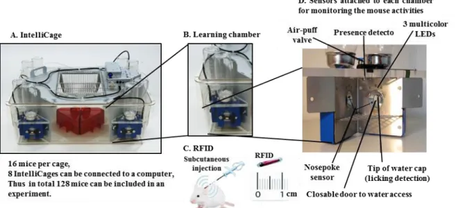
IntelliCage is a newly developed computer-based, fully automated testing apparatus that allows automated cognitive and behavioral screening of mutant or treated mice living in social groups. It is a large standard plastic cage (55 × 37.5 × 20.5 cm3) (Figure 8A) equipped with four triangular operant learning chambers (15 × 15 × 21 cm3) (Figure 8B) that fit into each corner of
28
the cage. Subcutaneously injected radio frequency identification (RFID) readers (Figure 8C) and other type of sensors (Figure 8D) allow simultaneous monitoring of up to 16 transponder-tagged mice living in the same cage. Mice have access to enter the corner through a short narrow tunnel that functions as an RFID antenna. In this unit, only one mouse can enter a corner at a time because of the limited size of the corner and tunnel. In the inner space of the corner, there are two nose-poke holes with an infrared beam-break response detector. Nose-poke triggers the opening of a motorized access gate to water-bottle spouts. In IntelliCage, the time and duration of each behavioral event (number of visits in each corner, duration of stay in each corner, number of nose-pokes, number of licks and licking duration), mouse ID and corner ID are automatically recorded through RFID readers, infrared sensors and lickometers respectively. Thus it provides the real time measure of the mouse activities, which is a unique feature of the IntelliCage over the traditional cages. Figure 8 was adapted from (“Info - Home - TSE Systems,”)
Several features of IntelliCage have made this automated system as a powerful tool for the behavioral characterization of mice or rats.
1. It is possible to achieve a sensitive and highly standardized experiment by minimizing the artifacts that arise from unavoidable differences among experimenters or other laboratory-specific conditions.
2. Long term monitoring of mouse behavior can be performed in less stressful environment. Figure 8: Overview of the IntelliCage system: (A) IntelliCage system apparatus: mice are group-housed in the cage and their behavioral responses (corner visits, nosepokes, and lickings) are monitored in a fully automated manner. (B) Learning chamber: each corner of the IntelliCage contains a learning chamber that holds 2 bottles for drinking, and other monitoring sensors. Only one mouse can enter the chamber at a time through a hole. (C) Radio frequency identification reader (RFID) of 1 cm in length is subcutaneously injected into the mouse shoulder. Each RFID has a unique identification number that is registered each time in response to the activity of the mouse. (D) Interior of the learning chamber: multiple sensors are attached in each chamber. Each time a transponder-tagged mouse enters the learning chamber; all activities are recorded by the system which can further be analyzed using the designated IntelliCage software modules.