HAL Id: tel-01674198
https://tel.archives-ouvertes.fr/tel-01674198v2
Submitted on 2 Jan 2018HAL is a multi-disciplinary open access archive for the deposit and dissemination of sci-entific research documents, whether they are pub-lished or not. The documents may come from teaching and research institutions in France or abroad, or from public or private research centers.
L’archive ouverte pluridisciplinaire HAL, est destinée au dépôt et à la diffusion de documents scientifiques de niveau recherche, publiés ou non, émanant des établissements d’enseignement et de recherche français ou étrangers, des laboratoires publics ou privés.
and ACKR3/CXCR7
Pasquale Cutolo
To cite this version:
Pasquale Cutolo. Structural and functional study of the interaction between CXCL12 chemokine and its receptors : CXCR4 and ACKR3/CXCR7. Immunotherapy. Université Paris Saclay (COmUE), 2016. English. �NNT : 2016SACLS550�. �tel-01674198v2�
Thèse de doctorat
De
L’Université Paris-Saclay
Ecole Doctorale n° 569INNOVATION THÉRAPEUTIQUE : DU FONDAMENTAL À L’APPLIQUÉ
Spécialité de doctorat :
IMMUNOLOGIE ET BIOTHÉRAPIES Présentée par
Pasquale CUTOLO
Etude de l’interaction structurelle et fonctionnelle entre la chimiokine
CXCL12 et ses récepteurs : CXCR4 et ACKR3/CXCR7
Thèse présentée et soutenue à Châtenay-Malabry, le 16 septembre 2016
Composition du Jury :
Françoise BACHELERIE DR, Inserm UMR-S 996, Clamart Directrice de thèse
Esther KELLENBERGER Prof., Faculté de Pharmacie de Strasbourg Rapportrice
Eric REITER DR, INRA, Tour Rapporteur
Philippe DETERRE DR, Inserm UMR-S 945, Paris Examinateur
Thierry DURROUX DR, CNRS – IGF, Montpellier Examinateur
INDEX
Abbreviations ... 4
Introduction ... 7
Chapter I - Chemokines structure and interactions ... 7
Preamble on Chemokines ... 7
I.1 Primary structure and classification of Chemokines (CKs) ... 9
I.2 Topology and Folding of CKs ...12
I.3 Oligomerization of CKs ...14
I.4 Interaction with Glycosaminoglycans (GAGs)...18
I.5 Interaction of CKs with receptors ...24
Chapter II - G-protein Coupled Receptors (GPCRs) structural basis ... 38
Preamble on GPCRs ...38
II.1 Current Chemokine Receptors (CKRs) Crystal Structures ...40
II.2 Oligomerization of GPCRs ...41
II.3 Atypical Chemokine Receptors (ACKRs)...47
II.4 CXCR4 receptor-dependent signaling pathways ...53
Chapter III - Nanobodies (NBs) as new innovative tools ... 59
Preamble on NBs: the smallest natural antigen-binding fragment ...59
III.1 Antibodies for biomedical applications ...63
III.2 NBs in structural biology ...64
III.3 CXCR4- and CXCR7/ACKR3- targeting NBs ...67
Objectives ... 68
Results ... 69
Paper n°1 ... 72
Paper n°2 ... 118
Discussion and perspectives ... 196
I - Mechanism of the interaction and stoichiometry of CXCL12 with CXCR4 and CXCR7/ACKR3 receptors ... 197
I.1 Modeling the three-dimensional structure of GPCRs ... 197
I.2 Modeling the binding of ligands to GPCRs ... 199
I.3 Conclusions ... 201
II – Nanobodies ... 203
II.1 Characterization of NBs against CXCR4 ... 203
II.2 NBs applications... 203 II.3 Conclusions ... 204 References ... 207 Annexes ... 238 Curriculum Vitae ... 243 Acknowledgement ... 238
“Eppur si muove”
"And yet it moves" or "Albeit it does move", one of the most famous Galileo Galilei’s citation for Earth movement, is the good sentence to introduce my thesis.
The Italian scientist spoke about the movement of our planet and its “migration” around the Sun, people are moving around the world during their life, but deeply inside this movement is also the base of our biology, from the embryogenesis to the adult life.
How can our cells “migrate” and decide where and when “be or not to be”?
The answer is in small molecules called chemokines and their fascinating and highly intricate networks with chemokine receptors.
Abbreviations
7-TM Seven transmembrane
TM Transmembrane
BRET Bioluminescent resonance energy transfer C-terminus Carboxyl-terminal extremity
CDRs Complementary determining regions CFP Cyan fluorescent protein
CKs Chemokines
CKRs Chemokines Receptors CRS1 Chemokine recognition site 1 CRS2 Chemokine recognition site 2 CS Chondroitin sulfate
CX3R, CX3C chemokine receptors CXCR CXC chemokine receptor Cyt Cytoplasmic region DS Dermatan sulfate ECL Extracellular loop ECM Extracellular matrix ELR Glu-Leu-Arg sequences
Fig Figure
FRET Förster Resonance Energy Transfer GAGs Glycosaminoglycans
GPCRs G-protein coupled receptors HA Hyaluronic acid
HCAbs Heavy-chain antibodies
HIV Human Immunodeficiency Virus HS Heparan sulfate
HTRF Homogeneous time-resolved FRET ICL Intracellular loop
KS Keratin sulfate
mAbs Monoclonal antibodies MOR mu opioid receptor
N-terminus Amino-terminal extremity
NMR Nuclear magnetic resonance PDB Protein DATA Bank sTyr Sulfotyrosine
Tab Table
TM Transmembrane region TNF Tumor necrosis factor
VHH Camelid heavy-chain antibody YFP Yellow fluorescent protein
- Table of Amino Acid abbreviations:
Name 3-Letters 1-Letter
Alanine Ala A
Arginine Arg R
Asparagine Asn N
Aspartic acid Asp D
Cysteine Cys C
Glutamic acid Glu E
Glutamine Gln Q Glycine Gly G Histidine His H Isoleucine Ile I Leucine Leu L Lysine Lys K Methionine Met M Phenylalanine Phe F Proline Pro P Serine Ser S Threonine Thr T Tryptophan Trp W Tyrosine Tyr Y Valine Val V
Introduction
Chapter I: Chemokines structure and interactions
Chapter II: G-Protein Coupled Receptors structural basis
Introduction
Chapter I
Preamble on Chemokines
Proteins called “chemokines” are cytokines, usually of a molecular weight between 8 and 14 kDa with a high structural homology, and the peculiar chemo-attractive function (CHEMOtactic cytoKINES).
We can define the chemotaxis as the oriented cell migration through the concentration gradient of a chemical signal (called chemotactic or chemo-attractive signal, (Mc 1946, Harris 1953, Harris 1953).
These proteins have historically been known under several other names including the SIS family of cytokines, SIG family of cytokines, SCY family of cytokines, Platelet factor-4 superfamily or intercrines. In 1977 the first one was described as a molecule capable to activate platelets during platelet aggregation and to promote blood coagulation (platelet factor 4, PF4 or now according to the new nomenclature CXCL4) and today 44 chemokines (CKs) are known in all vertebrates and are conserved for their different functions notably on immune responses (naïve and adaptive), inflammation, hematopoiesis and angiogenesis. They exert these functions by interacting with chemokine receptors (CKRs) that are 7-transmembrane (7-TM) -domains G-protein coupled receptors (GPCRs). To date 19 receptors have been identified, forming a highly intricate and precisely regulated network (Bonecchi, Galliera et al. 2009, Bachelerie, Ben-Baruch et al. 2014).
Expression of the CKs can be induced by cytokines (for example interferons (IFNs), tumor necrosis factor (TNF)-α, in inflammatory conditions) in a variety of immune and non-immune cells. These various cell types non-exhaustively include fibroblasts, epithelial, endothelial and smooth muscle cells, as well as mononuclear leukocytes and granulocytes. (Brown, Gerritsen et al. 1994)
On the basis of their expression and thereafter their function, CKs were initially classified in two main categories. Some are considered as homeostatic being produced constitutively (ex. CCL19, CXCL13), and generally involved in lymphocyte trafficking, immune surveillance and localization of immune cells in the lymphatic system (Fernandez and Lolis 2002), but
also for the central nervous system cells disposition and angiogenesis (Strieter, Polverini et al. 1995, de Haas, van Weering et al. 2007).
Other CKs were considered as being only produced during infection or following a pro-inflammatory stimulus and involved in immune cells recruitment to damaged tissues (ex. CXCL8, CXCL10). For instance, in case of skin wound, such inflammatory CKs can also activate cells to raise an immune response and start the wound healing process (Rollins 1997). This functional classification was based on the idea that cells are producing CKs either constitutively or under different pathogenic conditions and in a soluble secreted form. However, the knowledge built up over years indicates that CKs instead of being secreted as soluble proteins are rather interacting with extracellular matrix (ECM) glycosaminoglycans (GAGs) and the concept that adhesion events between cells not only provide a method of cellular interaction, but also may aid in the activation of the cells and induce the production of CKs in non-inflammatory conditions (Rot 1993).
Moreover, CKs are also involved in other functions than chemoattraction. A good example is CXCL12, a chemokine also involved in angiogenesis and considered as a growth factor. Moreover, this CK has been shown to be able to act as a chemo-repulsive agent. For instance, high concentrations of CXCL12 have been shown i) to repulse human thymocytes in vitro, a “run away” process that could be abolished using a neutralizing CXCL12 antibody (Poznansky, Olszak et al. 2000) and ii) to permit the firm adhesion of CD4 and CD8 T cells to microvascular endothelium from pancreatic islet thus resulting in a decrease of T-cell integrin activation in a CXCR4-independent manner (Sharp, Huang et al. 2008).
Such a process might account for the decreased chemotaxis observed at high concentration of a CK evidenced by the typically bell shaped response upon CK concentration. However the exact molecular mechanisms of CK-induced chemorepulsion are still undefined. Zlatopolskiy and Laurence (Zlatopolskiy and Laurence 2001) postulated that CK-mediated repulsion would be triggered by an excess of free ligand in the vicinity of the cell that would lead to a dimerization of the receptor, followed by an internalization of the ligand/receptor complexes. Internalization, degradation of the ligand, and recycling of the receptors would be realized under the same way than during the chemoattraction process. The difference between these two processes would take place through the localization of the recycled receptors. Appearance of the internalized receptors may occur not on the apical side of the cell but on the basal side resulting in a reverse movement. Summarizing, the direction of a CK-triggered movement is gradient dependent and concentration dependent.
I.1 Primary structure and classification of CKs
As mentioned above, expression of CKs being both constitutive and inducible (ex. CXCL12, CCL20) rendered obsolete the use of a “functional nomenclature”. Based on their structural features, namely the position of two cysteines located at the N-terminal part of the peptide chain, it is possible to distinguish four different families. CKs primary structure is composed of a single polypeptide chain of 70-100 aminoacid residues in length, with high variable sequence identity to each other (20-95%), including the conserved cysteine residues that have also been the basis for subfamily nomenclature.
Four subfamilies of CKs can be distinguished by the pattern of these four near-identical cysteine residues position. Two principals cysteines are located at the N-terminal part and are involved in the formation of two important disulfide bonds with two (or one) distal cysteines for the protein folding (Fig 1).
Based on this structural characteristic we can divide CKs in:
CXC or alpha family, grouping CKs with one amino acid between the two first cysteines
CC or beta family, for CKs with none residues between the two cysteines XC or gamma family, for CKs with only one cysteine and one disulfide bond
CX3C or delta family, which has only one known member (CX3CL1 or “fractalkine” or “neurotactin”), with three amino acids between the two cysteines.
CK genes are clustered on specific chromosomal regions and there are two major gene clusters comprising exclusively either CXC or CC genes on chromosome 4q13.3-q21.1 and 17q12, respectively (Tab 1). These major clusters can be subdivided into two regions. For the CXC gene cluster, the regions are named GRO and IP10 while the regions of the CC gene cluster are called MCP and MIP (Nomiyama, Osada et al. 2010). The GRO region contains the CXCL1–CXCL8 genes and the IP10 region the CXCL9–CXCL13 genes, respectively (with the exception of CXCL12 that is located on chromosome 10).
In the CC major cluster, the MCP and MIP regions comprise 6 and 12 genes, respectively (CCL2, CCL7, CCL11, CCL8, CCL13, CCL1 versus CCL5, CCL16, CCL14, CCL15, CCL23, CCL18, CCL3, CCL4, CCL3L3, CCL4L1, CCL3L1, CCL4L2). In addition to the two major clusters, a CC “mini”-cluster is found on chromosome 7 (comprising the CCL26 and CCL24 genes), on chromosome 9 (CCL27, CCL19, CCL21), and on chromosome 16 (CCL22, CX3CL1, and CCL17), respectively. Both XCL1 and XCL2 are also found in a “mini”-cluster on chromosome 1 (Maho, Carter et al. 1999, Nomiyama, Fukuda et al. 1999, O'Donovan, Galvin et al. 1999).
The CX3C CK is unusual because it is part of a cell surface receptor: the CK comprises a CK module (related in structure to the other families), as well as a stalk, a transmembrane region (TM), and a short cytoplasmic region (Cyt).
The relative proportions of these components are not drawn to scale in Fig 1, as the stalk comprises approximately 75% of the CX3C CK.
I.2 Topology and Folding of CKs
As mentioned above the primary sequence homology between CKs is highly variable, ranging from less than 20% to over 90%, but all share very similar structures. These proteins are produced in both membrane-bound and soluble forms and can act not only as chemo-attractants, but also as adhesion molecules, at least in the case of CX3CL1.
The first CK structure to be determined was that of CXCL8 in 1990 (Clore, Appella et al. 1990). Subsequent to the structures of CXCL8 and other CXC CKs, the structure of the CC CK, CCL4, was solved (Lodi, Garrett et al. 1994), followed by CCL5 (Skelton, Aspiras et al. 1995) and CCL2 (Lubkowski, Bujacz et al. 1997) shortly thereafter. Today in the Protein Data Bank (PDB) we can find multiple structures of 24 CKs that have been solved by solution NMR (60 different structures) or X-ray crystallography (86 different structures, 7 with less than 1.5 A of resolution) and have revealed that despite low sequence homology, CKs adopt a remarkably conserved folding (Fig 2).
The CK topology consists of an elongated N-terminus that precedes the first cysteine, with no particular structural features and in most cases was unobservable by high-resolution structural studies.
A loop of approximately ten residues follows the first two cysteines, known as the N loop, which plays functional roles and is succeeded by one strand of a 310 helix (Crump, Rajarathnam et al. 1998, Nomiyama, Mera et al. 2001). Three β-strands single-turn succeed the 310 helix and the C-terminus part forms an α-helix structure. Each secondary structural unit is connected by turns known as the 30s, 40s, and 50s loops (Eigenbrot, Lowman et al. 1997, Rajarathnam, Crump et al. 1999), which reflects the numbering of residues in the mature protein. In addition of having important roles in connecting secondary structures, the 30s and 50s loops possess the latter two of the four cysteines characteristic of the family. The first two cysteines following the N-terminal region limit the flexibility of the N-terminus, owing to the disulfides bounds with the third cysteine on the 30s loop and the fourth cysteine in the 50s loop, respectively.
Despite the presence of the two cysteines following the N-terminus, NMR dynamics studies indicate that the flexibility of the N loop is greater than the flexibility of other regions of the protein (excluding the N- and C- termini). This flexibility may play a role in the mechanism of binding to and/or activation of CKRs (Keizer, Crump et al. 2000).
Fig 2. The CK fold consists of a flexible N-terminus, an N-loop, occasionally a 310 helix, an antiparallel stranded β-sheet, and a C-terminus α-helix. Within the antiparallel three-stranded β-sheet, the β1-strand is connected to β2-strand by the 30s loop and β2-strand is linked to β3-strand by the 40s loop.
I.3 Oligomerization of CKs
One peculiarity of many CKs is their reported capacity to form dimers or higher-order oligomers in solution or upon binding to proteins and other components of the ECM, as GAGs (Johnson, Handel et al 2005, Handel et al 2004). CKs can interact with themselves (homo-oligomerization) or between them (hetero-oligomerization).
I.3a Homo-Oligomerization
These interactions are proposed to be different into the two main subfamilies of CKs (CC and CXC).
CC CKs (Fig 3) associate in an elongated structure with a considerable flexibility between the two subunits. The interaction is established through the formation of an antiparallel β-sheet structure involving residues near the N-terminus, including the first two cysteines (for example, residues 9–12 in CCL2). Although the structure of CCL2 was initially solved as a dimer (in solution by NMR), subsequent crystallographic studies revealed the potential formation of both dimers and tetramers from two different crystal forms (Lubkowski, Bujacz et al. 1997, Sticht, Escher et al. 1999, Blaszczyk, Coillie et al. 2000).
Other CKs such as CCL5, CCL3, and CCL4 can aggregate into even higher- order oligomers. Tetramers are likely to be the next level of organization, as point mutants of both CCL5 and CCL3 have been identified that form predominantly CC dimers or tetramers of as yet unknown structures. Thus, the higher-order structures are likely organized assemblies rather than random precipitates (Shaw, Johnson et al. 2004, Dias, Losberger et al. 2009).
Fig 3. A typical CK monomeric unit is shown at the top (CCL20; PDB ID 2JYO). The CC-type dimer is formed from contacts between the N-terminal fragments of each subunit to create an elongated shape (CCL2 homodimer; PDB ID 1DOM). The CXC-type dimer is created by the continuation of the β-sheets via their first strands and a small reorientation of the α-helices, running anti-parallel to each other (CXCL8; PDB ID 1IL8). Heterodimerization is also possible, modeled here from two subunits of CXCL8 and CXCL4 (PDB ID 1IL8 and 1RHP, respectively) (adapted from Nguyen et al 2012).
Regarding the propensity of CXC CKs to oligomerize, it was reported that residues in the first strand of the β-sheet form hydrogen bonds with the same strand from a second subunit, forming one extended six-stranded sheet (Fig 3)(Wang, Sharp et al. 2013). To stabilize these conformations, interactions between the ends of the C-terminus α-helices with the β-sheet of the opposing subunit are formed. When compare with CC dimers, the shapes of the CXC ones are much more globular and the overall topology is effectively a β-sheet platform topped by two α-helices with a slight cavity between the helices. As for CC CKs, studies of CXC structures (i.e. CXCL4 and CXCL10) suggest that the “tetrameric architecture” is the dominant form of the proteins in solution. In the case of CXCL10, two different tetramers were reported by crystallography, one similar to the CCL2 and CXCL4 tetramers and the other consisting of a novel 12-stranded β-sheet structure (Swaminathan GJ, 2003) (Kuo, Chen et al. 2013).
Despite the propensity of CKs to oligomerize, the prevalent concept is that CKs interact with receptors as monomers, at least in the context of their directed-migration function. This finding was highly suggested by structure-based design of mutants that are obligate monomers.
Clark-Lewis’group made a synthetic variant of CXCL8 containing a methyl group on the amide of Leu25 in the central β-strands of opposing subunits in the dimer which is normally involved in H bonding across the dimer interface (Crump, Rajarathnam et al. 1998, Rajarathnam, Crump et al. 1999). The mutant with Leu25 methylated does not dimerize, but its binding affinity and ability to induce cell migration and elastase release in vitro were found to be equivalent to those of the wild-type CK thus strongly suggesting that a monomeric form of CXCL8 is sufficient for receptor binding and full in vitro biological activity.
Subsequently, a similar chemical modification of Thr8 in the N-terminus of CCL5 produced a monomeric variant with in vitro activity equivalent to the wild-type protein (Proudfoot, Handel et al. 2003). A different strategy was used with CCL2 and CCL4. In the recombinant forms of these CKs, mutation of Pro8 (N-terminus), an amino acid that flanks the dimer interface and is present in many CC CKs, results in variants that do not dimerize (Paavola, Hemmerich et al. 1998, Laurence, Blanpain et al. 2000). Like for CXCL8 and CCL5, these mutants also displayed in vitro binding and chemotaxis equivalent to their wild-type counterparts. With data from these four CKs, it is tempting to generalize the monomer-binds-receptor hypothesis to most if not all CKs, at least with respect to the induction of cell
migration. However oligomerization seems to be required for the chemotactic function of some CKs and was correlated with the ability of the CKs to bind GAGs.
Thus, the propensity of CKs to oligomerize can be beneficial for some CKs depending the cells, the tissues and the physiological contexts and can also add some diversity with regard to receptors activation and signaling, most notably in the case of CKs hetero-oligomerization.
I.3b Hetero-Oligomerization
The finding that CKs can form hetero-oligomers is not entirely surprising since all CKs have a similar folding and use a limited set of dimer interface motifs. Additionally, because structural studies have revealed tetrameric complexes in which both CC and CXC dimer–like interfaces and alternative interfaces occur (Swaminathan et al. 2003(Lau, Paavola et al. 2004), formation of CC/CXC hetero-oligomers is not out of the question (Fig 3). Indeed, heterodimerization has been proposed for CCL3/CCL4, CXCL4/CXCL8, CCL21/CXCL13, CXCL4/CCL5, and CCR2 ligands, particularly CCL2 and CCL8 (Nesmelova IV, (Paoletti, Petkovic et al. 2005).
The formation of hetero-oligomers may be a common property of CKs and possibly important modulate their functions.
Indeed, hetero-oligomers binding to receptors might have cooperative or, conversely, inhibitory consequences on receptors functions, or induce the emergence of new signaling pathways downstream activated receptors and potentially new physiological responses of CK-stimulated cells.
We also have to consider that the effects of hetero-oligomerization can be indirect (Paoletti, Petkovic et al. 2005, Allen, Crown et al. 2007, Koenen, von Hundelshausen et al. 2009). For example, heterodimers may regulate the pre-binding quantities and availability of the CKs, as well as changes in the affinity and/or selectivity of CKs for GAGs relative to homodimers. CKs hetero-dimerization may also negatively regulate receptor activation by sequestering CKs into non-functional heterodimers, somewhat akin to the role of scavenger receptors in dampening the immune response. In the context of receptor signaling, two CKs may act independently at the cell surface, although their signals integrate downstream to modulate the response of the cell. On the other hand, in contrast to the monomer-binds-receptor paradigm, it remains entirely possible that some hetero-oligomers of CKs could bind directly to
receptors via a different interaction surface than do the CKs monomers, or they may act through alternate receptors (than their cognate ones) to produce novel responses. For both homo- and hetero- oligomerization, much work needs to be done to determine the diversity of their functional effects, the supra structures involved (in relationship with their receptors) and most important, their in vivo existence.
I.4 Interaction with glycosaminoglycans (GAGs)
I.4a Early evidence
The importance of CKs binding to endothelium was suggested soon after the family was identified by the demonstration that neutrophils migrated toward immobilized CXCL8 by a mechanism called haptotaxis (Rot 1993). Interestingly, this concept was strengthened with the identification of CXCL4 upon heparin-sepharose affinity chromatography (Deuel, Keim et al. 1977). While from this earlier works, it was well accepted that CKs are immobilized on the endothelial surface through their interaction with GAGs in order to provide a directional signal, direct evidence of this binding process and its functional consequences were more recently provided by the use of CK mutants with abrogated GAGs-binding capacity and that consequently have lost their ability to recruit cells in vivo (Proudfoot, Handel et al. 2003). Formation of CK gradients on cell surfaces is considered to be critical for haptotactic cell migration (Middleton, Neil et al. 1997, Patel and Haynes 2001) and the absence of such gradients leads to impaired migration either because of the lack of directional signals (Weber, Hauschild et al. 2013) or due to bulk receptor desensitization (Ali, O'Boyle et al. 2007).
I.4b GAGs structure
GAGs or mucopolysaccharides are long unbranched chains composed of repeating disaccharide units. These carbohydrate structures found on the surfaces of virtually all cells, are classified into several families on the basis of the identity of their repeating disaccharide units and the variability in their lengths (Fig 4). GAGs have high degrees of heterogeneity with regards to molecular mass, disaccharide construction, and sulfation due to the fact that their synthesis, unlike that of proteins or nucleic acids, is not template driven, and dynamically modulated by processing enzymes. CKs binding to GAGs can be determined by
several methods. These include affinity chromatography to heparin sepharose, binding assays, isothermal fluorescence titration, and surface plasmon resonance to name a few, which were described in detail elsewhere (Hamel, Sielaff et al. 2009). It should be noted that the majority of these assays use heparin as a GAGs prototypic model since it is readily available commercially and is less expensive than the other classes of GAGs. There are six major classes of GAGs, which include heparin, heparan sulfate (HS), chondroitin sulfate (CS), dermatan sulfate (DS), keratin sulfate (KS), and hyaluronic acid (HA). Heparin and HA are soluble GAGs, whereas HS, DS, KS, and CS are usually covalently attached to a protein core, referred to as proteoglycans. It should be noted that heparin is more highly sulfated than HS, the most abundant GAGs, which is found on almost every cell in the body. CKs therefore exist both in the fluid phase in the circulation and as a bound form immobilized on proteoglycans-expressing GAGs. In some cases, the CKs/GAGs interaction may not just provide a mechanism for localization, but could also induce modification in the receptor activation that may differentially contribute to some steps of cell migration in vivo, such as leukocyte arrest that cannot be easily modeled in vitro.
I.4c Functions of GAGs
Recent methodological improvements including studies of transendothelial migration under shear flow (Cinamon, Shinder et al. 2001) have notably improved our understanding of the critical role for GAGs in the presentation and functions of CKs for lymphocyte interactions with vasculature. It was shown that blood vessels create steep gradients of HS between their lumenal and basolateral surfaces and that further inflammation significantly increases HS deposition in the ECM, thereby providing a mechanism for patterning CKs gradients (Stoler-Barak, Moussion et al. 2014). Moreover, several studies have shown that the ability of some CKs to bind GAGs is important for their function using in vivo models of inflammation (Proudfoot, Handel et al. 2003, Ali, Robertson et al. 2005) where CKs–GAGs interactions are involved in the transport of CKs across endothelial cells from their site of production at inflammatory foci (Wang, Fuster et al. 2005). CKs–GAGs interactions were also involved in the storage and release of CKs from T cells (Wagner, Kurtin et al. 1998) and in the secretion of CKs from tumor cells (Soria, Lebel-Haziv et al. 2012).
I.4d Affinity and structural determinants for GAGs binding
While the importance of these interactions has motivated numerous studies to determine binding affinities of CKs with GAGs, there are few quantitative comparisons of the affinities of CKs for GAGs apart from some early studies (Hoogewerf, Kuschert et al. 1997, Kuschert, Coulin et al. 1999). Compiling comparisons based on studies of individual CKs is, however, challenging due to the use of a wide range of techniques, solution conditions and types/sources of GAGs in these studies (Hamel, Sielaff et al. 2009). Nevertheless, such comparisons are important as they may reveal differences in the specificity of CKs for GAGs and/or be relevant to the role of such interactions in CK function. The molecular details of how CKs bind to GAGs are also poorly understood and the characterization of GAGs binding sites remains a complex issue.
Among studies based on heparin binding protein sequence comparison, an early work led to the identification of two binding consensus sequences, XBBXBX or XBBBXXBX clusters, where B stands for a basic residue and X for any others (Cardin and Weintraub 1989). However, recent analyses have challenged the universality of this paradigm. Site directed mutagenesis, structural characterization of protein/heparin complexes and the development of a new approach, which relies on the proteolytic digestion of protein/heparin complexes and the subsequent identification of the heparin bound peptides by N-terminus sequencing (Vives, Crublet et al. 2004), clearly indicate that binding sites are not exclusively composed of linear sequences, but can also include conformational epitopes comprising distant amino acids organized in a precise spatial orientation through the folding of the protein.
For CKs, amino acids involved in GAGs recognition are more or less scattered along the polypeptide chain, however, they systematically form well-defined clusters at the surface of the folded protein (Lortat-Jacob, Grosdidier et al. 2002). Four of these clusters have been characterized, one for each of the CC and CX3C type of CKs and two within the CXC family (Fig 5). Cluster 1, characteristic of CXCL8 and most CXC CKs is created by the residues of the C-terminal α-helix together with the loop connecting the extended N-terminal strand region with the first β-strand. Cluster 2, which has only been observed in CXCL12, forms a crevasse at the interface between the β-strands, where three basic amino acids in both b(1) and b(2) characterize the binding site. Cluster 3 is observed in most CC CKs and mainly consists of the loop between b(2) and b(3) strands with a typical BBXB conserved motif. Basic amino acids located at the beginning and the end of the loop connecting b(N) and b(1) also
participate in the establishment of such a cluster. Finally, cluster 4 (CX3C CK) comprises a flat area made up of loops between b(1) and b(2) and loop connecting b(3) and a(c). Except for a single Lys, shared by cluster 2 and cluster 3, these GAGs binding sites are not overlapping and thus represent specific binding signature of each group of CKs.
Focusing on CXCL12, we observed that data on this CK have been obtained from two isoforms (CXCL12α and β), arising from the alternative splicing of a single gene. CXCL12α binding to HS critically involves amino acids K24 and K27, which together with R41 form the essential part of the HS-binding site and are distinct from those required for binding to CXCR4 (Fig. 6) (Sadir, Baleux et al. 2001). A novel isoform, CXCL12γ, has been identified more recently (Laguri, Arenzana-Seisdedos et al. 2008) and is characterized by a distinctive 30 amino acids long C-terminus peptide. This peptide contains as much as 18 basic residues, nine of which are clustered into three putative ‘BBXB’ HS-binding domains. As shown by NMR spectroscopy, this C-terminal peptide (residues 69–98) is characterized by an important flexibility, and was highly disordered in solution, while the first 68 residues of CXCL12γ have a structure very similar to that of CXCL12α with a typical CK fold (Laguri, Arenzana-Seisdedos et al. 2008). Binding assays in which reducing end biotinylated HP, HS or DS were captured on top of a streptavidin coated sensor chip, showed that CXCL12γ interacts not only with HP and HS (as the α and ß isoforms) but also with DS.
On the CXCL12γ core region, the most perturbed residues form a continuous surface, from R20 to R41. On the C-terminal extension, most of the residues were perturbed by the interaction in particular residues 83–97, demonstrating that the C-terminal extension was strongly involved in GAGs recognition, and specific mutations within these domains were found to decrease the binding reaction. Specific mutations within both the core domain and the C-terminal sequence were found to inhibit the binding (Laguri, Arenzana-Seisdedos et al. 2008).
The biochemical characterization of the CXCL12-HS complex showed that, in that particular system, the GAGs binding domain and the receptor binding site are spatially distant, and any mutations that prevent binding to either one did not affect the recognition of the other. In addition, the observation that CXCL12α and γ mostly differ by their ability to bind GAGs provides a useful system of investigation. The comparison of the activity of these two naturally occurring variants, without the need of using mutant forms of the CK should give
rise to a better understanding of the importance of GAGs in mediating CK localization in vivo (Rueda, Richart et al. 2012).
Fig 5. Binding epitopes on GAGs. The figure shows surface topology and ribbon diagrams for CKs that were biochemically characterized to identify the domain important for GAGs binding. Those residues are labeled and highlighted in cyan, magenta, green and blue.
I.5 Interaction of CKs with receptors
CKs act by binding specialized receptors on the target cell surface, named CKRs (ChemoKines Receptors). These receptors are also grouped into four families, CXCR, CCR, XCR, and CX3R, based on the CK family they bind (Bachelerie, Ben-Baruch et al. 2014). Similar to the challenges associated with structurally characterizing CKs-GAGs interactions, determining structures of CKs in complexes with CKRs is also challenging, in this case because the receptors are conformationally flexible 7-TM proteins (see below in II.4b). Thus mutagenesis coupled with binding and functional assays have dominated efforts to determine molecular details of binding and activation. Moreover this question is further confounded by the established promiscuity found in the ligand-receptor interactions that may occur between the multiple members of the CK family. As shown in Table 2, CKs may bind to various receptors, but it may not be too surprising that sharing the same structural basis may give rise to some degree of CKR promiscuity (Kunkel 1999).
Early work on CXCL8 by Clark-Lewis revealed the critical role of the CK N-terminus in receptor activation and that receptor binding and activation could be uncoupled (Clark-Lewis, Schumacher et al. 1991). Specifically, N-terminal modifications (deletions or mutations) were identified that converted CXCL8 from an agonist into an antagonist (Moser, Dewald et al. 1993). Subsequent studies of many other CKs also supported the generality of this phenomenon (Clark-Lewis, Kim et al. 1995). Moreover, naturally occurring proteolytic modification of CKs N-termini was discovered as a natural mechanism for regulating CKs functions (Moelants, Mortier et al. 2013). On the receptor side, mutagenesis studies revealed the general trend that the N-termini of GPCRs are also important for binding to the structured CK “core domain” (Monteclaro and Charo 1997, Pease, Wang et al. 1998). Together, these findings gave rise to a paradigm referred to as the two-site model of receptor binding/activation, in which the receptor N-terminus interacts with the CK core domain (CK recognition site 1, CRS1), while the N-terminus of the CK interacts with the receptor ligand-binding pocket (CK recognition site 2, CRS2) (Fig 6). This paradigm has guided the field for many years, even in the absence of high-resolution structural information. Consistent with this model, an NMR study of CXCL12 in the presence of detergent solubilized-CXCR4 demonstrated the ability of the small molecule antagonist compound, AMD3100, to specifically dislodge the CXCL12 N-terminus from its binding site on CXCR4 without
displacing the bound CK core domain (Kofuku, Yoshiura et al. 2009). Since AMD3100 binds to the TM binding pocket (Gerlach, Skerlj et al. 2001), the logical conclusion was that the CXCL12 N-terminus binds in the pocket as well, and that the CRS1 core domain and CRS2 N-terminal interactions can be at least partially decoupled.
I.5a Structural determinants of CK-CKR interaction
Since the earliest studies of CXCL8 by Clark-Lewis’ group (Clark-Lewis, Schumacher et al. 1991) and then on CXCL12 (Crump, Gong et al. 1997), the general concept has emerged that the N-termini of CKs are key signaling domains. It was also proposed that the N-termini sequences can mark the CKs function with Glu-Leu-Arg (ELR) sequences for CXC angiogenic CKs (e.g. CXCL1–8) whereas most non-ELR CXC CKs are angiostatic (e.g., CXCL9–11, CXCL4, CXCL13) with the notable exception of CXCL12 which contradicts this assumption (Strieter, Polverini et al. 1995).
Nevertheless, studies of CCL2, CCL5, CCL9, CCL19, CXCL10 and CXCL12 CK variants have confirmed the importance of the N-terminus of these CKs for inducing signaling but not binding to their respective receptors some signaling-death CK mutants retaining high-affinity interactions with their receptors.
For example, deletion of seven residues from the N-terminus of CCL2 results in a CCR2 antagonist (Hemmerich, Paavola et al. 1999). The N-terminus of all CKs studied to date is believed to activate the receptor subsequent to the recognition and binding steps. Although cleavage of the N-terminal first lysine of CXCL12 results as expected in a loss of function that does not induce Ca2+ mobilization, ß-arrestin recruitment and arrestin-dependent MAP kinase phosphorylation, surprisingly, the mutant CK maintains the potency to induce CXCR4-dependent G-alpha-i protein activation thus acting as a partial agonist (Levoye et al.
unpublished)
.
In CCL5, if the N-terminal serine is preceded by a methionine the resulting molecule is a potent antagonist (Proudfoot, Power et al. 1996). Likewise, for the CCR2-binding CCL2, the whole 10-residues in the N-terminus preceding the first cysteine are involved in receptor binding and activation. Deletion of the N-terminal glutamate results in a marked reduction in activity and that of the first two residues results in the conversion from an agonist to an antagonist. Interestingly, deletion of the first N-terminal residue leads to a mutant CCL2 protein that acquires a novel activity toward eosinophils, which become responsive to the
mutant CK but presumably through binding to CCR3 (i.e. Ca2+ mobilization and actin polymerization) (Zhang, Rutledge et al. 1994).
Certain motifs in the N-terminus of CKs were found to correlate with specific function: CXC CKs containing N-terminal ELR sequences are angiogenic (e.g., CXCL1–8), whereas most non-ELR CXC CKs are angiostatic (e.g., CXCL9–11, CXCL4, CXCL13), with one notable exception: CXCL12.
In the CK, the N-loop region that follows the first two cysteines and connects the N-terminus to the β-sheet region through the single turn of a 310 helix (Fig 2) is the major receptor-binding site, and the sequence therein confers receptor specificity. In CXCL8 residues YSKPF (13-17) confer slightly greater specificity toward CXCR1 over CXCR2 (Schraufstatter, Barritt et al. 1993, Williams, Borkakoti et al. 1996). In CXCL1, the residues LQGI (15-18) confer specificity only to CXCR2. Switching these regions in CXCL8 and CXCL1 results in a reversal of receptor binding and activation by the chimeric proteins (Lowman, Slagle et al. 1996). In CXCL12, the sequence RFFESH (12-17) confers specificity to CXCR4. A chimeric molecule generated by replacing the N-terminus and N-loop region of CXCL1 with that of CXCL12 results in a CXCR4 agonist with only sevenfold less potency than wild-type CXCL12 (Crump, Gong et al. 1997).
The type I or type III turn connecting the first and second β-strands have also been implicated in receptor binding (Beall, Mahajan et al. 1996, Hemmerich, Paavola et al. 1999). Following the 30s loop is the second β-strand, which has a significant number of cationic residues. In most CXC and CC CKs, the C-terminus of the second β-strand has a lysine or arginine. This region of the CKs is speculated to be the GAGs binding site (as mentioned in I.4) (Sadir, Baleux et al. 2001).
Little is known about the involvement of the third β-strand in CK activity. Finally, the C-terminal α-helix of CKs has been shown to modulate the activity of at least three CKs, CCL2 (Zhang, Rutledge et al. 1994), CXCL1 (Roby and Page 1995) and CXCL12 (Luo, Luo et al. 1999), but in general it is not believed to be involved in receptor activation.
An alternative approach toward defining receptor-binding epitopes includes the characterization of the electrostatic surface potential of the CK, an approach that has been used to probe the growth hormone agonist-receptor system (Clackson and Wells 1995).
Changes in the surface area of this region brought about by mutating residues that contribute to the bulge, result in lowered binding affinities of the CK agonist for its receptor, particularly for CCL5 and CXCL12 (Clark-Lewis, Kim et al. 1995, Pakianathan, Kuta et al. 1997).
Fig 6. Two-steps mechanism for CK-CKR interaction (Crump, Gong et al. 1997).
I.5b CK recognition site CRS1 and CRS2 interactions with the receptor
As described above, the first interaction thought to occur between CK and receptor is binding of the receptor N-terminus to the CK core domain (CRS1). CRS1 docking of the CK is then thought to orient the CK N-terminal signaling domain in a manner that enables it to bind to the CRS2 TM pocket (Fig 6). Ideally, one would have structures of CKs in complex with intact receptors to understand this recognition process. However, the challenge of working with membrane receptors, has historically led to a divide and conquer approach, with focus on the more tractable CRS1 interactions that can be recapitulated as soluble complexes. These studies have provided important insights into the structural role of tyrosine sulfation (sTyr), a frequent post-translational modification observed in the N-termini of many CKRs. Specifically many groups have utilized NMR methods to investigate CRS1 interactions of CKs with peptides corresponding to CKR N-termini (Skelton, Quan et al. 1999, Veldkamp, Seibert et al. 2008, Millard, Ludeman et al. 2014).
In all studies, the CKR peptides bind on the same face of the CK, and show interactions with the N-loop as expected from mutagenesis studies. Skelton and coworkers were able to determine a structure of CXCL8 in complex with a peptide from the N-terminus of CXCR1, which was modified with hexanoic acid to increase its affinity (Skelton, Quan et al. 1999). Two subsequent studies utilized sulfated receptor peptides; sulfation generally increases the affinity of CKs for their receptors and thereby permitted structure determination (Veldkamp, Seibert et al. 2008, Millard, Ludeman et al. 2014). Veldkamp and coworkers reported the structure of an N-terminal sulfopeptide derived from CXCR4 bound to a “disulfide-locked” dimer of CXCL12 in a 2:2 complex, while Millard and coworkers revealed the structure of a peptide from CCR3 bound to a CK monomer, CCL11, in a 1:1 complex.
In all three complexes, some common interactions were observed; specifically, the receptor peptides were found at a CK interface formed by the N-loop and β2-β3 strands and where present, the sTyrs formed salt bridge interactions with homologous basic residues in the β2-β3 hairpin of the CK (e.g. R47/K47). However the receptor peptides differ quite dramatically in their orientation on the CK surface (Stephens and Handel 2013). The CXCR1:CXCL8 structure is closest to what one might expect in order to accommodate CRS2 interactions in intact CKR:CK complexes since the receptor C-terminus points in the direction of the CK N-terminus. CXCR1:CXCL8 is also most similar to the orientation of the CXCR4 N-terminus on the surface of CK vMIP-II in the structure of an intact CXCR4:vMIP-II complex (described in the I.5c section) (Qin, Kufareva et al. 2015). Quite the opposite, the CXCR4:CXCL12 complex cannot be reconciled with the expected CRS2 interaction as it suggests that the CK N-terminal signaling domain points away from the receptor binding pocket (Kufareva, Stephens et al. 2014). One possible explanation for these differences is that structural rearrangements occur after binding CRS1 in order to engage CRS2, and that these rearrangements differ from complex to complex. Another possibility for the incompatible orientation of the receptor peptide in the CXCR4:CXCL12 structure is that it is derived from a disulfide-locked dimer structure. As dimers of CXC CKs have been shown to bind their respective receptors with high affinity and to function as partial agonists (Veldkamp, Peterson et al. 2005, Nasser, Raghuwanshi et al. 2009), the CXCR4:CXCL12 structure may therefore better reflect the CK dimer complex. In support of an alternative structure for the monomer bound form of CXCL12, a separate study using disulfide crosslinking to determine distance restraints between N-terminal residues in intact CXCR4 with monomeric CXCL12 led to a model in which the receptor peptide is oriented in the opposite direction from the dimeric
CXCR4:CXCL12 NMR structure, and more compatible with the expected interaction of the CK N-terminus with the TM CRS2 domain of the intact receptor (Kufareva, Stephens et al. 2014).
High-resolution structures of complexes with intact receptors will obviously be needed to understand how CK monomers and dimers bind their receptors. Nevertheless, the described NMR studies provide insight into the structural role of sTyr. Furthermore, the disulfide-locked dimer of CXCL12 is under investigation as an antimetastatic biotherapeutic, due to its oligomerization-enhanced serum stability over wild-type CXCL12 (Takekoshi, Ziarek et al. 2012). Finally, the CXCR4:CXCL12 complex motivated investigations of the druggability of the CRS1 interaction, however it remains to be seen whether CRS1-targeted compounds can be identified that have sufficient potency for overcoming the CRS2 interaction and sufficient specificity, given the sequence and structural homology of CKs.
I.5c Receptors structural determinant
The importance in the CKRs of conserved extracellular disulfide bridges and aromatic residues in their extracellular loop 2 (ECL2) for ligand binding and activation was also shown. For example in CCR8 receptor mutagenesis showed that the 7-TM receptor conserved disulfide bridge (7TM bridge) linking TM helix III and ECL2 is crucial for CK and small molecule action, whereas the conserved disulfide bridge between the N-terminus and TM-VII is needed only for CKs action. Furthermore, two distinct aromatic residues in ECL2, Y184 (Cys+1) and Y187 (Cys+4), are crucial for the binding of the CC CK CCL1 (agonist) and MC148 (antagonist), but not for that of the small molecule (Barington, Rummel et al. 2016). This aromatic cluster appears to be present in a large number of CC CKRs and thereby could play a more general role to be exploited in future drug development targeting these receptors. The same critical involvement of the second ECL and N-terminal domain for CK binding was showed for CCR5, whereas the TM helix bundle is involved in receptor activation. CK domains and residues important for CCR5 binding and/or activation have also been identified, but the precise way by which CKs interact with and activate CCR5 is presently unknown. The binding and functional properties of CK variants onto wild-type CCR5 and CCR5 point mutants on the extracellular domains (E172A, R168A, K191A, and D276A) strongly affected the binding of CCL3 but had little effect on CCL5 binding. However, a CCL3/CCL5 chimera, containing the CCL3 N-terminus and the CCL5 core, bound to these mutants with an affinity similar to that of CCL5 (Blanpain, Doranz et al. 2003). Several CCR5 mutants affecting TM
helices 2 and 3 (L104F, L104F/F109H/F112Y, F85L/L104F) reduced the potency of CCL3 by 10–100 fold with little effect on its activation by CCL5. However, the CCL3/CCL5 chimera, containing the CCL3 N-terminus and the CCL5 core, which retains the CCL5 affinity (Blanpain, Doranz et al. 2003), activated these CKRs mutants with a potency similar to that of CCL3. These results suggest that the core domains of CCL3 and CCL5 bind distinct residues in CCR5 extracellular domains, whereas the N-terminus of CKs mediates receptor activation by interacting with the TM helix bundle (CRS2) (Blanpain, Doranz et al. 2003).
I.5d CXCL12 - CXCR4 interactions
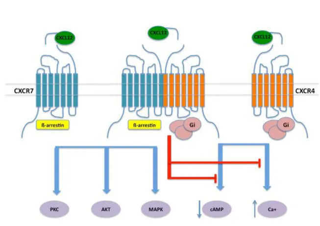
Binding of CXCL12 to CXCR4 triggers typical activation of G-proteins and ß-arrestins dependent pathways of a GPCR (see below in Ch. II), that cannot solely account for the wide spectrum of physiological activities of this axis (Busillo and Benovic 2007). It is rather hypothesized that such functional complexity originates from various determinants, including the cell context, the available receptor interactome (receptorosome), together with the reported ability of both CXCL12 and CXCR4 to oligomerize (Bachelerie, Ben-Baruch et al. 2014). This is also consistent with observations made in live cells, from the use of biophysical techniques, including bioluminescence and fluorescence resonance energy transfer, which indicate that monomers, dimers and higher-order oligomers of CXCR4 might coexist (Ferre, Casado et al. 2014). Such as emerged for other GPCRs, CXCR4 dimers might display unique ligand-binding properties and functional selectivity.
Theoretically, CXCR4 in dimer is able to accommodate one ligand or a dimer of ligands, forming a 2:1 or a 2:2 CXCR4-CXCL12 complex, respectively. However, a forced-dimeric CXCL12 was shown to behave as a partial agonist capable of inducing intracellular calcium mobilization but not chemotaxis (Veldkamp, Peterson et al. 2005). Thus, when considering the full agonist signaling process, the question of whether the CXCR4-CXCL12 complex has a 1:1 or 2:1 preferential stoichiometry with regard to the stability of their respective tridimensional structures and signaling properties remains an open question (Kufareva, Stephens et al. 2014) (Fig 7).
The interaction of CXCR4 with its agonist CXCL12 implicates residues in the N-terminus and ECL2 of the receptor. The three N-terminal residues Glu-14, Glu-15, and Tyr-21 of CXCR4 are of particular importance for the binding to CXCL12 (Doranz, Orsini et al. 1999, Brelot, Heveker et al. 2000). Residues in ECL2, especially the acidic sequence of Glu-179,
Ala-180, Asp-181, and Asp-182, are critical for activation of CXCR4 (Doranz, Orsini et al. 1999). CXCL12 has a high positive charges potential, and a significant amount of this positive charges, so it is possible that the interactions of the positive charges on CXCL12 with the negatively charged regions on CXCR4 contribute to the association of these two proteins. Negatively charged residues of CXCR4 also appear to be involved in the interactions with the basic V3 loop of gp120, the envelope glycoprotein of X4 HIV-1 strains (Kajumo, Thompson et al. 2000). Mutagenesis of specific residues on the CXCR4 N-terminus, ECL1, and ECL2 identified glutamate and aspartate residues, specifically Glu-15, Glu-32, Asp-97, Asp187, and Asp-193, as being important for interactions with X4 HIV-1 gp120 (Chabot, Zhang et al. 1999, Kajumo, Thompson et al. 2000). Other residues such as Asn-11, Arg-30, and Arg-188 have also been identified as binding determinants for HIV-1 gp120. Based on chimeric mutants that involved replacing ECL2 of CXCR2 with the corresponding loop from CXCR4, the observation was made that the CXCR4 ECL2 was able to confer the chimeric CXCR2 with HIV-1 coreceptor function (Lu, Berson et al. 1997). Since both CXCL12 and the V3 loop domain of X4 HIV-1 gp120 that interact with CXCR4 have a high positive potential, and since the interacting domains of CXCR4 are mostly negatively charged, it is likely that the interactions between CXCR4 and these molecules are highly driven by charges complementarity.
In the TM cavity of the receptor, several negative residues were found by site-directed mutagenesis studies to be important for CXCR4 signaling, including the region from Glu179 to Asp182 in ECL2, the residues Asp97 in TM helix 2 (TMH2), Asp187 in ECL2, Glu288 in TMH7, Tyr190 in ECL2 and Glu268 in ECL3 (Doranz, Orsini et al. 1999, Brelot, Heveker et al. 2000, Zhou, Luo et al. 2001, Tian, Choi et al. 2005).
It should be emphasized that the three residues Asp97, Asp187 and Glu288 of CXCR4 are also critical for the CK interaction with the CRS2 region (Brelot, Heveker et al. 2000). More specifically, they probably make contacts with the CXCL12 first two residues Lys1 and Pro2 (Heveker, Montes et al. 1998), from the disordered N-terminus (residues 1–8) critical for CXCR4 activation, as suggested by the recent crystal structure of CXCR4 with the viral CK vMIP-II (Qin, Kufareva et al. 2015).
These evidences are consistent with a two-site, two-step proposed model for the CXCR4-CXCL12 interactions, where the CXCR4-CXCL12 motif RFFESH first binds the receptor N-terminus, and then the CK N-terminus KPVSLSYR enters the buried cavity within the CXCR4 TM
helices, triggering receptor activation, probably mediated by a change in the conformation of the receptor TM helices (Crump, Gong et al. 1997, Kofuku, Yoshiura et al. 2009). Nevertheless, despite the important recent structural data, there is not yet a comprehensive characterization of the quaternary structures and dynamics of the CXCR4-CXCL12 associations, as the receptor like other GPCRs is difficult to express in sufficient quantities and to purify, because of its instability and ability to aggregate in detergent, and displays a reduce polar surface area for crystallization. With few exceptions, ligand-receptor structures require appropriate ligands that provide sufficient stability for purification and stabilization into well-defined crystallizable constructs, making the best targets those identified in drug discovery with high affinity.
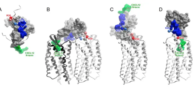
Fig 7. Molecular models and experimental designs of CXCR4:CXCL12 interaction. (A) NMR structure of CXCL12 (skin mesh) in complex with the N terminus of CXCR4 (residues M1– K38, ribbon). CK N-terminus (green) and N-loop (blue) correspond to the expected interactions with the receptor in CRS2 and CRS1, respectively. Receptor residues K25–R30 are shown as spheres, labeled, and colored in order from blue to red. (B) A 2:1 model of the receptor:CK interaction accommodates both NMR proximity restraints (black lines) and the mutagenesis data. (C) A hydrid 1:1 model that accommodates NMR proximity restraints (black lines) is inconsistent with mutagenesis and with the two-site interaction hypothesis, because the N terminus of the CK invariably points away from the receptor CRS2. (D) A 1:1 model consistent with the two-site interaction hypothesis contradicts NMR proximity restraints, as receptor residues K25–R30 are directed along the CK loop toward its N-terminus. (Kufareva, Stephens et al. 2014)
Given the clear role of CXCR4 in pathogenesis (exp. HIV and cancer metastasis), much efforts have been deployed successfully permitting the first structure determination of a CKR in 2010. CXCR4 was crystallized with two different synthetic ligands, a small molecule antagonist, IT1t, and a 16-residue cyclic peptide antagonist, CVX15 (Wu, Chien et al. 2010). While the overall geometry of the receptor resembled structures of other GPCRs with the typical seven-helix topology, CXCR4 revealed for the first time the large acidic binding pocket of the receptor relative to other solved GPCR structures, which is consistent with the fact that the natural ligands are basic proteins. IT1t occupies a small part of the cavity (430 Å3) in the minor sub-pocket defined by TM helices I, II, III and VII and is stabilized by contacts with a number of polar side chains implicated in binding CXCL12. CVX15 fills a much larger fraction of the binding pocket (2200 Å3), particularly the major subpocket (TM helices III–VII). It is stabilized by interactions with some similar but also many different receptor side chains as IT1t, including residues identified as important for binding CXCL12. Along with pharmacological data, these structures suggest that IT1t and CVX15 act as orthosteric antagonists by interfering with the CXCR4-CXCL12 CRS2-driven interaction, while occupying very different parts and fractional volumes of the binding pocket. As described below, the CXCR4:CVX15 complex provided insight for the modeling of the interactions of CXCR4 with HIV-1 gp120 (Wu, Chien et al. 2010).
In total, five structures of IT1t and CVX15 complexes were solved, and all demonstrated that CXCR4 forms dimers with a roughly consistent dimer interface involving TM helices V and VI. These finding corroborated a wealth of cell-based studies suggesting that CXCR4 forms homo- and heterodimers (Stephens and Handel 2013) as proposed for other class A GPCRs, although mostly by the way of overexpression systems (Stephens and Handel 2013).
They also raised the question of whether the stoichiometry of CKR-CK complexes is 1:1 as historically envisioned, or 2:1, which also seemed feasible and consistent with the two-site model, but contradict by a study which favored the 1:1 CKR-CK stochiometry and by extension the binding of two CKs on one CXCR4 dimer (Kufareva, Stephens et al. 2014). Thus the structure of CXCR4 and more generally CKRs dimers and their contribution to the receptors functions remain an open question awaiting proofs for their existence in physiological settings.
I.5e Structure of CXCR4 bound to the viral CK antagonist, vMIP-II
In 2015, the first structure of a CKR, CXCR4, in complex with a CK (vMIP-II) was solved, and provided detailed insight into the recognition of CKRs by their natural ligands (Qin, Kufareva et al. 2015). vMIP-II is a virally encoded high affinity antagonist for CXCR4 and was chosen over the main CK ligand, CXCL12, because the CK requires G-protein coupling for high affinity binding (Nijmeijer, Leurs et al. 2010), but antagonists generally do not. Additionally, vMIP-II binds promiscuously to both CC and CXC receptors, and is a CC like-CK; thus along with the solved structure of CCR5, the CXCR4vMIP-II structure was expected to provide insight into the specificity of CKs for CC and CXC CKRs.
In the vMIP-II-bound state, CXCR4 formed dimers that were spatially similar to those in the IT1t- and CVX15-bound structures; however, the structure of the complex confirmed the 1:1 stoichiometry anticipated from prior studies (Kufareva, Stephens et al. 2014), with ligand occupancy of both receptor subunits (Qin, Kufareva et al. 2015). The structure generally conforms to the concept of the two-site model in that the CK core domain interacts with the receptor N-terminus (CRS1) while the CK N-terminus binds in the CRS2 TM binding pocket of the receptor. Surprisingly, however, in contrast to the two-site model, where the expectation was that these two sites would be decoupled, the interaction between CXCR4 and vMIP-II involves an extensive contiguous interface, necessitating the introduction of an intermediate region termed CRS1.5 (Fig 8). In fact, every residue of the CK N terminal domain and N-loop (residue 1–16) as well as residues in the third β-strand, interact with the receptor (Qin, Kufareva et al. 2015).
Part of the CXCR4 N-terminus (residues 1–22 involving the key sulfated tyrosine, sTyr21), is missing from the electron density. Nevertheless, the visible CRS1 region (CXCR4 residues 23–27) showed interactions with the CK N-loop, as expected from numerous mutagenesis studies, and with the β3-strand in the CK core domain. Moreover, as the electron density stops just before sTyr21, it was fairly straightforward to generate a model containing sTyr21, which suggested a compelling interaction with nearby R46 of the CK, similar to the basic residue interactions reported for sTyr in (CXCR4:CXCL12)2 and CCR3:CCL11 complexes. In CRS1.5, CXCR4 forms an anti-parallel β-sheet with the di-cysteine motif of vMIP-II. Finally, in CRS2 the CK makes numerous interactions within the binding pocket including many residues known as determinants of vMIP-II binding or CXCR4:CXCL12 binding and activation. The N-terminus of vMIP-II overlaps with the binding site of IT1t in the
CXCR4:IT1t complex, while it only partially overlaps with CVX15, reflecting the structural plasticity of CXCR4 and its ability to accommodate diverse ligands via overlapping but distinct interfaces (macrophage migration inhibitory factor (MIF), extracellular ubiquitin (eUb), antimicrobial protein human β3-defensin (HBD-3), HIV (Human Immunodeficiency Virus) protein gp120 (Pawig et al. 2015)).
Fig 8. Interaction between CXCR4 and vMIP-II. The interaction is mediated by a contiguous interface containing CRS1 (green), CRS2 (red) and CRS1.5 (blue). The receptor is shown as a ribbon, receptor residues making substantial contacts with CK are shown as sticks, and vMIP-II is shown as a surface mesh (Qin, Kufareva et al. 2015).
vMIP-II is also a high affinity antagonist of CCR5, and the availability of the CCR5:Maraviroc (Lagane, Garcia-Perez et al. 2013) structure along with CXCR4:vMIP-II facilitated modeling of the complex (Qin, Kufareva et al. 2015). An important sequence difference relates to the presence of two adjacent sTyrs (sTyr14 and sTyr15) (Farzan, Mirzabekov et al. 1999) proximal to the conserved N-terminal Cys residue in the two receptors. These CCR5 sTyrs along with E18 are predicted to interact with the basic residues in the vMIP-II N-loop (K17 and R18) and β2-β3 loop (R46 and R48). By comparison, CXCR4 has only thr single proximal sTyr21, but in concert with two acidic groups (D22 and E26), makes similar interactions with the basic residues on the vMIP-II surface. These CRS1 models thus provide a plausible explanation for the unusual ability of vMIP-II to interact with both a CC and CXC CKR. By contrast, the CRS1 interaction between CXCR4 and the CXCL12 monomer is predicted to be quite different due to absence of basic residues in the CXCL12 N- and β2-β3 loops. Instead, the backbone of CXCR4 N-terminal residues S23 and M24 lie in a groove formed by the N- and β2-β3- loops, and position sTyr21 at the top of the core domain where it interacts with backbone amides of the CXCL12 residues R20 and A21. Notably, these interactions mimic the placement of a small molecule CXCR4:CXCL12 inhibitor (Smith, Ogert et al. 2014) as well as sulfate groups and ions observed in several CXCL12 X-ray structures (Murphy, Yuan et al. 2010), which adds support for the CRS1 predictions.
However, additional structures will be required to confirm the above-mentioned models and also to better understand the structural basis for the generally strict recognition of CC CKs for CC receptors and CXC CKs for CXC receptors.
Chapter II
Preamble on GPCRs
All CKRs are G-protein-coupled receptors (GPCRs) (Fig 9), a superfamily of 7-TM helixes proteins, which encompases approximately 791 genes encoding for the six different receptor subtypes. Currently, all 19 known CKRs belong to the class A (rhodopsin-like) family of GPCRs and are classified into four main subfamilies based upon which CKs they bind: CC, CXC, XC, and CX3C receptors. Many of the CKRs are promiscuous and bind to several CKs within their family and allow for tailored CK response and redundancy (Bachelerie, Ben-Baruch et al. 2014).
In this section it will be discussed the structure and oligomerization of GPCRs, focusing on CXCR4 and CXCR7/ACKR3 receptors of the CXCL12 CK and the drug design on these receptors using structural information. To simplify we will talk about the hypothesis of dimer forms, but higher form of oligomers organization can be formed.
Fig 9. The GPCRs 7-TM helixes class A subfamily illustrated by the ß-adrenergic receptor structure coupled to heterotrimeric G-proteins (Rasmussen, Choi et al. 2007).
II.1 Current CK Receptors Crystal Structures
In 2010, the first CKRs (CXCR4) was crystallized at the Scripps Research Institute (Wu, Chien et al. 2010). The CXCR4 crystal structure was the first peptide GPCR to be solved and represented a major breakthrough in this field. It is important to note that several structural changes were used in order to stabilize the receptor for crystallography (Rasmussen, Choi et al. 2007, Jaakola, Griffith et al. 2008, Warne, Serrano-Vega et al. 2008, Wu, Chien et al. 2010). Using the T4 Lysozyme (T4L) strategy, intracellular loop 3 (ICL3) of CXCR4 was replaced with T4L along with truncating its C-terminus and using point mutations for stabilizing the receptor structure (Wu, Chien et al. 2010). While these techniques have been used successfully to crystallize GPCRs, they may introduce or induce unnatural receptor conformations (Fanelli and De Benedetti 2011, Arnatt and Zhang 2013). However, GPCR crystal structures have provided a wealth of knowledge concerning ligand binding that have confirmed or disproved modeling and mutagenesis data. More importantly, CXCR4 was crystallized in two dimeric forms with resolutions of 2.5 and 3.2 Å and these two dimers had either a TM5 and TM6 interface or a TM3 and TM4 interface respectively (Wu, Chien et al. 2010).
The antagonist ligands used in the CXCR4 crystallization process were the small molecule antagonist IT1t and a cyclic peptide antagonist CVX15. In the presence of either ligand the binding pocket was shown to be much larger than expected and comparable to that of class A aminergic receptors, which accommodates large endogenous peptide agonists. The increase in the size of the binding pocket presumably led to both antagonists binding shallowly near ECL2, which is important in ligand recognition and receptor activation (Clark-Lewis, Kim et al. 1995). While the large binding pocket may make computational modeling and docking difficult, the CXCR4 crystal structure has permitted subsequently numerous studies using structural-based drug design in order to make new CXCR4 ligands (Xu, Zhao et al. 2015). In 2013 the CKR CCR5 (CCR5) was crystallized by Tan et al. and revealed both similarities and differences within the CKRs family (Tan, Zhu et al. 2013). Like for CXCR4, the binding pocket of CCR5 was large due to its endogenous peptide agonists, but the co-crystallized antagonist, maraviroc, occupied a deeper domain into the pocket as compared to CXCR4 in complex with IT1t or CVX15. Due to the depth of the maraviroc binding-pocket, ECL2 did not play a role in binding, which contrasts with the CXCR4 structure (Tan, Zhu et al. 2013). Such variation could reflect differences in the mechanisms of binding of the used antagonists