HAL Id: hal-00857502
https://hal.archives-ouvertes.fr/hal-00857502
Submitted on 3 Sep 2013
HAL is a multi-disciplinary open access
archive for the deposit and dissemination of
sci-entific research documents, whether they are
pub-lished or not. The documents may come from
teaching and research institutions in France or
abroad, or from public or private research centers.
L’archive ouverte pluridisciplinaire HAL, est
destinée au dépôt et à la diffusion de documents
scientifiques de niveau recherche, publiés ou non,
émanant des établissements d’enseignement et de
recherche français ou étrangers, des laboratoires
publics ou privés.
Progress on the preparation of nanocrystalline apatites
and surface characterization: Overview of fundamental
and applied aspects
Jaime Gómez-Morales, Michele Iafisco, José Manuel Delgado-López, Stéphanie
Sarda, Christophe Drouet
To cite this version:
Jaime Gómez-Morales, Michele Iafisco, José Manuel Delgado-López, Stéphanie Sarda, Christophe
Drouet.
Progress on the preparation of nanocrystalline apatites and surface characterization:
Overview of fundamental and applied aspects. Progress in Crystal Growth and Characterization
of Materials, Elsevier, 2013, vol. 59, pp. 1-46. �10.1016/j.pcrysgrow.2012.11.001�. �hal-00857502�
This is an author-deposited version published in:
http://oatao.univ-toulouse.fr/
Eprints ID: 9162
To link to this article: DOI: 10.1016/j.pcrysgrow.2012.11.001
URL:
http://dx.doi.org/10.1016/j.pcrysgrow.2012.11.001
To cite this version:
Gómez-Morales, Jaime and Iafisco, Michele and
Delgado-López, José Manuel and Sarda, Stéphanie and Drouet, Christophe
Progress on the preparation of nanocrystalline apatites and surface
characterization: Overview of fundamental and applied aspects. (2013)
Progress in Crystal Growth and Characterization of Materials, vol. 59 (n°
1). pp. 1-46. ISSN 0960-8974
!O
pen
A
rchive
T
oulouse
A
rchive
O
uverte (
OATAO
)
OATAO is an open access repository that collects the work of Toulouse researchers and
makes it freely available over the web where possible.
Any correspondence concerning this service should be sent to the repository
administrator:
staff-oatao@listes-diff.inp-toulouse.fr
!Progress on the preparation of nanocrystalline
apatites and surface characterization: Overview
of fundamental and applied aspects
Jaime Gómez-Morales
a, Michele Iafisco
b,c,1, José Manuel Delgado-López
a,
Stéphanie Sarda
d, Christophe Drouet
d,*aLaboratorio de Estudios Cristalográficos (IACT-CSIC-UGR), Avda. Las Palmeras, n!4, 18100 Armilla, Granada, Spain b
Alma Mater Studiorum Università di Bologna, Dipartimento di Chimica “G. Ciamician”, Via Selmi 2, 40126 Bologna, Italy cUniversità del Piemonte Orientale, Dipartimento di Scienze Mediche, Via Solaroli 4, 28100 Novara, Italy
dCIRIMAT Carnot Institute, University of Toulouse, UMR CNRS/INPT/UPS, ENSIACET, 4 allée Emile Monso, 31030 Toulouse cedex 4, France
Keywords:
Biomimetic apatites and biomineralization Crystallization
Surface characterization and reactivity (adsorption, ion exchange) Bone tissue engineering
Drug delivery Medical imaging
a b s t r a c t
Nanocrystalline calcium phosphate apatites constitute the main inorganic part of hard tissues, and a growing focus is devoted to prepare synthetic analogs, so-called “biomimetic”, able to precisely mimic the morphological and physico-chemical features of bio-logical apatite compounds. Both from fundamental and applied viewpoints, an accurate characterization of nanocrystalline apatites, including their peculiar surface features, and a deep knowledge of crystallization aspects are prerequisites to attempt understanding mineralization phenomena in vivo as well as for designing innovative bioactive materials that may then find applications in bone tissue engineering, either as self-supported scaffolds and fillers or in the form of coatings, but also in other domains such as drug delivery or else medical imaging. Also, interfacial phenomena are of prime importance for getting a better insight of biomineralization and for following the behavior of biomaterials in or close to their final conditions of use. In this view, both adsorption and ion exchange represent essential processes involving the surface of apatite nanocrystals, possibly doped with foreign elements or functionalized with organic molecules of
* Corresponding author. Tel.: þ33 (0) 5 34 32 34 11; fax: þ33 (0) 5 34 32 34 99.
E-mail addresses:jaime@lec.csic.es(J. Gómez-Morales),michele.iafisco@unibo.it(M. Iafisco),jmdl@lec.csic.es(J.M. Delgado-López),stephanie.sarda@iut-tlse3.fr(S. Sarda),christophe.drouet@ensiacet.fr(C. Drouet).
1 Present address: Laboratory of Bioceramics and Bio-hybrid Composites, Institute of Science and Technology for Ceramics (ISTEC), National Research Council (CNR), Via Granarolo 64, 48018 Faenza, Italy.
interest. In this review paper, we will address these various points in details based on a large literature survey. We will also underline the fundamental physico-chemical and behavioral differences that exist between nanocrystalline apatites (whether of biological origin or their synthetic biomimetic analogs) and stoichiometric hydroxyapatite.
1. Introduction
Nanocrystalline calcium phosphate apatites play an important role in biomineralization and in the biomaterials field. Biological nanocrystalline apatites are the main inorganic components of hard tissues in mammals (bone and tooth, with the exception of enamel which is closer to stoichiometric hydroxyapatite) and are involved in several pathological calcifications such as dental calculi, salivary stones, blood vessel calcification, etc[1–5]. In comparison with hydroxyapatite [HA, Ca10(PO4)6(OH)2]
which is a stoichiometric apatitic phase that is the most stable and least soluble calcium phosphate at ambient conditions, nanocrystalline apatites are nonstoichiometric, calcium- (and OH-) deficient and may incorporate substituted ions in their nanosized crystals. Their calcium and hydroxide deficiencies are responsible for a higher solubility than HA. Besides, they have the property of being able to mature when submitted to humid environments[5]. Due to this maturation process, “mature” bone crystals for vertebrates are less soluble and reactive than embryonic bone mineral crystals[6]. The small size and nonstoichiometry of apatite nanocrystals fulfill an important biological function in bone since they presumably bestow the mineral phase with the solubility needed for resorption of the bone by oste-oclasts (bone resorbing cells), and enable bone mineral to act as an ion “reservoir” capable of either capturing or releasing ions (or small molecules) under the control of the body for ensuring homeo-stasis. Owing to these special properties, bone is thus a living tissue, far from being inert, which continuously undergoes remodeling and repair processes[7]. In addition, nanocrystalline apatites play a decisive role in the biological activity of implants (biomaterials) since the formation of a nano-crystalline bonelike apatite layer in their surface, when immersed in a simulated body fluid solution, can determine whether these materials can adequately integrate into the host living tissue[8].
Synthetic apatites exhibit excellent biological properties such as biocompatibility, bioactivity, lack of toxicity or inflammatory and immunitary responses, and also a relatively high bioresorbability. These properties can be significantly enhanced by improving their biomimetism [9], that is, by preparing them with similar dimensions, morphology, (nano)structural and chemical characteristics as for biological ones. Many different synthetic strategies have been employed to prepare nanosized apatite crystals[5]; however, the preparation of actually biomimetic nanocrystalline apatites may still be seen as a scientific and technological challenge[10]. It is important to underline that most important characteristics conferring the special properties to nanocrystalline apatites are the nanosized dimensions, their large surface-to-volume ratio, and the existence of a surface hydrated layer, non-apatitic in nature, which is essentially related to the formation process in solution. In fact this layer is bound to progressively disappear as the stable apatite domains (in the core of the crystals) develop with time (so-called maturation process). This hydrated layer exhibits a great ionic mobility, as well as ion exchange and adsorption capacities, and could participate in the interaction with macromolecules and drugs[4]. Thus, as mentioned above, this layer on bone mineral nanocrystals is thought to actively participate in homeostasis and other regulation processes [6]. Because of their excellent physico-chemical and surface properties, nanocrystalline apatites then found applications as bio-inspired apatite-based bioceramics for bone substitution, repair or augmentation, or else as carrier vehicles for proteins, drugs and gene delivery[4,11]or even in medical imaging.
The present paper reviews the recent progresses on the preparation of nanocrystalline apatites with tailored surface properties, which are based on the exploitation of their surface hydrated layer (as for biological apatites), as well as the characterization of nanocrystals surfaces and surface interactions with ions and macromolecules.
The review is structured in five sections. Section 1is devoted to the description of biologically-related calcium phosphates and in particular to the recent studies and findings in terms of bio-mineralization related to the formation of bone and teeth. Section 2deals with the latest published works regarding the preparation and crystallization mechanisms of biomimetic apatites with tailored surfaces properties. In Section 3we report on the recent advances in the characterization of nano-crystalline apatite surfaces and their interactions with macromolecules and ions, of superior impor-tance regarding their biological activity. Finally, Section 4 reviews some of the main advanced biomedical applications, related in particular to the emerging field of nanomedicine such as medical imaging, the setup of nano-carriers for drugs, proteins and gene delivery, or else bioactive coatings of metal implants.
Based on this review of the state-of-the-art in relation with biomimetic nanocrystalline apatites, there are excellent and exciting perspectives for the development of advanced bioactive bioceramics for bone tissue engineering, as well as for the setup of biomimetic nanoparticles with new applications based on the tailoring of their surface properties, for example by functionalizing the surface of the nanocrystals with specific molecules and ions or by using them to produce multifunctional engineered nanoparticles.
2. Biologically-related calcium phosphates and recent advances in biomineralization related to bone and tooth formation
As previously explained, in biological systems, calcium phosphates are the principal inorganic constituent of normal (bones, teeth, fish enameloid, deer antlers and some species of shells) and pathological (dental and urinary calculus and stones, atherosclerotic lesions) calcifications. Except for small portions of the inner ear, all hard tissues of the human body are formed of calcium phosphates. Structurally, with the exception of enamel, they occur in the form of poorly crystallized non-stoichiometric and carbonate-substituted nanocrystalline apatites[12,13].
2.1. Bone mineralization
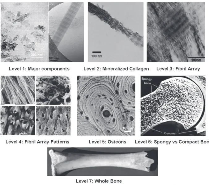
Bones are rigid organs that form part of the endoskeleton of vertebrates. Their function is to move, support and protect the various organs of the body, produce red and white blood cells and store minerals [14,15]. Bones appear in a variety of shapes and have a complex internal and external structure; they are lightweight, yet strong and hard, in addition to satisfying their many other func-tions. Bone is a hierarchically-structured biomineral that has captivated scientists because of its particular structure and unique mechanical properties and remodeling capability. To understand its complex architecture, several hierarchical models have been proposed. The most known is the model of Weiner and Wagner[16]who described seven levels of hierarchy that range from the nanoscale to the macroscopic scale (Fig. 1).
The first level consists of molecular and crystalline components such as collagen, apatite, water and the rest of molecules. The second level is formed by the mineralized collagen fibrils. The third level is composed of arrays of mineralized collagen fibrils. These fibrils are almost always associated as bundles or other arrangements, often aligned along their long axis. The fourth level is composed of the different patterns of fibril arrays. These patterns can be parallel arrays, woven arrangements, plywood like structures, and radial arrays. Cylindrical structures called osteons make up the fifth level. The sixth level is formed by spongy (trabecular or cancellous) or compact (cortical) bone tissues. Cancellous bone is extremely porous (75–95 wt.% porosity) and provides space for marrow and blood vessels, but has much lower compressive strength. Cortical bone is otherwise the dense outer layer (5–10 wt.% porosity) that allows many functions of bone. The seventh level is simply the whole bone. S. Mann presented a similar structural hierarchy containing six levels[1].
Bone is made of about 65 wt.% of mineral phase (nanosized crystals of apatite), 25 wt.% of organic phase (basically type-I collagen, non-collagenous proteins (NCPs), minor organic molecules such as citrate) and 10 wt.% of water [2,11,16]. Apatite nanocrystals are calcium- (and hydroxide-) deficient, with a ratio of Ca/P < 1.67, which is the theoretical value for the stoichiometric hydroxyapatite mineral (Ca10(PO4)6(OH)2). Other interesting features of bone apatite are the poor crystallinity degree, the
presence of ionic substituents in its crystalline structure (4–6 wt.% carbonate, 0.9 wt.% Na, 0.5 wt.% Mg and others) and a typical plate-like morphology. The reported dimensions of nanocrystals vary according to the technique employed (Table 1). Using TEM, XRD and SAXS, the characterized dimen-sions vary in the following ranges: length (20–50 nm), width (15–30 nm) and thickness (1.5–4 nm)
[2,17–21]. Studies by AFM, however, found that widths and lengths of bone crystals range from 30 to 200 nm[1]. Although apatite nanocrystals are usually presented as an inert single chemical compound with a passive role in the mechanical properties of bone, it must be underlined that the nanocrystals fulfill in fact a biological role, including through the “ion reservoir” function. It is thought that they may participate actively in homeostasis. This role has been attributed to their very high surface area and to
Fig. 1. Seven hierarchical levels of bone organization. Reprinted with permission from Ref.[13]. Copyright 2010 Dove Medical Press Ltd.
Table 1
Characterization of bone crystallites dimensions using different analytical methods. Analytical method Crystal dimensions
TEM 20 nm long # 3–6 nm wide
XRD 10–35 nm long
mXRD (14–17) nm # (3–5) nm
SAXS 50 nm # 25 nm # 1.5–4 nm
SAXS and TEM 30 nm # 20 nm # 1.5–2 nm
the presence of a hydrated surface layer with strong abilities for ion exchange and adsorption of organic molecules. Due to their variable and complex composition, biological apatites were sometimes considered as a mixture of phases, which in consequence can be characterized by X-ray diffraction techniques. However, these techniques cannot provide information on the fine structural details of their surface. Recent progress on wet surface characterization of synthetic nanocrystalline apatites has occurred thanks to the use of spectroscopic methods. The data obtained by FTIR and solid state NMR revealed that freshly precipitated nanocrystals show specific lines that are lost after drying and are assigned to the existence of a hydrated structured surface layer, non-apatitic in nature. Some of their spectroscopic features are surprisingly analogous to those of triclinic octacalcium phosphate (noted OCP or, more precisely, OCPt). This layer becomes progressively transformed into the more stable
apatitic lattice upon maturation in aqueous media. On the other hand the loosely-bound ions of this layer can be easily and reversibly substituted. Another feature is that the adsorption properties and reactivity of nanocrystalline apatites strongly depend on the composition of this layer[22].
Besides the above surface features, it is generally assumed that the very small particle size and the nonstoichiometry are bulk physicochemical features that presumably bestow the mineral phase with the solubility needed for resorption of the bone by osteoclasts, while their small thickness favors the mechanical properties, likely preventing crack propagation. Recently, citrate molecules strongly bound to the nanocrystalline apatite surfaces (1 molecule per every 2 nm2) have been identified by advanced solid-state NMR and proposed to be responsible for the inhibition of crystal thickening and also for the stabilization of nanocrystals within the collagen matrix[23]. NMR studies have revealed that citrate represents 5.5 wt.% of the organic matrix of bone[23]. Due to its excellent chemical and biochemical properties, like its excellent Ca-complexing power and its bioavailability, some authors of the present review employed citrate molecules for the preparation of synthetic apatite nanoparticles that incor-porated the molecules to their surfaces[24].
In addition to the recent interest for the characterization of surface properties of bone nanocrystals, the mineralization of collagen (the second level of hierarchy) has been the subject of intense inves-tigation during the last decade. In this respect the description of bone nanostructure deserves some few words. Bone nanostructure is made of a self-assembled fibrillar collagen matrix mineralized with oriented apatite nanocrystals at both intrafibrillar and interfibrillar zones. During bone formation the collagen molecules self-assemble into triple helical structures called tropocollagen. The interaction between tropocollagen units leads to self-organization into fibrillar structures. Type I collagen mole-cules assemble their tropocollagen units giving rise to holes and overlapping areas that can be observed as a periodic banding pattern along the 67 nm repeat and a less dense 40 nm-long gap zone (when stained for observation by TEM), the so-called D-band pattern (Fig. 2).
The holes appear mineralized and forming mineral bridges between them. The non-collagenous proteins (NCPs) appear linked to collagen and close to the mineralization front. Most of them are polyanionic and hold high amounts of carboxylate and phosphates groups which are arising from phosphoserine and aspartic and glutamic residues, with high affinity for Ca ions[26].
During many years there was controversy on whether it is collagen or NCPs that initiate intrafibrillar mineralization and on the mechanisms producing it at molecular level. It was thought that NCPs, while bonded to collagen fibers, could act as promoters of intrafibrillar nucleation. Such a hypothesis was supported by reports showing that NCPs appear during the formation of new bone in a very specific spatiotemporal timeline [27]and also that type-I collagen alone, dissociated from NCPs, is unable to produce bone mineralization [28]. On the other hand, it was proven that NCPs such as osteopontin, vitronectinand albumin, when they are dissociated from collagen, are inhibitors of calcium phosphate nucleation in solution[29–31].
Recently, in vitro intrafibrillar mineralization of collagen was achieved by substituting NCPs with polyaspartic acid (pAsp)[7,32]. Price et al.[33]proposed a mechanism for fibril mineralization based on inhibitor exclusion, by which macromolecular inhibitors of apatite growth favor fibril mineralization by selectively inhibiting crystal growth in the solution outside of the fibril. These authors tested this mechanism by using fetuin, a 48-kDa inhibitor of apatite growth. In the absence of fetuin, mineral growth occurred primarily in the solution outside of bone collagen, whereas in the presence of fetuin, mineral growth occurred almost exclusively within bone collagen. Only very recently, in 2010, the group of Sommerdijk [34] based on previous results [7,32] has shown that collagen functions in
synergy with inhibitors of hydroxyapatite nucleation to actively control mineralization by a mecha-nism of electrostatic interactions. These researchers used cryo-TEM and cryogenic electron tomog-raphy with molecular modeling to study this phenomenon and to follow the mineralization mechanism. In their experiments, Sommerdijk’s group used type-I collagen and either polyaspartic acid (pAsp) or fetuin instead of NCPs as soluble inhibitors macromolecules. According to their results amorphous calcium phosphate (ACP) nucleate on polyaspartic acid forming negatively-charged ACP-pAsp complexes. The mineral infiltration inside the fibrils is promoted by the net positive charge close to the C-terminal region of the collagen molecules at the gap and overlap areas. Furthermore, the charged aminoacids in these zones form nucleation sites controlling the conversion of ACP into a parallel array of oriented apatite nanocrystals. Although there are some open questions such as the role of the citrate molecules in this process, that could explain how these molecules participate in the biological regulation of nanocrystalline apatite formation, it can be said that these investigations have opened the door to the fabrication of intra-mineralized collagen bone analogs.
2.2. Teeth mineralization
Teeth consist of a bulk of dentin covered with (inorganic) enamel on the crown and cementum on the root surface[35]. Thick collagen bundles, called periodontal ligaments (PDL), attach to cementum at one end and the alveolar bone at the other end. The alveolar bone is supported by the jaw. These mineralized tissues are deposited by cells differentiating through interactions between inner neural crest-derived ectomesenchymal cells and overlying epithelial cells. Dentin and bone are similar in inorganic content (about 70 wt.% and 67 wt.%, respectively), crystal size and crystallinity and organic composition, including the type-I and type-V collagen heterofibril framework[36,37], but these two
Fig. 2. Cryo-TEM images of (A) loosely packed collagen bundle (arrow, 1) and fully assembled fibril (arrowhead, 2) at pH 7.4. Scale bar: 100 nm. (B) Assembled fibril showing clear D-band pattern. The dashed rectangle indicates the area used to calculate the profiles depicted in (C). (C) Intensity profile of the collagen fibril along the long axis, showing D-band pattern. Reprinted with permission from Ref.[25]. Copyright 2011 Royal Society of Chemistry.
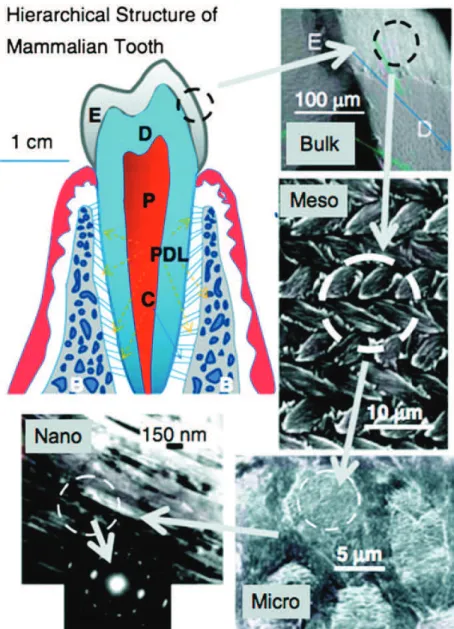
tissues are histogenetically different. Dentin is characterized by closely-packed tubules extending throughout its entire thickness. These tubules contain cell processes of odontoblasts (dentin-forming cells), and their cell bodies are typically aligned along the inner edge of dentin, the dental pulp[35]. In contrast, bone usually has cells (osteocytes) within the matrix and either active or quiescent bone-forming cells (osteoblasts) on the surface. On the contrary, enamel is distinct from bone and dentin in its tissue origin, mineralization matrix, and mode of mineralization [38]. Enamel is the hardest materials and with most robust mechanical properties formed by vertebrates and it is the most highly mineralized skeletal tissue present in the body. Mature enamel is composed of 95–97 wt.% carbonated HA with less than 1 wt.% organic material. Compared to dentin, enamel is uniquely composed of extremely long and narrow crystals (around 100
m
m # 50m
m,[39]), packed into parallel arrays, called enamel rods, which can form intricate interwoven patterns. The high degree of mineralization makes enamel a fascinating model for understanding fundamental mineralization processes and processes that occur within an extracellular matrix. It is distinct from bone in terms of architecture, pathology and the biological mechanisms mediating its formation[40]. Additionally, unlike other biomineralized tissues, mature enamel is acellular and does not resorb or remodel. As a result, enamel regeneration cannot occur in vivo following failure and is therefore an attractive target for future biomimetic and therapeutic approaches.Enamel formation, or amelogenesis, is a highly regulated process involving precise genetic control as well as protein–protein interactions, protein–mineral interactions, and interactions involving the cell membrane. Enamel forms by HA crystallization on a non-collagenous protein matrix secreted from ameloblasts of epithelial origin, whereas bone and dentin form on a collagenous matrix deposited by cells of mesenchymal origin. The enamel matrix mineralizes immediately after secretion[41]. During the initial stages (secretory stage) of enamel formation, long thin ribbons of enamel mineral are formed almost immediately as the ameloblast lays down enamel matrix proteins [42,43], suggesting that enamel mineralization does not take place within a preformed matrix [41]. This process is in sharp contrast to bone and dentin, both of which mineralize on preformed unmineralized matrix, called osteoid and predentin respectively. Although these mineral ribbons are extremely long (>100
m
m) and may extend the full thickness of the enamel layer[39,44], they are only a few unit cells thick (i.e., in the order of 10 nm)[45,46], if the initial mineral phase is considered to be HA or OCP. The c-axis of these crystals always coincides with their long axis [47], whereas, a- and b-crystallographic axes coincide with two other (thickness and width) morphological axes of the crystal. The mineral phase of secretory enamel is approximately 10–20 vol.%, with the remaining portion occupied by matrix protein and water[48]. It is important to notice that these ribbon-like crystals are organized in parallel arrays that ultimately dictate the highly-ordered arrangement of bundles of enamel crystals (i.e., enamel rods) found in mature enamel. In developing enamel[42,49], the c-axes (long axes) of these crystals are co-aligned and generally run in parallel to the overall direction of the enamel rod. Subsequently, the enamel matrix matures into a hypermineralized inorganic tissue (about 96% inorganic content). Specialized proteins, amelogenin, ameloblastin, and enamelin, constitute the mineralizing enamel matrix. During the maturation stage, coincident with the almost complete removal of enamel proteins by resident proteases, these mineral ribbons grow rapidly in thickness and width, resulting in a mineralized tissue that is >95% mineral (by weight), with only 1–2% remaining protein (the missing percentage is mostly water), in a way that maintains the structural organization of the enamel crystals established during the secretory stage[50]. These findings suggest a functional relationship between organic matrix removal and subsequent mineral growth. The transient nature of the enamel matrix is unique and distinguishes enamel formation from other systems in biomineralization.Similar to bone, enamel possesses a complex architecture, which can be broken into several hier-archical levels from the nanoscale to the macroscale[51](Fig. 3). On the nanoscale, the protein–protein and protein–mineral interactions in the presence of supersaturated calcium phosphate solutions create a highly organized array of HA crystallites that grow preferentially along the c-axis[52]. The sizes of these crystallites depend on the stage of the mineralization. The crystallites grow primarily in length during the secretory stage and continue to grow in width and thickness during the maturation stage. The assembly of amelogenin has been shown to be crucial for the proper development of enamel crystallites [53,54]. Disruption of the assembly alters the formation process on the nanoscale, subse-quently affecting larger length scales and giving rise to a diseased or malformed enamel phenotype. On
the mesoscale level, there are three main structural components: the rod, the interrod and the aprismatic enamel. The main component of enamel on the mesoscale includes rods, which are bundles of aligned crystallites that are “woven” into intricate architectures that are approximately 3–5
m
m in diameter [55]. The second structural component of the enamel matrix is the interrod (or inter-prismatic) enamel, which surrounds and packs between the rods. The difference between the rod and the interrod is the orientation of HA crystals; the rod contains aligned crystallites, whereas the mineral in the interrod is less ordered. These structures coalesce to form the tough tissue of enamel, which can withstand high forces and resist damage by crack deflection. The third structure, aprismatic enamel, refers to the structures containing HA crystals that show no mesoscale or macroscale alignment. The macroscale architecture includes specific zones of enamel that have unique characteristics, which contribute to the whole tissue. The enamel adjacent to the dentin–enamel junction (DEJ) exhibits a gradual transition from dentin to enamel. Aprismatic regions of enamel have been proposed to be primitive areas of the tooth serving as a toughening mechanism due to their flexible nature [56].Fig. 3. Hierarchical architecture of mammalian enamel. Enamel (E) is the outermost layer at the crown of the tooth and resides above the dentin (D). The pulp (P) contains nerves and blood vessels, while the cementum (C) is the outermost layer of mineralized tissue surrounding the root of the tooth allowing the tooth to be anchored to the jawbone through the periodontal ligament (PDL). The bulk image depicts the E organ, the transition across the D–E junction, and the D below. On the mesoscale level, prismatic E consisting of weaving of rods (or prisms) that range from 3 to 5mm in diameter can be visualized. Upon further magnification, the micrometer scale shows the composition of a single rod. The nanometer scale reveals a highly organized array of individual HA crystallites (approximately 30 nm thick, 60 nm wide, and several millimeters in length), which are preferentially aligned along the c-axis. Reprinted with permission from Ref.[58]. Copyright 2008 Cambridge University Press.
Several authors have identified these aprismatic areas to be located adjacent to the DEJ and at the incisal surface of both deciduous and permanent human enamel[57].
Over the last 40 years, a remarkable effort has been made to elucidate the mechanism by which enamel matrix proteins regulate the formation and organization of the enamel tissue. Although significant advances have clearly been made during this time, the complete mechanism of enamel formation is still unknown. Such progress has led investigators to suggest that the predominant enamel matrix protein, amelogenin, self-assembles to form organized supramolecular structures that facilitate crystal organization[59,60], prior to its subsequent removal during tissue maturation. This suggestion was based, in part, on the detection of chains of nanometer-sized spheres by using TEM observations of both dehydrated resin-embedded[61]and non-dehydrated freeze-fractured sections of forming dental enamel [62]. It was further suggested that such aggregates, alone or in combination with other proteins, facilitate the nucleation and organization of mineral phases[41]. Although this conceptual-ization is very interesting, especially given its similarity to mineralconceptual-ization mechanisms in collagen-based tissues, the precise mechanism by which enamel matrix proteins regulate such processes is not well-understood. Nevertheless, evidences suggest that both protein–protein interactions and protein–mineral interactions (in particular amelogenin proteins) play crucial roles in the regulation of enamel mineral formation and organization.
To better comprehend the enamel formation in addition to the protein–mineral interactions, the role of fluoride has been investigated. In the last five decades, fluoride has been used for the treat-ment of dental erosion and caries prevention through different modalities[63]. In addition, the effect of fluoride on the nanostructure of remineralized apatite has been well documented [64]. However, despite its use in humans as remineralizing agent, its benefits are not still clear due to the fact that the optimal dose of fluoride resulting in a positive effect on enamel remineralization with minimum risk of toxicity has not been well documented. In this respect several recent studies have demon-strated the excellent properties as remineralizing agent of the apatite as such (especially nanosized apatite) [65–67].
Several works have suggested that enamel crystal growth comprises two events: the two-dimensional growth of an OCP-like precursor in a narrow outermost zone adjacent to the amelo-blasts and the subsequent overgrowth of apatite units on the template under discrete fluid environ-ment in the underlying region distant from the cell layer. In this way, the presence of 0.1–2 mg/l fluoride in the mineralizing solution is known to promote hydrolysis of OCP to apatite [68,69]and fluoride can accelerate epitaxial growth of apatite crystals on the OCP precursor, changing the crystal morphology. It has also been shown that at 0.1–1 ppm fluoride the ribbon-like OCP changes to an interlayered structure whereas at 2 mg/l needle-shaped apatite crystals were produced [68,70]. Fluoride was hypothesized to reduce growth of (100) face of OCP and to cause needle-like crystal structure formation. Recently Fan et al. have demonstrated that 1 mg/l fluoride was effective in altering the crystal nanostructure from porous plate-like OCP to a 20–50 nm diameter needle-like array of fluoridated apatite nanocrystals [71]. The biomimetic mineralization system used in this work (supersaturated calcification solutions were used to initiate the growth of calcium phosphate crystals on the etched enamel surfaces), at best of our knowledge, is a first step toward developing a clinically-applicable means of forming an enamel-mimicking mineralized layer, under minimal fluoride expo-sure, on eroded or damaged tooth enamel. However, direct evidence of the effect of F ion on the morphology and nanostructure of crystal growth at a nanoscale level on an enamel surface has not been reported yet.
3. Crystallization of apatites with tailored surface properties 3.1. Precipitation of amorphous calcium phosphate (ACP)
Amorphous calcium phosphate is thermodynamically unstable which usually spontaneously transforms into crystalline calcium phosphate, mainly apatite. However, this instability and the easy transformation to crystalline phases are of a great biological relevance. Actually, ACP has been considered as a precursor phase of bone mineral in vertebrates which play a key role in intrafibrillar mineralization of collagen fibrils[72]. Recently, Weiner et al. reported that ACP is a major component of
the first-formed mineral phase of bone[73]. Additionally, Nancollas et al. on the basis of the work of Hu et al.[23]have suggested that citrate can strongly influence early ACP formation[74]. They proposed that at an early stage of nucleation in the presence of citrate, only some citrate molecules can partly bind with the cluster surface, inhibiting their further aggregation, and thus increasing the induction time[74]. In the later nucleation stage, the ACP clusters aggregation can be promoted by the presence of non-collagenous protein. Although some citrate molecules can interact with the amorphous clusters, the binding area density is low, owing to the continuing mismatch between the spacing of the terminal carboxylate groups in citrate and the structural parameter of the amorphous clusters. In fact, the interaction of the citrate molecules and ACP clusters can slow down but not inhibit the transformation process to crystalline apatite. On the other hand, the area density of citrate binding on the crystal surfaces increases because the spacing of carboxylate groups in citrate matches that of calcium ions of the ð1010Þ planes in apatite [23]. Thus, the crystal growth is inhibited along the [100] direction (reducing so the thickness of the nanoparticle) but it grows along the c-axis. Hence, it is clear that citrate and non-collagenous proteins play a key role on the tailoring of not only the transition from ACP to apatite but also the thickness and crystal orientation of apatite on the collagen fibrils.
3.2. Crystallization methods of apatites
Numerous methods have been used for the synthesis of apatite crystals. They can be classified into the following categories: wet chemical precipitation [75–77], sol–gel synthesis [78–80], co-precipitation [24,81], hydrothermal synthesis [82–84], rapid or continuous precipitation from solu-tion [85], mechanochemical synthesis [86], microwave processing [87–90], vapor diffusion [91–93], silica gel template[94], emulsion-based syntheses[95], electrospraying[96], electrospinning[97], flux cooling[98] and other methodologies producing (nano)crystals of various shapes and sizes[99,100]. Dorozhkin[5]has recently produced a review of those methods producing nanocrystalline apatites and has underlined that the interest for nanocrystalline apatites was quite recent. In fact the systematic investigation on these materials did not commence until 1994.
Using as criteria the control of apatite structure and morphology, the crystallization methods can be divided into high and low temperature approaches. The synthesis at high temperature usually involves the homogenization of precursor compounds, such as Ca3(PO4)2and Ca(OH)2, and their annealing at
about 1000!C. The advantage is the possibility to set the final stoichiometry of the product, whereas
the main downsides are the long processing times and high annealing temperatures. In general, when using this method the Ca/P ratio is a crucial parameter; in fact, if the initial molar ratio of Ca/P is not well set to 1.67, extraneous phases could appear such as
a
- orb
-TCP at values lower than the stoi-chiometric Ca/P ratio and typically CaO at higher values. Thea
-TCP phase is normally formed at temperatures around 1200!C whereas theb
-TCP phase is formed at lower temperatures, up to 900!C.In addition to the high levels of energy consumption, another major downside of high temperature methods is the difficulty to produce uniform and nanosized crystals[101].
The synthetic methods at low temperature involving precipitation from solution offer the advan-tage that they can produce nanosized crystals, but a disadvanadvan-tage may be in some cases the presence of transient and metastable phases in the final product. The samples produced by these methods are then generally nonstoichiometric and poorly crystallized. Indeed, the analyses carried out on several samples allow to assess that they have the typical features of biological apatites such as a poor degree of crystallinity, the existence of ionic surface disorder (non-apatitic surface layer) and surface compositions different from the bulk in nanocrystals, which consists on the presence of non-apatitic anionic and cationic chemical environments and sometimes labile carbonates.
As explained previously, biological apatites are known for their high content of defects, caused in part by a relatively large percentage of ionic substituents, all of which affect the lattice parameters, crystal morphology, crystallinity, solubility, and the thermal stability of the material. Biological apatite crystal surface is thus rarely smooth, which is also linked to biological significance. The exceptional roughness, comprising surface irregularities in the order of size of single unit cells, hypothetically corresponds to the tendencies to increase protein binding in the process of biomineralization. More-over, recent studies have shown that rough surfaces improve biocompatibility of the material and have a positive effect on inflammatory reactions [102]. For this reason, evolution in the preparation
techniques has led to methods for obtaining apatite in nanosized forms, mimicking those naturally occurring in bone. Structure is also important for biomimetism, as the mineral component of bone has well-defined features in relation to bone tissue function and age[103]. In this respect, the possibility to turn quite smoothly the bulk structure from highly to poorly crystalline by changing preparation conditions, such as temperature, pH, presence of anionic and/or cationic substituents, or else nucle-ation on a substrate such as collagen fibers, is well-known[104].
To investigate the effect of temperature on nanosized apatite, Sakhno et al. compared two types of samples synthesized at 40!C and 95!C[105]. The apatite synthesized at low temperature displayed
platelet-like morphology and are constituted by a crystalline core coated by an amorphous surface layer 1–2 nm thick (Fig. 4). By increasing the preparation temperature, the platelet morphology was retained but apatite nanoparticles exhibited a higher degree of crystallinity (evaluated by X-ray diffraction techniques). HRTEM observations revealed that, in this case, the crystalline order was extended up to the particles surfaces, exhibiting the planes (010), (100), and (001). IR spectroscopy was used to investigate the surface hydration of both materials, in terms of adsorbed H2O molecules and
surface hydroxyl groups, as well as the Lewis acidity of surface cations, by removing water and adsorbing CO[106]. For both features, strong similarities between amorphous and crystalline surfaces were found. However, interestingly the apatite synthesized at 95!C with crystalline surfaces appeared
able to physisorb multilayers of water in a larger extent than less crystallized samples.
Nowadays, the interest for nanocrystalline apatites is still rising thanks to their new applications in biomedicine requiring control of shape and size distribution as well as tailored surface properties (see in particular Section4). Although there are numerous methods to produce nanocrystalline apatites in a controlled manner, the preparation of actually biomimetic and surface- or bulk-tailored nanocrystals is still a scientific and technological challenge. In this sense variations of already established meth-odologies such as the method based on precipitation from metastable Ca/citrate/phosphate solutions
[24]or new methodologies, not yet explored, are being investigated to get these materials.
Delgado-López et al. have prepared citrate-functionalized carbonate-apatite nanoparticles with
mean lengths ranging from 20 to 100 nm by a thermal-decomplexing batch method [107]. This
crystallization method consists on the thermal decomplexing of metastable calcium/citrate/phos-phate/carbonate solutions that produces a gradual and homogeneous release of Ca2þ ions in the solution producing, thus the precipitation of nanocrystalline apatites. They also studied the effect of the maturation time and the presence of sodium carbonate in the solution on the physico-chemical and structural properties of the nanoparticles. They found these two experimental conditions play an important role in tailoring both the physicochemical and the morphological properties of nano-crystalline apatites such as size, composition, and crystallinity. At short precipitation time, poorly crystalline 100 nm-mean length apatites with low carbonation degree (1.5% w/w, mainly as B substitution) and high citrate content (5.9% w/w) were precipitated. Interestingly, this citrate content is close to that recently measured on bone apatite[23]. When increasing the precipitation time up to 96 h the mean length and the citrate content progressively decrease and at the same time the nanoparticles
Fig. 4. (Left) High resolution TEM image of a portion of apatite synthesized at 40!C (main panel, left), related FT and zoomed view of two border regions (right panels). Original magnification: 800k#. (Right) High resolution TEM image of a portion of apatite synthesized at 95!C (main panels A and B), related FT (right, bottom). Panels A, A0: zoomed view of two enframed border regions in panel A. Original magnification: 800k#. Reprinted with permission from Ref.[105]. Copyright 2010 American Chemical Society.
become more crystalline. The presence of carbonate in the solution favors its incorporation in the crystal lattice up to 3.1% (both, in A and B positions) giving rise also to shorter and more isometric nanoparticles. Hence, this work provides new insights on the role of citrate to tailor the apatite nanocrystal size. The nanoparticles were composed of a well-ordered carbonate-substituted apatitic core embedded in a non-apatitic hydrated layer containing citrate ions. This layer progressively transforms into more stable apatite domain upon maturation in aqueous media. Moreover, the nanoparticles prepared by this method displayed excellent compatibility properties in cell biology systems, since they were not cytotoxic to a mouse carcinoma cell line, when added until a final concentration of 100
m
g mL'1.One of the most interesting system to produce apatite nanocrystals at low temperature is the mechanochemical–hydrothermal (M–H) method. The main advantages of this kind of synthesis of ceramic powders are simplicity and low cost since conventional milling equipment can be used. The (M–H) technique is located at the intersection of hydrothermal and mechanochemical processing. Mechanochemical powder synthesis is a solid-state synthesis method that uses the perturbation of surface-bonded species by pressure to enhance thermodynamic and kinetic reactions between solids. Pressure can be applied at room temperature by milling equipment ranging from low-energy ball mills to high-energy stirred mills. M–H synthesis (sometimes called “wet” mechanochemical), takes advantage of the presence of an aqueous solution in the system. The aqueous solution actively participates in the mechanochemical reaction by acceleration of dissolution, diffusion, adsorption, reaction rate, and crystallization. The mechano-chemical activation of slurries can generate local zones of high temperatures (up to 700!C) and high pressures due to friction effects and adiabatic heating of
gas bubbles, while the overall temperature is close to the room temperature. The M–H technique produces comparable amounts of calcium phosphates powder as the hydrothermal processing, but it requires lower temperature than the hydrothermal process. Shuk et al. crystallized for the first time agglomerates of nanocrystalline apatite (about 20 nm in size) from heterogeneous reaction between Ca(OH)2powders and (NH4)2HPO4solutions via the M–H route[108]. More recently, several authors
improved the basic knowledge about this method and investigated the effects of pH, surfactant and polymer on the features of synthesized apatite nanocrystals [109–111]. Moreover the possibility to produce substituted apatites with carbonate, magnesium and fluoride by M–H technique was also studied[112,113,114].
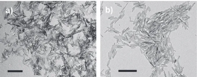
Recently, Iafisco et al. reported a new methodology, based on the vapor diffusion sitting drop micro-method, to precipitate carbonate-substituted apatite nanoparticles[91,92](Fig. 5a). The method was developed by using an innovative device called the “crystallization mushroom”[115,116], which offers the advantage of a reduced consumption of reagents, since the volume of micro-droplets is around 40
m
L and the high reproducibility due to the possibility of running 12 batches for each experiment. Therefore, this setup is suitable to evaluate interactions and/or the co-crystallization of apatite with small amounts of proteins, polymers, or drugs for studies in the fields of biomineralization and biomaterials. By using this methodology it has been found that mixtures containing 50 mM Ca(CH3-COO)2and 30 mM (NH4)2HPO4in micro-droplets and 3 mL of a 40 mM NH4HCO3solution in the gas
generation chamber were the optimal concentrations to precipitate carbonate-apatite nanocrystals after 7 days of reaction. The nanocrystals were produced by solvent mediated phase transformation of octacalcium phosphate (OCP) to apatite, with OCP most probably acting as a temporal template for the heterogeneous nucleation of apatite nuclei. The obtained crystals displayed nanometric dimensions, carbonate ions in the crystal lattice, plate-like morphology, and low crystallinity degree, closely resembling the inorganic phase of “young bones”. Nassif et al.[93]also precipitated carbonate-apatite by the vapor diffusion method. They used CaCl2–NaH2PO4 mixed solutions in the volume range of
milliliters (macro-method) and either a NH4OH and NaHCO3solution or solid (NH4)2CO3to generate
the gas phase, which led respectively to the precipitation of B- or A-type carbonate-apatite phases. They concluded that the best similarity between synthetic and natural apatite was obtained by using an aqueous carbonate precursor. This result agrees with those obtained by Iafisco et al.[92].
The analysis of the existing literature reveals that optimal routes to synthesize nanocrystalline apatites analogous to biogenic apatites are the low temperature methods, and also that in order to optimize their specific biomedical applications the particle dimensions, porosity, morphology and surface properties are major physical-chemical features that should be tailored. An approach consists
in using methods of precipitation in the presence of small amounts of additives [117,118]. The inter-actions between the growing inorganic particles and certain additives are governed mainly by elec-trostatic forces which can be tuned by controlling the zeta potential of colloidal particles. These forces are crucial for setting the optimal conditions for the growth of nanocrystals in different sizes and morphologies. Elongated apatite particles were for example obtained in the presence of poly(L-lysine),
whereas in the presence of more charged poly(L-glutamic acid), small nanocrystals were precipitated [119]. These additives thus act as inhibitors for the crystallization of apatitic crystals. A similar inhib-iting effect was found for other molecules such as dimethyl acetamide[120], polyvinyl alcohol[121]
and several other (bio)polymers [122], among others (more details on this matter are also given on Section4).
Among the additives, aminoacids are biological molecules of particular interest since they are the smallest molecular components of proteins. The effects of hydroxyproline, tyrosine, serine, glycine, cysteine, cystine, glutamine and lysine on apatite crystallization have been studied by the “Chemo-stat method” by Koutsopoulos et al. [123–125]. Diverse inhibiting activities were then observed as a function of the different aminoacid side groups. All these studies have utilized biomolecules as simple growth inhibitors of HA crystallization, rather than considering their use as a strategy to fine-tune the bioactivity of the apatite crystals. In fact, considering that the presence of proteins in biological materials is intrinsic to the bioactivity of apatite, aminoacids can be considered as agents that can increase the bioactivity of synthetic apatites. This idea was first developed by Gonzalez-McQuire et al.
[126], who obtained, by hydrothermal crystallization, stable aqueous colloids of positively charged aminoacid-functionalized apatite nanorods of less than 80 nm in length and ca. 5 nm in width. Greatly elongated apatite nanorods, of ca. 150 # 3.5 nm, were prepared with aspartic acid. The above work inspired further investigations into the nature and stability of the interaction of a series of aminoacids with apatite [127–129]. Together with the “Chemo-stat method”, these studies also suggested that crystallization kinetics is affected through the inhibition by aminoacids of the active growth sites of the apatite crystals surface. These sites could be constituted by surface calcium or phosphate ions (often protonated), and less probably by a few exposed hydroxyl groups. These experiments suggest that there may be a complex multi-site adsorption process, that may be “switched” on or off by changing the pH, since it affects the formation of different surface complexes between COO' and Ca2þ at
different sites.
Biomimetic nonstoichiometric apatite nanocrystals with aminoacid (alanine, arginine and aspartic acid) surface functionalities and different morphologies (depending on the aminoacid used as co-reagent during the synthesis) have been obtained by Palazzo et al. [130]. A self-assembly mecha-nism could be supposed, considering that the crystals domain size appeared slightly decreased by the aminoacid presence, while the nanocrystal grows unidirectionally. Zeta potential measurements then showed that the aminoacid-functionalized apatite surface charge is inverted, being shifted toward neutrality with respect to that of the apatite (Table 2).
Fig. 5. TEM images of carbonate-hydroxyapatite crystals grown by the sitting-drop vapor diffusion (a) and by the batch precipitation from metastable Ca/citrate/phosphate solutions. (b) Scale bars are 200 nm.
This chemical surface modification is found to dramatically affect the biological properties of the apatites, and should thus offer the potential for the nanocrystals to be used as carriers of bioactive molecules linked to the aminoacidic residue. Aminoacid is the anchoring agent between apatite and bioactive molecules, dramatically affecting the adsorption and release kinetics. It was found that, while the introduction of the aminoacids during the apatite nanocrystal synthesis induces a modification in the primary crystallite domains along the c direction and along its transverse directions, only polar aminoacids induce a morphological and dimensional variation in the apatite nanocrystals. Considering that the crystal domains sizes appear slightly decreased by the presence of the aminoacid while the nanocrystal grows unidirectionally, a self-assembly mechanism of nanocrystal formation can be supposed.
As was said earlier, zeta potential measurements showed that the surface charge of aminoacid-functionalized apatite was inverted compared to the unaminoacid-functionalized apatite shifting toward neutrality. The role of the lateral residue in the binding mechanisms, involving either the carboxylate group (in the case of alanine and aspartic acid) or the amino lateral group in the case of arginine, has been underlined.
Although each of the reported approaches to produce nanosized apatites has both a scientific and a practical relevance, only little attention has been dedicated to-date to the physico-chemical aspects involved in the careful control of the particle size distribution and particle shape. Indeed, in the case of particle size distribution, most of the reported ways to synthesize nanosized apatites really produce a mixture of particles with a wide size distribution from tens to hundreds of nanometers. In fact, the control of nanoparticle shape is another problem for these methods, which commonly result in pin-like or irregular particles. The size-controlled synthesis of materials can be achieved by using limited reaction spaces; namely microemulsions[131], micelles[132] and reverse micelles [133] have been successfully applied to the synthesis of nanosized apatites. In some cases, special polymers can be used as spatial reaction vessels for their fabrication. Mann et al. pioneered the use of organized molecular systems such as microemulsions to mimic the basic biomineralisation process in which mineral phase nucleation and growth is coupled with self-organized construction [134]. The micrometer-sized droplets of supersaturated solution stabilized in oil by surfactant molecules consist of microreactors where the reactions take place, and the nucleation and growth of mineral phase occurs specially at the surfactant headgroups segregated at the oil–water interface. Such systems can be used for the synthesis of apatitic nanocrystals, their growth (from about 10 nm thickness), occurred in a preferential direction oriented on the organized molecular support[131,135].
3.3. Crystallization mechanisms of nanocrystalline apatites
When using low temperature synthetic methods to produce nanocrystalline apatites, a disadvan-tage in some cases is the presence of transient phases in the final precipitates. To explain the possible presence of these extraneous phases the already published solubility phase diagrams can be consid-ered. The one published in 1992 by Johnsson and Nancollas[136] was performed at 37!C and ionic
strength 0.1 M. According to this diagram, the calcium phosphates phases decrease their solubilities with the increase of pH. At pH above 4.0 HA is the most stable phase followed by TCP and OCP, whereas at pH values lower than 4.0 the DCPD phase is more stable than HA. The position of the curves will change with the change in temperature and the ionic strength. On the other hand, the one published in 2009 [H.B. Pan and B.W. Darvell, Crystal Growth Des, 2009, 9, 2, 639–645] considers that DCPD is not the most stable phase below pH w4.2, since calcium-deficient HA is less soluble. The
Table 2
Zeta potential measurements for nonstoichiometric apatite synthesized in the absence of aminoacids and aminoacid-functionalized nonstoichiometric apatite nanocrystals.
pH of suspensions Zeta potential (mV)
Not-stoichiometric apatite 7.5 '21.2 ( 1.4
Alanine-apatite 7.8 '5.8 ( 1.5
Aspartic acid-apatite 7.6 '6.9 ( 1.2
misunderstanding arises from the metastability of DCPD, which nucleates much more easily than HA at low pH. This fact highlights the importance of kinetic factors when defining the pH domains in which a certain CaP phase can be formed in a precipitation experiment.
Thus, parameters such as pH, ionic strength, temperature, aging time, concentration and type of additives, Ca/P molar ratios and supersaturation have to be considered for the control of the formation of apatites [137]. The pH significantly affects the precipitation of HA because of its influence on the amount of free OH'groups and on the balance of phosphate species. A shift to low pH will decrease the
saturation level by diminishing the concentration of free OH' groups and shifting the balance of
phosphate species from PO43'to HPO42'to H2PO4'to H3PO4. Hence, at low pH, the phosphate groups
are protonated, and the precipitation is less favored. Moreover, the pH can shift the surface charge of the interacting particles by changing the distribution of proton and hydroxyl groups at the interface. Although H3Oþand OH'are usually considered as charge-determining ions in the case of HA particles,
other ions can adjust the surface charge, and in particular, in calcium-containing solutions, Ca2þions may bind to the negatively-charged HA surface at pH > IEP (isoelectric point), leaving the surface neutral rather than negative. The opposite effect can take place in phosphate-rich solutions when binding of HPO42'species at pH < IEP may result in a negatively-charged HA particle surface rather
than the positive.
One of the best accepted models for the crystallization of HA is the Ostwald–Lussac model which predicts the highest nucleation rate for the least stable phase for which the supersaturation limit is exceeded under given conditions. This model implies that an amorphous calcium phosphate (ACP) will be the first phase to precipitate, followed by the solid state transformation to OCP and then to HA. Ca(OH)2or TCP may be secondary phases depending on the exact stoichiometric ratio between the
precursor Ca2þ and phosphate ions in the solution. However, the exact chemical pathways, the transformation mechanism (dissolution/recrystallization or bulk rearrangement of ions within the prime crystal lattice), the transient compounds, and time frames for the nucleation and growth of each one of the phases are still subject to uncertainty.
The most important parameter used to describe the precipitation of HA (at a given temperature) is the supersaturation ratio S ¼ Q/Ksp, where Q is the product of ionic activities of precursor ions in the
solution for the given stoichiometry (for half a unit cell of HA, Q ¼ ½Ca2þ+5½PO43'+3½OH'+), and where
Kspis the product of ionic activities for the given compound at the saturation level (solubility product).
In view of the continuous transfer of the matter across the solid/liquid interface in both directions, Ksp
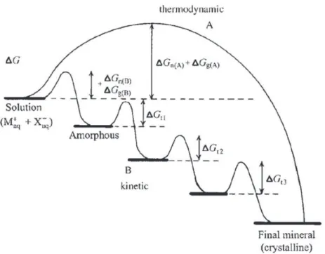
could be also defined as the product of activities of dissolved ions of a solid substance in equilibrium between the dissolved ions precipitating and the precipitated ions dissolving. Ostwald–Lussac rule postulates that the most soluble phase (i.e., the least stable) for which S > 1 is the first to precipitate, which will be successively followed by precipitation of less soluble phases. The reason for this is that the thermodynamic barrier between the state occupied by dissolved ionic species and the solid phase will be the lowest for the thermodynamically most unstable phase (Fig. 6)[138,139].
An important contributor to this effect is the surface/interfacial energy, that is, the work required to increase the surface area of a substance by one area unit, which is the obstacle that must be overcome when forming a solid phase. An amorphous phase is less ordered than a crystalline phase and it has a lower interfacial energy (particularly if it is hydrated, it is most similar in chemistry to the surrounding aqueous environment) which implies that it tends to be the first to precipitate prior to subsequently transforming into a more stable, crystalline modification. In the case of the precipitation of calcium phosphates, this means that HA could be the last phase to form. The initial precipitation of ACP is normally followed by nucleation of OCP at a certain stage. However, only the phases with S > 1 are involved in this successive precipitation. Those for which S < 1 are assumed not to be precipitated at any stage of this process, and their appearance may only be transitory during phase transitions that involve rearrangements of ions in the solid state. This kinetic rule was empirically observed, although it can be nowadays supported by theoretical arguments. In one such calculation[140], it was shown that in simulated body fluid (SBF), whose S normalized per growth unit (n ¼ 9 for HA) equals 19.5 with respect to HA, the nucleation rate of OCP is higher than the one for HA, implying that OCP would be the preceding crystalline phase to form.
Intermediate phases are thus expected to play a crucial role in defining the morphology, the interfacial properties and the growth mechanism of the HA. Hence, the first phase that should form
upon precipitation of calcium phosphates is an amorphous phase. The particles initially formed would be agglomerates of amorphous calcium phosphates units. Only in the following stage, the trans-formation of this phase into apatite and any other phase with S > 1 under the given conditions would take place. Under the conditions that resemble the physiological ones (pH about 7.4 and T ¼ 37!C), the
precipitated phases change over aging time (OCP is generally accepted to be the transient phase in physiological conditions and at pH < 9, whereas the mechanism at higher pH is not clearly defined yet). The solubility isotherms for brushite (DCPD) and OCP at room temperature and for Ca/P molar ratio of 1.16 (resembling the Ca/P ratio within the initially precipitated amorphous phase) interconnect at pH 6.7 (above this value OCP is a more stable phase and the trend is inverted at lower pH). This implies that according to Ostwald–Lussac rule, the transformation of the amorphous phase to apatite should follow the OCP to DCPD to apatite route at pH < 6.7, and the DCPD to OCP to apatite route at pH > 6.7[141]. The formation mechanisms of HA in pure solutions have been deeply investigated in the last years. An aggregation model of roughly spherical units of composition Ca9(PO4)6 called “Posner’s
clusters” (PC) was proposed in the 60’s to explain the formation of HA via an amorphous precursor, when working in neutral to basic conditions. The chemical analysis of the precursor amorphous phase indicated to be a hydrated calcium phosphate (Ca3(PO4)2.xH2O) with a Ca/P ratio 1.50,
con-sisting of close-packed Posner’s clusters to form larger spherical particles with water in the inter-stices [142–144].
After these pioneering works several researchers found evidences that strongly support an aggregation mechanism to explain HA formation. The mechanism proposed by Melikhov[145]can be divided into the following steps: (i) homogeneous nucleation of ACP; (ii) aggregation of primary ACP particles into typically spherical units; (iii) aggregation of spheres into chain-like structures; (iv) growth of these structures; (v) secondary precipitation and phase transformation. The initially precipitated particles of the amorphous phase were observed to be round-shaped with 20–30 nm in size (although they can reach 120 nm in size)[145], but composed of smaller particles of 4 nm in size on average. It was also observed that an increase in the ripening time implied aggregation of spherical singlets and formation of needle-shaped calcium phosphates particles of about 20 nm in length[146]. Rodríguez Clemente et al.[147]proposed a model of nucleation-directed aggregation–agglomer-ation-growth mechanism to explain the formation HA with micrometer size. The mechanism is based on empirical results of the precipitation of HA in batch and continuous process at 85 !C as well as Fig. 6. Crystallization pathways under thermodynamic and kinetic control. Whether a system follows a one-step route to the final mineral phase (pathway A) or proceeds by sequential precipitation (pathway B), depends on the free energy of activation (DG) associated with nucleation (n), growth (g), and phase transformation (t). Amorphous phases are common under kinetic conditions. Reprinted with permission from Ref.[138]. Copyright 2003 John Wiley & Sons, Inc.
HRTEM characterizations of the microparticles and the inconsistency of the classical nucleation-growth-agglomeration mechanism to explain the crystal size distributions of the HA obtained in their precipitation experiments. According to this model, the formation mechanism of particles starts with a nucleation episode, followed by the aggregation of supercritical nuclei ruled by surface energy excess, which counterbalance the repulsive forces between particles. The aggregates agglomerate, forming solid bridges, and these agglomerates continue to grow as distinct particles due to repulsive forces. This model allows to explain, for example, the precipitation of nanocrystalline apatites from complexed Ca–citrate–phosphate solutions. The specific surface area measurements (BET method), XRD, FTIR and microscopy analyses (TEM and SEM) showed that in the case of precipitation from Ca/ citrate/phosphate using sodium salts as reagents, the particle formation mechanism involves the initial precipitation of Na3cit.4H2O, which acts as a temporary template. The heterogeneous nucleation of
nanosized apatite in this system is interpreted in terms of surface free energy excess minimization by the interaction of the supercritical nuclei with this template. Afterwards, these supercritical nuclei grow to their final nanometer size, the template–particle interaction prevents the formation of apatite aggregates and, thus, allows the formation of primary nanosized particles at the time that the template dissolves. When using potassium salts or EDTA instead of sodium citrate as complexing agent, there was not an initial precipitation of a template and then apatite particles grew to final submicrometer or micrometer sizes. It is interesting to note that precipitation of a metastable phase which later dissolves, or the coprecipitation of both phases simultaneously, offers a unique way to obtain nanosized solid phases in natural and artificial crystallization from solution, which would otherwise be impossible to obtain due to surface energy excess.
Very recently the group of Sommerdijk revealed that ACP formation proceed via aggregation of prenucleation clusters before the development of oriented apatite crystals. High-resolution cryoTEM demonstrated that at 25 !C the simulated body fluid (SBF) solution that they employed contained
isolated nanometer-sized prenucleation clusters. After increasing the temperature to 37 !C these
clusters were present partially as individual entities or as small aggregates, but mainly as loosely aggregated networks within the bulk solution[148]. Size analysis of the observed clusters showed an average diameter of 0.87 nm, in line with previously reported cluster sizes (0.70–1.00) nm[149]and the theoretical size of Posner’s clusters (0.95 nm)[144].
4. Advances in the characterization of nanocrystalline apatite surface – study of surface interactions
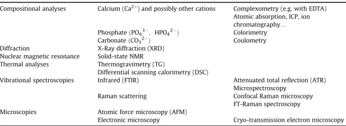
4.1. Introductive remarks on the need for precise and complete characterization data
Generally surfaces are difficult to characterize since they are easily contaminated and often exhibit great ion mobility, reactivity or metastability. This holds especially with calcium phosphate-based biomaterials. Therefore, great care has to be dedicated to the characterization of such systems. Indeed, significant departure from stoichiometry can be evidenced and is bound to have a noticeable impact on most of the sample properties. The full description of titration methods is not the object of this paper and has been addressed elsewhere[4], however we will summarize here the main aspects. The determination of calcium, total phosphate and carbonate ions can be performed nowadays with good accuracy using various methods. All these methods are based on the dissolution of the apatitic powder in acidic solution before the analysis (calcium and orthophosphate ions determination) or during the analysis (carbonate ions). Calcium contents can be assessed chemically by way of com-plexometry with EDTA [150] and orthophosphate ions contents ðPO43'and HPO42'Þ using a yellow
phosphovanadomolybdenum complex formed in acidic conditions[150]. The Ca/P mole ratio of apatite compounds is then the result of these analyses, and this ratio is often used to compare samples. Relative uncertainties on calcium and phosphorus concentrations obtained this way are of the order of 0.5%. Alternative methods as for example ICP or atomic absorption can measure lower ionic concen-trations than the current chemical methods, but phosphate concenconcen-trations are more difficult to asses on an ICP basis due to possible matrix effects in the presence of other ions in the systems. The titration of HPO42'ions only is, on the contrary, an indirect method exploiting the condensation of HPO42'ions