HAL Id: hal-02351122
https://hal.archives-ouvertes.fr/hal-02351122
Submitted on 6 May 2020
HAL is a multi-disciplinary open access
archive for the deposit and dissemination of
sci-entific research documents, whether they are
pub-lished or not. The documents may come from
teaching and research institutions in France or
abroad, or from public or private research centers.
L’archive ouverte pluridisciplinaire HAL, est
destinée au dépôt et à la diffusion de documents
scientifiques de niveau recherche, publiés ou non,
émanant des établissements d’enseignement et de
recherche français ou étrangers, des laboratoires
publics ou privés.
PAX5-ELN oncoprotein promotes multistep B-cell acute
lymphoblastic leukemia in mice
Laura Jamrog, Guillaume Chemin, Vincent Fregona, Lucie Coster, Marlène
Pasquet, Chloé Oudinet, Nelly Rouquié, Nais Prade, Stéphanie Lagarde,
Charlotte Cresson, et al.
To cite this version:
Laura Jamrog, Guillaume Chemin, Vincent Fregona, Lucie Coster, Marlène Pasquet, et al..
PAX5-ELN oncoprotein promotes multistep B-cell acute lymphoblastic leukemia in mice. Proceedings of the
National Academy of Sciences of the United States of America , National Academy of Sciences, 2018,
115 (41), pp.10357-10362. �10.1073/pnas.1721678115�. �hal-02351122�
PAX5-ELN oncoprotein promotes multistep B-cell acute
lymphoblastic leukemia in mice
Laura Jamroga,1, Guillaume Cheminb,1, Vincent Fregonaa, Lucie Costerc, Marlène Pasquetd, Chloé Oudinetb, Nelly Rouquiéa, Naïs Pradea,c, Stéphanie Lagardea,c, Charlotte Cressona, Sylvie Hébrarda, Ngoc Sa Nguyen Huub, Marina Bousquete, Cathy Quelene, Pierre Broussete, Stéphane J. C. Mancinif, Eric Delabessea,c,
Ahmed Amine Khamlichib,2, Bastien Gerbya,2,3, and Cyril Broccardoa,2,3
aCentre de Recherches en Cancérologie de Toulouse (CRCT), Université de Toulouse, Institut National de la Santé et de la Recherche Médicale (INSERM),
Université Toulouse III Paul Sabatier (UPS), 31037 Toulouse, France;bInstitut de Pharmacologie et de Biologie Structurale (IPBS), Université de Toulouse,
Centre National de la Recherche Scientifique (CNRS), UPS, 31077 Toulouse, France;cLaboratoire d’Hématologie, Centre Hospitalier Universitaire (CHU) de
Toulouse, 31000 Toulouse, France;dDepartment of Pediatric Hematology, CHU de Toulouse, 31000 Toulouse, France;eCRCT, INSERM, UPS, ERL5294 CNRS,
Laboratoire d’Excellence Toulouse Cancer (TOUCAN), 31037 Toulouse, France; andfAix Marseille University, CNRS, INSERM, Institut Paoli-Calmettes (IPC),
Centre de Recherche en Cancérologie de Marseille (CRCM), 13009 Marseille, France
Edited by Brian J. Druker, Oregon Health & Science University, Portland, OR, and approved August 29, 2018 (received for review December 14, 2017) PAX5 is a well-known haploinsufficient tumor suppressor gene in
human B-cell precursor acute lymphoblastic leukemia (B-ALL) and is involved in various chromosomal translocations that fuse a part of PAX5 with other partners. However, the role of PAX5 fusion proteins in B-ALL initiation and transformation is ill-known. We previously reported a new recurrent t(7;9)(q11;p13) chromosomal translocation
in human B-ALL that juxtaposedPAX5 to the coding sequence of
elastin (ELN). To study the function of the resulting PAX5-ELN fusion
protein in B-ALL development, we generated a knockin mouse model
in which thePAX5-ELN transgene is expressed specifically in B cells.
PAX5-ELN–expressing mice efficiently developed B-ALL with an
in-cidence of 80%. Leukemic transformation was associated with
recur-rent secondary mutations onPtpn11, Kras, Pax5, and Jak3 genes
affecting key signaling pathways required for cell proliferation. Our functional studies demonstrate that PAX5-ELN affected B-cell development in vitro and in vivo featuring an aberrant expansion of the pro-B cell compartment at the preleukemic stage. Finally, our molecular and computational approaches identified PAX5-ELN–regu-lated gene candidates that establish the molecular bases of the pre-leukemic state to drive B-ALL initiation. Hence, our study provides a new in vivo model of human B-ALL and strongly implicates PAX5 fusion proteins as potent oncoproteins in leukemia development.
B-cell acute lymphoblastic leukemia
|
engineered mouse models|
PAX5 fusion proteins
|
leukemia initiation|
oncogenic transformationB
-cell precursor acute lymphoblastic leukemia (B-ALL) is the most common pediatric cancer. B-ALL is characterized by a blockade of B-cell differentiation combined with an uncontrolled proliferation of blastic cells. Current chemotherapy is efficient at inducing long-term remission in childhood B-ALL, but the most common cause of treatment failure remains relapse that occurs in 15 to 20% of patients (1). The prognosis is even worse in adult B-ALL, as only 30% of adults achieve long-term disease-free survival (2).B-cell development is initiated by the entry of hematopoietic progenitors into the B-cell lineage transcription program and the concomitant sequential rearrangement of Ig genes through V(D)J recombination, ultimately leading to the generation of immunocompetent plasma cells. B-cell development can be dissected into pre-pro-B, pro-B, pre-B, immature B, and mature B-cell populations corresponding to different stages of differ-entiation (3). PAX5 is critical from early stages of B-cell devel-opment up to mature B cells (4). B-cell differentiation is completely blocked at the pro-B stage in Pax5 knockout mice, revealing its importance for early B lymphogenesis (5). Indeed, PAX5 plays a critical role in B-cell lineage commitment by ac-tivating the transcription of B cell-specific genes such as CD19 and BLK and suppressing alternative lineage choices (6–8).
PAX5 is the main target of genetic alterations in B-ALL. Heterozygous deletions and loss-of-function mutations of PAX5 are
found in more than one-third of human B-ALL (9–11). These al-terations result in loss of PAX5 expression and impairment of DNA-binding activity and/or transcriptional activity of PAX5. PAX5 is also rearranged in 2.6% of pediatric B-ALL cases, being fused to various fusion partners (9, 12–14). PAX5 translocations have been associated with a blockade of B-cell differentiation, as illustrated by PAX5-ETV6 and PAX5-FOXP1, which fuse the PAX5 paired do-main to ETV6 and FOXP1 transcription factors, respectively (15). We previously reported the molecular characterization of a new chromosomal t(7;9)(q11;p13) translocation in two cases of adult B-ALL. This translocation juxtaposed the 5′ region of PAX5 and almost the entire sequence of elastin (ELN) (13). The resulting PAX5-ELN fusion protein had conserved the nuclear localization sequence (NLS) and the DNA-binding paired-box domain of PAX5, and could therefore act as a constitutive repressor of the residual wild-type PAX5. Indeed, similar to PAX5-ETV6 and PAX5-FOXP1, transient expression of PAX5-ELN displayed a dominant-negative
Significance
Engineered mouse models of acute leukemia are critical to understanding the biological mechanisms by which a primary oncogene induces disease. While PAX5 fusion proteins are considered primary oncogenic events in B-ALL, their role in leukemia development is ill-known due to the lack of animal models. This report provides a novel and accurate in vivo model for B-ALL induced by PAX5-ELN fusion protein that es-tablishes a preleukemic phase and recapitulates the key fea-tures of human disease, including acquired mutations in genes of the JAK/STAT and RAS/MAPK pathways. This study is of general interest, as it allows a better understanding of the biological mechanism by which an oncoprotein perturbs nor-mal B-cell development and leads to pathological B-ALL.
Author contributions: L.J., G.C., V.F., L.C., M.P., C.O., N.R., N.P., S.L., C.C., S.H., N.S.N.H., M.B., C.Q., P.B., S.J.C.M., E.D., A.A.K., B.G., and C.B. designed research; L.J., G.C., V.F., L.C., M.P., C.O., N.R., N.P., S.L., C.C., S.H., N.S.N.H., M.B., C.Q., B.G., and C.B. performed re-search; M.P. provided pediatric B-ALL samples; L.J., G.C., V.F., L.C., M.P., C.O., N.R., N.P., S.L., C.C., S.H., N.S.N.H., M.B., C.Q., S.J.C.M., E.D., A.A.K., B.G., and C.B. analyzed data; and A.A.K., B.G., and C.B. wrote the paper.
The authors declare no conflict of interest. This article is a PNAS Direct Submission. Published under thePNAS license.
1L.J. and G.C. contributed equally to this work. 2A.A.K., B.G., and C.B. contributed equally to this work.
3To whom correspondence may be addressed. Email: [email protected] or cyril.
This article contains supporting information online atwww.pnas.org/lookup/suppl/doi:10. 1073/pnas.1721678115/-/DCSupplemental.
Published online September 26, 2018.
CELL
BIO
effect on PAX5 by down-regulating the transcription of PAX5 target genes (11, 13, 16, 17). However, the generality of the dominant-negative effect was recently questioned in vivo (15).
A detailed and dynamic analysis of the PAX5-ELN effect on B-ALL initiation and transformation requires the availability of a mouse model that allows a B-cell restricted expression of PAX5-ELN. Here, we demonstrate, by using a knockin (KI) mouse line, that PAX5-ELN is an initiating oncogenic event that perturbs normal B-cell development by aberrantly expanding pro-B cells at the earliest steps of leukemogenesis. Nonetheless, complete B-ALL transformation induced by PAX5-ELN involved the ac-quisition of additional mutations and the deregulated expression of multiple genes. Our study provides novel insights into the functional role of PAX5-ELN as a potent oncoprotein in B-ALL development, and should help to elucidate the molecular mechanisms that underlie B-ALL initiation and transformation. Results
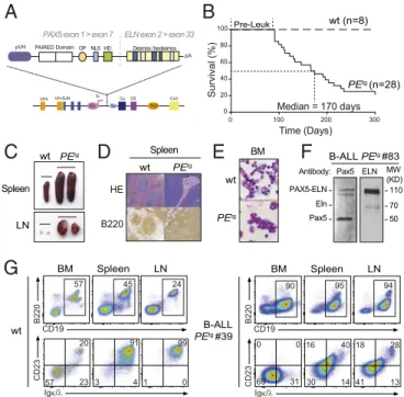
Constitutive Expression of Human PAX5-ELN Leads to B-ALL
Development.To investigate the B-leukemic potential of
PAX5-ELN, we developed a KI mouse model in which the cDNA encoding the human PAX5-ELN fusion protein was expressed in B-cell progenitors. This was achieved by inserting the human cDNA at the IgH locus under the control of a VH promoter (PVH) and the endogenous Eμ enhancer whose activity is trig-gered early in B-cell development (18). To avoid transcriptional readthrough from upstream promoters at different devel-opmental stages, a pause/polyadenylation site (19) was added upstream of the ectopic PVH promoter (Fig. 1A and SI Ap-pendix, Fig. S1). Unless otherwise indicated, the experiments have been performed on heterozygous mice.
Mutant mice that constitutively expressed PAX5-ELN (here-after PEtg mice) efficiently developed leukemia with a
pene-trance of 80% at 300 d (Fig. 1B). Leukemic development was associated with a splenomegaly (Fig. 1C, Upper) characterized by a massive infiltration of B220+cells and a dramatic perturbation of the spleen architecture (Fig. 1D). Moreover, PEtg mice
de-veloped a lymphadenopathy (Fig. 1C, Lower) and exhibited blast cells in the bone marrow (BM) (Fig. 1E). Importantly, although the expression of PAX5-ELN was driven by IgH regulatory se-quences, immunoblot analysis of protein extracts with a PAX5 paired domain-specific antibody revealed that the abundance of PAX5-ELN was not higher than that of endogenous PAX5 (Fig. 1F). This observation indicates that the reported effects on PEtg mice cannot be ascribed to high expression levels of PAX5-ELN fusion protein. Immunophenotypic characterization showed that BM, spleen, and lymph nodes (LNs) of leukemic PEtg mice contained an aberrant proportion of CD19+B cells associated with an aberrant and variable expression of CD23 and Igκ/λ markers (Fig. 1G andSI Appendix, Fig. S2).
Clonal Transformation and Collaborating Events with PAX5-ELN
Oncoprotein.The IgH variable region can broadly be divided
in-to the VHdomain, including the distal VHJ558and the proximal
VH7183gene families, and the DHJHdomain, comprising a dozen
DHsegments followed by four JHsegments (Fig. 2A, Upper) (20).
Assembly of the IgH variable region involves two recombination steps: first DHto JH, followed by VHto DHJH. To determine the
rearrangement status of the IgH locus in leukemic cells, we performed a qPCR-based V(D)J recombination assay (21) on genomic DNA purified from blasts of five independent B-ALL mice (Fig. 2A, Upper). The data reveal prominent DHJH and
VHDHJHrearrangements, involving both proximal and distal VH
segments, in all B-ALL samples (Fig. 2A, Lower). These results indicate that PAX5-ELN fusion protein induces the clonal transformation of a B-cell progenitor that has already rearranged its IgH locus, and support the notion that PAX5-ELN acts as a potent B-ALL oncoprotein.
Our observations on B-ALL transformation delay (>90 d) (Fig. 1B) led us to suspect potential acquisition of secondary mutations. To further identify these additional genetic alterations that po-tentially cooperate with PAX5-ELN, we performed whole murine exome sequencing in five PEtgleukemias and identified recurrent somatic mutations in Ptpn11, Kras, Jak3, and Pax5 genes. These mutations were also found after specific sequencing of these loci in an additional 11 PEtg leukemias (Fig. 2B). These findings strengthen the notion that aberrant activation of JAK/STAT and/ or RAS/MAPK signaling pathways is required for an overt B-cell leukemia transformation in our model.
To address the critical question of the recurrence of these so-matic mutations in human B-ALL, we performed the targeted se-quencing of several exons of PAX5, PTPN11, NRAS, KRAS, JAK3, and JAK2 genes in a cohort of 101 pediatric B-ALL patients (Fig. 2C andDataset S1). Importantly, we detected recurrent mutations in PAX5 (9/101), PTPN11 (15/101), NRAS (33/101), and KRAS (33/ 101) genes in all of the different B-ALL oncogenic subtypes (Fig. 2C). Interestingly, the patient cohort contained five PAX5-rearranged B-ALL patients, including one with the PAX5-ELN translocation that exhibited, just as in our mouse line, PAX5, NRAS, KRAS, and JAK3 mutations (Fig. 2C andDataset S1). To-gether, the above data establish that our PAX5-ELN mouse model recapitulates the multistep pathogenesis of human B-ALL.
A
PAiRED Domain pVH VHnVH-DJH Eμ Sμ CμCδ Cγ3 Sγ3 Iμ pAB
Survival (%) 0 100 200 300 0 20 40 60 80 100 Time (Days) PEtg (n=28) wt (n=8)E
Median = 170 daysC
D
wt PEtg wt PEtg wt PEtg HE B220 Spleen LN BM Spleen CD19 B220 CD23 wt BM Spleen LN 57 45 24 57 23 20 3 4 91 1 0 99 Igκ/λG
Pre-Leuk OP NLS HD Desmos / IsodesmosPAX5 exon 1 > exon 7 ELN exon 2 > exon 33
F
ELN Pax5 Pax5 110 50 MW (KD) PAX5-ELN B-ALL PEtg #83 Antibody: 70 Eln CD19 B220 Igκ/λ CD23 90 69 31 0 0 95 94 30 14 16 40 41 13 18 28 B-ALL PEtg #39 BM Spleen LNFig. 1. Human PAX5-ELN expression induces efficient B-ALL development.
(A) Generation of knockin mouse model expressing PAX5-ELN fusion protein. The sequence encoding the human PAX5-ELN fusion protein was inserted
downstream of Eμ enhancer. Desmos, Desmosine; HD, homeodomain; OP,
octapeptide; pVH, VH gene promoter. (B) Kaplan–Meier curves of the time to
leukemia for cohorts of PEtgmice (n= 28). WT mice (n = 8) were used as
controls. Pre-Leuk, preleukemic time. (C–E) PAX5-ELN induces B-ALL devel-opment characterized by leukemic cell invasion in the bone marrow, spleen, and lymph nodes. (C) Pictures of spleens (Upper) and LNs (Lower) from WT
and leukemic PEtgmice are shown. (D) Staining with hematoxylin and eosin
(HE) and immunohistochemistry of B220 are shown of spleens from WT and
leukemic PEtgmice. (E) Pictures of May-Grünwald–Giemsa–stained cytospin of
BM cells from WT and leukemic PEtgmice. (F) Protein extract of leukemic cells
from a B-ALL PEtgmouse (no. 83) was subjected to immunoblotting with anti–
N-terminal Pax5 (N19) antibody for the detection of PAX5-ELN and endoge-nous Pax5 and with anti-ELN antibody for the detection of PAX5-ELN and endogenous Eln. (G) Total cells from the BM, spleen, and LNs of WT (Left)
and leukemic PEtg(Right) mouse (no. 39) were immunophenotyped using
the B220, CD19, CD23, and Igκ/λ markers.
PAX5-ELN Oncoprotein Perturbs Early B-Cell Progenitor Cell
Differentiation at Preleukemic Stage.The survival curve of PEtg
mice revealed a leukemia onset starting at 90 d after birth (Fig. 1B). This leukemia development latency allowed us to explore the effect of PAX5-ELN oncoprotein on the early steps of leukemogenesis. Cytological examination of the preleukemic PEtgmice confirmed the absence of blasts in the BM (SI Appendix, Fig. S3A) and any obvious perturbation of the main cell lineages (SI Appendix, Fig. S3B). Moreover, the absence of detection of Ptpn11, Kras, Pax5, and Jak3 mutations in the BM of preleukemic PEtgmice (Fig. 2B) allowed us to precisely address the role of PAX5-ELN in leukemia initiation, before the onset of clonal transformation induced by the acquisition of additional cooperating oncogenic events.
To identify the earliest cell types that are affected by PAX5-ELN oncoprotein, we analyzed B-cell progenitors (SI Appendix, Fig. S3C) in the BM of PEtg mice during early and late pre-leukemic stages, namely at 30 and 90 d after birth, respectively.
We observed that PAX5-ELN significantly induced a three- to fourfold expansion of the pro-B population at 30 and 90 d, whereas pre-pro-B and pre-B populations were not affected (Fig. 3 A and B). Interestingly, 30-d-old PEtg pro-B cells exhibited a
polyclonal profile for DHJHand VHDJHgene rearrangements that
was roughly comparable to their WT counterparts (Fig. 2A), in-dicating that PAX5-ELN–induced pro-B cell expansion was not associated with a clonal selection at the preleukemic stage. In addition, this pro-B cell expansion was associated with a reduction of the immature and circulating B-cell populations in the BM (Fig. 3 A and B), indicating that PAX5-ELN partially blocked B-cell differentiation at the preleukemic stage. Accordingly, we observed a moderate but significant reduction of spleen size in preleukemic
A
B
1 2 3 4 Low High Wt PEtg #110 #114 #83 #083 #39 0 0 1 0 0 1 0 0 1 0 0 1 0 0 1 0 0 1 0 0 1 0 0 1 100 100 100 100 0 2 1 8 6 3 4 3 3 3 6 1 1 2 0 3 8 3 1 3 2 1 105 32 57 275 0 0 0 4 5 9 6 0 0 0 0 1 0 0 2 0 7 9 1 0 0 3 6 4 0 0 0 1 0 0 1 0 0 0 0 0 2 0 5 2 7022953514 7 9 9 2 2 1 0 1 1 3 1 2231 25 5 6 4 0 5 1 5 9 0 3 1 0 2 0 3189 62 J558 E Igh locus 7183 dist.VH prox.VH ~ 12 DH 4 JH 4 3 2 1 ~ 200 VH 1 2 3 4 1 2 3 4 DHJH VHJ558DHJH VH7183DHJHE76G E76K E76Q D61V E69K A72V
G60R G12D R653H V670A R38C
Ptpn11 Kras Jak3 Pax5
PEtg #1 PEtg#2 #11150 #05823 45 #081 70 50 #083 57 49 #099 29 1 #005 46 4 #00643 #0148 3 #05333 #11434 23 #12630 1 #18636 8 #270 17 #27311 #026 44 #110 17 WES Targeted NGS
C
PEtg#3 PEtg#4 wt #1 wt #2 wt #3 B-ALL B-ALL Pre-leuk PAX5 PTPN11 NRAS KRAS JAK3 JAK2 TEL-AML1 E2A-PBX1 MLL Hypodiploidy Hyperdiploidy BCR-ABL1 BCR-ABL1 likePAX5 rea Other B-ALL
PAX5-ELN
Ptpn11 Ras Jak3 Pax5
Fig. 2. Clonal selection in PAX5-ELN–induced B-ALL. (A) Schematic of the
mouse IgH locus (Upper). The locus is composed of∼200 VH(red boxes),∼12
DH(blue), and 4 JH(green) segments that undergo recombination in B-cell
precursors to produce a functional VHDHJHunit. Genomic DNAs were
pre-pared from purified PEtgpro-B cells and five leukemic PEtgmice and subjected
to quantitative PCR to quantify DHJHand VHDHJHrearrangements using
pri-mers that bind the indicated gene segments. dVH, distal VH; pVH, proximal VH.
PCR of the HS5 element downstream of the 3′ regulatory region was
per-formed for normalization of DNA input. PCR was perper-formed in triplicate.
Quantification of DHJHand VHDHJHrearrangements is represented as a heat
map (Lower). DNA from purified WT pro-B cells was used as a control and normalization (100% of the signal) for each gene rearrangement ranked in each column. (B) Recurrent mutations in B-ALL blasts induced by PAX5-ELN. Whole-exome sequencing (WES) was performed on BM cells from five leukemic
PEtgmice. Mutations found on Ptpn11, Kras, Pax5, and Jak3 genes were verified
and screened for recurrence by targeted next-generation sequencing (NGS) on
BM cells from 11 leukemic, 3 WT, and 4 preleukemic 30-d-old PEtgmice. Each
row represents a leukemia sample, and each column represents a genetic al-teration. Colors indicate the position of the mutation, and numbers represent the variant allele frequency of each mutation. (C) Mutations on PTPN11, KRAS, NRAS, JAK3, JAK2, and PAX5 genes were screened for recurrence on 101 B-ALL patient samples. Each column represents a B-ALL sample classified according to the indicated oncogenic subtypes, and each row represents a genetic alteration.
Colored boxes indicate the presence of a mutation, listed inDataset S1.
A
B
Pre-pro-B Pro-B Pre-B
Immature-B Mature-B 15 10 5 0 20 0 40 60 80 15 10 5 0 20 25 15 10 5 0 20 Cell number (x10 5) 90 ns ns ns ns *** * * * Bone Marrow 15 10 5 0 20 25 *** ** wt PEtg Pre-pro-BB220+CD19+ Mature-B Immature-B Pre-B +Pro-B Pre-B Pro-B CD19 B220 wt PEtg Igκ/λ CD23 Kit Events 59 41 91 9 3 53 4 51 76 13 9 57 19 22 Lymphocytes 30 30 90 30 90 Days30 90 30 90 0 10 20 30 40 50 B engraftment (%) 6 4 2 0 8 Engrafted B (x10 7) Total B-cells CD45.2+ CD45.1+ Transplant 2.106 cells 5 wks FACS BM Spleen BM Spleen wt Pre-Leuk B-ALL PEtg wt Pre-Leuk B-ALL wt Pre-Leuk B-ALL 0 50 100 150 200 250 Spleen (mg) PEtg wt Pre-Leuk B-ALL PEtg wt Pre-Leuk B-ALL ns ** ** ** ns * * ** ns * wt CD23 BM CD45.2+ B220+ CD19+ B-cells Igκ/λ Events Kit Pre-Leuk 75 13 12 86 13 1 95 5 45 55 Pro-B 60 40 20 0 80 30 20 10 0 40 50 15 10 5 0 20 4 2 0 6 Pre-B * ns ns * Cell number (x10 5) BM CD45.2+ cells
C
D
E
F
G
3 2 1 0 4 Pre-pro-B ns Imma-B Mat-B wt Pre-Leuk wt Pre-Leuk wt Pre-Leuk wt Pre-Leuk wt Pre-Leuk PEtg PEtgFig. 3. Pro-B cells are expanded by PAX5-ELN at a preleukemic phase. (A and
B) Preleukemic PEtgpro-B cells exhibit an aberrant expansion potential in vivo.
Immunophenotyping of BM cells from 30-d-old WT and PEtgmice was
per-formed using B220, CD19, CD23, Igκ/λ, and Kit markers (A, Left), and a gating
strategy was applied to discriminate B-cell subpopulations (A, Right andSI
Appendix, Fig. S2). Absolute numbers of pre-pro-B, pro-B, pre-B, immature B,
and mature B cells from the BM of 30- and 90-d-old WT and PEtgmice were
calculated (B; n= 5 to 8 mice per condition). (C–E) Preleukemic and leukemic
PEtgB cells exhibit different engraftment potentials. Total B cells from the BM
of preleukemic and B-ALL PEtgmice (CD45.2+) were purified and i.v. transplanted in recipient mice (CD45.1+, n= 3, 2.106cells per mouse) pretreated with 30 mg/kg busulfan (C). The same number of total B cells from the BM of WT mice was
transplanted in parallel as control (C; n= 3). The proportion (D, Left) and
ab-solute numbers (D, Right) of donor-derived (CD45.2+) B cells in the recipient BM
were calculated 5 wk after transplantation. Spleen weights of engrafted mice
were compared (E). (F and G) Preleukemic PEtgpro-B cells expand after
trans-plantation. Immunophenotyping of engrafted B cells (CD45.2+B220+CD19+) in
the BM was performed (F), and absolute numbers of donor-derived (CD45.2+)
preleukemic ppro-B, pB, pro-B, immature B, and mature B cells in the re-cipient BM were calculated (G). Error bars represent standard deviations, ***P< 0.0005, **P< 0.005, and *P < 0.05; ns, nonsignificant.
CELL
BIO
PEtg mice compared with age-matched WT mice (SI Appendix, Fig. S4A), suggesting that PAX5-ELN retained B-cell loading in peripheral lymphoid organs. Indeed, while the overall distribution of B-cell populations was unaffected by PAX5-ELN in the spleen (SI Appendix, Fig. S4B), the absolute number of immature and mature splenic B cells was significantly reduced in preleukemic PEtgmice (SI Appendix, Fig. S4C). The above results indicate that PAX5-ELN oncoprotein expands preleukemic pro-B cells at the expense of subsequent stages of maturation in vivo. Finally, we confirmed that PAX5-ELN altered the intrinsic capacity of dif-ferentiation of pro-B cells using in vitro functional approaches, and had a restricted expression and effect on the B-cell lineage (SI Appendix, Text and Figs. S5 and S6).
PAX5-ELN Oncoprotein Confers an Aberrant Expansion Potential to
Preleukemic Pro-B Cells. We first evaluated the effect of
PAX5-ELN on B-cell progenitor turnover using a total BM trans-plantation assay. The data strongly suggested that PAX5-ELN in-duced an aberrant expansion potential to preleukemic pro-B/pre-B progenitors before B-ALL transformation (SI Appendix, Text and Fig. S7 A–D). To strengthen this notion, we compared the re-constitution potential of WT, preleukemic, and leukemic PEtg B cells. Equal numbers of total B cells purified from the BM of WT, preleukemic, and leukemic PEtgmice were transplanted into con-genic mice (Fig. 3C). Five weeks after transplantation, engraftment efficiencies of WT and preleukemic PEtgB cells were similarly low (Fig. 3D), and recipient mice did not exhibit splenomegaly (Fig. 3E). Interestingly, the analysis of engrafted B-cell compartments in the BM of recipient mice revealed an aberrant proportion of PEtg donor-derived pro-B cells specifically (65± 15-fold expansion) (Fig. 3 F and G andSI Appendix, Fig. S7E). This striking expansion was not associated with the acquisition of Ptpn11, Kras, Pax5, or Jak3 mutations (SI Appendix, Fig. S7F). In addition, consistent with our results in steady-state conditions, we observed a moderate but sig-nificant diminution of the engraftment efficiency of preleukemic PEtgB cells in the spleen compared with WT controls (Fig. 3D). This was due to a reduction of the number of donor-derived im-mature and im-mature B cells (SI Appendix, Fig. S7 G and H).
Thus, our transplantation experiments with preleukemic B cells provide additional evidence to support that PAX5-ELN induces an aberrant self-renewal activity to normal pro-B cells before clonal and malignant transformation. On the other hand, recipient mice transplanted with clonal leukemic PEtg cells exhibited a high level of engraftment in the BM (Fig. 3D) as-sociated with an important blast invasion in the spleen (Fig. 3 D and E), and with the presence of Ptpn11 mutations (SI Appendix, Fig. S7F), characteristic of the spreading of a clonal population. Combined, our functional approaches establish a sharp differ-ence in the reconstitution potential between clonal and nonclonal B-cell populations in our model. Furthermore, they indicate that the pro-B cell compartment is abnormally expanded by PAX5-ELN oncoprotein at the preleukemic stage, and therefore repre-sents the likely cellular target of leukemia initiation.
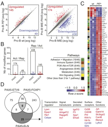
Gene Regulation by PAX5-ELN in Pro-B Cells. Based on transient
transfection assays, PAX5 fusion proteins including PAX5-ELN, PAX5-ETV6, and PAX5-FOXP1 were thought to act as dominant-negatives by affecting the transcriptional activity of WT PAX5 (11, 13, 16, 17). However, recent evidence in vivo revealed that PAX5-ETV6 or PAX5-FOXP1 fusion proteins marginally modified the expression of PAX5 target genes (15, 22). To address this hypothesis in our in vivo model, we first compared the gene expression profiles of purified pro-B cells from preleukemic PEtg mice and age-matched WT mice (SI Appendix, Fig. S8A). Transcriptome analysis identified 145 up-regulated and 49 down-up-regulated genes with an expression dif-ference of more than 1.5-fold and an adjusted P value of<0.05 in PAX5-ELN–expressing pro-B cells (Fig. 4A andDataset S2). In
parallel, we established a list of ex vivo PAX5-modified genes by comparing the gene expression profiles of Pax5−/− embryonic liver (E17.5) pro-B cells retrovirally transduced with either MIE or MIE-PAX5. This strategy led to the identification of 174 PAX5–up-regulated and 438 PAX5-repressed genes (Fig. 4A and Dataset S3). Interestingly, the gene set enrichment analysis of a well-established list of in vivo PAX5-regulated genes, arising from in vivo gene expression profiles of murine Pax5+/+ and Pax5−/−pro-B cells (22), with our new ex vivo PAX5 gene sig-nature demonstrates a global similarity between the two ap-proaches (SI Appendix, Fig. S8B). We determined the overlap of
−1.5 0.0 1.5
C
Row z-score Cd180 Il2rb Cebpe Ly6c2 Cybb Il2ra Lpxn Kdr Tgfbr3 Hip1 Cd38 Irs1 Prlr Ptgs1 Muc13 Rbms3 Pgf Nkd2 Rgs18 Trem1 Klrb1c Crhbp Rapgef3 Efna5 Sh3bgrl2 F5 Card6 Pde8a Tle6 Igsf10 Galnt14 Cd248 Spp1 Ly6d Arhgef12 Amotl1 Gimap3 Plk2 Fetub Asb10 Pcdh9 Htr1b Jyh Epas1 Sema6d Sema3g wt PEtgD
56 6 25 PAX5-ELN PAX5-ETV6 PAX5-FOXP1 75 243 8 7 Transcription regulator Signal transducers Secreted proteins Other functions Lpxn Epas1 Hip1 Cebpe Kdr Arhgef12 Rapgef3 Irs1 Igsf10 F5 Spp1 Efna5 Jyh Galnt14 Sh3bgrl2 Card6 Rbms3 Ly6c2 Surface receptors Cd38 Cd248 Prlr Klrb1c Htr1b Cd180 Il2rb 174 49 438 145 100 200 300 400 500 0 PAX5 1 1 Act / Rep Rep / Act 14 14 4 6 8 10 12 4 6 8 10 12 Upregulated 145 Downregulated 49 4 6 8 10 12 4 6 8 10 12Upregulated174 Downregulated 438 Pro-B PE tg (avg log)Pro-B wt(avg log) Pro-B MIE(avg log)
Pro-B MIE-P AX5 (avg log) Nb of modified genes PAX5-ELN PAX5 PAX5-ELN
A
B
Pathways Angiogenesis (6/46) Adhesion + Migration (19/46) PI3K-Akt-mTOR (8/46) Immune System (9/46) Metabolism (5/46) Other (less than 3 for 1 pathway) Wnt Signaling (3/46)Fig. 4. (A) Scatter plot of gene expression differences between in vivo
purified WT and PEtgpreleukemic pro-B cells (Left) and between ex vivo
E17.5 fetal liver Pax5−/−pro-B cells transduced with either MIE-PAX5 or MIE
retroviral vectors (Right) based on three independent microarray experi-ments. The normalized expression data of individual coding genes (indicated by dots) were plotted as the average log ratio (avg log). Up- and
down-regulated genes with an expression difference of>1.5- and >3-fold, and
an adjusted P value of<0.05, are colored in red or blue, respectively. (B)
Absence of a general dominant-negative effect of PAX5-ELN on ex vivo PAX5-regulated genes in pro-B cells. Comparison of PAX5-activated and
PAX5-ELN–repressed genes (Left) and of PAX5-repressed and
PAX5-ELN–ac-tivated genes (Right) in preleukemic pro-B cells. Overlap indicates that one gene was activated by PAX5 and repressed by PAX5-ELN and 14 genes of the PAX5-repressed genes were activated by PAX5-ELN as represented by
col-ored bars. (C) Heat map displaying the differential expression of PAX5-ELN–
activated (red) and–repressed (blue) genes in WT (n = 3) and PEtg(n= 3)
pro-B cells. The 46 PAX5-ELN–modified genes were selected on the basis of an
expression difference of>2-fold (P < 0.05) and for encoding a protein
im-plicated in one of the indicated pathways. The expression value of each gene is visualized according to the indicated scale. The pathway annotation is
shown (Left). (D) Venn diagram indicating the overlap between PAX5-ETV6–,
PAX5-FOXP1– (15), and PAX5-ELN–modified genes in preleukemic pro-B
cells, selected for an expression difference of>3-, 3-, and 2-fold,
respec-tively (Left). PAX5-ELN–modified genes that were included neither in
PAX5-ETV6 nor in PAX5-FOXP1 signatures are listed according to their biological functions as transcriptional regulators, signal transducers, secreting proteins,
and surface receptors. PAX5-ELN–activated and –repressed genes are
in-dicated in red and blue, respectively.
our ex vivo PAX5-activated genes with PAX5-ELN–repressed genes. This analysis revealed that only one gene was activated by PAX5 and repressed by PAX5-ELN. Conversely, only 14 of the PAX5-repressed genes were activated by PAX5-ELN in pro-B cells (Fig. 4B and Dataset S4A). Closely similar results were observed when we used the well-established list of in vivo PAX5-regulated genes (22) (SI Appendix, Fig. S8CandDataset S4B), further validating our approach. In addition, we observed by qPCR that ectopic expression of PAX5-ELN in preleukemic pro-B cells was not associated with the down-regulation of endoge-nous Pax5 and its two common target genes CD19 and CD79a (SI Appendix, Fig. S8D). Finally, we confirmed that the majority of the genes modified by PAX5-ETV6 and PAX5-FOXP1 in pro-B cells (15) did not overlap with our PAX5-regulated genes (SI Appendix, Fig. S8 E and FandDataset S4 C and D). Together, similar to PAX5-ETV6 and PAX5-FOXP1 (15), our results in-dicate that, in vivo, PAX5-ELN does not generally antagonize the normal functions of PAX5 in preleukemic pro-B cells.
To gain insight into the molecular regulation of PAX5-ELN in the early steps of B-ALL development, we focused on the genes presenting a twofold change of expression when comparing pre-leukemic PEtgwith WT pro-B cells. Forty-one activated and five
repressed PAX5-ELN genes were identified in preleukemic PEtg
pro-B cells (Fig. 4C andDataset S2). To identify shared molecular programs induced by ETV6, FOXP1, and PAX5-ELN, we overlapped the lists of their targets in preleukemic pro-B cells (15) (Fig. 4D). Notably, we found only six common genes, including genes encoding for a signal transducer (Gimap3), a secreted protein (Sema3g), and surface receptors (Il2rα and Trem1) (SI Appendix, Fig. S9A). In contrast, we identified 25 PAX5-ELN–modified genes, which were modified neither by PAX5-ETV6 nor by PAX5-FOXP1 (Fig. 4D). Interestingly, these genes encoded four signal transducers, four secreted proteins, and seven surface receptors but also the four transcriptional regulators Lpxn, Epas1, Hip1, and Cebpe (Fig. 4D).
The above results suggest that PAX5-ELN predominantly regulates an independent molecular program in preleukemic pro-B cells. To provide additional support to this notion, we categorized PAX5-ELN–modified genes according to pathways and identified activated (act) and repressed (rep) genes coding for proteins involved in the following processes: adhesion and migration (19 act), immune system (5 act, 4 rep), PI3K-mTOR signaling (7 act, 1 rep), angiogenesis (6 act), metabolism (5 act), and Wnt signaling (3 act) (Fig. 4C). Most of these pathways are strongly deregulated in PAX5-ETV6–induced B-ALL cells compared with their preleukemic counterparts (15), suggesting that PAX5-ELN activates at the preleukemic stage a molecular program required for B-ALL development in PAX5-ETV6tg mice. Interestingly, we identified six genes (Epas1, Arhgef12, Fetub, Rapgef3, Galnt14, and Cd248) activated by PAX5-ELN in preleukemic pro-B cells that were shown to be specifically acti-vated in B-ALL PAX5-ETV6 cells (SI Appendix, Fig. S9B).
In conclusion, we have identified regulated PAX5-ELN genes in preleukemic pro-B cells with important functions in several signaling pathways and gene candidates that establish the mo-lecular bases to drive B-ALL development.
Discussion
Genome-wide profiling identified the PAX5 gene as the most frequent target of somatic mutation in human B-ALL (11). Nonetheless, while PAX5 acts as a main tumor suppressor gene in B-ALL, B-cell development is normal in heterozygous Pax5+/− mice (5, 23), unless primed by chemical or retroviral mutagenesis (24), suggesting oncogenic cooperation for B-ALL trans-formation. Indeed, the tumor suppressor functions of Pax5 in B leukemogenesis were revealed under constitutive activation of STAT5, JAK1, and JAK3 (24, 25). PAX5 is also involved in re-ciprocal translocations leading to the fusion of its N-terminal
domain with the C-terminal sequence of a second transcription factor such as ETV6 and FOXP1 (11, 26). In contrast with heterozygous PAX5 deletions, which are considered to have a secondary role during late stages of leukemogenesis, it is gen-erally assumed that PAX5 fusion proteins act as primary onco-genic events altering normal B-cell development in the early steps of the disease (14). A wide diversity of fusion partners has been described (9, 14). Remarkably, they all conserve the N-terminal DNA-binding region of PAX5 and the NLS but lack the potent C-terminal transcriptional regulatory domains, sug-gesting that the PAX5 part of the chimeric protein is required for leukemogenesis.
The loss of PAX5 transcriptional domains in the chimeric proteins led to the notion that the fusion proteins might act as constitutive repressors, as shown in various reporter assays (11, 13, 16, 27). Consequently, it was inferred that B-ALL might arise as a result of an antagonizing effect of the fusion proteins on PAX5 activity. The situation may be more complex, as illustrated by the recent finding that PAX5-ETV6 and PAX5-FOXP1 do not actually modify the expression of PAX5 target genes but rather implicate pre-B cell receptor, migration, and adhesion signaling pathways (15). PAX5-ELN rearrangement is unusual because it does not involve a transcription factor but an extra-cellular matrix protein (13). Furthermore, PAX5-ELN is one of the chimeric proteins that conserves the largest part of PAX5. Although PAX5-ELN blocked the transcription of PAX5 target genes in transactivation assays (13), our results indicate that PAX5-ELN was able to repress only one of the PAX5-activated genes and to activate a small subset of the PAX5-repressed genes, strongly suggesting that PAX5-ELN does not generally antagonize the normal function of PAX5 in preleukemic pro-B cells. Thus, in line with PAX5-ETV6 and PAX5-FOXP1 models (15), our in vivo data do not support the generality of a dominant-negative mechanism for PAX5 fusion proteins.
Several hypotheses could be put forward to account for this discrepancy. One possible explanation could be merely quantita-tive. In heterozygous mice, thus mimicking the situation in pa-tients, the concentration of the fusion protein may be below the threshold required to exert a dominant-negative effect, whereas this threshold may be readily achieved in transient reporter assays. As mentioned above, expression of a small subset of PAX5-regulated genes was impacted by PAX5-ELN, suggesting that there are instances in vivo where the dominant-negative effect is exerted by the oncoprotein. Alternatively, though not mutually exclusively, fusion proteins may be subjected to different regu-latory mechanisms (half-life, posttranslational modifications, interacting partners, etc.) in vivo from the time lapse of transient expression. In this context, it should be noted that although the expression of PAX5-ELN cDNA was driven by the powerful IgH regulatory elements, PAX5-ELN levels were not higher than endogenous PAX5 levels, excluding an inappropriate ectopic expression of the fusion protein in our model. Importantly, al-though the insertion of Pax5 into the IgH locus led to T lym-phomas (28), the weak expression of PAX5-ELN in thymocytes did not perturb T-cell development, underlying the restricted effect of PAX5-ELN expression on the B-cell lineage.
Interestingly, while PAX5-ETV6 and PAX5-FOXP1 arrested lymphopoiesis at the pro-B/pre-B cell transition, they did not induce leukemia on their own in the corresponding mutant mice (15), suggesting the requirement of cooperating mutations for B-ALL transformation. Specifically, in contrast to the PAX5-ETV6 mouse model that required oncogenic cooperation (i.e., loss of the Cdkn2a/b tumor suppressor locus) for leukemia develop-ment (15), PAX5-ELN efficiently induced B-ALL in mice. In this context, most deregulated pathways in preleukemic PAX5-ELN pro-B cells were also strongly deregulated in PAX5-ETV6– induced B-ALL cells. Further analysis revealed that PAX5-ELN predominantly regulated an independent molecular
CELL
BIO
program in preleukemic pro-B cells, in which activation of six genes, also found activated in B-ALL PAX5-ETV6 cells (15), may explain the stronger potential of PAX5-ELN to induce B-ALL compared with PAX5-ETV6.
B cell-specific, constitutive expression of PAX5-ELN led to clonal transformation associated with the acquisition of additional mutations in key components of the JAK/STAT and RAS/MAPK pathways. These pathways represent two common targets of so-matic mutations in other oncogene-induced B-ALL mice (29, 30) and in human B-ALL (11), including PAX5-ELN B-ALL cases (31, 32). Our analysis of a B-ALL patient cohort revealed the presence of PAX5, PTPN11, NRAS, KRAS, and JAK3 mutations within different B-ALL oncogenic subtypes including PAX5-rearranged leukemias and a PAX5-ELN B-ALL case.
This study clearly demonstrated the effect of PAX5-ELN on leukemia initiation at the preleukemic stage, before malignant transformation. The partial blockade of differentiation induced by PAX5-ELN was associated with an aberrant expansion of pro-B cells, which was also revealed by transplantation assays. Im-portantly, this proliferative advantage was not associated with an obvious clonal selection and therefore suggests that PAX5-ELN confers self-renewal properties to pro-B cells, a situation that is reminiscent of the E2A-PBX1 mouse model (30). This obser-vation supports the view that aberrant self-renewal activity of lymphoid progenitors induced by a primary oncogene can be an initiating event in leukemia development. This establishes a preleukemic stage setting genetic instability and accumulated genetic alterations that cooperate to lead to fully transformed B-ALL (33). Emerging evidence suggests that primary genetic alteration can convert normal committed progenitors into pre-leukemic stem cells by reprogramming aberrant self-renewal
properties both in lymphoid and myeloid lineages (33–35). Im-portantly, recent findings proposed that long-lasting preleukemic stem cells were hidden within the bulk of leukemic cells, and served as a reservoir for disease relapse (36, 37). An important challenge is to identify and target these leukemic initiating cells that represent an extremely rare subpopulation in patients. In this respect, the availability of the PAX5-ELN mouse model should be valuable. Additionally, by recapitulating the different steps of the human disease, the model represents a major op-portunity to develop new therapeutic strategies on both B-ALL initiation and transformation, and a robust and reproducible tool for preclinical studies of drug screening and development. Experimental Procedures
Generation of the PAX5-ELN knockin mouse model, FACS analysis, Ig rear-rangement assay, whole-exome sequencing and specific resequencing, transplantation assay, microarray experiment, retroviral transduction, co-culture of pro-B cells, RT-PCR, Western blot, and statistical analysis are
de-scribed inSI Appendix.
ACKNOWLEDGMENTS. We acknowledge the cytometry and cell-sorting facility of the CRCT (INSERM U1037) and the IPBS animal facility for technical assistance. We are grateful to Manon Farcé and Laetitia Ligat from the facilities of the CRCT for assistance with flow cytometry and microscopy, respectively. We thank the Anexplo/Genotoul platforms for technical assis-tance (UMS006 and histology department). This research was supported by Institut National du Cancer (INCa) R11190BB-INCA 2011-131-PAX5, ARC Grant SFI20101201888, and FEDER Grant CITTIL. G.C., N.R., C.C., and N.S.N.H. were supported by INCa Grant R11190BB-INCA 2011-131-PAX5. The team is supported by association Laurette FUGAIN, Ligue nationale contre le
cancer,“111 des arts,” Association Capucine, Société Française des Cancers
de l’Enfant, and la région Occitanie. Work in the A.A.K. lab is supported by
INCa, ANR, and Fondation ARC.
1. Pui CH, Carroll WL, Meshinchi S, Arceci RJ (2011) Biology, risk stratification, and therapy of pediatric acute leukemias: An update. J Clin Oncol 29:551–565. 2. Armstrong SA, Look AT (2005) Molecular genetics of acute lymphoblastic leukemia.
J Clin Oncol 23:6306–6315.
3. Hardy RR, et al. (2000) B-cell commitment, development and selection. Immunol Rev 175:23–32.
4. Adams B, et al. (1992) Pax-5 encodes the transcription factor BSAP and is expressed in B lymphocytes, the developing CNS, and adult testis. Genes Dev 6:1589–1607. 5. Urbánek P, Wang ZQ, Fetka I, Wagner EF, Busslinger M (1994) Complete block of early
B cell differentiation and altered patterning of the posterior midbrain in mice lacking Pax5/BSAP. Cell 79:901–912.
6. Souabni A, Cobaleda C, Schebesta M, Busslinger M (2002) Pax5 promotes B lympho-poiesis and blocks T cell development by repressing Notch1. Immunity 17:781–793. 7. Nera KP, et al. (2006) Loss of Pax5 promotes plasma cell differentiation. Immunity 24:
283–293.
8. Nutt SL, Heavey B, Rolink AG, Busslinger M (2015) Pillars article: Commitment to the B-lymphoid lineage depends on the transcription factor Pax5. Nature. 1999. 401: 556– 562. J Immunol 195:766–772.
9. Nebral K, et al. (2009) Incidence and diversity of PAX5 fusion genes in childhood acute lymphoblastic leukemia. Leukemia 23:134–143.
10. Familiades J, et al. (2009) PAX5 mutations occur frequently in adult B-cell progenitor acute lymphoblastic leukemia and PAX5 haploinsufficiency is associated with BCR-ABL1 and TCF3-PBX1 fusion genes: A GRAALL study. Leukemia 23:1989–1998. 11. Mullighan CG, et al. (2007) Genome-wide analysis of genetic alterations in acute
lymphoblastic leukaemia. Nature 446:758–764.
12. Medvedovic J, Ebert A, Tagoh H, Busslinger M (2011) Pax5: A master regulator of B cell development and leukemogenesis. Adv Immunol 111:179–206.
13. Bousquet M, et al. (2007) A novel PAX5-ELN fusion protein identified in B-cell acute lymphoblastic leukemia acts as a dominant negative on wild-type PAX5. Blood 109: 3417–3423.
14. Coyaud E, et al. (2010) Wide diversity of PAX5 alterations in B-ALL: A Groupe Francophone de Cytogenetique Hematologique study. Blood 115:3089–3097. 15. Smeenk L, et al. (2017) Molecular role of the PAX5-ETV6 oncoprotein in promoting
B-cell acute lymphoblastic leukemia. EMBO J 36:718–735.
16. Kurahashi S, et al. (2011) PAX5-PML acts as a dual dominant-negative form of both PAX5 and PML. Oncogene 30:1822–1830.
17. Kawamata N, Pennella MA, Woo JL, Berk AJ, Koeffler HP (2012) Dominant-negative mechanism of leukemogenic PAX5 fusions. Oncogene 31:966–977.
18. Kemp DJ, Harris AW, Cory S, Adams JM (1980) Expression of the immunoglobulin C mu gene in mouse T and B lymphoid and myeloid cell lines. Proc Natl Acad Sci USA 77: 2876–2880.
19. Haddad D, et al. (2011) Sense transcription through the S region is essential for im-munoglobulin class switch recombination. EMBO J 30:1608–1620.
20. Jung D, Giallourakis C, Mostoslavsky R, Alt FW (2006) Mechanism and control of V(D)J recombination at the immunoglobulin heavy chain locus. Annu Rev Immunol 24: 541–570.
21. Braikia FZ, Chemin G, Moutahir M, Khamlichi AA (2014) Quantification of V(D)J re-combination by real-time quantitative PCR. Immunol Lett 162:119–123.
22. Revilla-I-Domingo R, et al. (2012) The B-cell identity factor Pax5 regulates distinct transcriptional programmes in early and late B lymphopoiesis. EMBO J 31:3130–3146. 23. Nutt SL, Heavey B, Rolink AG, Busslinger M (1999) Commitment to the B-lymphoid
lineage depends on the transcription factor Pax5. Nature 401:556–562.
24. Dang J, et al. (2015) PAX5 is a tumor suppressor in mouse mutagenesis models of acute lymphoblastic leukemia. Blood 125:3609–3617.
25. Heltemes-Harris LM, et al. (2011) Ebf1 or Pax5 haploinsufficiency synergizes with STAT5 activation to initiate acute lymphoblastic leukemia. J Exp Med 208:1135–1149. 26. Cazzaniga G, et al. (2001) The paired box domain gene PAX5 is fused to ETV6/TEL in
an acute lymphoblastic leukemia case. Cancer Res 61:4666–4670.
27. Fazio G, et al. (2013) PAX5/ETV6 alters the gene expression profile of precursor B cells with opposite dominant effect on endogenous PAX5. Leukemia 27:992–995. 28. Souabni A, Jochum W, Busslinger M (2007) Oncogenic role of Pax5 in the T-lymphoid
lineage upon ectopic expression from the immunoglobulin heavy-chain locus. Blood 109:281–289.
29. van der Weyden L, et al. (2015) Somatic drivers of B-ALL in a model of ETV6-RUNX1; Pax5(+/−) leukemia. BMC Cancer 15:585.
30. Duque-Afonso J, et al. (2015) Comparative genomics reveals multistep pathogenesis of E2A-PBX1 acute lymphoblastic leukemia. J Clin Invest 125:3667–3680.
31. Mullighan CG, et al. (2009) JAK mutations in high-risk childhood acute lymphoblastic leukemia. Proc Natl Acad Sci USA 106:9414–9418.
32. Denk D, Bradtke J, König M, Strehl S (2014) PAX5 fusion genes in t(7;9)(q11.2;p13) leukemia: A case report and review of the literature. Mol Cytogenet 7:13. 33. Hong D, et al. (2008) Initiating and cancer-propagating cells in TEL-AML1-associated
childhood leukemia. Science 319:336–339.
34. Gerby B, et al. (2014) SCL, LMO1 and Notch1 reprogram thymocytes into self-renewing cells. PLoS Genet 10:e1004768.
35. Krivtsov AV, et al. (2006) Transformation from committed progenitor to leukaemia stem cell initiated by MLL-AF9. Nature 442:818–822.
36. Shlush LI, et al.; HALT Pan-Leukemia Gene Panel Consortium (2014) Identification of pre-leukaemic haematopoietic stem cells in acute leukaemia. Nature 506:328–333, and erratum (2014) 508:420.
37. Mullighan CG, et al. (2008) Genomic analysis of the clonal origins of relapsed acute lymphoblastic leukemia. Science 322:1377–1380.