Application of In Vitro Erythropoiesis from Bone Marrow Derived
Progenitors to Detect and Study Genotoxicity
by Joseph F. Shuga B.S. Chemical Engineering The University of Texas at Austin, 2001
SUBMITTED TO THE DEPARTMENT OF CHEMICAL ENGINEERING IN PARTIAL
FULFILLMENT OF THE REQUIREMENTS FOR THE DEGREE OF
DOCTOR OF PHILOSOPHY
AT THE
MASSACHUSETTS INSTITUTE OF TECHNOLOGY
JUNE 2007
© 2007 Massachusetts Institute of Technology All rights reserved
A
Signature of. Certified by: Certified by: Accepted by: OF TECHNOLOGJUN 112001
LIBRARIE
Author:Ae-uthor:ýnlf Chemical Engineering
May
25,
2007
Linda G. Griffith Professor of Biological and Mechanical Engineering
i , Thesis Supervisor
S Leona D. Samson Professor of Biology and Biological Engineering Thesis Supervisor
E
S
William M. Deen Professor of Chemical Engineering Chairman, Committee for Graduate Students
Application of In Vitro Erythropoiesis from Bone Marrow Derived
Progenitors to Detect and Study Genotoxicity
by
Joseph F. Shuga
Submitted to the Department of Chemical Engineering
on May
25,
2007 in Partial Fulfillment of the
Requirements for the Degree of Doctor of Philosophy in
Chemical Engineering
ABSTRACT
Assays that predict toxicity are an essential part of drug development and there is a demand for
efficient models to better predict human responses. The in vivo micronucleus (MN) assay is a
robust toxicity test that assesses the genotoxic effect of drugs on adult bone marrow (BM) using
the metric of genotoxic damage to the reticulocyte population in mice. An in vitro correlate to
this assay might facilitate extension to human cells and thus provide a highly predictive
genotoxicity assay. As first steps in developing a toxicity assay, this thesis work (a) adapted a
fetal liver-based in vitro erythropoietic culture system to induce optimized erythropoietic growth
from the lineage-marker-negative (Lin) population in adult BM, as adult hematopoietic tissue is
ultimately a feasible source of cells; and (b) demonstrated that exposure to alkylating agents
induces physiological MN-formation in erythroid populations derived in vitro. The potential for
increased efficiency in this in vitro model depends on the ability to stimulate terminal erythroid
differentiation at an optimal level from adult BM. With this goal in mind, this thesis work
employed experimental design strategies, erythroid-specific growth measurements, and
multi-linear regression to model erythropoietic growth in this system and thus estimate the relative
sensitivity of Lin7 BM to erythropoietic growth parameters, including Erythropoietin, Stem Cell
Factor, p02, and Fibronectin, among others. From these erythroid-specific growth
measurements, it is estimated that >1500 MN assays can be conducted using the BM of a single
mouse. This throughput represents a significant improvement over the current in vivo test, which
assays a single condition per mouse. This thesis work then quantified the genotoxic response to
three alkylating agents (1,3-bis(2-chloroethyl)- 1-nitrosourea [BCNU], N-methyl-N
-nitro-N-nitrosoguanidine [MNNG], and methylmethane sulfonate [MMS]) in this culture system and
detected a significant cytotoxic response and concomitant increase in MN incidence in
reticulocytes. This increase in MN frequency provides a clear signal of the genotoxic events that
likely lead to global toxicity, and thus mimics the physiological hematopoietic response to
alkylating chemotherapeutics. In addition, this thesis work determined that DNA repair-deficient
(MGMT"
-) BM displayed sensitivity to genotoxic exposure in vivo compared with wild-type
(WT) BM, and that this phenotypic response was reflected in erythropoietic cultures. These
findings suggest that this in vitro erythroid MN assay is capable of screening for genotoxicity on
BM in a physiologically reflective manner. Finally, responses to genotoxicants during erythroid
differentiation varied with exposure time, facilitating the study of genotoxic effects at specific
developmental stages.
Thesis Supervisor: Linda G. Griffith
Title: Professor of Biological and Mechanical Engineering
Thesis Supervisor: Leona D. Samson
ACKNOWLEDGEMENTS
I must first thank Zoe Lillian Shuga, my beautiful, sweet, smart, and feisty three-month-old daughter. Zoe, you have changed my perspective forever and will continue to do so every day; much more of this world makes sense to me now that I've become a parent: thank you for that, Zoe. Mostly, though, thanks for providing me with a clear purpose in this crazy world. Then I must thank Laura, Zoe's mother and the love of my life. Come to think of it, you also changed my perspective forever, and continue to do so, but after nearly eight years together, I should just say that you have changed my life forever. Without your love and support I could not be happy; I might have stubbornly finished this degree, but not as a truly happy person. Thanks for your continued tolerance and understanding as I clumsily pursue my dreams. Next, I would like to thank my parents, Wunda and Tony Shuga, who first gave me life and then gave me the love, support, education, and challenges that molded me into what I have become. You sacrificed a lot for me, and I can now truly begin to appreciate it (thanks again, Z!). I would also like to thank my brother Doug, who I can always count on, and who appreciates my sense of humor and worldview more than any other. I have many grandparents, aunts, uncles, and cousins who have all influenced me greatly: thanks to Beryl, Joe, Mike, Leslie, Liz, Paul, Dean L., Marsha, Steve Jr., Nate (Weaver), Melissa, Andrew, Meaghan, Will, and Nate (Johnson) for comprising a great family. I also thank the best in-laws in the world, Martin, Cari, and Katie: I'm so glad to call you family. I would also like to thank my grandfather Dean Sr. and my uncle Steve Sr., both great men who left this world while I was studying at MIT: you are both deeply missed. Lastly, I would like to specially thank my maternal grandmother, Olive. You always believed in me, more than any other (and somewhat irrationally, I must say... you have such faith!). Thank you, Grandma, for your unbridled optimism, your great expectations, and your striking example. Next I would like to thank the many people that have taught, supported, and collaborated with me in academics. Thanks to my advisors, Linda, Harvey, and Leona: you all provided me with excellent advice and constant support for the last four years, thanks for your time and effort, and thanks for not giving up on me when things were tough. I'd like to also thank David Journeay and Sean Dennis, my excellent high school physiology and chemistry teachers, respectively, who laid the foundations for continued interest in those areas. I'd like to thank Prof. Christine
Schmidt, for luring me away from oil and specialty chemicals with a very interesting Tissue Engineering course and undergraduate research opportunity. I'd also like to thank Drs. Dharini Shah, Jing Zhang, Lisi Meira, and Chengcheng (Alec) Zhang for many helpful discussion regarding hematopoiesis and genotoxicity. I must thank Wes Overton, Sophia Kamran, and Catherine Moroski for technical assistance, and Glenn Paradis for expert technical assistance in flow cytometry studies; I also thank the Cambridge-MIT institute for financial support. Finally, I thank Fred Pearce, Shawdee Eshghi, Neil Kumar, Mauro Brigante, Alejandro Dominguez-Garcia, Eric Verploegen, and Shannan O'Shaughnessy: you are all more friends than collaborators, but it was discussions with you, as we all completed the PhD together, which helped me to preserve sanity and continue on in science. Of course, the fun times were great, too... long live 647. I also thank my oldest friends: thanks to Jeff Dunahoo, Ben Crawford, Matt McBrearty, and Michael Sladecek for staying close to me despite the 1692 miles and many major life events that separated us. Long live 23. I'd also like to thank Leo Crawford, Chiquita Crawford-Sladecek, Sayde Humphreys, and Mikey, Cody and Maggie Shuga for all the purrs, tail-wags, and
unconditional love. I also thank Scout McBrearty, who recently left this world after a quick bout with cancer; you were a great friend, and I hope that you have found many a rock to retrieve in the great beyond. Finally, I must thank Itai Suez, a great friend who might be the most generous and happy person that I know; Pradeep Penta, a considerate and special friend who has always been an inspiration to me; and Ashley Johnson, who brightens the lives of everyone around her.
TABLE OF CONTENTS Title Page Abstract Acknowledgements Table of Contents List of Figures List of Tables Chapter One: 1.1 1.2 1.3 1.4 1.5 1.6 Introduction General Introduction Motivation
Preclinical Genotoxicity Screens
in vitro Erythropoiesis
Model Genotoxicant: BCNU (Carmustine)
Effects of BCNU: Reactivity and Pharmacokinetics
Chapter Two: Identification of a suitable hematopoietic starting population and optimization of erythropoietic growth for increased assay throughput
2.1 Introduction 27
2.2 Results 31
2.2.1 Stimulating and Tracking Erythropoiesis in Erythroid Progenitors 31 2.2.2 The PCE Yield from Lin- BM Depends on Multiple 33
Environmental Cues
2.2.3 Multi-Linear Regression to Model the Late-Erythropoietic 37 Response
2.2.4 Differences Between Epo-only and Improved Culture 40
2.3 Discussion 43
2.4 Methods 45
2.4.1 Cells 45
2.4.2 Erythropoietic Culture 45
2.4.3 Immunostaining and Flow Cytometry to Analyze Erythroid 46 Differentiation
2.4.4 Cytospin Preparation and Cytological Staining 46 2.4.5 Viable Cell Counting, Histological Imaging and Quantification 46
2.4.6 Methylcellulose Colony Assays 47
2.4.7 Statistics, Design of Experiments, and Multi-Linear Regression 47 Chapter Three:
3.1 3.2
3.3 3.4
Genotoxic responses during erythropoiesis: detection of model genotoxic agents and study of DNA repair's effect on erythroid micronucleus formation
Introduction 49
Results 51
3.2.1 Stimulation of Terminal Erythropoiesis in Lin- BM 51 3.2.2 Detection of Genotoxic Exposure: in vitro Erythroid MN Assay 54 3.2.3 MGMT-' Mice Display Sensitivity to BCNU Exposure by the in 56
vivo MN assay
3.2.4 in vitro Erythroid MN Assay Reflects the in vivo Phenotype of 59 MGMT-/- Mice
Discussion 63
Materials and Methods 3.4.1 Cells
3.4.2 Erythropoietic Culture
3.4.3 Immunostaining and Flow Cytometry
3.4.4 Cytospin Preparation and Cytological Staining
3.4.5 in vivo Genotoxic Treatment and Cytology
3.4.6 Genotoxic Treatment of Erythropoietic Cultures 3.4.7 Viable Cell Counting
3.4.8 Histological Imaging and Quantification 3.4.9 Cell Cycle Analysis
3.4.10 Statistics Chapter Four:
4.1 4.2 4.3
Conclusions and Future Work Conclusions
Extension to Human Tissue
Automated High-Throughput Analysis Appendix I: MATLAB@ Script for Multi-Linear Regression Appendix II: References
LIST OF FIGURES CHAPTER ONE
Figure 1-1. 10
Hematopoiesis
Figure 1-2. 18
Erythropoiesis tracked by flow cytometry
Figure 1-3. 21
Reactive species generated by HENUs in aqueous solution CHAPTER TWO
Figure 2-1. 32
Terminal erythropoiesis in Lin- BM cells over three days in culture
Figure 2-2. 34
Effect of [Epo] on erythroid cell yield from Lin- and Ter- 119- BM
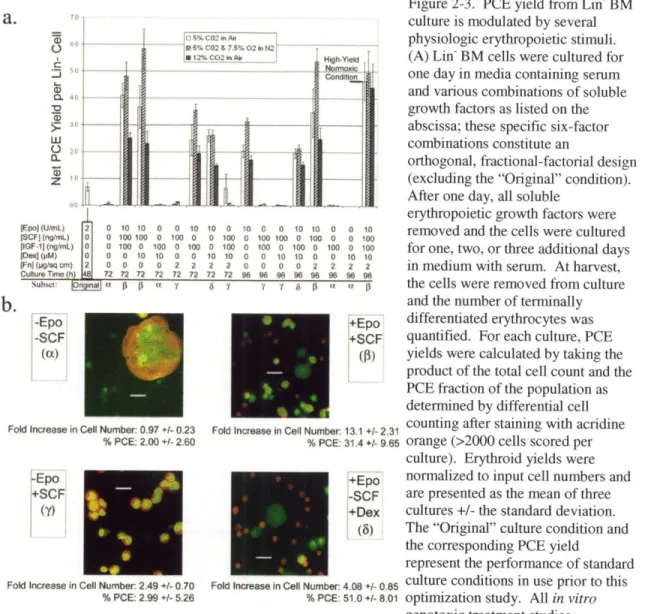
Figure 2-3. 36
PCE yield from Lin- BM culture is modulated by several physiologic erythropoietic stimuli
Figure 2-4. 41
Multi-linear regression to predict PCE yield and quantify relative parameter
effects
Figure 2-5. 42
Growth dynamics during improved and Epo-only erythropoietic cultures
Figure 2-6. 43
Erythropoietic populations culture under improved conditions contain CFU-Es well beyond the point at which Epo is withdrawn (24h)
CHAPTER THREE
Figure 3-1. 52
Terminal erythropoiesis stimulated in Lin- BM cells over three days in culture
Figure 3-2. 53
Dynamic analysis of Lin- bone marrow cultured for three days under improved erythropoietic conditions: flow cytometry and benzidine-Giemsa staining
Figure 3-3. 54
Growth and cell cycle dynamics during improved erythropoietic culture
Figure 3-4. 55
in vitro erythropoiesis continues after treatment with alkylating agents: flow
Figure 3-5.
Detection of genotoxicity through in vitro erythropoiesis
Figure 3-6. 57
MGMT - mice exhibit sensitivity to MN formation following in vivo exposure to BCNU: dynamic quantification of the frequency of PCEs and micronucleated PCEs in BM
Figure 3-7. 58
Aag expression has no significant effect on erythroid MN formation following
in vivo treatment with BCNU
Figure 3-8. 59
Dynamic analysis of MGMT - Lin- bone marrow cultured for three days under improved erythropoietic conditions: flow cytometry and benzidine-Giemsa staining
Figure 3-9. 60
Similar to in vivo responses, Lin- BM from MGMT-' mice exhibits sensitivity to MN-formation and decreased PCE yields when treated with BCNU during erythropoietic culture
Figure 3-10. 61
Early BCNU treatment of WT Lin- BM cultures leads to decreased viable cell numbers and diminished MN formation
Figure 3-11. 62
WT Lin- BM, differentiating in vitro, shows evidence of altered cell cycling following exposure to BCNU
CHAPTER FOUR
Figure 4-1. 71
Model of erythropoietic growth and genotoxic responses
Figure 4-2. 80
Representative images of PCEs identified by LSC
Figure 4-3. 80
LIST OF TABLES CHAPTER TWO
Table 2-1. 35
Flow cytometry shows that erythroid cell yield in vitro is modulated by physiologic erythropoietic stimuli
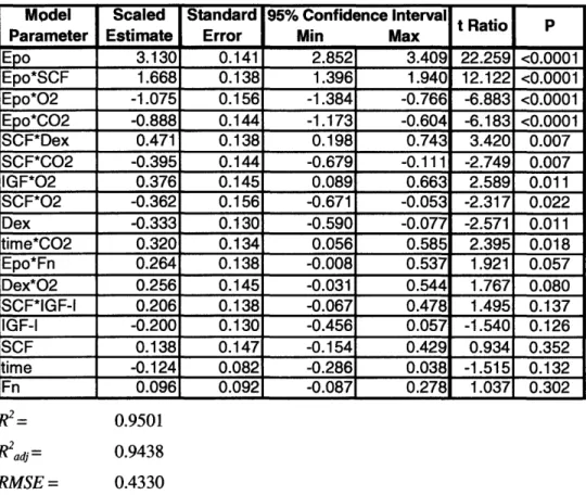
Table 2-2. 39
Parameter estimates derived from the complete model, including all primary and estimable secondary interaction parameters
Table 2-3. 40
Chapter One
1.1 GENERAL INTRODUCTION
This thesis describes the application of blood cell growth (hematopoiesis), and specifically red blood cell growth (erythropoiesis), to test for and study genotoxicity. It is fitting that the study of genotoxicity and hematopoiesis are joined in this thesis, as the field of hematopoiesis has long depended on genotoxic stresses (normally delivered in the form of radiation) to perturb and prime the hematopoietic compartment for donor-derived hematopoiesis. In what is largely considered to be the first report on hematopoietic repopulating activity, Lorenz et al. (1951) rescued lethally-irradiated mice by injecting bone marrow cells from a syngenic donor (1). Then, in 1956, Ford et
al. used repopulation after irradiation to demonstrate, based on karyotype differences between
donor and recipient cells, that animals protected using bone marrow transplants are hematopoietic chimeras, proving that long-term hematopoietic growth is directly derived from donor marrow
(2). Finally, in 1961, Till and McCulloch again used irradiation and repopulation to describe the multi-lineage spleen colony formation that has since been associated with the pluripotent
repopulating activity of hematopoietic stem cells (HSCs) (3). In the decades that followed, HSC research (founded on radiation-based studies) has made HSCs the most clearly-defined and understood adult stem cells; and bone marrow transplantation has become the most
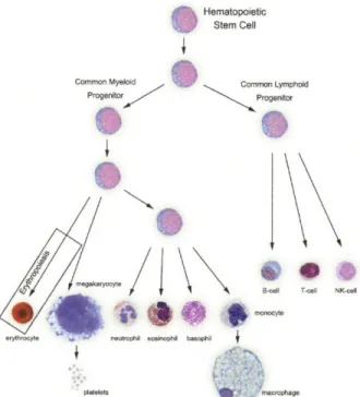
well-established and effective form of stem cell therapy. Today, the complete differentiation hierarchy of hematopoiesis, starting with the HSC, which is contained in the bone marrow at a frequency of approximately one in 105 cells, has been described (summarized in Fig. 1-1).
Hematopoietic
Stem Cell
Common Myeloid Common Lymphoid
Progenitor Progentor
iN
B-nll T-Cell NK-cell
platelets m ophage
Figure 1-1. Hematopoiesis. All the blood cell lineages in the body are derived from a single stem cell precursor (the HSC). The work in this thesis focused on the erythropoietic branch of hematopoiesis (far left). Image adapted from http://daley.med.harvard.edulassets/Willy/Willynoframes.htm.
The reason that genotoxic insult, delivered via radiation, has proven useful for perturbing normal hematopoiesis is that the hematopoietic compartment, and HSCs in particular, are sensitive to genotoxic damage. In typical HSC repopulation experiments, a rodent is given a lethal dose of radiation, and is then rescued with a bone marrow transplant. In the absence of rescue by HSC transplant, the animal succumbs to anemia and cytopenia less than a week after irradiation as terminally differentiated blood cells are lost and the compromised stem cells and progenitors in the marrow fail to produce sufficient replacement cells. Part of the reason that the hematopoietic tissue is intolerant of DNA damage is that this tissue is one of the most rapidly dividing tissues in the body, with approximately 10" blood cells formed each day in the adult human. Radiotherapy and chemotherapy are effective at treating cancers because cancerous cells divide rapidly, but the rapidly dividing bone marrow is also sensitive to these types of genotoxic damage, and
hematopoietic side effects are common.
The erythropoietic (red cell) lineage in the hematopoietic compartment is particularly useful for detecting certain types of genotoxic damage. After the final erythropoietic cell division,
erythroblasts extrude their nucleus to form enucleated reticulocytes (nascent red blood cells). If sufficient damage to erythroid progenitors has occurred prior to enucleation, then a small
"micronucleus," will be left behind after enucleation. These micronuclei are composed of broken DNA and entire chromosomes that have been excluded from the daughter nucleus due to
excessive DNA damage, and they are easily detected and quantified in reticulocytes because the remainder of the daughter nucleus has been extruded. That is, sub-2n particles left behind in reticulocytes are clearly detected because no other DNA is present to confound the signal provided by these particles. A mouse-based assay, based on this phenomenon, is a well-established part of modern drug development. This test, known as the in vivo micronucleus assay, is one of the most robust genotoxicity assays available (4-6).
1.2 MOTIVATION
Toxicity assays are an essential part of modem drug development. The therapeutic benefit of a new drug may be eclipsed by its toxic side-effects, and both the patient and the manufacturer are protected by mandatory screens in model systems. The cost of these tests is significant;
accordingly, there is a demand for more efficient toxicity tests to screen new drugs during early research and development. However, the most crucial quality of a toxicity test is its predictive ability. Therefore, modifications that increase efficiency should not decrease the sensitivity or relevance of the assay. The overarching goal of this thesis was to develop and characterize a
culture system that stimulates erythroid differentiation and micronucleus formation in vitro, using adult hematopoietic tissue, to provide an optimized corollary to the in vivo micronucleus (MN) genotoxicity assay. Further, this thesis aimed to apply this novel system to study the basic biology of the DNA damage-response in erythropoietic micronucleus formation and genotoxicity. Greater efficiency, compared with the in vivo assay, is possible because the bone marrow (BM) is harvested prior to treatment and used to initiate multiple erythroid-differentiation cultures, each of which then serves as a model for the in vivo erythropoietic system. In comparison to whole-animal dosage, this system provides a high-throughput system that might be further extended to conduct preclinical tests on human primary hematopoietic tissue.
Potential therapeutic agents are screened for toxic and carcinogenic effects before initiating clinical trials. This initial risk-assessment through toxicity tests is an essential part of modern drug development, and this step both protects patients and provides early data regarding the agent's biological effect. Studies in animals and in vitro are required by regulatory agencies around the globe to assess possible hazards prior to human exposure (7). However, there is a motive to apply toxicity screens early, to provide warning that a promising lead might test positive in a key regulatory test. Positive results in early toxicity screens identify problems that could ultimately lead to failure in clinical trials. Further, there is an interest in conducting
preclinical toxicity tests on primary human tissue, based on the premise that in vitro human might be closer than in vivo mouse to in vivo human. However, for this promise to be fulfilled, more in
vitro toxicity screens based on primary mammalian tissue must first be established.
1.3 PRECLINICAL GENOTOXICITY SCREENS
Key assays that hold a prominent place in regulatory considerations and genetic toxicology include the Ames test in Salmonella, the cytokinesis-block MN (CBMN) assay in human
lymphocytes, the analysis of chromosome aberrations in Chinese hamster ovary (CHO) cells, and the in vivo MN assay in rodent BM (7). The Ames assay screens mutagens using Salmonella strains that can only form colonies if mutation converts the strain from histidine-dependency back to prototrophy (8, 9). The Ames test is extremely well characterized, and liver microsomes can be added to the assay to screen for promutagens (latent mutagens that require metabolism by mammalian-specific pathways). The CBMN assay employs cytochalasin-B (Cyt-B) in vitro to interrupt cell division after telophase (10, 11). Cyt-B allows cells that have undergone a cell division to be identified by their binucleate appearance, and the presence of a nuclear body excluded from the daughter nuclei clearly indicates prior DNA damage. The CBMN test is
typically applied to cultured human lymphocytes or mammalian cell lines. Another common cytogenetic technique consists of exposing cells to a test agent during the DNA synthesis (S) phase of the cell cycle and then scoring chromosome aberrations during mitosis (7, 12). CHO cells have a stable, well-defined karyotype, a low number of large chromosomes, and a short cell cycle, making them ideal for visualization of chromosome aberrations in this last test.
Although each of these assays is capable of detecting some genotoxic agents, none of them is completely analogous to normal mammalian cellular biology. Mammalian cells and bacterial cells, such as those used in the Ames test, are known to differ in their response to genetic damage (13). Not surprisingly, then, model organisms and cell lines offer only modest predictive power for hematopoietic genotoxicity, and human genotoxicity in general (8-11, 13-15). Some bacterial repair enzymes, such as photolyase, do not function at all in placental mammals, and others enzymes, such as the adaptive (ada) gene-product, share only some functions with their mammalian homologs. The CBMN test, on the other hand, can be applied to human tissue.
However, CBMN test results are difficult to interpret because the test compound is always administered along with Cyt-B, which is also a toxin capable of fragmenting DNA (16). Finally, any test conducted in CHO cells or in other immortalized cell lines is imperfect because these cells carry mutations in genes that normally monitor genetic fidelity and regulate cell
proliferation. Therefore, some compounds yield anomalous results, testing negative in all of these in vitro systems before yielding a positive response in vivo (17).
The in vivo MN assay, however, is conducted on somatic mammalian tissue. Therefore, it is perhaps the most descriptive of the pivotal toxicity tests mentioned above. Micronuclei (MNs) were first described in erythrocytes by W.H. Howell and J.M. Jolly in the early 20" century; therefore, they are known to hematologists as Howell-Jolly bodies. Erythrocytes containing MNs are normally removed by the spleen, and their presence in the peripheral blood (PB) is an
indication that the spleen is either stressed beyond capacity, damaged, or absent. The in vivo MN assay is presently conducted in a manner very similar to that first described by Schmid and coworkers in the early 1970s (5, 6). Typically, the animal is exposed to the test substance by intraperitoneal injection and then sacrificed (24-48 h later) to harvest the BM. Conducting the analysis on newly-synthesized erythrocytes in the BM, rather than on those circulating in the PB, eliminates the complex influence that spleen function can have on the assay. The current work sought to conduct the initial treatment step of this assay with greater efficiency. That is,
delivering the dose to a whole animal to score only 2000 erythrocytes is inefficient, and significant improvement can be provided by dosing in vitro erythropoietic cultures instead.
The next steps of the in vivo test are fixation and staining of the BM. Finally, the slide is scored to determine the frequency of micronucleated polychromatic erythrocytes (MN-PCEs) within the PCE population. Newly-synthesized erythrocytes are described as "polychromatic" because they stain bluish, rather than pink, in May-Griinwald solution. Two main differences exist between PCEs and mature erythrocytes, which are termed "normochromatic" erythrocytes (NCEs). First, PCEs still contain ribosomes and mitochondria, which both give them their bluish tint after May-Grtinwald staining; in addition to rRNA, they contain also mRNA, which both stain orange after staining with acridine orange (AO). Since the original description of the MN test, technological advances have improved its sensitivity, and these technologies have been employed in this thesis work. One of the primary difficulties in scoring slides stained with May-Griinwald or Giemsa solutions is the inability to distinguish MN (consisting of nucleic acid) from artifacts of slide preparation, such as granular inclusions. The application of AO fluorescent staining in the MN test allows the scorer to clearly distinguish DNA from other debris: therefore, this technique provides greater confidence in data (18, 19). PCEs can be definitively identified using AO staining because they contain single-stranded nucleic acid (RNA) which stains bright orange. AO stains double-stranded nucleic acid (DNA), which is found in nuclei or MN, bright green. The RNA in PCEs is translated and degraded over the course of three to five days to produce NCEs, which stain a dull khaki/green color (please see Fig. 3-6 for representative micrographs).
There are at least three recognized mechanisms by which MNs can develop in PCEs: 1) loss of acentric fragments during mitosis, 2) chromosome breakage, and 3) loss of entire chromosomes during mitosis (20). Therefore, detection of increased MN-PCE frequency over normal levels is an indication that the test substance is either genotoxic or a mitotic spindle poison. The in vivo MN assay is established as an extremely reliable genotoxicity assay. A review conducted by the Collaborative Study Group of the Micronucleus Test (CSGMT) compiled MN assay results from several laboratories that collaborated to examine approximately 100 test substances. The
CSGMT study examined compounds from International Agency for Research on Cancer (IARC) Groups 1 (human carcinogen), 2A (probable human carcinogen), and 2B (possible human
carcinogen). They found that the positive rates in the MN test were 68.6%, 54.5%, and 45.6% for Groups 1, 2A, and 2B, respectively (4). The test is so robust that independent requirements for
the MN test have been established by regulatory authorities in Canada, the United States, the European Economic Community, and Japan (20).
Other technologies provide further improvements to the original MN assay. A high density of nucleated cells obscures effective scoring of MN-PCE frequency. To address this issue, erythrocytes can be purified from BM using a cellulose column that retains adherent, nucleated cells in the solid phase (21). It has also been demonstrated that the MN test can be conducted on
PB without a loss of sensitivity (22). The use of PB rather than BM permits repeated sampling from a single animal, though bleeding the animal perturbs erythropoiesis and may affect test results. Finally, automated methods for slide scoring have been developed to provide two main
improvements: 1) higher-throughput analysis, and 2) elimination of scorer-subjectivity from the test (23). Flow cytometric analysis based on erythrocyte markers and DNA stains is one
approach to automated scoring (24, 25). A second approach employs computerized image analysis to score MN-PCEs on slides by searching for regions with low integral, but high peak, DNA-fluorescence intensity (21, 26). In the current work, cellulose column separations were used for in vivo studies, but it was found that erythropoietic populations derived in vitro were already sufficiently pure for unobscured scoring. The use of laser-scanning cytometry and flow cytometry to score MN-PCEs was investigated in this thesis, but microscopic examination proved
more robust; accordingly, all slides were scored by differential cell counts (microscopy) after blind coding.
Although the MN assay is relatively robust, an incomplete understanding of the organism- and tissue-specific cellular responses to DNA damage can lead to unexpected genotoxic outcomes. Studies in a specific DNA-repair activity context have yielded anomalous results and provide evidence of an incomplete understanding of DNA-repair mechanisms. For example, Aag - mice displayed an unexpected resistance to alkylation in the bone marrow (27). A yeast strain expressing a mutant version of the Mec3 gene was found to only display defects in the GI checkpoint, rather than the expected defects in both the Gl and G2 checkpoints (28). Another report, comparing BRCA2-deficient and control cells, exposed an unanticipated role for BRCA2 in stabilizing DNA structures at stalled replication forks (29). As illustrated by these examples, it is difficult to predict the specific response to DNA damage in a particular cell-type and genotype, in part, because the function of repair-related genes in a given cellular context may not be completely understood.
The prediction of hematopoietic genotoxicity is further complicated by the fact that gene expression varies during hematopoietic differentiation (30, 31), and during the course of
developmental programs, in general. In this thesis, the data show that MGMT activity influences the erythropoietic response to BCNU, and it has been shown that MGMT activity varies between different hematopoietic stages (31). Therefore, it could be difficult to predict the erythroid response to BCNU using lymphocytes, which are often employed in the CBMN assay. Using the mature (CD34), progenitor (CD34+38÷), and stem cell (CD34'38-) fractions of human cord blood, Bracker and colleagues found that the expression and functional capacity of DNA damage
response genes varied both between hematopoietic stages and between individuals (30). The assay system described here provides phenotypic results from mouse tissue (Chapter Three); and, thus, might capture some of the differences between individuals if extended to incorporate primary tissue from individual patients. Assays conducted in CHO cells or Salmonella cannot be adapted to reflect individual patient responses.
Other phenotypic differences that are dependent on developmental stage have been observed. In one report, unexpected differences were observed between kidney tubular epithelium and
peripheral blood T lymphocytes, illustrating that mutation spectra and cellular responses can vary significantly depending on the cell type that is examined (32). In another study, it was found that the ATR protein was undetectable in peripheral blood mononuclear cells, which was an
unexpected finding because ATR was thought to be essential for the viability of somatic cells and for normal human and murine embryonic development (33). The authors of this study then go on to show that the ATR-p53 pathway is suppressed in noncycling lymphocytes via ATR
downregulation, and they hypothesize that this suppressed response to DNA damage may have evolved to protect quiescent lymphocytes from the potential for p53-dependent apoptosis in the face of some forms of tolerable genotoxic stress. These lymphocyte-specific characteristics, and cell-type specific differences, in general, may skew the results obtained in the CBMN assay. Of course, the assay described in this thesis is subject to those same limitations: it reflects the cellular biology of erythropoietic BM. However, erythropoietic BM may be a useful model of general marrow toxicity because a large portion of steady-state marrow growth is erythropoietic and because the in vivo MN assay is established as a useful indicator of genotoxicity.
As the studies described above illustrate, it is difficult to predict the cell type and genotype specific response to a given genotoxic agent. Assay systems, such as the one described in this thesis, which can reflect a physiologic DNA-repair activity context might provide more predictive
power in toxicity testing. While this thesis work did not test a complete panel of genotoxic agents, and thus does not establish the assay system described here as being more predictive than the Ames or CBMN assays; this work did use three model alkylating agents that differ in their chemical reactivity. The assay system described here detected all three, and more importantly, it provided an in vitro response that reflected a phenotype observed in vivo. The Salmonella strains used in the Ames assay differ significantly from differentiating BM, or other mammalian tissues that contribute to adverse effects in the clinic. Therefore, by modeling erythropoietic
development and defining specific treatment protocols for generating a MN response in vitro, this system has the potential to provide more physiologically-relevant results regarding BM toxicity.
1.4 IN VITRO ERYTHROPOIESIS
Recent advances in the stimulation and analysis of in vitro erythropoiesis provided the
opportunity to create an in vitro correlate to the in vivo MN assay using primary hematopoietic tissue. The work of Zhang, Socolovsky and coworkers established a culture environment for ex
vivo erythropoiesis and developed a flow cytometric technique to quantitatively analyze erythroid
differentiation (34, 35). Erythropoietin (Epo) is the primary hormone responsible for
erythropoietic growth, and it is a key component of the media formulation. Epo promotes the survival of colony-forming units erythrocyte (Es), providing the opportunity for these CFU-Es to differentiate into enucleated reticulocytes (36). Holo-transferrin is also included in the media, and carries iron into the cell via the transferrin receptor (CD71) where the iron serves as a cofactor for hemoglobin. Finally, a coat of Fibronectin (Fn) on the culture surface promotes adhesion of CFU-Es before they differentiate and lose the Fn receptor (37, 38).
Flow cytometric analysis of erythroid differentiation is achieved by immunostaining for CD71 and Ter-] 19 (Fig. 1-2). Ter-119 is a surface protein associated with glycophorin A, and Ter-119 is specifically expressed on the surface of cells in the late stages of erythroid differentiation (39). CD71 is only transiently expressed during late erythropoiesis. Therefore, Ter- 119-/CD7 1 cells represent the most primitive erythroid cells (CFU-Es and more primitive erythroid cells), along with some non-erythroid cells. As these cells differentiate, they first express CD7 1, and then
Ter-119, to briefly become Ter- 119÷/CD71 before gradually losing CD71 expression as reticulocytes become mature red blood cells. When this analysis is applied to fetal liver (FL) cells, harvested near embryonic day 14 (E14), Ter-1 19/CD7 1 cells comprise a relatively homogeneous
population of CFU-Es (35). During a two-day culture, the staining characteristics and flow cytometric qualities of this Ter- 119- population change as they undergo three to five terminal cell
divisions and differentiate into reticulocytes (36). In FL, Ter-119-/CD71- (also known as "RI") cells comprise a highly enriched population of Es (approximately 41% of RI cells are CFU-Es) (35). It should be noted that the boundaries of the R1 to R5 populations, defining regions of Ter-1 19 and CD71 expression, as applied by Zhang and colleagues during flow cytometry, do not exactly correspond to phenotypic changes occurring during exactly one cell division. Nor do these boundaries define a completely homogeneous population of cells. However, the flow cytometric technique used by these authors to distinguish the phases of erythroid differentiation serves as a useful model, dividing the continuum of expression levels into discrete stages of late-erythropoiesis (34, 35).
4
8
AM z; Na
0 CTE
R19-PE
Figure 1-2. Erythropoiesis tracked by flow cytometry
The culture and analytical technologies described above allow stimulation and quantification of erythropoiesis in vitro. However, these technologies were developed using highly-erythropoietic FL tissue. The BM is a more relevant target tissue for toxicity assays. Further, extending a system based on fetal tissue to primary human populations is ethically complex. Developing an analogous culture system from BM or other adult tissue presented two main challenges. First, although approximately half of the Ter-l 19-CD71- cells in FL are CFU-Es, a population with
these surface characteristics in the BM contains both the progenitors for a variety of hematopoietic lineages as well as their fully-differentiated progeny (B-cells, T-cells, etc.);
therefore, an appropriate starting population in the BM needed to be identified. The ideal starting population for the development of a high-throughput toxicity test provides the maximum number of PCEs per animal. Separation of the BM population to enrich the starting population for CFU-Es and removal of reticulocytes was essential to optimize erythrocyte production in vitro and to ensure that assayed reticulocytes were formed in vitro. Fortunately, many markers for CFU-Es in
BM and PB have been identified (40, 41). Although complex phenotypic definitions can be used to isolate relatively pure CFU-Es from BM, the simplest isolation strategy was preferred so that the developed technology could be applied in an industrial setting. Therefore, the potential of Ter-119- BM and lineage-marker-negative (Lin-) BM to yield PCEs in culture was examined (Chapter Two). Although these heterogeneous starting populations yield a variety of cell-types,
including many nucleated cells, the culture conditions select for erythropoietic cells, and the harvested populations contain a sufficient frequency of PCEs for cytological studies (42).
The specific media formulation used by Zhang and coworkers was simply a liquid version of the commercially available, methylcellulose-containing formulation used in erythroid colony assays. Although this formulation provides sufficient stimulation to induce some measure of terminal erythroid differentiation, one goal of this work was to maximize the production of reticulocytes, which then serve as indicators of genotoxic damage. Parameters that influence erythropoiesis were varied to define a growth condition that provided an improved PCE yield during culture. Although the ultimate metric for culture optimization is PCEs produced per primary BM cell, the number of Ter-l 19/CD7 1- cells in the post-culture population proved to be a useful surrogate marker for PCE content in preliminary studies. Flow cytometric analysis of cultured populations was preferred to slide preparation and scoring because it is faster and less subjective. Some of the most well-established erythropoietic modulators, examined in Chapter Two, include: 1) Epo concentration, 2) Stem Cell Factor (SCF) concentration (43, 44), 3) pH (45), and 4) oxygen tension (46-48). The timing of media changes and culture harvest were also considered. These quantitative studies identified Lin- BM as a sufficiently pure starting population, established a culture environment that provides a high yield of PCEs (per Lin- BM cell), and provided a reasonable estimate on the throughput of an in vitro correlate to the in vivo MN assay.
In mouse hematopoietic populations, a detailed understanding of which surface phenotypes and physical characteristics correspond to various stages of erythroid development exists. For
example, a BM subpopulation was recently identified that generates CFU-E colonies at an efficiency of approximately 70% (41). The investigators who identified this population refer to this CFU-E population as the erythroid progenitor (EP) population. This EP population is characterized as having a surface protein phenotype that is Lin-, c-Kit+, Sca- -, IL7-Ra-, IL-3Ra-, CD41-, and CD71+ and it comprises 0.41% of nucleated BM cells. Incorporation of this highly-enriched CFU-E population into the in vitro genotoxicity screen described here might provide an improved erythroid-specific model of the genotoxic response.
1.5 MODEL GENOTOXICANT: BCNU (Carmustine)
In the body, damage to cellular DNA is the combined result of environmental exposure, endogenous compounds, and foreign agents. The cell responds to these events either with damage tolerance, attempts at DNA repair, or apoptosis. Tolerance and repair are potentially mutagenic events, whereas apoptosis results in destruction of the damaged cell and its genome. Mechanisms of DNA repair include damage reversal, tolerance, and excision. Damage reversal, in placental mammals, mainly refers to the removal of alkyl adducts from monosubstituted bases or the ligation of strand breaks (49). Tolerance refers either to replicative bypass of template damage (resulting in recombination) or to translesion DNA synthesis. Finally, excision refers to a variety of mechanisms including base excision repair (BER), nucleotide excision repair (NER), and mismatch repair (MMR). Any attempt at excision repair can result in either an abasic site or a transient single-strand DNA break, and abasic sites can be converted into DNA strand breaks either due to their alkali lability or through the function of an apurinic endonuclease (50). Clastogenic events, whether arising due to excision repair or direct scission, can result in MN formation (20). Given the complexity of these varied cellular responses, it was important to choose a model genotoxicant that had been well-characterized.
The primary model genotoxicant used in this study, 1,3-bis(2-chloroethyl)-l-nitrosourea (BCNU), is a clinically-relevant chemotherapy drug. Damage by BCNU induces responses that are well-understood and that involve repair proteins for which transgenic mouse models were available. In particular, 06-methylGuanine-DNA methyltransferase (MGMT) had been studied extensively
in the Samson lab, and MGMT-knockout mice had been previously derived. BCNU is a DNA alkylating agent, like many other chemotherapeutic drugs, and MGMT acts to directly reverse alkylation at the 06 position of Guanine. Other examples of alkylating drugs include nitrogen mustards, nitrosoureas, aziridines, alkane sulfonates, platinum compounds, and methylating agents. Studies of BCNU examining its degradation in solution, pharmacokinetics in the body,
reactivity with DNA, and behavior in toxicity assays made BCNU an attractive model genotoxicant for the present work.
1.6 EFFECTS OF BCNU: REACTIVITY AND PHARMACOKINETICS
BCNU is a chemotherapy drug that is both clinically relevant and mechanistically understood. These qualities make it an ideal model compound for use in the present work. Existing reports on BCNU provide information regarding its behavior in solution, deposition in the body, and
reactivity with DNA. BCNU is a lipid-soluble alkylating agent that forms reactive species without metabolism, i.e. it is "direct-acting." Furthermore, BCNU is "bifunctional," meaning two reactive species are formed with each decomposition of the parent compound (51).
Haloethylnitrosoureas (HENUs), like BCNU, are hydrolyzed in aqueous media to form a variety of reactive products, three of which are depicted below in Fig. 1-3 (52). The first of these products is a haloethyldiazonium hydroxide that reacts with nucleophilic sites in DNA to give haloethyl adducts. Hydroxyethyl adducts are also produced when HENUs react with DNA, and decomposition via the second and third routes yields the probable intermediates that
hydroxyethylate DNA (52-57). Colorimetric analysis reveals that only 10% of the drug remains intact after five hours in solution at a pH of 7.8; however, at acidic pH (3.5), BCNU is relatively stable in aqueous solution (58). o
II
X VIC-, N R
I H
N=O
X N NOCONHR /O_ NR
Figure 1-3. Reactive species generated by haloethylnitrosoureas in aqueous solution
Although these simple chemical decompositions do occur in vivo, there is also evidence that metabolism, particularly by the liver, can contribute to the generation of reactive species from HENUs. BCNU acts as a substrate for enzymes in liver microsomes when incubated under conditions that prevent chemical decompositions (59). Furthermore, the authors of this study found that the rate of metabolism by these enzymes is sufficiently rapid for this mechanism to compete with chemical decomposition in generating reactive species. A more detailed study of HENU decomposition via enzymatic metabolism reveals that cytochrome P450 hydroxylates the parent compound (60). This finding is of some significance because hydroxylation of HENUs
during degradation has been shown to influence their alkylating activity (61). In addition, when applied in the Ames functional assay, incubation of BCNU with liver microsomes prior to dosing was shown to increase its mutagenic ability to generate revertants to prototrophy (62). Finally, a
study using thin layer chromatography (TLC) to analyze the in vivo decomposition products of BCNU in Wistar rats identified several other decomposition products in addition to the ones
shown in Fig. 1-3 (63).
Nucleophilic sites on DNA bases or phosphodiester linkages displace leaving groups on HENU-derived cationic intermediates to form DNA adducts. The most common site for DNA adduct formation by HENUs is the N7 position of Guanine, though additions may occur at any of the
nucleophilic sites in DNA (52). For example, analysis of modified DNA following exposure of a rat gliosarcoma (9L) cells to HENU revealed that DNA adducts were composed of 40.2%
phosphotriesters (PTEs), 21.7% N7-(2-hydroxyethyl)Guanine (N7-HOEtG), 19.9% N7
-(2-chloroethyl)Guanine (N7-CIEtG), 2.2% O6-(2-hydroxyethyl)deoxyguanosine (06-HOEtdG), along with several other minor products (64). Another study, using calf-thymus DNA in a cell-free system, found that HENU formed thirteen distinct alkylation products including 57.8% N7 -HOEtG, 13.5% PTEs, 7.9% N7-CIEtG, 1.3% 06-HOEtdG, as well as several other minor products
(65). Although additions to Guanine were predominant in the studies discussed above, it should be noted that modifications to all four DNA bases have been detected following exposure to
HENUs (52).
Following an initial chloroethylation at a nucleophilic site, a second step involving the
displacement of CI- by a nucleophilic site on an opposite DNA strand results in an ethyl bridge between strands (66). It has been shown that these interstrand crosslinks are formed over a period of hours and that they are derived from an initial alkylation at the 06 position of Guanine (67, 68). This haloethyl adduct then rearranges to form an exocyclic ring before finally forming an ethyl bridge between bases. Therefore, though it was not detected directly, the presence of 06-(2 -chloroethyl)deoxyguanosine (06-ClEtdG) adducts was indicated by detection of its downstream products, including both deoxyguanosine-deoxycytosine (dG-dC) crosslinks and
N'-(2-hydroxyethyl)-2-deoxyguanosine (N'-HOEtdG) (65). If dG-dC and N'-HOEtdG levels are summed to estimate the amount of 06-ClEtdG initially formed, it is found that the ratio of 06-ClEtdG to N7-CIEtG is 0.51. Furthermore, these authors also found that the ionic strength in the buffer influenced both the total amount of adduct formation and the product ratio of various
application based on the relative abundance of N7-HOEtG following exposure of DNA to HENU, high performance liquid chromatography with electrochemical detection (HPLC-ED) of this adduct has been used as an analytical method to quantify the delivery and effect of BCNU in patients (69, 70).
The MN assay, as performed in vivo, introduces the test compound into an environment of higher complexity than that of the cell culture or cell-free nucleic acid experiments described above. As discussed above, liver metabolism produces modified reactive species, but systemic metabolism and distribution might have led to difficulties in reproducing BCNU's induction of MN-PCEs in
vitro. General studies of the pharmacokinetics of BCNU, inspired by BCNU's clinical relevance,
have been published (58, 63, 71-81). However, despite the high-sensitivity of BM to alkylating agents, none of the studies cited have examined the BM specifically. In the current work, the in
vitro BCNU concentrations that stimulated erythroid MN formation (-20giM, Chapter Three)
were found to be similar to the initial plasma levels (-27 giM) following intravenous bolus injection (10mg/kg BCNU) in rats (81).
Though the pharmacokinetic studies previously mentioned did not consider the BM directly, they did provide information about the physiological deposition and clearance of BCNU. Using radiolabeled BCNU in mice, DeVita and colleagues found that 36% of the label was recovered in urine within one hour and up to 62% was recovered by four hours, indicating rapid excretion (58). Furthermore, these authors found that, both at one hour and at 24 hours, more radiolabel was found in the liver and small intestine than in the nine other organs analyzed. However, at both of these times the "residual carcass," which contained the BM, contained approximately the same radioactivity as that found in the liver and small intestine. A study in rats using the Gliadel® wafer, a biodegradable ['"C]BCNU-loaded polymer matrix used to treat malignant glioma, again found that BCNU was primarily excreted through the urine and that BCNU was primarily
distributed to the brain (site of implant), liver, and residual carcass (73, 82). Several other studies also show rapid clearance of the drug from rats and a predominant distribution to the liver, small intestine, and kidney, but they are of limited relevance due to the use of intravenous (IV)
injection, which allows more rapid distribution than the use of IP or Gliadel®-mediated deliveries
(74-77, 79). In consideration of this information, it seemed likely that in vitro dosing of
erythropoietic tissue must be brief (on the order of hours) and, again, that an initial incubation of BCNU with liver microsomes might lead to increased genotoxicity in vitro.
Although the N7-position of Guanine is the most common site for adduct formation by BCNU, enzymatic studies have emphasized the biological importance of alkylation at the 0 6-position of
Guanine. A study by Erickson and colleagues revealed the involvement of MGMT in preventing interstrand crosslink formation following exposure to HENUs (67). Specifically, these authors found that cells possessing MGMT activity contain fewer DNA crosslinks and are resistant to cell
killing. Samson and colleagues examined the effect of the E. coli homolog to the MGMT gene, ada, when expressed (along with the alkB gene) in a human cell line (Mer HeLa) (83). This cell line is deficient in alkyltransferase activity, and these authors found that exogenous expression of the bacterial enzyme resulted in resistance to both cell killing and induction of sister chromatid exchange (SCE) by BCNU. Finally, competitive irreversible inhibition of MGMT activity by
06-benzylGuanine (06-BeG) potentiates both the in vivo toxicity (measured by colony assay and cell count) and the clastogenicity (assayed via the MN test) of BCNU in mouse BM (84, 85). These reports serve to establish the ability of direct repair by MGMT to provide protection against
BCNU's effects. Further, they suggest a role for MGMT in MN formation.
Though direct repair is one of the cellular responses to damage by BCNU, it is also known that cellular responses include repair by recombination, a variety of repair mechanisms leading to
strand breaks, and apoptosis. It has been found that cellular responses including alkylation frequency, SCE induction, and cell killing, are linearly correlated to HENU dose (64). This evidence suggests that the cellular responses of SCE and cytotoxicity are highly correlated events
that are direct responses to alkylation. Another report, which examined the time-courses of intracellular BCNU concentration, interstrand crosslinks, and cell death also suggested a
relationship between these events in a murine cell line (86). Finally, it has been established that treatment with BCNU can lead to DNA strand breaks, which indicates a clastogenic effect that is essential to MN formation (87). These studies show that cellular exposure to BCNU results in
recombination (resulting in SCEs), cleavage of damaged regions (resulting in strand breaks), and apoptosis (resulting in cytotoxicity).
There is also information indicating that MMR may be involved in the clastogenic effect of BCNU. 06-methylGuanine (06-MeG) can be misread by DNA polymerases during replication to
yield incorrect 06-MeG-Thymine (T) pairing. It has been suggested that both 06-MeG.T and
06-MeG-C, which is correct pairing, are identified as errors, ensuring that attempted MMR is futile (50). Although BCNU does not produce 06-MeG adducts, there is indirect evidence that MMR mechanisms mediate MN induction by BCNU. An investigation examining the kinetics of MN
induction and chromosome breakage revealed that rates of effect by BCNU differed depending on the dose used (88). At higher doses, induction of MN-PCEs was less efficient and began after a longer latency period, suggesting that the mechanism generating strand breaks at these doses is indirect and involves repair enzymes. Finally, a more detailed study of MN-induction showed that bifunctional alkylating agents not only induce MN-PCEs during the first cell division, but that they also induce MN formation after the third division (89). Due to differences in the kinetics observed between different damaging agents, these authors conclude that MMR may be involved in the late induction of MN-PCEs by BCNU.
BER and translesion DNA synthesis (damage tolerance) have also been implicated in cellular response to BCNU. A report by Allen and colleagues found that cells bearing homozygous null mutations in the gene for 3-methyladenine DNA glycosylase (Aag) were more sensitive to BCNU (90). These authors showed that Aag protects against the cytotoxic and clastogenic effects of BCNU as measured by cell killing, SCE, and chromosome aberration frequency. Roth and Samson observed a similar, yet more subtle, effect in Aag- - mice (27). Their results show that Aag null mice, in comparison to WT animals, showed slight increases both in ex vivo BM cell-killing and in in vivo MN-induction when treated with BCNU. Finally, the capacity for translesion synthesis was demonstrated using a synthetic oligonucleotide containing 1,N6 -ethanoadenine (EA), which is an exocyclic adduct formed from the reaction of DNA with
BCNU(91). This report found that pols a, 13, and t were primarily blocked by EA with only minor extension, but that pol rj incorporated all four nucleotides opposite EA in an error-prone manner.
In conclusion, cells respond to the covalent DNA modification made by BCNU via several repair mechanisms. There is evidence for the involvement of direct repair by MGMT, Aag-mediated BER, SCE by homologous recombination, induction of apoptosis, translesion synthesis, and MMR. However, the tendency for a given adduct to induce a certain repair response is not clearly established in all cases. Furthermore, the direct involvement of particular repair enzymes in effecting erythroid MN-induction has rarely been demonstrated. The simplified in vitro erythropoietic environment and flow cytometric techniques established here provided a unique tool that can be used to elucidate the process of MN-induction. Following the development of an
in vitro corollary to the in vivo MN assay, the current work focused on clarifying the role of
repair enzymes in the clastogenic response to alkylation damage by using DNA repair-deficient mouse models (Chapter Three).
Chapter Two
Identification of a suitable hematopoietic starting population and
optimization of erythropoietic growth for increased assay throughput
2.1 INTRODUCTION
In 105 nucleated Ter-1191 CD71- cells from E14.5 fetal liver there are approximately 4.1 x10⁴ colony-forming units erythroid (CFU-Es), making this relatively-pure starting population ideal for short-term terminal erythropoietic culture (35). However, fetal liver may not be as predictive of adult hematopoietic responses as bone marrow (BM), the physiological sight of normal adult erythropoiesis. Isolating erythroid progenitors from other adult murine tissues, such as peripheral blood or spleen, or from natal tissues, such as cord blood, is possible, but the isolation of
erythropoietic subpopulations from these alternative murine tissues is technically challenging and not common practice. Therefore, the first aim of this thesis was to identify a suitable BM
subpopulation and corresponding culture methodology for enhanced erythropoietic growth over short-term culture.
Accordingly, the ex vivo erythropoietic potentials of Ter- 119- BM and lineage-marker negative (Lin-) BM, obtained from C57BL/6J mice, were examined when cultured under a variety of conditions. These BM subpopulations were chosen to serve as model populations because they are adult erythropoietic populations that are readily available and easily isolated. It was found that some measure of erythropoietic growth, complete with terminal division and enucleation, can be stimulated from either of these simply-defined populations. However, Lin- BM displayed a larger erythropoietic response to various erythropoietic growth factors, including Erythropoietin (Epo) and Stem Cell Factor (SCF); thus, future growth studies, as well as the studies that established this culture system's ability to detect genotoxicity (Chapter Three), were conducted using Lin- BM.
The flow cytometric analysis of late-stage in vitro erythropoiesis, developed by Zhang,
Socolovsky and coworkers, provided a method to quantify the dynamics of erythropoiesis. Thus, flow cytometry served as a metric to guide the rational modification of culture conditions to facilitate high-throughput genotoxicity testing using the hematopoietic tissue of a single animal
(34, 35). As discussed further in Chapter One, flow cytometric analysis of erythrocyte
differentiation is achieved by double-staining for CD71 and Ter-1 19. Ter-119 is a molecule that is associated with Glycophorin A, and antibodies against Ter-1 19 specifically bind the surface of cells in late stages of erythroid differentiation (39). Late-stage erythropoietic cells also express CD71 briefly during differentiation (see Fig. 2-1). This flow cytometric technique was originally developed for E14.5 fetal liver, and was used here to identify a suitable BM-derived starting population for an analogous, adult tissue-based culture system. As described above, the flow
cytometric staining characteristics of a Ter- 119 murine erythroid progenitor population change in a predictable manner as its members undergo three to five terminal cell divisions and differentiate into reticulocytes, which are also known as polychromatic erythrocytes (PCEs) (36). The ability to track these last developmental divisions by flow cytometry allowed the net production of erythroid cells, at various stages of differentiation, to be quantified in a dynamic manner. Briefly, total cell counts were conducted on each population at harvest, and then the percentage of the population that fell within a given erythroid flow cytometric region, as depicted in Fig. 2-1, was determined. The combination of these two quantities allowed the net production of cells in a given, late-stage of erythroid development to be calculated. Through these quantitative studies, erythropoietic yield in different growth environments was estimated.
While approximately 41% of Ter-l 197CD7 1- cells in fetal liver are CFU-Es, this surface phenotype in BM cells also identifies the committed progenitors and differentiated progeny of a variety of hematopoietic lineages. Fortunately, a detailed knowledge of the cell-surface markers of murine CFU-Es exists (41). To develop improved culture technology, the potential of Ter-1 19-BM and Lin- 19-BM, obtained from C57BL/6J mice, to yield reticulocytes in culture was examined (Fig. 2-2). Initial studies revealed that either of these populations, when cultured in the presence of Epo for approximately 72 hours, could be induced to undergo some degree of terminal erythropoiesis. However, Lin- mouse BM displayed greater sensitivity to erythropoietic stimulatory factors and was thus used as a model erythropoietic tissue for the development of improved culture methodologies.
Epo, which is an essential component of the erythropoietic media formulation, must be added to the media during the first day of short-term culture. Epo promotes the survival of CFU-Es, thus facilitating their differentiation into reticulocytes (36). Flow cytometric analyses conducted on cultured Lin- BM found that the erythropoietic stimulatory effect of Epo reached a maximum at a concentration of 10U/mL (Fig. 2-2). There was also prior evidence that SCF has a stimulatory effect on erythropoietic growth; and the SCF receptor, c-Kit, is even used as a marker for the isolation of CFU-Es (41, 43, 44). Again, flow cytometric analyses revealed that SCF did increase erythropoietic growth up to the maximum SCF concentration tested (100ng/mL). Furthermore, coating the culture surface with Fibronectin (Fn) promotes stimulatory adhesion of CFU-Es before they differentiate and decrease expression of the Fn receptor (37, 38). In the case of Fn, erythropoietic flow cytometry revealed a small effect when coated plates were compared with
uncoated plates, but this effect could not be enhanced with increasing Fn concentration (Tbl. 2-1). That is, a coating of 2gpg/cm2 was found to provide approximately all of Fn's stimulatory effect.
Another stimulatory factor that is sometimes added to erythropoietic liquid culture is
Dexamethasone (Dex) (92). Dex has been found to have erythropoietic activity in vivo, and it is known that Dex stimulates the glucocorticoid receptor to induce a cooperative erythropoietic response stemming from simultaneous stimulation with Dex, Epo, and SCF (93, 94).
Furthermore, Insulin-like Growth Factor-I (IGF-I) has been found to have an erythropoietic stimulatory effect both in vivo and in vitro, and an in vitro study by Sawada and Krantz suggests that IGF-I stimulates erythroid progenitors directly, rather than through the action of accessory cells (95, 96). In addition, a multifactor analysis found that the stimulatory effects of SCF, Epo, Dex, and IGF-I could be employed, in concert, to provide an increasingly proliferative
erythropoietic environment (97). Therefore, flow cytometric methods were used to quantify the erythropoietic effect of Dex and IGF-I on Lin- BM from C57BL/6J mice, and these preliminary studies found that these factors also had an effect on erythroid cell numbers in the present experimental system.
Further, it is known that basic chemical properties, such as media pH and dissolved oxygen content, which depends partially on atmospheric oxygen content, can have an effect on
erythropoietic growth. In the literature, pH is often controlled by the addition of NaOH or HCI to culture media, and in this manner it was found that a pH near 7.6 induces greater erythropoietic growth than a pH of 7.35 or 7.1 (45). However, in this work it was found that the CO2 content of the incubation atmosphere had a dominant effect on pH over extended culture, and that control of the atmospheric CO2 can be used to tune pH in a more direct and robust manner. Experiments conducted at pH 7.6 (2.5% CO2) and pH 7.4 (5% CO2) revealed that pH, in the present
experimental system, induces a response contrary to that observed by McAdams, Miller et al. That is, more erythropoietic growth, as quantified by erythropoietic flow cytometry, was observed at the lower pH. In the case of atmospheric 02, it was found that a hypoxic ambient condition in the incubator (5-10% 02, 5% CO2, balance N2) can enhance erythropoietic growth, as measured by erythropoietic flow cytometry in the present experimental system. This finding regarding hypoxic culture is consistent with previous research reports (46-48). Finally, the duration of culture was found to have an effect on erythropoietic growth, with maximal cell numbers reaching the late stages of erythropoietic growth after 72-96 hours in culture.
The Lin- and Ter-1 19- BM subpopulations were cultured using various levels of the factors described above, and both the level and dynamics of the resulting erythropoietic growth were quantified. In these early studies, flow cytometry, combined with total cell counts, was used to quantify erythroid (Ter-119+) cells; further, these Ter-1 19' erythroid cells were subdivided based on their CD71 expression into approximate erythroblasts (CD7 1) and approximate reticulocytes (CD71-). It was found that, while both the Ter-l 19- and the Lin- BM subpopulations undergo some measure of erythropoietic growth in this cultures system, the response of Lin- BM was more pronounced, and provided a better signal-to-noise ratio for further optimization studies. Further, these initial flow cytometry studies revealed that several parameters had a significant
erythropoietic growth affect in this culture system.
These studies were conducted by stimulating Lin- mouse BM with specifically defined media formulations for 24 hours before then replacing the medium to a minimal erythroid-differentiation medium (EDM) and culturing for an additional two to three days. This EDM contains 20% fetal bovine serum (FBS), 2mM L-glutamine, and 0. 1mM 3-mercaptoethanol in Iscove's Modified Dulbecco's Medium (IMDM), whereas the "Day One" medium consists of IMDM with 15%
FBS, 2mM L-glutamine, 0.1mM 3-mercaptoethanol, 1% bovine serum albumin (BSA), 200 g holo-transferrin, and 10pg/mL insulin as well as further supplements (specific media formulations examined are listed in Tbl. 2-1 and Fig. 2-3). Upon harvest, the numbers of late-stage erythroid cells and enucleated erythrocytes produced over the culture period were analyzed. Flow cytometric methods, as described above, were used in initial effect screening studies to identify culture factors that had a pro-erythropoietic effect and to gauge the relative size of these effects. Although the most relevant endpoint for these cultures is histologically-distinguishable PCEs-produced per primary BM cell, the number of Ter-I 19+/CD71+ and Ter-1 19+/CD7 I1 cells in the post-culture population, as measured by flow cytometry, provided an approximate metric of PCE-content in these early screening studies.
However, a study that compared erythropoietic flow cytometry region statistics with PCE fractions, as determined by histological examination of population samples, revealed that flow cytometry does not provide a rigorous metric of PCE production. Furthermore, some variability between various Lin- BM isolations was observed over the course of preliminary flow cytometric studies. Therefore, a definitive study was conducted that used aliquots of a large, well-mixed Lin-population that was isolated from the combined BM of fifteen mice. Furthermore, in this study flow cytometry was replaced by histological analysis to provide more rigorous quantification of a


![Figure 2-2. Effect of [Epo] on erythroid cell yield from Lin- and Ter-1 19- BM](https://thumb-eu.123doks.com/thumbv2/123doknet/14034747.458501/34.918.156.767.179.607/figure-effect-epo-erythroid-cell-yield-lin-ter.webp)