Publisher’s version / Version de l'éditeur:
Scientific Reports, 8, 1, 2018-01-08
READ THESE TERMS AND CONDITIONS CAREFULLY BEFORE USING THIS WEBSITE. https://nrc-publications.canada.ca/eng/copyright
Vous avez des questions? Nous pouvons vous aider. Pour communiquer directement avec un auteur, consultez la première page de la revue dans laquelle son article a été publié afin de trouver ses coordonnées. Si vous n’arrivez pas à les repérer, communiquez avec nous à PublicationsArchive-ArchivesPublications@nrc-cnrc.gc.ca.
Questions? Contact the NRC Publications Archive team at
PublicationsArchive-ArchivesPublications@nrc-cnrc.gc.ca. If you wish to email the authors directly, please see the first page of the publication for their contact information.
Archives des publications du CNRC
This publication could be one of several versions: author’s original, accepted manuscript or the publisher’s version. / La version de cette publication peut être l’une des suivantes : la version prépublication de l’auteur, la version acceptée du manuscrit ou la version de l’éditeur.
For the publisher’s version, please access the DOI link below./ Pour consulter la version de l’éditeur, utilisez le lien DOI ci-dessous.
https://doi.org/10.1038/s41598-017-18392-w
Access and use of this website and the material on it are subject to the Terms and Conditions set forth at
Differential mobility-mass spectrometry double spike isotope dilution
study of release of β-methylaminoalanine and proteinogenic amino
acids during biological sample hydrolysis
Beach, Daniel G.; Kerrin, Elliott S.; Giddings, Sabrina D.; Quilliam, Michael
A.; Mccarron, Pearse
https://publications-cnrc.canada.ca/fra/droits
L’accès à ce site Web et l’utilisation de son contenu sont assujettis aux conditions présentées dans le site LISEZ CES CONDITIONS ATTENTIVEMENT AVANT D’UTILISER CE SITE WEB.
NRC Publications Record / Notice d'Archives des publications de CNRC:
https://nrc-publications.canada.ca/eng/view/object/?id=b86c33a9-2267-43bd-acdc-6ac24cef753b
https://publications-cnrc.canada.ca/fra/voir/objet/?id=b86c33a9-2267-43bd-acdc-6ac24cef753b
www.nature.com/scientificreports
Diferential Mobility-Mass
Spectrometry Double Spike
Isotope Dilution Study of Release
of β-Methylaminoalanine and
Proteinogenic Amino Acids during
Biological Sample Hydrolysis
Daniel G. Beach , Elliott S. Kerrin, Sabrina D. Giddings, Michael A. Quilliam & Pearse
McCarron
The non-protein amino acid β-methylamino-L-alanine (BMAA) has been linked to neurodegenerative disease and reported throughout the environment. Proposed mechanisms of bioaccumulation, trophic transfer and chronic toxicity of BMAA rely on the hypothesis of protein misincorporation. Poorly selective methods for BMAA analysis have led to controversy. Here, a recently reported highly selective method for BMAA quantitation using hydrophilic interaction liquid chromatography-diferential mobility spectrometry-tandem mass spectrometry (HILIC-DMS-MS/MS) is expanded to include proteinogenic amino acids from hydrolyzed biological samples. For BMAA quantitation, we present a double spiking isotope dilution approach using D3-BMAA and 13C15N2-BMAA. These methods were
applied to study release of BMAA during acid hydrolysis under a variety of conditions, revealing that the majority of BMAA can be extracted along with only a small proportion of protein. A time course hydrolysis of BMAA from mussel tissue was carried out to assess the recovery of BMAA during sample preparation. The majority of BMAA measured by typical methods was released before a signiicant proportion of protein was hydrolyzed. Little change was observed in protein hydrolysis beyond typical hydrolysis times but the concentration of BMAA increased linearly. These indings demonstrate protein misincorporation is not the predominant form of BMAA in cycad and shellish.
he non-protein amino acid β-methylamino-L-alanine (BMAA) has been implicated in the aetiology of neu-rodegenerative diseases, particularly amyotrophic lateral sclerosis/Parkinson’s disease complex (ALS/PCD) in humans1. BMAA was irst identiied in the 1960s as a natural product of the cycad plant (Cycas micronesica)
during the search for the cause of ALS/PCD on the Paciic island of Guam2. he possibility that BMAA was the
cause of ALS/PCD on Guam was later revived by hypotheses that BMAA could be biomagniied through its misincorporation into protein3 and that BMAA was a natural product of cyanobacteria growing symbiotically
in the roots of the cycad4. It was also reported that BMAA production by cyanobacteria was not limited to the
symbiotic Nostoc sp. in cycad roots but that it was produced in high levels by nearly all species of cyanobacteria investigated5. he validity of these indings3–5 have since been challenged by researchers in a number of ields
including epidemiology6, cycad biology7 and analytical chemistry8–10.
Analysis of BMAA was originally carried out using reverse phase liquid chromatography with luorescence detection (RPLC-FLD) for quantitation and single stage mass spectrometry (LC-MS) for conirmation of BMAA identity3–5. Derivatization chemistry was used to achieve retention of polar BMAA in RPLC and to attach a
lu-orescent tag enabling detection. his derivatization chemistry has broad speciicity for all primary and second-ary amino groups and has oten been used in combination with single stage mass spectrometry for qualitative Measurement Science and Standards, National Research Council Canada, Oxford St., Halifax, NS, B H Z , Canada. Correspondence and requests for materials should be addressed to D.G.B. (email: daniel.beach@nrc-cnrc.gc.ca) Received: 7 August 2017
Accepted: 16 November 2017 Published: xx xx xxxx
conirmation. Concentrations of BMAA as high as 1 g/kg in cyanobacteria5 and 3 g/kg in rodents4 have been
reported using RPLC-FLD. Methods have since been developed using tandem mass spectrometry (MS/MS) detection in combination with either RPLC ater derivatization11–16 or hydrophilic interaction liquid
chromatog-raphy (HILIC) without derivatization17–21. he detection of g/kg levels of BMAA in biological samples has never
been reproduced using these more selective methods, raising concerns about these original reports8,9. Results of
BMAA quantitation using more selective MS/MS methods have been in good agreement in shellish and diatoms with concentrations reported in the low- to sub- mg/kg. Even using MS/MS methods, inconsistencies in BMAA detection still exist for cyanobacteria and human tissue samples with some labs consistently reporting detec-tion11,22,23 and others reporting negative results9,18,24.
Since the original report of higher recovery of BMAA from biological samples ater strong acid hydrolysis3,
sample preparation for BMAA analysis has largely been based on classical methods of strong acid protein hydrol-ysis. Recently, evidence has been published to suggest that the speciation of BMAA in biological samples is more complex than just “free” and “protein associated” fractions, with a large proportion being present in a third, “sol-uble bound” fraction25,26. Rosén et al. carried out a fractionation of low and high molecular weight compounds
from neutral extracts of blue mussels and found that bound BMAA was predominantly present in the low molec-ular weight fraction26. Furthermore, the comparison of hydrolysis of these extracts using strong (6 M HCl) and
mild (0.02 M HCl) acid showed that BMAA was released from its bound form under conditions not expected to lead to protein hydrolysis. he presence of a “soluble bound” fraction in cycad, shellish and BMAA exposed zooplankton was simultaneously reported by an international collaborative study on BMAA analysis25. he
frac-tionation of BMAA can therefore been deined as “free BMAA”, measured by extraction without strong acid hydrolysis, “total BMAA”, measured by total sample hydrolysis or “soluble bound BMAA”, measured by hydrolysis ater sample extraction. he speciation of the “soluble bound” fraction remains unknown, and the reliable meas-urement of BMAA in biological samples remains a challenge.
Our group has recently reported two highly sensitive and selective new methods for quantitation of BMAA in biological samples, one using HILIC-diferential mobility spectrometry-MS/MS (HILIC-DMS-MS/MS)27 and
another using capillary electrophoresis-MS/MS (CE-MS/MS)28. Both these methods measure un-derivatized
BMAA ater total sample hydrolysis with limits of detection of 20 µg/kg dry sample. Compared with typical HILIC-MS/MS and RPLC-MS/MS methods used previously in our lab17,18, both HILIC-DMS-MS/MS and
CE-MS/MS methods ofer signiicant improvements in selectivity, limits of detection and conidence of positive identiication. In particular, the higher separation resolution of hyphenated HILIC-DMS or CE eliminated inter-ference from isomers and isobaric species, many of which are routinely observed in BMAA analysis12,27. Excellent
quantitative performance has been achieved with both methods using isotope dilution calibration to compensate for matrix efects in ESI. Our work has consistently detected BMAA in shellish and cycad, but all cyanobacteria samples we have analyzed to date have been negative for BMAA17,18,27,28.
DMS, with planar electrodes, or the similar high ield asymmetric waveform ion mobility spectrometry (FAIMS) with concentric cylindrical electrodes, are relatively new gas phase ion mobility separation techniques. Applied to analytical mass spectrometry, DMS acts as an ion ilter, separating ions produced by electrospray before they are detected in mass spectrometry. DMS and FAIMS have been used previously as primary separa-tion tools for amino acid analysis by mass spectrometry, but not in combinasepara-tion with liquid chromatography. he separation of leucine and isoleucine, a typical benchmark for ion mobility spectrometry resolution of small molecules, was demonstrated early on in the development of FAIMS as a separation tool for analytical mass spectrometry at the National Research Council Canada29 and then again later using DMS30. Quantitation of the
full suite of un-derivatized proteogenic amino acids in hydrolyzed biological samples has also been shown by ESI-FAIMS-MS/MS using low injection analysis31.
Here, the recently reported HILIC-DMS-MS/MS method27 is expanded to include 17 proteogenic amino acids
in order to make it useful in the investigation of the protein bound nature of BMAA in biological samples. We also developed a novel double spiking isotope dilution approach that allowed the study of BMAA extraction ei-ciency, hydrolytic release and stability in isolation from matrix efects in ESI. hrough a variety of fractionations and hydrolysis experiments, these methods were applied to a detailed study of the extraction, fractionation and hydrolytic release of BMAA in shellish and cycad.
Results and Discussion
We recently reported the highly selective quantitation of BMAA in cycad and shellish using HILIC-DMS-MS/ MS27. First, two important modiications to this method were required to allow for a detailed study of the
extrac-tion, fractionation and hydrolytic release of BMAA. Two diferent isotopically labelled standards are now availa-ble for BMAA; D3-BMAA used in our original studies and 13C15N2-BMAA. It was desirable to develop a method that could selectively measure these two internal standards, each with an [M + H]+ precursor ion of m/z 122, using tandem mass spectrometry. Second, in order to optimize BMAA release and study its relationship to protein hydrolysis, a method suitable for analysis of BMAA and proteinogenic amino acids was desirable.
BMAA Quantitation Using Double Spike Isotope Dilution.
Several factors can lead to biases in BMAA quantitation including extraction recovery, BMAA stability under hydrolytic conditions and ESI signal suppression from the sample matrix. Fortunately, isotopically labeled standards for BMAA are now readily avail-able and can be used to correct for one or all of these efects, depending on when during the protocol the labeled standard is spiked. A spike added to the initial sample corrects for most possible biases from sample preparation and analysis and is most oten used in BMAA quantitation11,19,25, but this approach does not provide anywww.nature.com/scientificreports/
conditions would itself introduce a bias on quantitation and free and bound BMAA could be expected to exhibit difering stability under hydrolytic conditions.
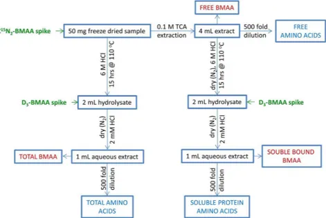
A double spiking isotope dilution protocol was therefore developed to obtain information about both the stability and release of BMAA during hydrolysis, as well as correcting for matrix efects in ESI. Figure 1 shows a summary of the worklows used in this study, including the two spikes of labelled standard. 13C15N
2-BMAA spiked prior to hydrolysis measured extraction recovery and BMAA degradation. D3-BMAA added ater hydrol-ysis corrected for matrix efects in ESI and additional sample preparation steps such as iltration and allowed for quantitation of degradation of the initial standard spike. he challenge in developing this approach was to achieve selectivity between the two labelled standards, both with the same nominal m/z of 122. Figure 2 shows the full scan and product ion spectra of BMAA and its two isotopologues. he absence of electrospray background ions in these spectra is notable and is the result of using DMS for standard infusion experiments, as suggested previ-ously32. From the data shown in Fig. 2, selective SRM conditions were developed to diferentiate the three
difer-ent isotopologues of BMAA, allowing them to be analyzed simultaneously. High resolution settings were required
Figure 1. Summary of sample preparation worklows used in this study.
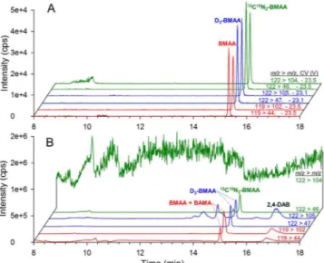
Figure 2. Full scan (A,B,C) and collision induced dissociation (D,E,F) spectra of protonated BMAA (A,D), D3 -BMAA (B,E) and 13C15N
2-BMAA (C,F). Product ions used in SRM experiment are shown in bold. Spectra were collected by infusion ESI-DMS-MS(/MS) using a collision energy spread of 15 V to 20 V.
in Q3 to selectively diferentiate product ions with adjacent m/z value such as ammonia loss from D3-BMAA at m/z 105 and from 13C15N
2-BMAA at m/z 104. A low injection analysis of the three isotopologues of BMAA was carried out to verify this selectivity and is shown in Fig. S1. his showed less than 0.5% interference arising from minor fragmentation pathways of 122 > 104 and 122 > 46 for 13C15N
2-BMAA that can be detected in D3-BMAA transitions.
Figure 3A shows HILIC-DMS-MS/MS chromatograms for a mussel tissue reference material (RM) spiked with 0.25 nmol 13C15N
2-BMAA, hydrolyzed for 12 h at 100 °C, and then spiked with 0.25 nmol D3-BMAA. he use of DMS ofers a signiicant beneit in selectivity and limits of detection compared to analysis of the same mussel tissue RM sample using an equivalent HILIC-MS/MS method without DMS (Fig. 3B). he m/z 119 > 44 transi-tion of BMAA, the m/z 122 > 104 transitransi-tion of 13C15N
2-BMAA and the m/z 122 > 105 transition of D3-BMAA all have a high background when DMS is not used. his high background, that can be observed in general in ESI-MS analysis at low m/z values, can be attributed to chemical background from ESI and consists of a poorly character-ized mixture of cluster ions, source fragments and electrochemical products of mobile phase components and sys-tem contaminants. he broader peak observed for BMAA in the m/z 119 > 102 transition in Fig. 3B is the result of an unresolved naturally occurring isomer of BMAA, β-amino-N-methylalanine (BAMA), interfering with BMAA detection. Using DMS, BAMA and BMAA are completely resolved and can each be selectively analyzed in the presence of a large excess of the other33. Without DMS, the more selective but less sensitive SRM transition
m/z 119 > m/z 76 for BMAA can be used19. In addition to improved selectivity of BMAA analysis from isomers
and chemical background, DMS also results in a signicant decrease in absolute ion counts detected, about 10-fold for BMAA in Fig. 3. Despide this decrease in absolute sensitivity, a signiicant increase in signal-to-noise ratio is observed and results in a corresponding decrease in limits of detection27.
Amino Acid Analysis by HILIC-DMS-MS/MS.
he irst step to incorporating proteinogenic amino acids into our existing HILIC-DMS-MS/MS method for BMAA was to optimize the separation of amino acid standards by DMS. Recent work has shown that low concentrations (<1%) of acetonitrile are broadly suitable for separation of small polar analytes by FAIMS and DMS, including BMAA and its isomers27,33–35. Carrier gas modiiers in DMShelp to amplify diferences in mobility by inducing clustering of ions with neutral solvent molecules, thereby increasing selectivity of the separation. Without modiier, little separation is observed in DMS between small polar analytes such as amino acids. Using a modiied carrier gas modiier solvent delivery system27 allowed us to
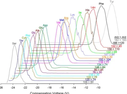
fully explore the efect of varying low concentrations of acetonitrile in the carrier gas on amino acid separation, along with the efect of varying dispersion voltage (DV) (Fig. S2). Analyte dependant minima in compensation voltage of transmission were observed for most amino acids between 0.2% and 0.4% acetonitrile (Fig. S2A). A general trend of increasing resolution of amino acids with increasing DV was observed, but both acetonitrile concentration and dispersion voltage had an impact on sensitivity of analysis as well. Beyond 0.35% acetonitrile or 2600 V, many amino acids exhibited a signiicant decrease in sensitivity of analysis. hese conditions, 0.35% acetonitrile and DV = 2600 V, correspond to those previously identiied as optimum for separation of BMAA isomers27, and it was possible to analyze proteinogenic amino acids under the same DMS conditions as BMAA.
Figure 4 shows the separation of 17 proteinogenic amino acids in a mixed standard by DMS-MS/MS using the SRM parameters in Table 1.
he established DMS-MS/MS parameters for amino acid analysis were combined with the existing HILIC gra-dient and DMS-MS/MS conditions for BMAA to develop a highly selective multidimensional HILIC-DMS-MS/ MS method for BMAA and proteinogenic amino acids. his method is suitable for trace analysis of BMAA and amino acids, but for the intended application of measuring protein hydrolysis in biological samples, a concen-tration diference between of several orders of magnitude between trace BMAA and protein amino acids made it
Figure 3. HILIC-DMS-MS/MS (A) and HILIC-MS/MS (B) chromatograms of naturally occurring BMAA,
125 nM spiked 13C15N
www.nature.com/scientificreports/
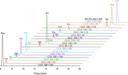
sub-optimal to measure both in a single run. his was primarily due to the poor HILIC peak shape and detector saturation observed for amino acids at these high concentrations. It was therefore desirable to analyze 500 fold dilutions of hydrolyzed samples in a separate injection for amino acid analysis, as shown in Fig. 5 for the hydro-lyzed mussel tissue RM. Amino acid standards showed good linearity up 1 µM concentration and amino acids in diluted samples were estimated at an order of magnitude lower concentration. hough not required for the current application, the developed HILIC-DMS-MS/MS methodology would also be highly suited to the sensitive and selective trace analysis of proteinogenic amino acids in complex environmental or biological samples.
Figure 4. Separation of 17 proteinogenic amino acids in a mixed standard solution by ESI-DMS-MS/MS.
Analyte Retention Time (min) Compensation Voltage (V) Precursor ion m/z Product ion m/z Collision Energy (V)
BMAA 15.7 −23.5 119 102 13 44 25 13C15N 2-BMAA 15.7 −23.5 122 105 13 47 25 D3-BMAA 15.7 −23.1 122 104 13 46 25 Phenylalanine 6.3 −14.4 166 120 17 Leucine 6.4 −15.5 132 43 33 Isoleucine 6.6 −16.6 132 69 21 Methionine 7.2 −18.3 150 104 15 Valine 7.5 −18.6 118 72 17 Tyrosine 7.8 −13.0 182 165 14 Proline 7.9 −22.3 116 70 23 Alanine 9.1 −21.4 90 44 18 Glutamic acid 9.9 −21.0 148 84 20 Glycine 10.0 −21.9 76 30 20 Cysteine 10.1 −22.7 122 76 14 hreonine 10.1 −22.9 120 74 15 Aspartic acid 10.1 −20.9 134 74 19 Serine 11.1 −23.5 106 60 16 Histidine 17.4 −15.8 156 110 20 Arginine 17.7 −18.0 175 70 30 Lysine 18.1 −17.6 147 84 23
Table 1. HILIC-DMS-MS/MS parameters for analysis of BMAA and proteogenic amino acids. he datasets
Extraction and Fractionation of BMAA and Protein.
he goal of the current work was to apply the developed HILIC-DMS-MS/MS methodology to study the hydrolytic release of BMAA and proteinogenic amino acids during strong acid hydrolysis of representative shellish and cycad samples, while using a cyanobacterial RM negative for BMAA as a control sample. he developed methodology was suitable for measurement of the full suite of proteinogenic amino acids, but not all amino acids liberated from protein are stable to hydrolysis conditions. Based on previous reports of accurate, metrological protein quantitation by hydrolysis and amino acid analysis, proline, valine, phenylalanine, leucine and isoleucine were chosen as the most stable36,37. he relativepeak area of these amino acids was compared across diferent sample preparation and hydrolysis procedures to give a relative measure of the amount of protein hydrolyzed.
hree diferent sample preparation procedures were investigated: analysis of free BMAA, hydrolyzed soluble BMAA (soluble bound) and total sample hydrolysis without extraction, as summarized in Fig. 1. hese were applied in triplicate to cycad and shellish samples for analysis of BMAA and proteinogenic amino acids using the developed HILIC-DMS-MS/MS methodology (Fig. 6). Signiicant diferences are observed between the distribu-tion of BMAA in cycad and shellish tissues. For both lobster and mussel, BMAA is predominantly present in the “soluble bound” fraction, with close to 10% present as free BMAA. For cycad seed, which contained much higher BMAA concentration than shellish, about 75% of BMAA was present as free BMAA, but a signiicant amount of soluble bound BMAA was also detected. his result is consistent with recent reports of signiicant soluble bound fraction in mussels25,26, but showed a much lower proportion of soluble bound BMAA to free BMAA in cycad
seed than reported previously3,25.
Figure 6 also demonstrates the signiicant diference between the extraction of protein and BMAA from all samples. In this respect, shellish and cycad seed show a similar distribution, with the majority of protein remain-ing un-extracted in the pellet ater TCA extraction. his result suggests that BMAA is not predominantly associ-ated with protein in the extracts investigassoci-ated. Instead, these results suggest the presence of some other unknown precursor to BMAA, which is released during hydrolysis. his notion is further supported by the observation that the majority of “soluble bound” BMAA in mussels is found in the low molecular weight fraction ater iltration with a molecular weight cut-of ilter (3000 Da), as reported previously26 and repeated here for shellish and cycad
(Fig. S3).
Hydrolysis procedures used in BMAA sample preparation have not been fully optimized for BMAA recovery, but are rather based on existing methods of protein hydrolysis. Considering BMAA appears not to be associated with protein in the samples investigated here, sample preparation conditions optimized for the hydrolysis of pro-tein may not be optimal for BMAA analysis. A time course experiment was therefore carried out on the hydrolysis of the mussel tissue RM, with the goal of optimizing BMAA recovery from shellish samples. Samples hydrolyzed for between 0.5 to 120 h were analyzed for BMAA and proteinogenic amino acids using the methods developed.
Figure 7A shows the release of BMAA during the time course experiment. While hydrolysis times between 9–15 h gave BMAA concentrations of around 4 mg/kg, consistent with our previous analyses of this material, this represented only a fraction of the BMAA that was detected over longer hydrolysis times. In a series of increasingly longer experiments, our original 24 h experiment was extended to 120 h and BMAA levels continued to increase. Good reproducibility between up to 6 replicate samples prepared at each hydrolysis time was also observed. he BMAA concentrations shown in Fig. 7A are calculated using the 13C15N
2-BMAA internal standards spiked into dry mussel tissue prior to hydrolysis. A constantly increasing concentration by this method of calibration would be expected in the case of internal standard degradation during hydrolysis . However, all samples were also spiked with D3-BMAA ater hydrolysis and this spike was used to monitor the stability of 13C15N2-BMAA by directly measuring its concentration in each sample, as shown in Fig. 6C. his showed excellent stability of the internal standard over the 120 h time course experiment. he D3-BMAA spike added ater hydrolysis was also used to monitor changes in ESI matrix efects throughout the time course experiment (Fig. S4). his showed a minor
Figure 5. HILIC-DMS-MS/MS analysis of proteinogenic amino acids in a 500 fold dilution of a mussel tissue
www.nature.com/scientificreports/
matrix efect of about 10% suppression in extractions of free BMAA from mussel tissue, but signiicant suppres-sion of about 65% ater protein hydrolysis was complete. his supports the previous suggestion that matrix efects observed in BMAA analysis in hydrolyzed samples by HILIC are predominantly due to high levels of proteino-genic amino acids17.
he developed methodology was also used to monitor the release of proteinogenic amino acids during the time course experiment. his showed an expected trend of increasing amino acid release for the irst 12 to 48 h of hydrolysis, depending on the amino acid. Figure 7B shows the release of proline, which is stable under hydrolytic conditions. he four other stable amino acids, valine, leucine, isoleucine and phenylalanine gave similar results (Fig. S5). Two important diferences can be observed between the release of BMAA (Fig. 7A) and that of proline (Fig. 7B). For the irst 12 h of the time course experiment, BMAA is released much more quickly than protein is hydrolyzed. By about 18 h proline reaches a plateau, but beyond that BMAA continues to increase, with a nearly linear increase ater 24 h for the remainder of the study period. his result was further conirmed by analysis of the same samples using a recently reported CE-MS/MS method28, which showed the same trend of increasing
BMAA concentration.
Together, these results suggest two diferent compartments of bound BMAA in mussel tissue, the specia-tion of each difering signiicantly from non-speciic protein incorporaspecia-tion. he rapid release of BMAA early in the hydrolysis is consistent with the previous suggestion of an unknown, low molecular weight conjugate that releases BMAA under mild hydrolysis conditions26. he continuous formation of additional BMAA at time
points beyond protein hydrolysis may be unrelated and possible artefactual formation of BMAA during sample hydrolysis should be investigated. It is important to note however, that extended hydrolysis (48 hrs) of the control cyanobacterial RM did not lead to detection of BMAA.
Conclusion
Here we have introduced new methodology that is highly suitable for studying the sample preparation and hydro-lytic release of BMAA from biological samples. he double spiking isotope dilution approach for BMAA quanti-tation allowed BMAA stability during strong acid hydrolysis and ESI matrix efects to be studied independently. his will be of ongoing utility in BMAA method development where it is desirable to examine diferences in recovery or stability in isolation from changing matrix efects in ESI. HILIC-DMS-MS/MS is also shown to be
Figure 6. Distribution of BMAA (A) and protein as measured by hydrolytic release of proline (B) between free
(unhydrolyzed), soluble (hydrolyzed) and total (hydrolyzed) cycad seed, mussel tissue RM and lobster samples. Error bars show standard deviations of triplicate sample preparations.
well suited to the selective analysis of protein amino acids and the hydrolytic release of stable amino acids is used to study the fractionation of protein and progress of hydrolysis. hese methods could equally be applied to help elucidate the speciation of BMAA in other important biological samples such as microalgae and human tissue where BMAA has been reported previously.
he results presented clearly demonstrate that BMAA in shellish and cycad seed is not broadly misincorpo-rated into protein. It is instead present in some other form that appears to include at least two diferent sources or compartments. he rapid release of BMAA ater only 0.5 h of hydrolysis suggests an unidentiied, low molecular weight conjugate and is analogous to the recent observation that BMAA was released from mussels under milder acidic conditions26. Given that no maximum BMAA concentration from mussels could be observed, even ater
5 days of strong acid hydrolysis, the possibility of artefactual formation of BMAA during the hydrolytic reaction should also be considered. It has also been suggested that BMAA could selectively bind to and be incorporated
Figure 7. Release of BMAA (A), proline (B) and 13C15N
2-BMAA internal standard stability (C) during strong acid hydrolysis of a mussel tissue RM. BMAA concentration determined by double isotope dilution using 13C15N
2-BMAA spiked before hydrolysis. Error bars represent standard deviation of multiple sample preparations (2 ≤ N ≤ 6). Insets expand results from the irst 12 h of the experiment.
www.nature.com/scientificreports/
into other biopolymers, speciically the pigment melanin38. It is notable that melanin is also present in shellish
such as the mussels examined here and that the binding of polar neurotoxic paralytic shellish toxins to melanin has also been previously shown39. However, the low aqueous solubility and high molecular weight of melanin
make it an unlikely source of BMAA in the current study. Additional work is required for molecular identiication of possible low molecular weight precursor to BMAA as well as to investigate the possibility of its artefactual formation during sample preparation.
Methods
Chemicals, Reagents and Samples.
Optima LC-MS grade acetonitrile and a mixed L-amino acid stand-ard (2.5 mM alanine, arginine, aspartic acid, glutamic acid, glycine, histidine, isoleucine, leucine, lysine hydro-chloride, methionine, phenylalanine, proline, serine, threonine, tyrosine, valine and 1.2 mM L-cysteine in 0.1 M HCl) were purchased from hermo Fisher Scientiic (Mississauga, ON). Formic acid (>98% ACS grade) was purchased from Sigma Aldrich (Oakville, ON). β-N-Methylamino-L-alanine hydrochloride was obtained from Tocris Bioscience (Minneapolis, MN). D3-BMAA was initially synthesized in-house, as described previously27 and later purchased from Abraxis LLC (Warminster, PA). 13C15N2-BMAA dihydrochloride was purchased from Isoscience (King of Prussia, PA). he structure, purity and concentration of BMAA and internal standards was veriied by 1H nuclear magnetic resonance spectroscopy (NMR)40. NMR stock solutions were diluted to 10 µM
for BMAA and 25 µM for the internal standards and aliquoted into argon purged, lame sealed ampoules for use as stock solutions to prepare calibration standards.
Seeds of Cycas thouarsii were obtained from rarepalmseeds.com (Muenchen, Germany) and the endosperm of one seed was homogenized in a ball mill for 30 min, freeze-dried, and ground in a mortar and pestle. A steamed lobster (Homarus americanus) sample was purchased from a local supermarket (Halifax, Canada; May, 2016). Muscle tissue from the legs and tail were homogenized in a blender, freeze dried, and ground by mortar and pes-tle. A mussel (Mytilus edulis) tissue RM certiied for several marine algal biotoxins (CRM-FDMT) was acquired from the National Research Council Canada (Halifax, Canada)41–44. A recently developed pilot scale
cyanobac-terial RM that has been extensively characterized for a wide range of cyanotoxins was used as a negative control for BMAA45.
Sample Preparation.
Two diferent methods were used for sample hydrolysis: A total tissue hydrolysis and a soluble fraction hydrolysis. he total tissue hydrolysis procedure was modiied from our previous report27.Triplicate samples of 50 mg freeze-dried tissue were weighed into 5 mL amber glass ampoules with 2 mL 6 M HCl, spiked with 100 µL of 2.5 µM 13C15N
2-BMAA and purged with argon before lame sealing. Hydrolysis was carried out in an oven at 110 °C for 15 h, unless otherwise speciied. Ater hydrolysis, samples were spiked with 100 µL of 2.5 µM D3-BMAA, evaporated to dryness at 60 °C under nitrogen, reconstituted in 1 mL of 2 mM HCl, and iltered to 0.22 µm using a Millipore Ultrafree-MC spin ilter. he soluble fraction hydrolysis procedure was adapted from Reveillon et al19. Triplicate samples of 50 mg freeze-dried tissue were weighed into 5 mL reaction vials with 0.5 g
of 75 µm acid washed glass beads (Sigma Aldrich, Oakville, ON) and 2 mL of 0.1 M trichloroacetic acid (TCA), spiked with 100 µL of 2.5 µM 13C15N
2-BMAA and then shaken at 30 Hz for 30 min. Samples were centrifuged at 3100 × g and 15 °C for 10 min. Supernatant was transferred to a 5 mL amber ampoule and the extraction was repeated with an additional 2 mL of 0.1 M TCA. he pooled extract was either analyzed directly to measure free BMAA or was evaporated to dryness at 60 °C with a gentle stream of nitrogen and hydrolysed using the same conditions used in the total extraction prior to analysis. hese sample preparation worklows are summarized graphically in Fig. 1.
For analysis of protein amino acids, a 500 fold sequential dilutions of sample hydrolysates in water were car-ried out using a Gilson Aspec GX 271 liquid handler (Mandel Scientiic, Guelph, ON).
HILIC-DMS-MS/MS Methods.
LC separation was performed using a 5 µm TSKgel Amide-80 column (250 mm × 2 mm i.d.) (Tosoh, Grove City, OH, USA) held at 40 °C and a mobile phase of 50 mM formic acid in both water (A) or 95% acetonitrile (B) at a low rate of 300 µL min−1. A linear gradient ran from 10 to 35% A over 15 min followed by a 6 min isocratic period, a linear gradient to 45% A over 1 min, an 8 min isocratic period, a 5 min column lush at 90% A and a 10 min equilibration at 10% A17,27.Detection was carried out using an AB Sciex (Concord, ON, Canada) QTRAP 5500 mass spectrometer equipped with a Turbospray ionization source and SelexION diferential mobility spectrometer. he DMS carrier gas modiier solvent delivery system was adapted, as described previously, to allow for delivery of a full range of modiier concentrations and better solvent evaporation and mixing27. Turbospray parameters included a spray
voltage of + 5500 V, curtain gas of 20 psi N2, collision gas of 5 psi N2, desolvation gases of 45 psi (GS1) and 50 psi (GS2) N2, and a spray temperature of 400 °C. Mass spectrometer settings included a declustering potential of 140 V, an entrance potential of 10 V and exit potential of 13 V, DMS settings included separation (dispersion) volt-age of 2600 V, a carrier gas modiier of 0.35% acetonitrile, a DMS temperature of 150 °C, and a DMS ofset of −3 V. DMS conditions for each amino acid were optimized by infusing the diluted amino acid standard at 10 µL min−1 into a 350 µL min−1 low of 1:1 MeCN:H
2O with 50 mM formic acid and collecting compensation voltage scans using selected reaction monitoring (SRM) with the transitions for each amino acid reported previ-ously46. he concentration of acetonitrile in the N
2 carrier gas was varied over a range of 0.04 to 2.5% and separa-tion voltage was varied over a range from 2000 V to 4000 V. Optimized retensepara-tion times, SRM transisepara-tions, collision energy, and DMS compensation voltages used for sample analysis are listed in Table 1.
References
1. Masseret, E. et al. Dietary BMAA Exposure in an Amyotrophic Lateral Sclerosis Cluster from Southern France. PLOS ONE 8, e83406, https://doi.org/10.1371/journal.pone.0083406 (2013).
2. Vega, A. & Bell, E. A. α-Amino-β-methylaminopropionic acid, a new amino acid from seeds of Cycas circinalis. Phytochemistry 6, 759–762, https://doi.org/10.1016/s0031-9422(00)86018-5 (1967).
3. Murch, S. J., Cox, P. A. & Banack, S. A. A mechanism for slow release of biomagnified cyanobacterial neurotoxins and neurodegenerative disease in Guam. Proc. Natl. Acad. Sci. USA 101, 12228–12231, https://doi.org/10.1073/pnas.0404926101 (2004). 4. Cox, P. A., Banack, S. A. & Murch, S. J. Biomagniication of cyanobacterial neurotoxins and neurodegenerative disease among the
Chamorro people of Guam. Proc. Natl. Acad. Sci. USA 100, 13380–13383, https://doi.org/10.1073/pnas.2235808100 (2003). 5. Cox, P. A. et al. Diverse taxa of cyanobacteria produce beta-n-methylamino-l-alanine, a neurotoxic amino acid. Proceedings of the
National Academy of Sciences of the United States of America 102, 5074–5078 (2005).
6. Chen, K.-M., Craig, U. K. & Lee, C.-T. & Haddock, R. Cycad neurotoxin, consumption of lying foxes, and ALSIPDC disease in Guam. Neurology 59, 1664–1665 (2002).
7. Marler, T. E., Snyder, L. R. & Shaw, C. A. Cycas micronesica (Cycadales) plants devoid of endophytic cyanobacteria increase in β-methylamino-L-alanine. Toxicon 56, 563–568, https://doi.org/10.1016/j.toxicon.2010.05.015 (2010).
8. Faassen, E. J. Presence of the neurotoxin BMAA in aquatic ecosystems: what do we really know? Toxins (Basel) 6, 1109–1138, https:// doi.org/10.3390/toxins6031109 (2014).
9. Faassen, E. J., Gillissen, F. & Lurling, M. A comparative study on three analytical methods for the determination of the neurotoxin BMAA in cyanobacteria. PLoS One 7, e36667 (2012).
10. Kruger, T., Oelmuller, R. & Luckas, B. he origin of β-N-methylamino-L-alanine (BMAA): Cycads and/ or cyanobacteria? Journal of Endocytobiosis and Cell Research 22, 29–36 (2012).
11. Glover, W. B., Baker, T. C., Murch, S. J. & Brown, P. N. Determination of beta-N-methylamino-L-alanine, N-(2-aminoethyl)glycine, and 2,4-diaminobutyric acid in Food Products Containing Cyanobacteria by Ultra-Performance Liquid Chromatography and Tandem Mass Spectrometry: Single-Laboratory Validation. J AOAC Int 98, 1559–1565, https://doi.org/10.5740/jaoacint.15-084
(2015).
12. Jiang, L., Aigret, B., De Borggraeve, W. M., Spacil, Z. & Ilag, L. L. Selective LC-MS/MS method for the identiication of BMAA from its isomers in biological samples. Analytical and Bioanalytical Chemistry 403, 1719–1730 (2012).
13. Jiang, L., Johnston, E., Aaberg, K. M., Nilsson, U. & Ilag, L. L. Strategy for quantifying trace levels of BMAA in cyanobacteria by LC/ MS/MS. Anal. Bioanal. Chem. 405, 1283–1292, https://doi.org/10.1007/s00216-012-6550-1 (2013).
14. Lage, S. et al. BMAA extraction of cyanobacteria samples: which method to choose? Environmental Science and Pollution Research 23, 338–350, https://doi.org/10.1007/s11356-015-5266-0 (2016).
15. Lampinen Salomonsson, M., Hansson, A. & Bondesson, U. Development and in-house validation of a method for quantiication of BMAA in mussels using dansyl chloride derivatization and ultra performance liquid chromatography tandem mass spectrometry. Analytical Methods 5, 4865, https://doi.org/10.1039/c3ay40657a (2013).
16. Spacil, Z. et al. Analytical protocol for identiication of BMAA and DAB in biological samples. Analyst (Cambridge, UK) 135, 127–132, https://doi.org/10.1039/b921048b (2010).
17. Li, A. et al. Elucidation of matrix efects and performance of solid-phase extraction for LC-MS/MS analysis of β-N-methylamino-L-alanine (BMAA) and 2,4-diaminobutyric acid (DAB) neurotoxins in cyanobacteria. Analyst (Cambridge, UK) 137, 1210–1219,
https://doi.org/10.1039/c2an15887f (2012).
18. McCarron, P., Logan, A. C., Giddings, S. D. & Quilliam, M. A. Analysis of β-N-methylamino-L-alanine (BMAA) in spirulina-containing supplements by liquid chromatography-tandem mass spectrometry. Aquatic Biosystems 10, 1–7 (2014).
19. Reveillon, D. et al. Beta-N-methylamino-L-alanine: LC-MS/MS optimization, screening of cyanobacterial strains and occurrence in shellish from hau, a French Mediterranean lagoon. Mar Drugs 12, 5441–5467, https://doi.org/10.3390/md12115441 (2014). 20. Reveillon, D. et al. beta-N-methylamino-l-alanine (BMAA) and isomers: Distribution in diferent food web compartments of hau
lagoon, French Mediterranean Sea. Mar Environ Res 110, 8–18, https://doi.org/10.1016/j.marenvres.2015.07.015 (2015). 21. Reveillon, D., Sechet, V., Hess, P. & Amzil, Z. Production of BMAA and DAB by diatoms (Phaeodactylum tricornutum, Chaetoceros
sp., Chaetoceros calcitrans and, halassiosira pseudonana) and bacteria isolated from a diatom culture. Harmful Algae 58, 45–50,
https://doi.org/10.1016/j.hal.2016.07.008 (2016).
22. Berntzon, L., Ronnevi, L. O., Bergman, B. & Eriksson, J. Detection of BMAA in the human central nervous system. Neuroscience 292, 137–147, https://doi.org/10.1016/j.neuroscience.2015.02.032 (2015).
23. Downing, S., Banack, S. A., Metcalf, J. S., Cox, P. A. & Downing, T. G. Nitrogen starvation of cyanobacteria results in the production of β-N-methylamino-L-alanine. Toxicon 58, 187–194, https://doi.org/10.1016/j.toxicon.2011.05.017 (2011).
24. Meneely, J. P. et al. beta-methylamino-L-alanine (BMAA) is not found in the brains of patients with conirmed Alzheimer’s disease. Sci Rep 6, 36363, https://doi.org/10.1038/srep36363 (2016).
25. Faassen, E. J. et al. A Collaborative Evaluation of LC-MS/MS Based Methods for BMAA Analysis: Soluble Bound BMAA Found to Be an Important Fraction. Mar Drugs 14, https://doi.org/10.3390/md14030045 (2016).
26. Rosen, J., Westerberg, E., Schmiedt, S. & Hellenas, K. E. BMAA detected as neither free nor protein bound amino acid in blue mussels. Toxicon 109, 45–50, https://doi.org/10.1016/j.toxicon.2015.11.008 (2016).
27. Beach, D. G., Kerrin, E. S. & Quilliam, M. A. Selective quantitation of the neurotoxin BMAA by use of hydrophilic-interaction liquid chromatography–differential mobility spectrometry–tandem mass spectrometry (HILIC–DMS–MS/MS). Analytical and Bioanalytical Chemistry 407, 8397–8409, https://doi.org/10.1007/s00216-015-9012-8 (2015).
28. Kerrin, E. S., White, R. L. & Quilliam, M. A. Quantitative determination of the neurotoxin beta-N-methylamino-L-alanine (BMAA) by capillary electrophoresis-tandem mass spectrometry. Anal Bioanal Chem 409, 1481–1491, https://doi.org/10.1007/s00216-016-0091-y (2017).
29. David, A. & Barnett, B. E. Roger Guevremont, Randy W. Purves. Separation of Leucine and Isoleucine by Electrospray Ionization–High Field Asymmetric Waveform Ion Mobility Spectrometry–Mass Spectrometry. Journal of he American Society for Mass Spectrometry 10, 1279–1284 (1999).
30. Shvatsburg, A. A., Prior, D. C., Tang, K. & Smith, R. D. High-Resolution Differential Ion Mobility Separations Using Planar Analyzers at Elevated Dispersion Fields. Analytical Chemistry 82, 7649–7655 (2010).
31. McCooeye, M. & Mester, Z. Comparison of low injection analysis electrospray mass spectrometry and tandem mass spectrometry and electrospray high-field asymmetric waveform ion mobility mass spectrometry and tandem mass spectrometry for the determination of underivatized amino acids. Rapid Commun Mass Spectrom 20, 1801–1808, https://doi.org/10.1002/rcm.2515
(2006).
32. Beach, D. G. & Gabryelski, W. Revisiting the reactivity of uracil during collision induced dissociation: tautomerism and charge-directed processes. J Am Soc Mass Spectrom 23, 858–868, https://doi.org/10.1007/s13361-012-0343-9 (2012).
33. Beach, D. G. Diferential Mobility Spectrometry for Improved Selectivity in Hydrophilic Interaction Liquid Chromatography-Tandem Mass Spectrometry Analysis of Paralytic Shellish Toxins. J Am Soc Mass Spectrom 28, 1518–1530, https://doi.org/10.1007/ s13361-017-1651-x (2017).
34. Beach, D. G., Melanson, J. E. & Purves, R. W. Analysis of paralytic shellish toxins using high-ield asymmetric waveform ion mobility spectrometry with liquid chromatography-mass spectrometry. Analytical and Bioanalytical Chemistry 407, 2473–2484,
www.nature.com/scientificreports/
35. Purves, R. W. et al. Using Gas Modiiers to Signiicantly Improve Sensitivity and Selectivity in a Cylindrical FAIMS Device. Journal of he American Society for Mass Spectrometry 25, 1274–1284, https://doi.org/10.1007/s13361-014-0878-z (2014).
36. Burkitt, W. I. et al. Toward Systeme International d’Unite-traceable protein quantiication: from amino acids to proteins. Anal Biochem 376, 242–251, https://doi.org/10.1016/j.ab.2008.02.010 (2008).
37. Fountoulakis, M. & Lahm, H.-W. Hydrolysis and amino acid composition analysis of proteins. Journal of Chromatography A 826, 109–134 (1998).
38. Karlsson, O., Berg, C., Brittebo, E. B. & Lindquist, N. G. Retention of the cyanobacterial neurotoxin β-N-methylamino-L-alanine in melanin and neuromelanin-containing cells - a possible link between Parkinson-dementia complex and pigmentary retinopathy. Pigm. Cell Melanoma Res. 22, 120–130, https://doi.org/10.1111/j.1755-148X.2008.00508.x (2009).
39. Price, R. J. & Lee, J. S. Paralytic Shellish Poison and Melanin Distribution in tr’ractions oi Toxic Butter Clam (Saxidomus giganteus) Siphon. Journal of the Fisheries Research Board of Canada 29, 1657–1658 (1972).
40. Burton, I. W., Quilliam, M. A. & Walter, J. A. Quantitative 1H NMR with external standards: use in preparation of calibration solutions for algal toxins and other natural products. Anal. Chem. 77, 3123–3131 (2005).
41. McCarron, P., Emteborg, H., Giddings, S. D., Wright, E. & Quilliam, M. A. A mussel tissue certiied reference material for multiple phycotoxins. Part 3: homogeneity and stability. Anal. Bioanal. Chem. 400, 847–858, https://doi.org/10.1007/s00216-011-4787-8
(2011).
42. McCarron, P. et al. A mussel tissue certiied reference material for multiple phycotoxins. Part 1: design and preparation. Anal. Bioanal. Chem. 400, 821–833, https://doi.org/10.1007/s00216-011-4786-9 (2011).
43. McCarron, P., Giddings, S. D. & Quilliam, M. A. A mussel tissue certiied reference material for multiple phycotoxins. Part 2: liquid chromatography-mass spectrometry, sample extraction and quantitation procedures. Anal. Bioanal. Chem. 400, 835–846, https://doi. org/10.1007/s00216-011-4803-z (2011).
44. McCarron, P., Wright, E., Emteborg, H. & Quilliam, M. A. A mussel tissue certiied reference material for multiple phycotoxins. Part 4: certiication. Anal Bioanal Chem 409, 95–106, https://doi.org/10.1007/s00216-016-0004-0 (2017).
45. Hollingdale, C. et al. Feasibility study on production of a matrix reference material for cyanobacterial toxins. Analytical and Bioanalytical Chemistry 407, 5353–5363, https://doi.org/10.1007/s00216-015-8695-1) (2015).
46. Petritis, K., Chaimbault, P., Elfakir, C. & Dreux, M. Parameter optimization for the analysis of underivatized protein amino acids by liquid chromatography and ionspray tandem mass spectrometry. Journal of Chromatography A 896, 253–263, https://doi. org/10.1016/S0021-9673(00)00582-3 (2000).
Acknowledgements
he authors would like to thank Sheila Crain for technical assistance with NMR measurements.
Author Contributions
All authors helped plan the experiments, E.K. and S.G. carried out the experiments, D.B. and E.K. analyzed the data, D.B. prepared the manuscript and all authors provided editorial input.
Additional Information
Supplementary information accompanies this paper at https://doi.org/10.1038/s41598-017-18392-w.
Competing Interests: he authors declare that they have no competing interests.
Publisher's note: Springer Nature remains neutral with regard to jurisdictional claims in published maps and
institutional ailiations.
Open Access This article is licensed under a Creative Commons Attribution 4.0 International
License, which permits use, sharing, adaptation, distribution and reproduction in any medium or format, as long as you give appropriate credit to the original author(s) and the source, provide a link to the Cre-ative Commons license, and indicate if changes were made. he images or other third party material in this article are included in the article’s Creative Commons license, unless indicated otherwise in a credit line to the material. If material is not included in the article’s Creative Commons license and your intended use is not per-mitted by statutory regulation or exceeds the perper-mitted use, you will need to obtain permission directly from the copyright holder. To view a copy of this license, visit http://creativecommons.org/licenses/by/4.0/.